
Abstract
All kinds of commercially available wound dressings are clinically used as fleshly obstacles and therapeutic materials in opposition to microbial incursion. Few researches focused on effective-bleeding and anti-bacteria at the same time. In order to better solve this problem, two hydrogels were synthetized in this study. One is phosphate buffer solution-activated dopamine-modified-γ-poly glutamic acid (PBS-PD) hydrogel, the other one is cirsium setosum extracts-activated dopamine-modified-γ-poly glutamic acid (CSE-PD) hydrogel. The two hydrogels are prepared by applying an enzyme-catalyzed crosslinking means in the presence of horseradish peroxidase (HRP) and hydrogen peroxide (H2O2). The chemical structures were characterized through 1H-NMR and FT-IR. In conclusion, both PBS-PD and CSE-PD hydrogels exhibit superior tissue adhesion properties, and remarkable anti-infection quality. In addition, these two hydrogels manifest prominent hemostatic efficiency. The bio adhesion performance can achieve 30 kPa, meanwhile the CSE-PD hydrogels show good germicidal properties, and the antibacterial rate can reach 98%. The hydrogels could reduce blood loss without any obvious side effect, and present a new prospect in the field of hemostasis rapidly.
Introduction
Skin is the first natural safeguard for humans, playing a crucial part in sheltering the tissues and organs from outer damages, such as physical, chemical, and involuntary impair, morbific germs, radioactivity, or excessive temperatures, and maintains electrolytes and nutritional components, while preventing body dehydration.1,2 As we all know, at one time the skin undergoes severe damage, and the wound extreme bleeding generally causes hemorrhagic shock coagulopathy, multiple organ failures, infection, and even threatening people's life and health. 3 Recently, several kinds of wound dressings have been developed including microspheres, foam, bandage, film, and hydrogel and so on.4–7 In these dressings, hydrogel can maintain a moist wound environment, absorb tissue exudates, permit oxygen into penetrate and chill the wound cover to relief pain for human beings. 8 Especially, in situ fabricating hydrogel can be equal to the extracellular matrix of native organizations, and these hydrogels are comfortable, highly compliant and convenient, which can be combined with irregular shapes of wounds;5,9 therefore, they have been arranged as a kind of hopeful wound materials. Besides, the first stage of wound healing is rapid hemostasis, and in situ fabricating adhesive hydrogels can prevent bleeding.6,10
In situ forming gel derived from mussel belongs to a type of classical tissue adhesives materials, and they have gained tremendous attention in biological territory.11–13 The mussel’s tough adhesion ability is benefitted from the catechol, which contains ample amino acid. Especially in 3,4-dihydroxyphenylalanine (DOPA), many researchers found that the adhesive proteins were secreted by foot of mussels. 14 It has been reported that strong adhesion of mussel is contributed by DOPA, since it can interact with the interface of the organization by various ways, including hydrogen bond, metal chelation, π-π or cation-π.13,15 Based on the similarity of DOPA, dopamine (DA) is the prime candidate to embellish γ-poly glutamic acid (γ-PGA). More importantly, DA is a biomaterial with nature antibacterial property and possesses many other superiorities in biological field. 16 γ-PGA belongs to biopolymer that possesses green, water-soluble, comestible and avirulent to human and environment. Moreover, in the field of bioadhesion, it can be a promising composite to improve the swelling ratio and mechanical strength because of its high wetting ability;17–20 also, it can provide a watery wound benefit for accelerating wound healing process. However, moist environment is also growing a large number of bacteria that can cause wound infection, two of the major bacteria are Staphylococcus aureus (S. aureus) and Escherichia coli (E. coli). 21 The hydrogel possesses releasable antibacterial agents, which will present inherent antibacterial properties and can continuously release drugs with relatively low side effect. Thus, investigators have exploited germicidal and astrictive wound dressings by introducing bactericide into the dressings or straightforwardly make use of materials with inherent bactericide to solve this problem.22,23 In addition to depending on the adhesion of the material, hemostasis can also be achieved by adding some hemostatic agents such as thrombin, proteins, polysaccharides and inorganic soils.24,25 Besides, cirsium setosum (CS) has been used in the field of treat bleeding and inflammation for thousands of years in the Chinese Pharmacopoeia. 26 CS plays a vital role in many ways, such as hemostasis, 27 sterilization, 28 anti-inflammatory, and mitigative functions. 29 Many studies have shown that CS has good biological activity, particularly, caffeic acid and chlorogenic acid in CS could promote platelet aggregation and adhesion.30,31 Buddleoside has been recorded to possess analgesic, anti-inflammatory and neuroprotective action, 32 and rutin could weaken vascular permeability. 33 Other study has indicated that tyramine of CS via regulate α-ARs could induce vasoconstriction. 34 Although the previous studies have some advantages, but their strong toxic side effects and poor adhesion inhibit intimate combine with tissue surfaces, resulting in excessive loss of blood. Furthermore, previous studies often designed materials for a certain point, which could not achieve the effect of high adhesion, high antibacterial, and fast hemostasis, and what we can do is to reach these three points at the same time. Therefore, we constructed the PD hydrogels to achieve the goal simultaneously: (1) tough wet tissue-affinity, (2) anti-infection and (3) hemostasis.
In the work, we devised two bio-inspired hydrogels containing both robust adhesion and antibacterial capabilities; they are manufactured via DA to modify γ-PGA. One is phosphate buffer solution-activated dopamine-modified-γ-poly glutamic acid (PBS-PD) hydrogel, and the other is CS extracts-activated dopamine-modified-γ-poly glutamic acid (CSE-PD) hydrogel. We adopted enzyme catalyzed cross-linking method along with horseradish peroxidase (HRP) and H2O2. In waterborne polymers, polymerization reaction was performed by oxidizing phenol hydroxyl or coupling aniline groups. In this system, on the one hand, as a crosslinker, H2O2 plays an essential role and is decomposed with water and O2 molecules by HRP. On the other hand, the residuary H2O2 in hydrogel acts as bactericidal agent. 5 In the present study, it is more fascinating that cirsium setosum extracts (CSEs) were introduced into the gelling system, which can improve the antibacterial and hemostatic properties. Furthermore, the PD hydrogels have antibacterial activity via the pivotal synergistic interplay of H2O2 and CSE. Besides, we also testified in vitro hemostatic ability of the hydrogels.
Experimental section
Materials
γ-PGA (Molecular weight: 1000–2000 kDa), 1-ethyl-3–(3-dimethylaminopropyl)-carbodiimide (EDC), N-hydroxysuccinimide (NHS), Hydrogen Peroxide (H2O2), HRP and DA were bought from Aladdin Reagent. Phosphate-buffered saline (PBS, pH 7.4) was purchased from Solarbio. CSE was collected from Shihezi University. All other chemical solvents were used of the highest available purity, unless stated they were used as received.
Synthesis and characterization of dopamine-modified-γ-poly glutamic acid
(PD) conjugates
The PD conjugate was prepared according to the previous report. 3 In brief, DA and γ-PGA were activated by EDC and NHS in water as a solvent of N, N, N-dimethylformamide (DMF), stirred for half an hour at room temperature, then γ-PGA was added to the mixed solution tardily. Subsequently, the solution was transformed into an ice bath under nitrogen protection atmosphere for 4 h. The prepared samples were dialyzed continuously by deionized water. After the dialysis, the PD conjugates can be obtained by freeze-drying. The structure and composition of PD were confirmed by 1H NMR (Bruker Avance III HD 400M) and UV-vis.
Preparation of the CSE
The CSE was obtained based on reported literature.34,35 CS was put in a cutting mill to obtain coarse powder shape, then air-dried at 60°C. The dry samples were extracted three times with 150 mL ethanol by high shear, each time for 10 min at room temperature. The supernatant was separated by centrifugation, combined with ethanol solution, filtration; subsequently, the solvent in the CSE was removed by spin evaporation method.
Preparation of two PD hydrogels
Hydrogels were prepared in bottles under normal temperature. Briefly, the PD polymer (9 wt.%) was dissolved in 2 mL PBS (pH 7.4) solution or CSE solution, then the solution was split into half, 1 mL solution was added to the HRP (0.05 mg mL−1), the other 1 mL solution was added to 0.1 mL different H2O2 concentrations (2%–3 wt.%). Adding one part to the other part gently forms the in situ gel; gelling process only needs 2 s by the vial tilting method. 36
Morphology properties
The micromorphology of PD hydrogels was characterized by SEM (LEO 1430VP). Before test, the PD hydrogels were flushed several times by distilled water to remove the residual unreacted molecules, then freeze-dried and goldcoated to observe the cross-sectional morphologies.
Rheological properties and mechanical properties test
Measurement of elastic modulus (G′) of the PD hydrogel is done by Disc Rheometer (Anton Paar MCR501), of which the diameter of the parallel plate is 25 mm. We prepared three groups of PD hydrogels with different concentrations of H2O2 to study their rheological properties. We placed the PD hydrogel on the disc mold and kept the two parallel plates at 1 mm intervals, and silicone oil was coated around the parallel plates to prevent the evaporation of the PD hydrogels. In dynamic vibration mode, 0.1 Hz frequency and a strain of 0.1% were used to measure at 37°C.
The compressive stress test was conducted with a dynamic thermo mechanical analysis instrument (DMA) (Q800, TA). The PD hydrogels were prepared with different concentrations of H2O2 in the cylindrical mold (5 mm diameter, 5 mm height). The stress–strain curves of the PD hydrogels were determined at constant ratio of 10%/min at room temperature. The compressive stress (σ) was calculated as σ = F/A, where F represents the load and A stands for the original cross area of the specimen.
Tissue adhesion strength
The adhesion properties test about the hydrogel was conducted by universal testing machine (INSTRON 3366, USA). To simulate human skin, we picked porcine skin as substrate for this experiment. Subsequently, porcine skin was cut into regular rectangles, where 1 mL PD solution was spread on pigskin with an area of 1.5 cm2, another identical skin was put on the gel at once. Then the associative part compressed for 10 min at closed environment. Subsequently, the adhesive strength was measured at a cross-head speed of 5 mm min−1. Each measurement was repeated three times.
Assessment of in vitro antibacterial performance of the PD hydrogels
In order to detect the broad-spectrum antibacterial effect of the PD hydrogel, we chose the most representative E. coli and S. aureus as Gram-negative bacteria and Gram-positive bacteria. Two methods were mainly used in the experiment: quantitative method (living bacteria counting) and qualitative method (zones of inhibition were measured);37,38 the growth medium adopted Luria–Bertani (LB) agar. In the qualitative method, the antibacterial activity of the PD hydrogels was evaluated by inhibition zone assay. In the beginning, 100 mL LB medium was prepared by 1 g tryptone, 0.5 g yeast extract, 1 g sodium chloride, and 1 g agar and autoclaved for 20 min at 121°C. When cooled to 60°C, the medium was inflood with Petri dishes until harden, then punch one round orifice (0.5 cm in diameter). About 1 mL of CSE-PD or PBS-PD bioadhesive hydrogel was used to fill the cavity; each hydrogel was lightly rinsed several times with sterilized water to remove the unreacted H2O2. Subsequently, 0.2 mL of bacteria was sprayed on the agar plates to ensure the surfaces of hydrogels were uniformly covered with bacteria, and then incubated at 35°C about 18 h; the distance between the total bacteriostatic ring and the hydrogel was called the inhibition zone diameter.
As for the quantitative method, it is regarding counting the number of living bacteria. Bacterial cells were about 106 colony forming units (CFUs)/mL in medium, and after the bacterial cells were allowed serial dilution to a visible number, the three groups were incubated at 35°C for 18 h respectively. The first group was without any hydrogels as a contrast, the second group was PBS-PD hydrogel, the third group was CSE-PD hydrogel. Bacterial viability by counting CFU, and the calculation method of antibacterial rate were determined: antibacterial rate=a–b/a × 100% (a: the total number of bacteria with contrast group; b: the number of living bacteria with hydrogel group).
Whole blood clotting and platelet activation studies of PD hydrogels
The experiment of whole blood coagulation was conducted based on previous report, 39 then analyzed the zymoplastic capability of PD hydrogel. Fresh sheep blood was mixed with acid citrate dextrose (ACD) according to a certain ratio, and the blood was regarded as negative control in the study. Firstly, the prepared hydrogel was placed in Petri dish and the whole blood was added to the hydrogel, secondly, the PD hydrogel was incubated several minutes at 37°C. Thirdly, a certain volume of distilled water was added slowly without damaging the blood clot. Afterwards, the solution was centrifuged at 1000 r/min for 5 min and taken the supernatant for an hour at 37°C; 200 μL of this solution was transferred to a 96-well plate, then used a plate reader (Thermo varioskan flash) to measure the optical density at 540 nm.
Platelet activation study was conducted as previous report. 40 Platelet-rich plasma (PRP) was isolated from the blood by centrifugation of blood at 5000 r/min for 2 min. Two hundred microliters of PRP were poured into the PD hydrogel and incubated at 37°C about several minutes. The hydrogels were then washed three times by PBS solution and fixed using 0.1% glutaraldehyde solution. Afterwards, the hydrogels were freeze-dried and SEM images were taken out of them.
Results and discussion
Synthesis and characterization of PD conjugates
The PD conjugates were prepared by grafting γ-PGA on the DA under the nitrogen-filling atmosphere. As shown in Figure 1(a), the chemical structure of γ-PGA and DA was characterized by 1H NMR, FTIR and UV-vis; as shown in Figure 1(b), in comparison with the DA and γ-PGA, the specific peaks of PD at around 7 ppm indicate the conjugation of DA with the polymer backbone successfully. As shown in Figure 1(c), the broad absorption peak at 1250 cm−1 is owed to the new stretching vibration of the C–N group of the PD conjugates amide bond. Meanwhile, the PPD conjugate exhibits a marked absorption peak in the UV–vis (UV-3200PCS) absorption spectra at 280 nm, which then confirmed the successful introduction of DA moieties (Figure 1(d)).
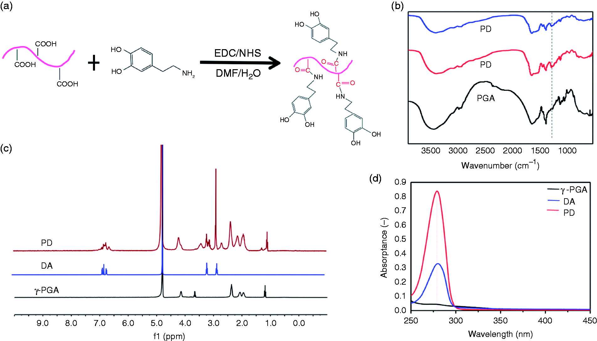
(a) Schematic of the preparation of PD conjugate. (b) FTIR spectra of γ-PGA and PD conjugate. (c) 1H NMR spectra of PD conjugate, DA, γ-PGA (solvent: D2O). (d) UV-visible spectra of synthesized PD conjugate and unmodified γ-PGA and DA.
Mechanism diagram and schematic diagram of the PD hydrogel formation
The PD hydrogels were prepared in situ by HRP catalyzed crosslinking in the presence of H2O2 (Figure 2(a)). The enzymatic crosslink theory mainly attributes to phenols cross-linking, through either the C–C linkage between the ortho-carbons of the aromatic ring or the C–O linkage between the ortho-carbon and phenolic oxygen. The color of the solution changed from colorless to dark brown at once representing the formation of the hydrogel, and the gel position remained unchanged after inversion (Figure 2(b)), the reason for this is that phenol hydroxyl structure is oxidized to quinine.
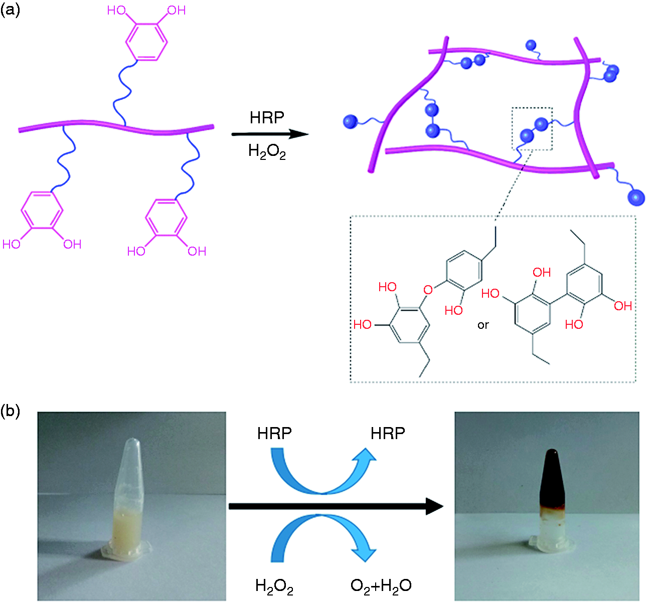
(a) Sketch map of the PD hydrogel formation. (b) Macroscopical view of the PD hydrogel forming, before (left) and after (right) gelation.
Morphology characterization
The scanning electron microscopy (SEM) was employed to investigate the effects of H2O2 on the inner microstructure. We observed the micromorphology of the PD hydrogels prepared from different concentrations of H2O2 (Figure 3). It is gratifying to note that all pore sizes are very uniform, and the density of pores becomes compact and small with the increase of H2O2 concentration. The result further shows that the mechanical strength of the PD hydrogel can be achieved by regulating the concentration of H2O2.
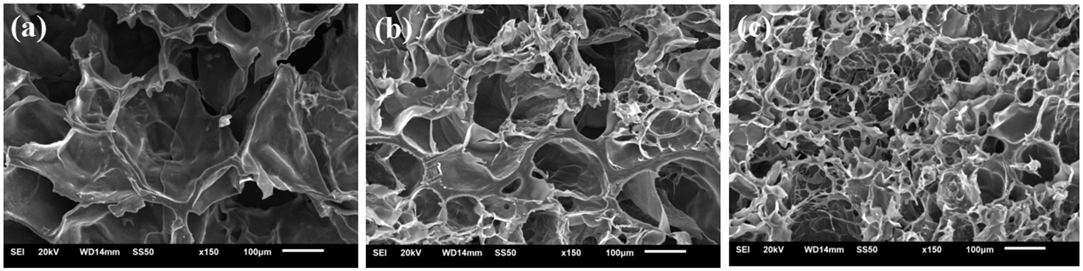
((a), (b), (c)) Scanning electron micrographs of the cross section of PD hydrogels formed with different H2O2 concentrations (a: 2%; b: 2.5%; c: 3%). The final concentrations of PD and HRP in the hydrogels were fixed at 10 wt.% and 0.05 mg mL−1, respectively.
Analysis of rheological properties and mechanical properties
A rheological rheological experiment was carried out to study the mechanical properties of the PD hydrogels with different H2O2 concentrations, which fixed the amount of polymer solution (10 wt.%) and HRP (0.05 mg mL−1) in PBS at 37°C. As shown in Figure 4(a), with the increase of H2O2 concentration, the modulus of elasticity increases gradually. In turn, the elastic modulus can be adjusted by H2O2 concentration. The result shows that the crosslinking density was increased with the increasing of the phenolic hydroxyl content in the enzyme catalyzed reaction, and the energy storage modulus increases, which are in agreement with the result of the SEM.
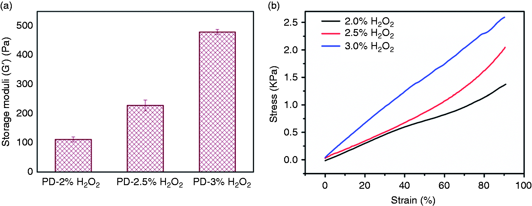
(a) Storage moduli (G′) of PD hydrogels with different H2O2 concentrations. (b) Stress–strain curve of the PD hydrogels with different concentrations of H2O2 concentrations at 37°C.
In order to study the mechanical properties of hydrogels with different concentrations of H2O2, DMA was used to detect the mechanical properties at room temperature. The stress–strain curves have been shown that the stress increases with the increasing of strain, and the rate of stress increases with the increasing of H2O2 concentration. The maximum strain of the three groups of PD hydrogels is above 80%. These characteristics indicate that the PD hydrogels were suitable for the application of wound dressing.
Tissue adhesive strength
The adhesive strength of the PD hydrogels was evaluated at different H2O2 concentrations. To imitate human skin, we used moist pig skins as the substrate materials. As previously described, the gel was prepared in situ by enzyme catalysis, and the gel was coated on the base of pig skin. 36 Figure 5(a) shows a schematic map of adherence test about the PD hydrogels, and Figure 5(b) illustrates the adhesive force of PD hydrogels varied from 13 kPa to 30 kPa, which shows that they have super adhesion. It has been reported that cationic polymers can interact closely with epithelial cells of tissue and interact through dynamic bond cross-linking reversible opening and remodeling processes. 39 Meanwhile, it can be seen from Figure 5 that the adhesive force of PD hydrogels increased with the increasing of H2O2 concentration. The reason for this phenomenon because of that the different microstructures of the gels, a higher cross-linking density can often enhance the adhesive strength of polymers, and a tighter gel network can provide stronger cohesive interactions.
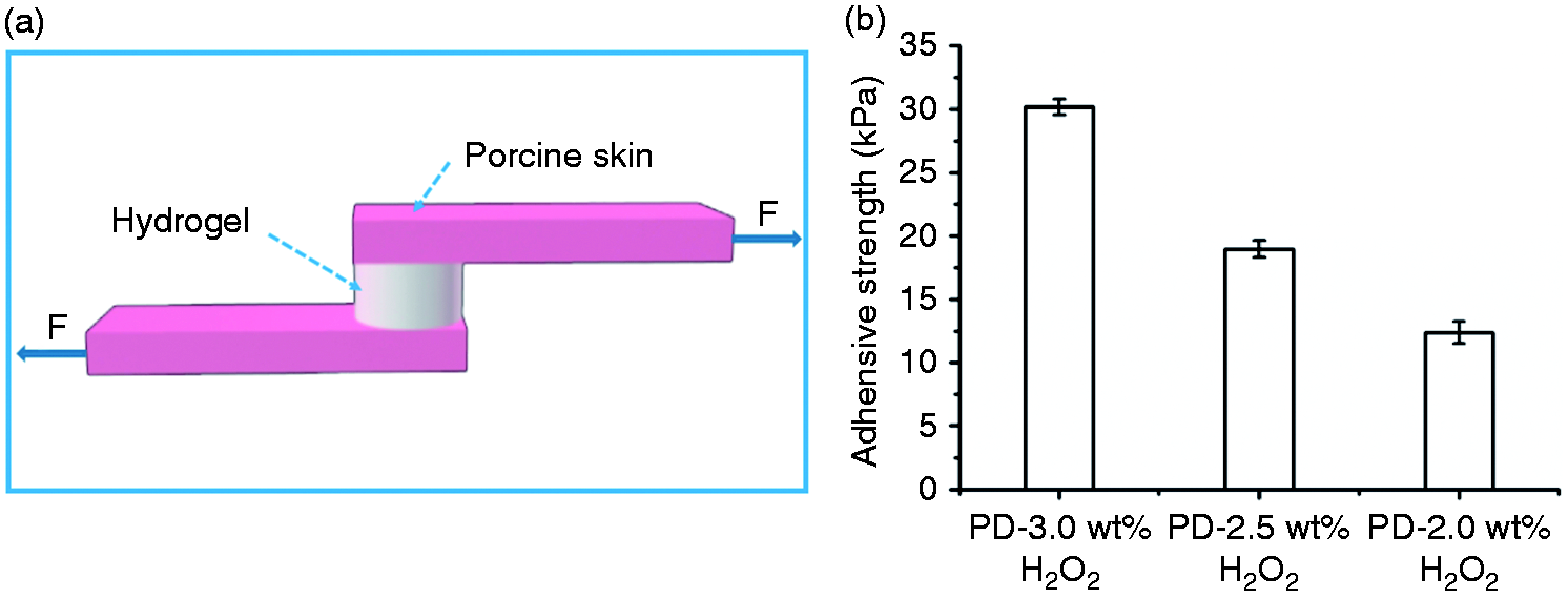
Adhesive property of PD hydrogel. (a) Schematic diagram of the lap shear test with porcine skin. (b) Adhesion strength of PD hydrogels on porcine at various concentrations of H2O2.
Antibacterial properties
Among many wound dressings, gelatin belongs to a class of biomaterial, which is due to its humid environment and promote the growth of epithelial cells during the wound healing stage. Regrettably, a humid environment can also cause bacterial propagation to the wound. The antibacterial effectiveness of hydrogels was evaluated with E. coli and S. aureus by visual inhibition zone and bacterial mortality. After cultivating for 18 h, the hydrogels possessed antibacterial activity clearly, and inhibition zone of the hydrogel could be seen intuitively (Figure 6(a) to (d)). As we can see, the bacteriostasis of CSE-PD hydrogels is obviously larger than that of PBS-PD hydrogels, whether it is E. coli or S. aureus. Meanwhile, it is suggested that the PD hydrogels inhibit completely the growth of E. coli and S. aureus. From Figure 6(e), as we can see, the PBS-PD hydrogels possess excellent antibacterial properties, and the antibacterial rate can reach 98%. The efficient antibacterial activity could be attributed to the presence of residual H2O2 and CSE in the hydrogel matrix.
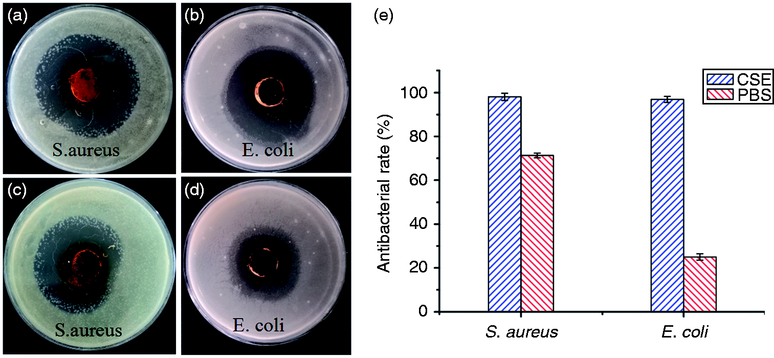
Antimicrobial activities of PD hydrogels direct at E. coli and S. aureus. (a, b) Photographs show the inhibition zone of CSE-PD hydrogels. (c, d) Photographs show the inhibition zone of PBS-PD hydrogels. (e) The antibacterial rates of CSE-PD hydrogels and PBS-PD hydrogels with 3% H2O2.
In vitro hemostatic capacity
In order to evaluate whether CSE-PD hydrogel and PBS-PD hydrogel can increase the rate of blood clotting, whole blood was contacted with hydrogels surface for 10 min. Figure 7(a) to (c) shows representative photographs of the blood clotting caused by PD hydrogels. PD control and PD hydrogels showed enhanced blood clotting ability in comparison with blood at different concentration of H2O2 (Figure 7(d)), a lower absorbance value of the hemoglobin solution thus indicates a quicker clotting. 40 In sharp contrast, the CSE-PD hydrogel led to significantly lower absorbance values than the PBS-PD hydrogel. This result was further confirmed by the platelet activation analysis by SEM. Significantly, more platelets adhered to the CSE-PD hydrogel (Figure 7(g) and (h)) than PBS-PD hydrogel (Figure 7(e) and (f)). Besides, these adhesive platelets all remained in their regular shapes and did not flatten, indicating good biocompatibility.
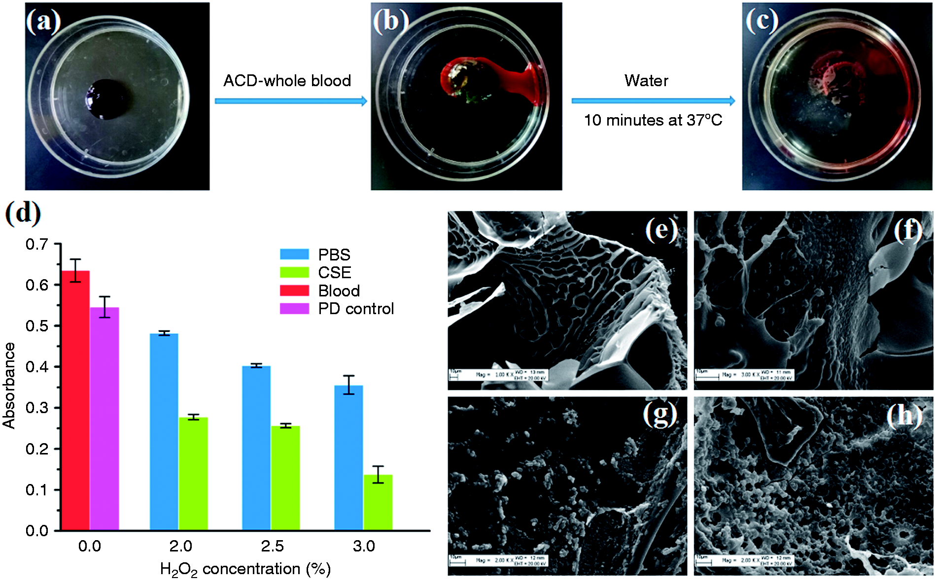
((a), (b), (c)) Photographs of the PD hydrogels, blood on the PD hydrogels, clotted blood on the PD hydrogels, respectively. (d) Whole-blood clotting evaluation of PD hydrogels with different solvent (PBS and CSE) at different concentrations of H2O2. (n = 10, mean ± SD) ((e), (f), (g), (h)) SEM micrographs of platelet activation of PD hydrogels.
Conclusions
In short, we have engineered two novel PBS-PD and CSE-PD hydrogels, which were inspired by mussels. The hydrogels robust bio adhesion performance can achieve 30 kPa, sealing the bleeding wound gloriously as a potential stuff in terms of hemostasis. Additionally, the CSE-PD hydrogels have great germicidal properties, and the antibacterial rate can reach 98%. The quantity of bacteria on the PBS-PD hydrogels is approximately 1.5 times than the CSE-PD hydrogels in terms of inhibition zone. What’s more, the PBS and CSE can evoke remarkable platelet adhere on the PD hydrogels interface. Hence, the PD hydrogels have a wide range of potential application in the field of hemostasis, wound repair, drug delivery, and tissue engineering. Furthermore, the injectable PD hydrogels prepared by the simple bio-inspired method will be useful as a tissue adhesive for a variety of surgical applications.
Footnotes
Declaration of Conflicting Interests
The author(s) declared no potential conflicts of interest with respect to the research, authorship, and/or publication of this article.
Funding
The author(s) disclosed receipt of the following financial support for the research, authorship, and/or publication of this article: This work was supported financially by funding from the National Natural Science Foundation of China (51662036 & 21467024) and Bingtuan Innovation Team in Key Areas (2015BD003).