
Abstract
Thixotropic clays have favorable properties for tissue regeneration. Hypoxia mimetic agents showed promising results in pre-clinical models for hard and soft tissue regeneration. It is unclear if clays can be used as carrier for hypoxia mimetic agent in a periodontal regenerative setting. Here, we tested the response of human fibroblasts of the periodontal soft tissue to synthetic clay hydrogels and assessed hypoxia mimetic agent release. Cells were cultured on synthetic clay hydrogels (5.00%–0.15%). We assessed viability and differentiation capacity with resazurin-based toxicity assays, MTT staining, Live-Dead staining, and alkaline phosphatase staining. To reveal the response of fibroblasts to hypoxia mimetic agent-loaded clay hydrogels, cells were exposed to clay supplemented with dimethyloxalylglycine, deferoxamine,
Introduction
Microinvasive strategies for periodontal and oral surgery rely on injectable carrier materials which are biocompatible and show a promising release profile for biologicals. Several hydrogels on synthetical and biological basis have been tested. Among these materials are collagen and fibrin which have been applied for various tissue engineering approaches in periodontology and oral surgery.1–4
Synthetic clays, such as LAPONITE®, are a novel kind of material with promising properties.5–8 Synthetic clay in combination with water can form hydrogels which are thixotropic and have the ability to adsorb proteins.5,6 Synthetic clay-based hydrogels have been successfully applied as carrier materials for signaling molecules including vascular endothelial growth factor (VEGF) and bone morphogenetic proteins (BMPs) which favor angiogenesis and osteogenic differentiation, respectively.6,9 Also the combination of biological scaffold material with synthetic clay showed promising results. Collagen matrices supplemented with clay hydrogel lead to a prolonged release of VEGF. 6 Recently, clay-based hydrogels have been developed to allow additive manufacturing for tissue engineering approaches in the muscular skeletal field using 3D printers.10–12 Although all these properties make synthetic clay a promising candidate as biomaterial for regenerative periodontology there is no available data on the effect of clay on cells of oral soft tissue and periodontal ligament and the feasibility for applications in periodontal surgery.
Promising novel regenerative approaches target the cellular oxygen sensors by application of prolyl hydroxylase inhibitors also known as hypoxia mimetic agents (HMAs).13–15 Among these are dimethyloxalylglycine (DMOG), deferoxamine (DFO),
In the present study, we assessed the impact of synthetic clay hydrogel on primary human fibroblasts of the periodontal soft tissue in vitro. Furthermore, the impact of synthetic clay hydrogels loaded with HMAs DMOG, DFO, L-MIM, and CoCl2 was assessed by an established in vitro bioassay. 13
Materials and methods
Synthetic clay hydrogel preparation
Synthetic clay hydrogels were prepared with the synthetic clay LAPONITE® XLG (IMCD South East Europe GmbH, Vienna, Austria) by application of the synthetic clay powder to water at 5%. The hydrogel was sterilized by autoclaving. The sterile 5% stock hydrogel was diluted to 2.5%, 1.25%, 0.62%, 0.31%, and 0.15%. Synthetic clay hydrogels at 2.5% containing DMOG, DFO, L-MIM, and CoCl2 all at 1 mM were prepared.
Isolation and culture of fibroblasts from the periodontal soft tissue
Primary human fibroblasts from the periodontal soft tissue were isolated from extracted third molars based on a previously described protocol 30 after informed consent was given by the donors (1065/2013, Ethics Committee of the Medical University of Vienna, Vienna, Austria). In brief, the periodontal soft tissue adhering to the tooth neck was scraped off and collected. We performed explant cultures in α-minimal essential medium (α-MEM, Invitrogen Corporation, Carlsbad, CA) supplemented with 10% fetal bovine serum (FBS, PAA Laboratories, Linz, Upper Austria, Austria) and antibiotics at 37°C, 5% CO2, and 95% atmospheric moisture. For the experiments cells were seeded at 50,000 cells/cm2 on the indicated materials and were incubated as stated in the Results section, figure legends, and table legends for 24–72 h.
Evaluation of release kinetics of the HMAs from clay hydrogel
Clay hydrogels at 2.5% synthetic clay supplemented with the HMAs DMOG, DFO, L-MIM, or CoCl2, all at 1 mM, were added to 96 well plates in 100 µl per well. These specimens were incubated with 100 µl medium and the supernatants were harvested and replaced with medium at hour 1, 3, 6, 24, 48, and 72. The hypoxia mimetic capacity of the released HMAs was evaluated in bioassays where human fibroblasts of the periodontal soft tissue were exposed to the supernatants for 24 h and immunoassays for VEGF were performed.
MTT staining
Fibroblasts cultured on the hydrogels or in the presence of conditioned medium were incubated with 1 mg/ml MTT (3–(4,5-dimethylthiazol-2-yl)-2,5-diphenyltetrazolium bromide; Sigma–Aldrich, St. Louis, MO) at 37°C for 2 h. The formation of formazan was evaluated under a light microscope and images were taken.
Live-Dead staining
Human fibroblasts from the periodontal soft tissue were cultured on synthetic clay hydrogels were stained with the Live-Dead Cell Staining Kit (Enzo Life Sciences AG, Lausen, BL, Switzerland) following the protocol provided by the manufacturer. The cells were evaluated by fluorescence microscopy for green and red, with a B-2A filter (excitation filter wavelengths: 450–490 nm). Vital cells were stained green while dead cells were stained red.
Histochemical staining for alkaline phosphatase
Human fibroblasts of the periodontal soft tissue were cultured on the synthetic clay hydrogels, in differentiation medium (α-MEM at 10% FCS supplemented with 50 mmol/L L-ascorbic acid and 10 mmol/L b-glycerophosphate (Sigma–Aldrich) and antibiotics). After 7 days of culture, the fibroblasts were fixed with neutral buffered formalin. Then, cells were stained with the substrate solution which contained Naphthol AS-TR phosphate disodium salt and Fast Blue BB Salt (Sigma–Aldrich) and images were taken.
Resazurin-based toxicity assay
The resazurin-based toxicity assay was done following the protocol of the manufacturer. Resazurin dye solution (Sigma–Aldrich) was applied at 10% of the culture medium. Fluorescence was evaluated using a Synergy HTX multimode reader (BioTek, Winooski, VT) at a wavelength of 590 nm, using an excitation wavelength of 560 nm.
Immunoassays for VEGF
The culture medium or synthetic clay supernatants were subjected to VEGF ELISA (Standard ABTS ELISA Development Kit for human VEGF, Peprotech, Rocky Hill, NJ) which was performed according to the protocol of the manufacturer. The optical density obtained from the samples was measured in a Synergy HTX multi-mode reader (IBM Corporation, Armonk, NY). The VEGF concentration was calculated with the standard curve method.
Statistical analysis
Data was analyzed with IBM SPSS Statistics Version 23 (IBM Corporation, Armonk, NY) by applying the Kruskal–Wallis test post hoc Mann–Whitney test. The level of significance was set at p ≤ 0.05.
Results
The impact of synthetic clay hydrogel on cellular activity of human fibroblasts of periodontal soft tissue
The impact of the synthetic clay concentration in hydrogels on cell viability of human fibroblast of the periodontal soft tissue was assessed using the resazurin-based toxicity assay. Fibroblasts cultured on synthetic clay hydrogels at 2.5%–0.15% converted resazurin, indicating vital cells (Figure 1(a)). Synthetic clay hydrogel at 5% was not feasible for in vitro application in cell culture due to the high viscosity. We found no significant reduction of the conversion of resazurin with the clay hydrogels at 2.5%--0.15% (Figure 1(a)). Furthermore, a dose–response experiment over 72 h was run. While cells remained vital over time there was a trend for a decrease in overall viability at 72 h (Table 1). In agreement with the results of the resazurin-based toxicity assay human fibroblasts of periodontal soft tissue maintained their ability to form formazan when cultured on synthetic clay hydrogels (Figure 1(b)). Fibroblasts cultured on clay hydrogel at 2.5% appeared dark violet. After Life-Dead staining fibroblasts appeared green under fluorescence microscopy suggesting that cells are vital. No cells appeared prominently red stained (Figure 1(c)). Via histochemical staining cells positive for alkaline phosphatase (AP) were found, while also cells negative for AP were observed (Figure 1(d)).
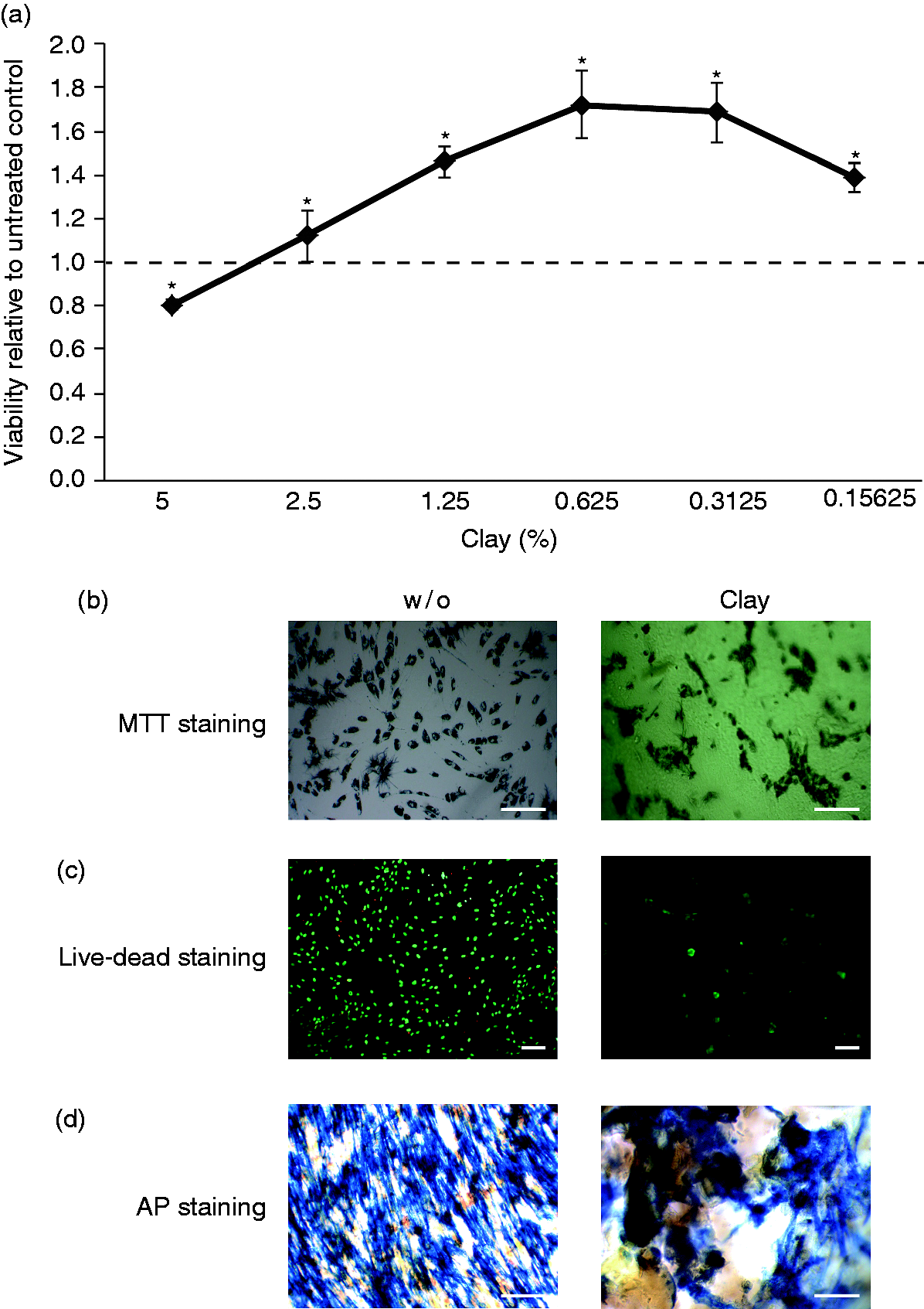
Viability and cell activity of primary human fibroblasts of the periodontal soft tissue on synthetic clay hydrogels. Primary human human fibroblasts of the periodontal soft tissue were incubated on synthetic clay hydrogels of 5%–0.15625%. (a) Viability of the fibroblasts was assessed based on conversion of resazurin. Data points represent mean ± standard deviation. Two independent experiments were performed with three donors. *p ≤ 0.05 versus control (dashed line). (b) MTT staining and (c) Live-Dead staining were performed, both on 2.5% synthetic clay hydrogel. (d) To assess osteoblastic differentiation, the cells cultured on 2.5% synthetic clay hydrogel were exposed to differentiation medium for 7 days and histochemical staining for alkaline phosphatase (AP) was done. The white bar represents 100 µm.
Dose–response of cell viability to various concentrations of synthetic clay hydrogels when normalized to 24 h.
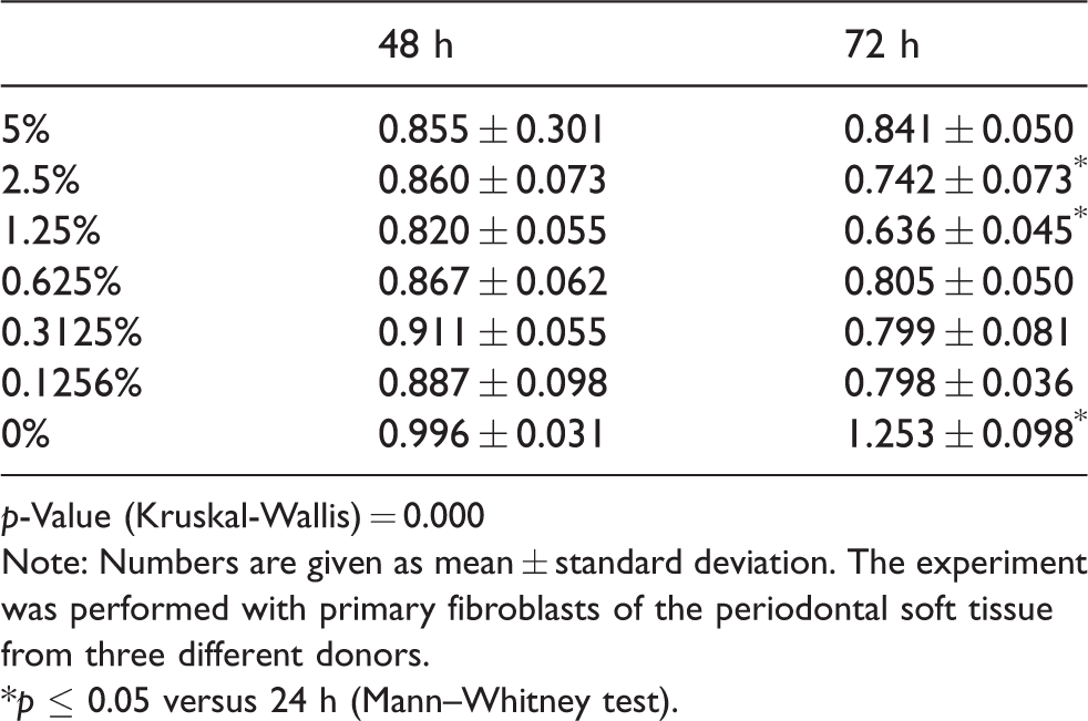
p-Value (Kruskal-Wallis) = 0.000Note: Numbers are given as mean ± standard deviation. The experiment was performed with primary fibroblasts of the periodontal soft tissue from three different donors.
*p ≤ 0.05 versus 24 h (Mann–Whitney test).
Evaluation of the cellular response to synthetic clay hydrogels loaded with HMAs
To reveal the impact of clay hydrogels loaded with HMAs on the activity of human fibroblasts of periodontal soft tissue the cells were cultured on the synthetic clay hydrogels which were supplemented with DMOG, DFO, L-MIM, and CoCl2. Fibroblasts remained vital when cultured on the 2.5% clay hydrogels in all preparations (Table 2). Furthermore, when normalized to 24 h, the fibroblasts also remained vital over a period of 72 h when cultured on hydrogels with the four different HMAs (Table 3). Levels of VEGF in the culture medium were not increased when clay hydrogels supplemented with HMAs were compared to clay hydrogel without hypoxia-mimetic agents (Table 2). As positive control for the bioactivity of the HMAs we exposed the cells to HMA solutions at 0.5 mM which represent the maximum concentration when all HMAs would be released within the first hour. Our data show that HMA can increase VEGF production (Table 4) as previously published.13–15 To evaluate if the produced VEGF binds to the hydrogel we incubated the cells with DMOG and collected the culture medium containing the produced VEGF. Then, the culture medium was incubated with synthetic clay hydrogels. Measurement of VEGF after the 24-h incubation period revealed significantly less VEGF in the culture medium compared to the culture medium which was incubated with the respective volume of water. These data indicate that LAPONITE® XLG binds the VEGF in the culture medium (Table 5).
Evaluation of the cellular response to synthetic clay hydrogels loaded with hypoxia mimetic agents in primary human fibroblasts of the periodontal soft tissue.
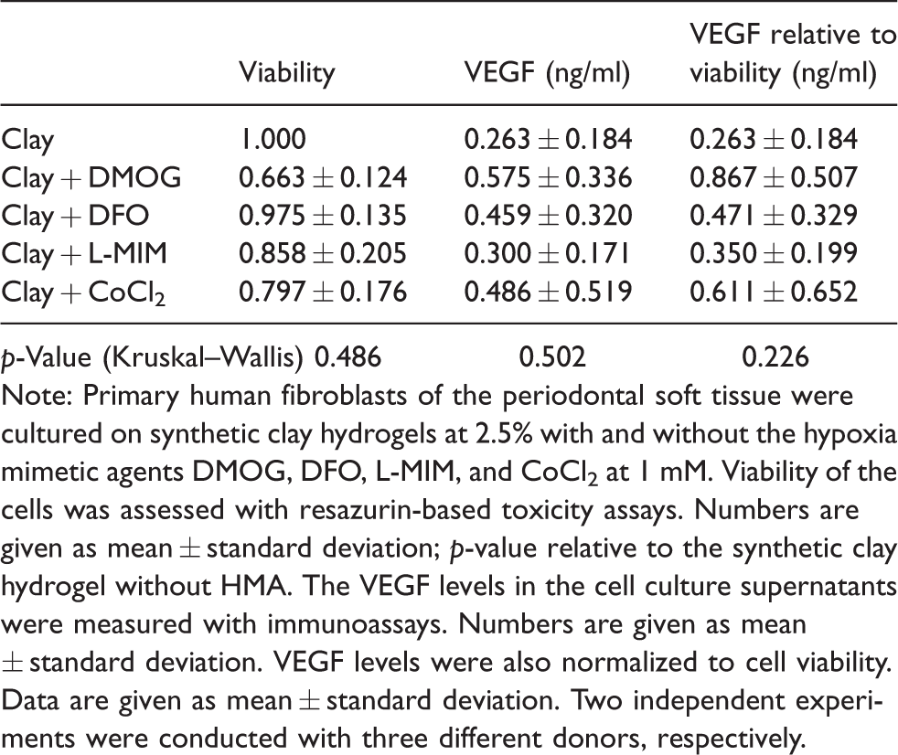
p-Value (Kruskal–Wallis) 0.4860.5020.226Note: Primary human fibroblasts of the periodontal soft tissue were cultured on synthetic clay hydrogels at 2.5% with and without the hypoxia mimetic agents DMOG, DFO, L-MIM, and CoCl2 at 1 mM. Viability of the cells was assessed with resazurin-based toxicity assays. Numbers are given as mean ± standard deviation; p-value relative to the synthetic clay hydrogel without HMA. The VEGF levels in the cell culture supernatants were measured with immunoassays. Numbers are given as mean ± standard deviation. VEGF levels were also normalized to cell viability. Data are given as mean ± standard deviation. Two independent experiments were conducted with three different donors, respectively.
Viability and VEGF levels in supernatants of fibroblasts of the periodontal soft tissue when treated with hypoxia mimetic agents at 0.5 mM normalized to untreated control.
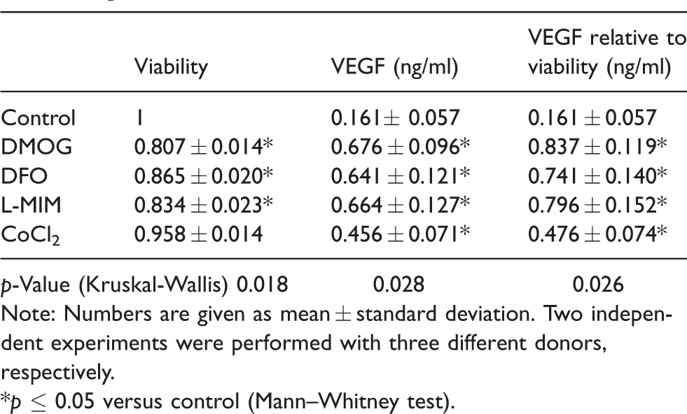
p-Value (Kruskal-Wallis) 0.018 0.028 0.026Note: Numbers are given as mean ± standard deviation. Two independent experiments were performed with three different donors, respectively.
*p ≤ 0.05 versus control (Mann–Whitney test).
Cell viability of primary human fibroblasts of the periodontal soft tissue when cultured on synthetic clay hydrogel with and without hypoxia mimetic agents.
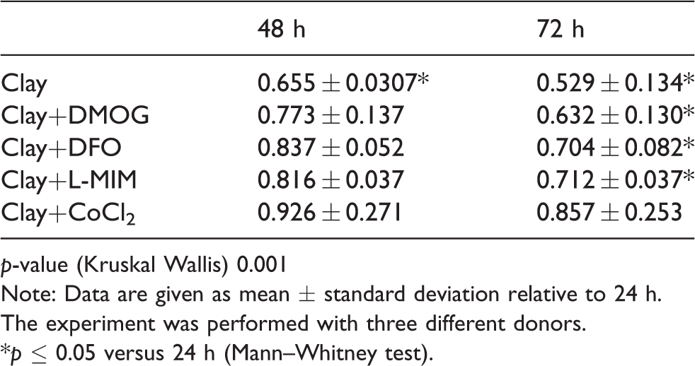
p-value (Kruskal Wallis) 0.001Note: Data are given as mean ± standard deviation relative to 24 h. The experiment was performed with three different donors.
*p ≤ 0.05 versus 24 h (Mann–Whitney test).
Synthetic clay hydrogel can bind VEGF in the culture medium.

Note: Culture medium of H2O exposed cells incubated with synthetic clay hydrogel (CM DMOG + H2O); culture medium of DMOG exposed cells incubated with synthetic clay hydrogel (CM DMOG + synthetic clay hydrogel). We incubated the cells with DMOG and collected the culture medium containing the produced VEGF. Then the culture medium was incubated with synthetic clay hydrogels or the respective volume of water. VEGF was measured after 24 h. Data are given as mean ± standard deviation. The experiment was performed with medium from three different donors.
*p ≤ 0.05 versus CM DMOG + H2O.
To exclude the possibility that synthetic clay binds the produced VEGF we uncoupled the cell culture from the synthetic clay hydrogels by generation of supernatants from synthetic clay and exposing the cells to these supernatants. In these cultures, the VEGF levels upon treatment with supernatants harvested after 1 h of incubation with clay hydrogel supplemented with DMOG were increased when compared to the supernatants from clay hydrogel without HMAs. No significant impact was observed on cell viability (Figure 2(a) and (b)).
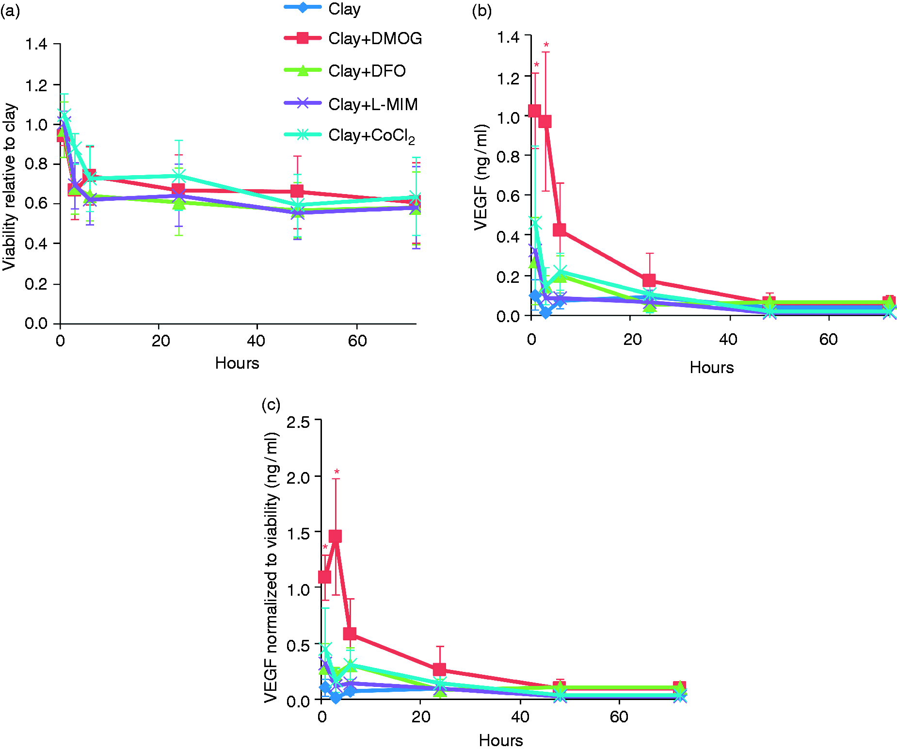
Release of hypoxia mimetic agents from synthetic clay based on a bioassay with primary human fibroblasts of the periodontal soft tissue. To reveal release kinetics of the synthetic clay hydrogels (2.5%) supplemented with hypoxia mimetic agents DMOG, DFO, L-MIM, and CoCl2 supernatants of the hydrogels were collected and replaced at hour 1, 3, 6, 24, 48, and 72. Primary human fibroblasts of the periodontal soft tissue were then treated with these supernatants. (a) Cell viability was assessed with resazurin-based toxicity assays. Data points represent mean ± standard deviation, relative to the synthetic clay hydrogel without HMA. Experiments were conducted twice with three different donors, respectively. (b) The proangiogenic response to the hypoxia mimetic agents was measured based on VEGF levels in the culture media. Data points represent mean ± standard deviation. Experiments were conducted twice with three different donors, respectively. and (c) VEGF levels were normalized to cell viability. Data points represent mean ± standard deviation. Two independent experiments were conducted with two different donors, respectively. *p ≤ 0.05 versus control.
Taken together our data show that synthetic clay releases the HMA DMOG which can induce VEGF production in human fibroblasts of the periodontal soft tissue. Furthermore, the data suggest that the released VEGF can then be adsorbed on the synthetic hydrogel.
Discussion
Synthetic clay is a promising biomaterial for tissue regeneration which has been proposed to have feasible properties for bone regeneration.6,9 Novel studies highlight successful application as hydrogel and for additive manufacturing using 3D printing in tissue engineering approaches.10--12 Due to the anatomical situation and difficult accessibility in periodontal surgery injectable carrier materials are of great interest in this field. Thus, we here proposed synthetic clay as a novel scaffold material for regenerative periodontics. Clay, furthermore, has the important ability to bind growth factors and other proteins6,9 and can bind and release HMAs with different kinetics as we showed in this study.
We showed by resazurin-based toxicity assays, MTT staining, and Live-Dead staining that human fibroblasts of the periodontal soft tissue treated with clay remained vital. This is in line with the favorable properties that have been described for bone regeneration approaches.6,9,12
Due to the structure of the hydrogel, the cells were not all in the same focus plane on the hydrogel. Also, the different concentrations of the hydrogel lead to a difference in the distribution of the cells as at lower concentrations the cells sink in the less viscose gel. Thus, in a quantification, the number of vital cells might be underestimated. In the resazurin-based assay no reduction was observed in overall cell viability. We also did not observe a prominent red staining in the live-dead staining, which would have indicated dead cells. What we can say based on our Life-Dead staining is that vital cells are present on the hydrogel. Time– and dose–response experiments indicate that cell proliferation is not increased in the presence of the synthetic clay hydrogels with and without the presence of HMAs.
Also, differentiation in alkaline phosphatase positive cells was observed. This experiment was carried out as cells of the periodontal soft tissue contain stem cells which have been applied for bone tissue engineering. Thus, for these applications it would be important to know whether cells in synthetic clay based hydrogels are compromised in their capacity to differentiate into osteoblastic cells. 31 While we also aimed to retrieve RNA to perform the expression of osteogenic markers, it was not possible to collect RNA in a reasonable quality. However, our data on alkaline phosphatase staining show that synthetic clay does not prevent the differentiation of cells from the periodontal soft tissue into alkaline phosphatase positive cells.
Hypoxia-based strategies are a novel approach in regenerative periodontics. 15 By applying HMAs cellular oxygen sensors are targeted to boost angiogenesis by an increase of VEGF and stimulate the regeneration process under hypoxia.14,32,33 Cells from the periodontal soft tissue have been shown to be target cells of HMAs. 14
We evaluated the capability of synthetic clay to act as a feasible carrier for HMAs. When synthetic clay was loaded with HMAs, viability was significantly decreased to approximately 80% in all but the DFO containing group in the resazurin-based toxicity assay. When we assessed the pro-angiogenic response of fibroblasts of the periodontal soft tissue based on the release of VEGF we found no significant impact of the HMA-loaded synthetic clay hydrogels on the levels of VEGF in the culture medium. As VEGF levels did not increase when the fibroblasts were cultured on synthetic clay it is possible that the released VEGF bound to clay. Therefore, we used an established in vitro release model where we uncoupled the release of HMAs from the cell culture.13,34,35 Using a bioassay we measured VEGF production after incubating the human fibroblasts from the periodontal soft tissue with supernatants harvested of synthetic clay mixed with DMOG, DFO, L-MIM, and CoCl2. Only DMOG lead to an increased VEGF production in the first 3 h. This possible discrepancy could be explained by the outstanding binding capacity of synthetic clay. Thus, it is reasonable to suggest that the VEGF produced when the cells were cultured directly on the synthetic clay hydrogel loaded with the DMOG may have been adsorbed by the synthetic clay.
As to why only DMOG of the four HMAs lead to a significant increase in VEGF levels is unknown yet. Overall, our data are in line with our results from dental pulp-derived cells.36 The four tested HMAs DMOG, DFO, L-MIM, and CoCl2 all vary in their nature and mode of action. L-MIM, DMOG, and DFO are amino acid derivatives and CoCl2 contains a metal iron. Due to the properties of the synthetic clay LAPONITE® XLG having different charges on the rim and the surface of its discs, the subunit, as well as changing its charge depending on the pH may all have an influence on varying release kinetics between the four HMAs.
We assessed the biological activity of the released HMAs and no direct quantification of the HMAs was performed. This is a limitation of the study, however, for application purposes the biological activity of the HMAs is more relevant than the absolute concentration.
Future studies will need to reveal if the supplementation of synthetic clay with HMAs can improve the efficiency of clay used as hydrogel or for additive manufacturing for regenerative purposes and if these approaches are feasible for periodontal regeneration.
Here, we found that human fibroblasts from periodontal soft tissue remain vital when exposed to synthetic clay hydrogels and that while it can be loaded with various HMAs only DMOG increases VEGF production in the cells significantly. Our in vitro results gave first insights into the effect of synthetic clay on cells from periodontal tissue and provide the basis for future studies in the field of regenerative periodontics.
Footnotes
Acknowledgements
The authors thank Manuela Pensch for skillful technical assistance. The authors deny any conflict of interest. Our research on HMAs was supported by the Osteology Foundation (10-063). LAPONITE® XLG, BYK Additives Ltd., Wesel, Germany was kindly provided by IMCD South East Europe GmbH, Vienna, Austria. Anna Müller received the research fellowship “Wissenschaftsstipendium für Waldviertler WissenschaftlerInnen mit sozialer Kompetenz Wissenschaft -- solide wie Waldviertler Granit” by the GEA Akademie (Schrems, Austria).
Declaration of Conflicting Interests
The author(s) declared no potential conflicts of interest with respect to the research, authorship, and/or publication of this article.
Funding
The author(s) disclosed receipt of the following financial support for the research, authorship, and/ or publication of this article: Our research on HMAs was supported by the Osteology Foundation (10-063). LAPONITE® XLG, BYK Additives Ltd., Wesel, Germany was kindly provided by IMCD South East Europe GmbH, Vienna, Austria. Anna Müller received the research fellowship “Wissenschaftsstipendium für Waldviertler WissenschaftlerInnen mit sozialer Kompetenz Wissenschaft -- solide wie Waldviertler Granit” by the GEA Akademie (Schrems, Austria).