
Abstract
Extracellular matrix materials mechanically dissociated into submillimeter particles have a larger surface area than sheet materials and enhanced cellular attachment. Decellularized porcine mesothelial extracellular matrix microparticles were seeded with bone marrow-derived mesenchymal stromal cells and cultured in a rotating bioreactor. The mesenchymal stromal cells attached and grew to confluency on the microparticles. The cell-seeded microparticles were then encapsulated in varying concentrations of fibrin glue, and the cells migrated rapidly off the microparticles. The combination of microparticles and mesenchymal stromal cells was then applied to a splinted full-thickness cutaneous in vivo wound model. There was evidence of increased cell infiltration and collagen deposition in mesenchymal stromal cells-treated wounds. Cell-seeded microparticles have potential as a cell delivery and paracrine therapy in impaired healing environments.
Introduction
Decellularized extracellular matrix (ECM) materials are an ideal candidate for a variety of regenerative medicine applications. These biomaterials preserve ECM proteins and retain significant biologic activity including stimulating cells grown on the material to secrete increased cytokines.1,2 A variety of cells have been grown on decellularized materials,3–6 including human bone marrow-derived mesenchymal stromal cells (MSCs) which do not spontaneously differentiate in vitro. 7 MSCs are an advantageous cell source due to their easy accessibility from bone marrow, early ECM production and robust secretion of cytokines which promotes the growth, migration, and recruitment of cells.8,9 Complex three-dimensional scaffolds seeded with MSCs have been tested in vitro,10–12 while pre-seeded sheet materials in vivo have shown improved results compared to un-seeded materials in cutaneous wounds 13 and hernia models. 14 These studies are the next step toward developing cell-seeded decellularized ECM materials for clinical use.
To date, clinical applications of decellularized ECM materials have focused on sheet-based applications, but as a powder, the materials could be used in a wider range of applications. Submillimeter particles produced from decellularized ECM materials 15 yield a much larger surface area per unit weight than the sheets and have been shown to enhance the cellular attachment to material. 16 These cell-seeded microparticles have improved the tensile strength of an in vitro anastomoses model 16 and improved the dermal regeneration of split-thickness skin grafts in vivo. 17 The addition of fibrin glue with this combined therapy of cell-seeded microparticles may help to secure the treatment in place as well as help to sustain the survival of implanted MSCs and protect their growth factors from rapid degradation.18,19 These cell-seeded microparticles encapsulated in fibrin glue can be used for cell delivery or paracrine therapies where a high number of cells in a small volume may augment the healing process.
Wound healing is a potential clinical application for the combined therapy of MSCs and ECM microparticles. The retained architecture and protein composition of ECM microparticles are believed to help in healing, and clinically in a case study, unseeded ECM microparticles were used in severe chronic wounds with encouraging results. 20 A number of studies have also looked at the effect of cell delivery in supporting cell recruitment, migration, and angiogenesis to augment healing in full-thickness excisional wounds.8,21–23 The most commonly used wound model is in rodents; however, their wounds heal predominantly through contraction. Therefore, a splinted model is often used to prevent contracture and to allow the wounds to heal through granulation and epithelialization, similar to humans.24,25 Given their ability to deliver a high number of cells in a small volume, MSC-seeded microparticles may be an improved treatment for full-thickness wounds.
In this study, we examined the seeding and growth of MSCs on submillimeter decellularized ECM microparticles. We also investigated the in vitro migration of cells off these seeded microparticles in fibrin glue as a potential cell delivery application. Lastly, an in vivo pilot study explored the use of cell-seeded microparticles in a splinted full-thickness cutaneous wound model.
Materials and methods
The Institutional Animal Care and Use Committee of the Massachusetts General Hospital (MGH) approved all the animal procedures. The Institutional Review Board of MGH approved the collection of human bone marrow aspirate. All materials were purchased from Sigma (St. Louis, MO) unless otherwise noted.
Microparticles from decellularized ECM sheet material
Decellularized porcine mesothelial ECM sheet material 1 (DSM, Exton, PA) was frozen and then mechanically dissociated into submillimeter diameter particles using a knife mill. The resulting powder was sieved through 850 µm and then 425 µm screen filters to remove large clumps. The resultant ECM microparticles (MP) averaged 216.3 ± 119.9 µm in diameter.
Cell seeding and culture on ECM microparticles
Human bone marrow-derived MSCs were harvested from adult bone marrow aspirate, isolated using a Ficoll density gradient 26 and then expanded in culture. Prior to seeding, the decellularized ECM microparticles were sterilized with 100% ethylene oxide and hydrated in cell growth media containing alphaMEM (Invitrogen, Carlsbad, CA) with 10% FBS (Invitrogen) and 0.1% gentamicin for 60 min. Following soaking in media, the MP remains as particles and, qualitatively, their volume has approximately doubled. The MP also retain their fibrous architecture and variable porosity after soaking. 16 The hydration media was removed and MSCs in 1 mL of cell growth media were added to the microparticles in a 1.5 mL centrifuge tube. The tube was placed on an oscillating table (Barnstead International, Dubuque, IA) set at 30 r/min in the incubator for seeding. To determine the seeding capacity of the microparticles, the MSCs were seeded for 60 min at densities of 1 × 10⁵, 3 × 10⁵, 5 × 10⁵, 1 × 10⁶, and 2 × 10⁶ cells per 10 mg of ECM MP (n = 7 each). After seeding, the supernatant was removed from the microparticles and counted using Trypan blue to quantitatively determine the number of adherent cells.
The MSC-seeded ECM microparticles were then transferred to CELLSTAR® CELLreactor™ tubes (Greiner Bio-one, Monroe, NC) and placed on a roller (Stovall Life Science, Greensboro, NC) in the incubator. Cell-seeded scaffolds were cultured for 14 days at a concentration of 10 mg of MP in 50 mL of cell growth media. Media changes were performed every three to four days. After 7 days and 14 days of in vitro culture, the MSCs were stained with calcein and ethidium homodimer (LIVE/DEAD® cell viability assay, Invitrogen) to assess the cell viability and morphology on the microparticles. Live cells are marked by green fluorescence and dead cells with red. Cells were imaged on a Nikon Eclipse TE2000-U (Nikon, Melville, NY) microscope.
Cell migration off ECM microparticles in fibrin gels
To analyze the cell migration, MSC-seeded ECM microparticles were encapsulated in fibrin gels and seeded on tissue culture plastic (TCP) in a 24-multiwell plate. Based on the results of the seeding capacity work, MSCs were seeded at 1 × 10⁶ cells per 10 mg of MP for 60 min in motion and cultured for three days in rotatory culture. Then the appropriate volume of bovine fibrinogen was added to 10 mg of microparticles depending on the desired concentration and transferred to one side of a dual syringe (RDS Acuflow, Holliston, MA) using a pipette. An equal volume of bovine thrombin was added to the opposite side of the dual syringe. The dual syringe was inverted a couple times until the microparticles were visually distributed in the fibrinogen, and 0.5 mL of fibrin gel was placed in the center of each well (n = 4 for each group).
Two different densities of microparticles in fibrin gel were tested: (1) a low density of 5 mg of MP per 1 mL of fibrin gel, and (2) a high density of 10 mg of MP per 1 mL of fibrin gel. In addition, three different fibrin gel concentrations were examined for each density: (1) 2.5 mg/mL fibrinogen + 7.8125 U/mL thrombin; (2) 5 mg/mL fibrinogen + 15.625 U/mL thrombin; and (3) 10 mg/mL fibrinogen + 31.2 U/mL thrombin. Cell growth media was added on top of the encapsulated microparticles in gel and changed every three to four days. The constructs were cultured for 14 days in vitro. MSCs harvested from the same human bone marrow aspirate sample were used for all experimental conditions.
Cell migration was monitored using the bright field microscopy at 1, 3, 5, 7, 10, and 14 days post-encapsulation of the microparticles in fibrin gel. Using light microscopy images in Adobe Photoshop, migration of the MSCs through the fibrin was quantified by first locating the cells migrating off the microparticles and then measuring the distance from the edge of the MP to the far edge of the cell. For each measurement, it was noted whether the cell was migrating toward other MP and cells or toward the free space at the edge of the well, where there were no cells or MP. At each time point, 14 total measurements were taken from 4 wells for each experimental condition. After 14 days, the MSC-seeded microparticles in the fibrin gels were stained with calcein and ethidium homodimer (Invitrogen) to assess the cell viability with live and dead cells fluorescing green and red, respectively. In addition, constructs were stained with phalloidin (Invitrogen) to visualize the F-actin in red fluorescence and qualitatively examine the structure of the cytoskeleton. Briefly, each well was fixed in 4% PFA for 10 min, blocked with 0.1% Triton X-100 in PBS for 5 min, and stained with 10 units fluorescent phallotoxins per mL for 25 min. Cells were imaged on a Nikon Eclipse TE2000-U (Nikon) microscope.
As controls, MSCs were seeded on TCP at a density of 2 × 10⁴ per cm2 concurrently with the seeded microparticles and cultured for the duration of the migration studies. After the microparticles in gels were cultured for 14 days, the MSCs on TCP were stained with calcein and ethidium homodimer to assess the cell viability and morphology.
Full-thickness cutaneous wound pilot study in murine model
To examine the healing response of ECM scaffolds and biologics in vivo, a full-thickness cutaneous wound pilot study was performed in mice. Five different experimental groups were examined: (1) MSC-seeded microparticles; (2) ECM microparticles alone; (3) ECM sheet alone; and (4) control of no additional scaffold. The ECM sheets (DSM) used were the same decellularized porcine mesothelium used to make the microparticles. Prior to injury creation, ECM microparticles were seeded with MSCs at a concentration of 1 × 10⁶ cells per 10 mg of MP for 60 min in motion and cultured in rotatory motion for three days prior to use, as done in the migration study. The groups of microparticles and ECM sheets alone were soaked in normal saline for 10 min prior to applying to the wound.
Two 0.5 cm2 full-thickness skin wounds were created on the back of each nude mouse (nu/nu mice; Massachusetts General Hospital) using an 8 mm biopsy punch.24,25 The four different experimental groups were then placed on the wounds (n = 4 per group at each time point due to the early investigational nature of the pilot study). The two microparticle groups were each capped with fibrin gel, ∼50 µL, at a concentration of 10 mg/mL fibrinogen and 31.25 U/mL thrombin. An ECM sheet was tacked down on top using 8 interrupted 8–0 prolene stitches to keep the scaffolds in place. The ECM sheet alone was also tacked down along the edges using 8–0 prolene. A donut-shaped silicone splint (Grace Bio-Labs, Bend, OR), 16 mm OD and 9 mm ID, was centered on each wound and secured with cyanoacrylate glue (Krazy Glue, Westerville, OH) and 8 interrupted 6–0 nylon sutures.24,25 As an example, Figure 1 shows the step-by-step process of applying the MSC-seeded microparticles to the wound.
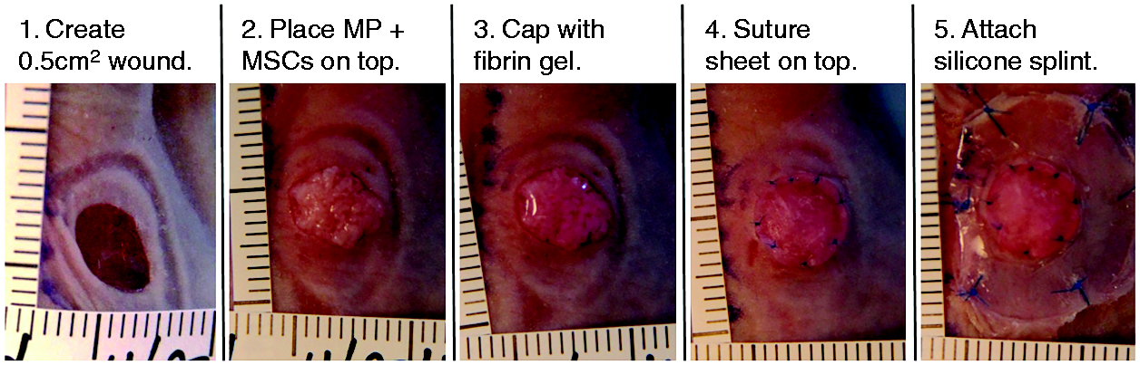
Step-by-step treatment of full-thickness excisional wound in murine model with MSC-seeded MP and fibrin gel. Each ruler graduation is equal to 1 mm.
A secondary dressing of xeroform petrolatum dressing and gauze was placed on top of the primary dressing and splints to keep the wounds moist. Then a butterfly harness (Lomir Biomedical, Malone, NY) was used to keep the dressings in place and intact on the mice. All scaffolds and splints were sterilized prior to use. Every three to four days post-injury, the secondary dressings were changed on the mice. The wounds were harvested at 7 days and 14 days post-injury.
Histological analysis of wounds
The harvested wounds were fixed in 10% phosphate-buffered formalin for 24 h, embedded in paraffin, and sectioned. Sections from all experimental groups at both time points were stained with hematoxylin and eosin (H&E) and Masson’s trichrome to qualitatively assess the wound healing. After deparaffinization and hydration, the sections were stained with routine protocols of H&E and Masson’s trichrome. Whole slide images of the stained sections were taken using a NanoZoomer (Hamamatsu, Bridgewater, NJ). These images were then used to quantify the quality of wound healing at 7 days and 14 days post-injury by 2 blinded reviewers using a scoring system to assess the epithelial maturation and dermal differentiation as defined by Sun et al. 27 The grading for epithelial maturation was as follows: 1, thin with no reticulation; 2, occasional reticulation; 3, moderate reticulation; and 4, thick and with complex reticulation. For dermal differentiation, the grading was as follows: 1, thin, dense, and monotonous fibrosis; 2, thicker but still dense and monotonous fibrosis; 3, two layer but not completely discrete; and 4, two discrete layers of papillary, superficial fibrosis, and reticular, loose alveolar tissue within the deep layer dermis.
Statistical analysis
Results are expressed as mean ± standard deviation. For each density of microparticles in gel, the migration distances for all the three gel concentrations at each time point were compared using the ANOVA with post hoc Tukey test. The MP-to-MP and MP-to-free space migration distances were compared using a Student’s t-test with unpaired data and unequal variance. The 4-point histology scores of epithelial maturation and dermal differentiation were compared using the ANOVA with post hoc Tukey test. The level of significance was set at p < 0.05 in all statistical tests. GraphPad Prism (San Diego, CA) was used for all statistical tests and graphs.
Results
Seeding efficiency
The average seeding efficiency of MSCs on ECM microparticles (MPs) in a 60-min seeding time was greater than 90% adherence for densities larger than 5 × 10⁵ cells on 10 mg MP, as shown in Figure 2(a). The initial seeding density of 1 × 10⁶ cells on 10 mg MP resulted in the highest attachment percentage of 97%. At this ideal seeding density, there was excellent viability and coverage of the cells on the scaffold after 7 and 14 days of in vitro culture (Figure 2(b) and (c)).
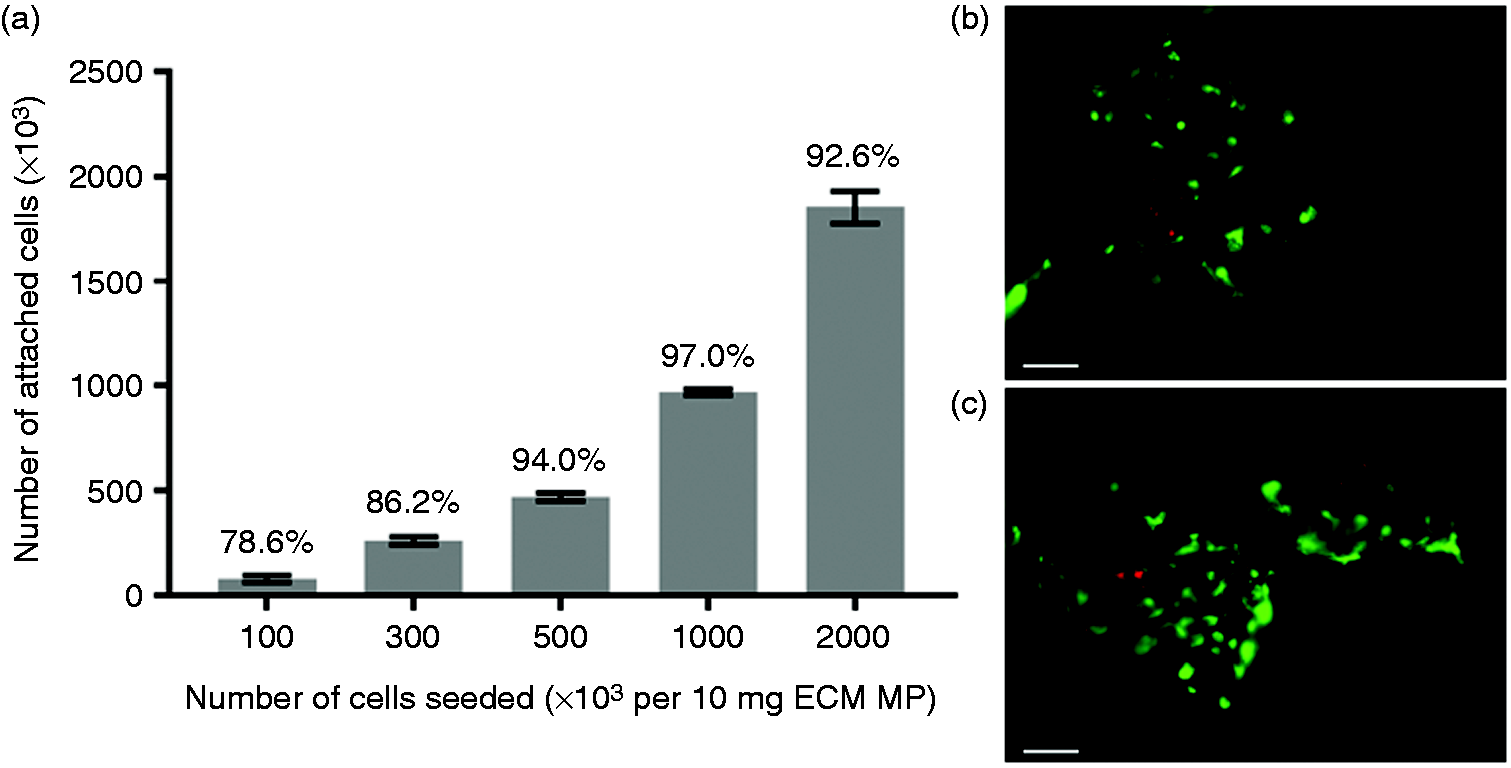
Evaluation of seeding and culture of cells on collagen MP. Seeding efficiency of hbMSCs on ECM MP for increasing cell number (a). At an initial seeding density of 1 × 10⁶ MSCs per 10 mg MP, the highest seeding efficiency showed excellent cell viability and coverage of the MP at 7 days (b) and 14 days (c) in live–dead images. There is minimal autofluorescence from the MP. Error bars = SD. Scale bars = 100 µm.
Migration of MSCs off the microparticles
Migration of MSCs from ECM microparticles in fibrin gels began in less than 24 h and steadily increased over time for both densities of 5 mg (low) and 10 mg (high) of MP per mL of gel (Figure 3). The average MSC migration distance increased with decreasing fibrinogen and thrombin concentrations. For the low density, the 2.5 mg/mL fibrin gel migration distance was statistically greater than the other gel concentrations at day 1 and statistically greater than the 10 mg/mL gel at day 14. At the high density, the migration distances of the 10 mg/ml fibrin gel concentration were statistically lower than 2.5 mg/mL fibrin gel at day 1 and day 5 and significantly lower than 5 mg/mL fibrin gel at day 5.
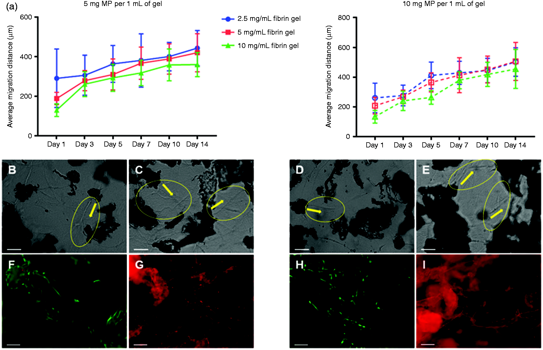
Cell migration of MSCs off cell-seeded MP. Average migration distances of MSCs off ECM MP in varying concentrations of fibrin gel increased over 14 days for both densities of 5 mg MP per 1 mL of gel and 10 mg MP per 1 mL of gel (a). Light microscopy images of MSCs migrating in 10 mg/mL fibrin gel show more cells migrating at 14 days (c, e) compared to 5 days (b, d) for both densities. MP is seen as dark clumps, yellow circles highlight the areas of visible cell migration, and yellow arrows denote the direction of cell migration off the MP. After 14 days in culture, live–dead (f, h) and F-actin (g, i) staining showed excellent MSC viability and extensive cytoskeleton structure between the microparticles in 10 mg/mL fibrin gel for both densities. MP autofluoresces slightly, and cell migration is highlighted by green, viable cells across MP and bright red, F-actin lines between MP. Error bars = SD. Scale bars = 100 µm.
The light microscopy images at 5 days (Figure 3(b) and (d)) and 14 days (Figure 3(c) and (e) highlighted the increasing number of MSCs migrating and the longer migration distances with continued in vitro culture. There was excellent viability of the cells encapsulating the microparticles (Figure 3(f) and (h). The positive F-actin stain showed extensive cytoskeleton structure of thin, parallel filament bundles indicative of undifferentiated MSCs and evidence of cell movement between the microparticles (Figure 3(g) and (i)). 28 The live–dead stain of the TCP control showed alive, healthy MSCs after 14 days of culture (results not shown).
There were statistically significant differences in the direction of MSC migration (Figure 4). At the lower density of 5 mg MP per 1 mL of fibrin gel, the cells in 2.5 mg/mL and 5 mg/mL fibrin gels migrated significantly farther distances toward other microparticles than into free space. At the higher density of 10 mg MP per 1 mL of fibrin gel, cells in all three fibrin gel concentrations migrated significantly longer distances toward other scaffolds than toward empty space.
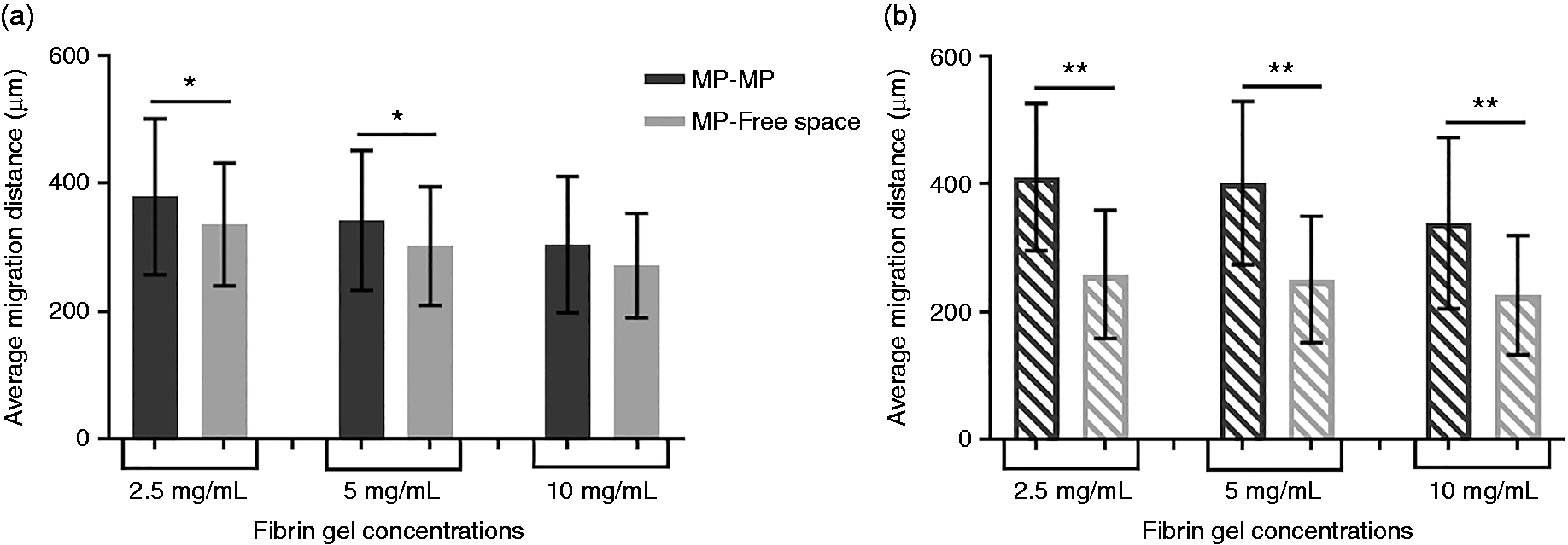
Comparison of MP-to-MP and MP-to-free space migration of cells off ECM microparticles (MP) in fibrin gels. At a density of 5 mg MP per 1 mL of fibrin gel (a) and at a density of 10 mg MP per 1 mL of fibrin gel (b), all but one of the fibrin gel concentrations resulted in MSCs migrating a significantly farther distance toward other microparticles than into free space. Migration distances are averaged across 14 days of culture. Error bars = SD. (*p < 0.01, **p<0.0001)
Histological analysis of full-thickness cutaneous wounds
The splinting of the full-thickness cutaneous wounds was successful in preventing wound contracture and allowing the wounds to heal through epithelialization and granulation (Figure 5(a)). At seven days post-injury, it was challenging to see epithelialization in the microparticle groups because the scaffold was in the wound bed but a thick dermal layer was present. As seen in the H&E images of Figure 5(a), there was increased cell infiltration in the MSC-seeded MP group and signs of epithelialization in the sheet-only group. There was moderate epithelial maturation of the control wounds at seven days, which was statistically higher than the three groups containing microparticles as shown in Figure 5(b). The dermal layer of the control wounds was statistically less developed than the two microparticle groups and the sheet-only group was significantly lower than the MSC-seeded MP group at seven days post-injury (Figure 5(c)).
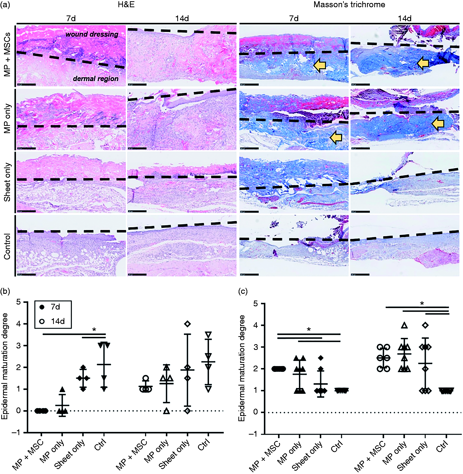
Histological evaluation of full-thickness excisional wounds in murine model using H&E and Masson’s trichrome staining (a). Black dotted lines approximate the top of the epidermis and interface between the wound and treatment; thus, above the line is the wound dressing and below the line is the dermal region. In the MSCs-seeded MP group, there was increased cell infiltration and a thick dermal layer present at 7 days post-injury and by 14 days, there was evidence of epidermal and dermal maturation. Yellow arrows denote the thicker dermal region present in all groups with MP. Scale bars = 250 µm H&E, 500 µm trichrome. Using a graded scoring system, dot plots of the epidermal maturation (b) show that the degree of epithelialization varies widely both within and between groups. The degree of dermal differentiation (c) at 7 days and 14 days post-injury was significantly higher in groups incorporating MP compared to control wounds. *p < 0.05.
By 14 days, the microparticles and sheets began to lift off the wound beds and there was epidermal maturation in all groups (Figure 5(a)). Although some microparticles were still embedded in the wound bed of both MP groups, the edges of those wounds consistently showed remodeling of the microparticles and differentiation into the two dermal layers. This is demonstrated by the increased dermal differentiation degree of both MP groups compared to seven days. All groups incorporating an ECM scaffold had a statistically higher dermal differentiation degree compared to control wounds at 14 days (Figure 5(c)).
Discussion
A number of clinical applications could utilize the submillimeter ECM particles seeded with MSCs as a cell delivery or paracrine therapy. Because of the increased number of exposed ECM proteins and large surface area compared to sheet materials, a high number of cells can be incorporated in a small volume. 16 In this study, we have developed a potential application of microparticles seeded with MSCs for use as a cell delivery and paracrine therapy.
For the potential clinical application of this technology, it is important to understand the cellular capacity of the ECM microparticles and the ideal gel concentration to allow cell migration off the scaffold while maintaining its adhesive strength. An initial seeding density of 1 × 10⁶ cells per 10 mg of microparticles appears to be the ideal density to achieve excellent cellular attachment in 60 min and nearly 100% confluence at seven days in vitro (Figure 2). Fibrin gels are routinely used clinically at a concentration of 40 mg/mL fibrinogen; however, pilot work at this concentration showed that it was too high for efficient migration of cells (not shown). The lower fibrin gel concentrations allowed steadily increasing migration of MSCs off the ECM microparticles over 14 days (Figure 3). Although the migration distances of the 2.5 mg/mL fibrin gel were significantly improved compared to the other concentrations at a few of the earlier time points, this inconsistent observation may not offset the lower adhesive strength of the gel. Therefore, a 5 to 10 mg/mL fibrin gel may be the best concentration to maintain glue adhesion and allow cell migration.
The advantage of this combined therapeutic is that the cell-seeded microparticles provide cells and paracrine factors to aid in the healing process, while being protected by the fibrin gel, as well as the mechanical strength and support from both the fibrin gel and the cells migrating through the gel.18,19 Therefore, it may be advantageous to utilize the higher density of microparticles and increase the number of cells delivered to significantly improve the paracrine effect in vivo. A 10 mg/mL fibrin gel with 10 mg/ml concentration of seeded microparticles may give the optimal balance of fibrin gel robustness, paracrine delivery, and potential for cell delivery to the adjacent tissue from cells migrating through the gel.
Another important aspect of the seeded microparticle technology is the cytokine gradient established within the system, both within the gel as well as between the cells. As seen in Figure 4, there was a significantly greater MSC migration observed between the microparticles as opposed to off microparticles and into free space. The higher density of microparticles in fibrin gel, which implies a higher density of cells, showed statistically greater migration differences in all gel concentrations Figure 4(b). This difference in cells migrating toward each other could be due to the paracrine effect of cells on each other due to the stimulus of a cytokine gradient established in the gel.
The in vivo pilot study investigating the use of cell-seeded microparticles for full-thickness cutaneous wounds showed promising results. The 10 mg/mL fibrin gel concentration used in the mouse study was based on the results of the in vitro migration work, and 10 mg of microparticles were placed on each wound to deliver approximately 1 × 10⁶ cells for the paracrine effect. It was also a reasonable amount of microparticles given the area of the wound. The MSC-seeded MP-treated wounds showed increased cell infiltration and increased dermal thickness at 7 and 14 days. Although the microparticle only group also showed increased dermal thickness compared to control wounds, there was no evidence of the same cell infiltration seen in the MSC MP groups. The improved collagen layer in MP groups was quantified in significantly higher degrees of dermal differentiation compared to control wounds (Figure 5(c)).
Given the degree of cell infiltration and robust collagen deposition seen in the MSC-seeded MP group, it is likely that a lower dose of paracrine factors would result in a more physiologic amount of wound healing. This study did not look at a dose response for the MSC-seeded microparticles, but this can be addressed in the future with titration of the overall cell dose. Development of cell-seeded microparticles as a therapeutic will also be aided by tracking the MSCs during healing as well as quantifying the inflammatory cell response in future studies. Additionally, there may be an advantage in washing the microparticles off the wound after several days, once their biologic impact is less, to prevent the microparticles from incorporating into the healing wound or impairing epithelialization. This therapeutic system has the flexibility to tailor the overall cell dose, microparticle dose and microparticle concentration within the fibrin gel to each specific clinical application.
There are a number of clinical scenarios including traumatic injuries, wounds of diabetes and peripheral vascular disease, chronic surgical wounds and high risk surgical anastomoses such as the airway and gastrointestinal which may benefit from a technology which stimulates robust collagen production and vascularization to aid in wound healing. The application of MSCs can aid in providing early ECM and promoting the growth and migration of adjacent cells to augment healing as has been shown in vivo 13 and clinically. 29 An acellular microparticle dressing was shown to improve the wound healing in a clinical case series. 20 The preliminary results of our in vivo study combining these two therapies – MSCs and microparticles – with fibrin gel show robust collagen deposition compared to no treatment and this may exhibit additional advantages at longer time points and in impaired healing environments.
In future studies, it will be important to determine the ideal treatment protocol as well as to better understand the mechanisms of dermal differentiation and epithelialization maturation. A treatment time study will explore the preferred duration of microparticles on the wound to aid in healing without impairing the epithelialization. It will also be important to better understand the inflammatory response of the technology through immunohistochemistry and to examine the dosing of MSCs to find the ideal concentration of MSCs that maximizes the paracrine response without depositing a detrimental abundance of collagen. Additionally, a future aim is to optimize the delivery of paracrine factors to aid in balanced dermal differentiation and epithelialization maturation. Evaluation of the paracrine response of MSCs on ECM over time and comparing to known cytokine responses in wound healing will allow us to better design the dose and time of biologic treatment.
Clinical translation of this novel technology presents numerous challenges. The addition of a biologic component to a material increases the regulatory requirements. Additionally, the cost is increased because culturing MSCs on the microparticles prior to application requires Good Manufacturing Practice certified facilities. However, a potential method to eliminate that increased cost would be to isolate mononuclear fraction (MNF) from freshly aspirated autologous bone marrow and seed the MNF on the microparticles in the operating room. The development of new MNF isolation techniques makes this is a promising future clinical scenario. 30
In conclusion, this study demonstrated the viability of seeding bone marrow-derived MSCs on microparticles of decellularized ECM material. There is a robust migration of cells off these microparticles when encapsulated in fibrin gel, and an in vivo pilot study exhibited a large collagen deposition in full-thickness cutaneous wounds treated with MSC-encapsulated microparticles. Future murine in vivo studies will explore longer time points of splinted wounds and the effect on diabetic wounds as continued work toward the clinical application.
Footnotes
Acknowledgements
The authors thank Tricia Della Pelle and the MGH Histopathology Research Core for their work in histological processing.
Declaration of Conflicting Interests
The author(s) declared the following potential conflicts of interest with respect to the research, authorship, and/or publication of this article: Gino Bradica and Scott Goldman are employees of DSM Biomedical, which manufactures the decellularized biomaterial used in this work. David Hoganson received research support and was a consultant for DSM Biomedical. Funding for this project was provided by DSM Biomedical. DSM Biomedical had no role in the study design, interpretation of data, writing of the report, or decision to submit the paper for publication.
Funding
The author(s) received no financial support for the research, authorship, and/or publication of this article.