
Abstract
Highly uniform silicon nanopore membranes were developed for applications in implantable bioartificial organs. A robust, readily scalable, non-fouling surface coating is required to enhance silicon nanopore membrane hemocompatibility. However, the coating must be ultrathin to keep the nanopores from occluding. Recently, zwitterionic brush polymers have demonstrated significantly lower fouling under biological conditions. In this study, we explore ultrathin zwitterionic poly(sulfobetaine methacrylate) (pSBMA) surface coating at sub-5 nm thickness. Membrane hydraulic permeability was measured before and after surface modification of silicon nanopore membranes, and pores were found to be patent and in agreement with coating thickness measurements. Coating stability was analyzed under biological shear as well as under blood flow in vitro and in vivo. Following exposure to shear over 24 h, coatings were characterized via X-ray photoelectron spectroscopy, goniometry, and ellipsometry, and found to survive biological shear. In vitro blood experiments with fresh human blood as well as in vivo 7-day and 26-day implants in a porcine model demonstrate minimal platelet adhesion and activation with pSBMA surface modification compared to unmodified silicon exposed to fresh human blood in vitro. These results demonstrate that ultrathin pSBMA surface modification is a viable choice for application in blood contacting implants with critical nanoscale features.
Keywords
Introduction
Silicon-based bio-microelectromechanical systems (bioMEMS) are commonly utilized in a diverse set of implantable medical devices, such as neuroelectrodes,1,2 drug delivery systems,3–5 biosensors, and diagnostic devices.6–8 One such bioMEMS component is the silicon nanopore membrane (SNM), which offers highly uniform and controllable pores with variation <1% across the wafer. 9 As shown in Figure 1, low resistance SNMs with ∼10 nm pore size have been developed for blood filtration and immunoisolation applications in renal replacement9–11 and islet therapies.12,13 These applications bring silicon in direct contact with blood flow, and therefore, biocompatibility and resistance to fouling is critical. Previous research has shown silicon to be non-cytotoxic.14,15 Additionally, various materials pertinent to implantable bioMEMS including silicon, polysilicon, and silicon dioxide have been tested against a battery of tests laid out in ISO 10993 and were found to be non-leaching and non-irritant. 16 However, the blood-contacting surfaces must also be non-activating and non-fouling for long-term functionality under blood flow.
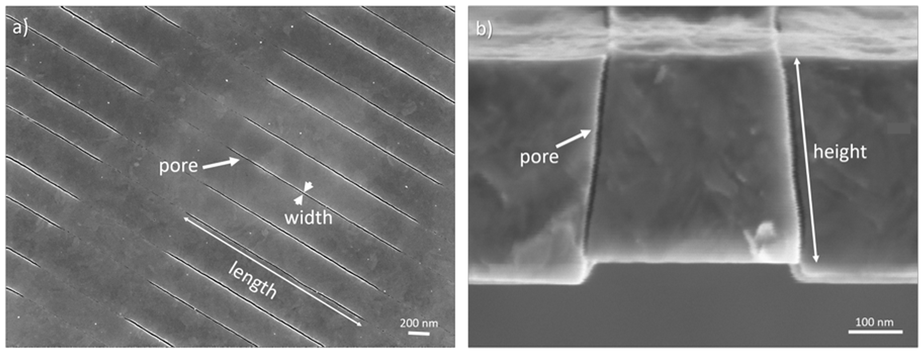
(a) Top view and (b) side view of silicon nanopore membrane (SNM) with pore length of 2 μm, width of 7 nm, and height of 360 nm. Figure is reproduced with permission from Iqbal et al., 17 ©Wiley Periodicals, Inc.
To improve hemocompatibility of a substrate, the surface is often modified. 18 Among surface coatings, hydrophilic, polymeric brush structures on the surface have shown enhanced hemocompatibility. One such coating is polyethylene glycol (PEG) and oligoethylene glycol, respectively, which has demonstrated excellent resistance to protein adsorption and platelet adhesion.19,20 However, PEG is known to degrade under biological conditions,21,22 making it impractical for long-term implants. Recently, hydrophilic zwitterionic polymer surface modifications have demonstrated significantly reduced protein fouling23–27 and thrombosis formation.28–30 Several biomimetic zwitterions, such as sulfobetaine, carboxybetaine, and phosphorylcholine, have been used to produce polymer brushes and hydrogels.30–32 It is hypothesized that the zwitterions coordinate water molecules and produce a stable hydration layer due to its hydrogen bonding and electrostatic interactions.29,31 This hydration layer allows the surface to be in stealth mode and reduces protein adsorption and cell adhesion.
Use of sulfobetaine and its derivatives is increasing due to its low cost and ease of production: its non-fouling properties have found application in the medical field in glucose biosensors, 33 and extracorporeal membrane oxygenation 34 as well as in general application such as reducing fouling in wastewater treatment.35,36 Mimicking taurine, which is a compound commonly found in animal tissue and bile, poly(sulfobetaine methacrylate) (pSBMA) is non-cytotoxic 37 and has demonstrated ultra-low protein fouling (i.e. <0.3 ng/cm2 of fibrinogen) with a significant reduction in platelet adhesion and activation.28,31,38–44 Additionally, pSBMA is highly resistant to bacterial adhesion and biofilm formation.41,45–47 These properties make them well suited for implant applications.
Surfaces have been successfully modified with pSBMA using various methods ranging from atom transfer radical polymerization (ATRP),38,43,45,48 reverse ATRP, 35 plasma-induced polymerization 49 as well as simple grafting-to method via use of a catechol 41 or click chemistry. 50 Utilizing these methods, pSBMA has been applied to gold,40,45 graphene, 35 glass, 43 silica nanoparticles, 51 silicon,26,44 as well as other polymers such as PDMS,34,52,53 cellulose,38,42 polyurethane, 39 poly(vinylidene fluoride). 49
For use in implant application, long-term stability of the polymers is required. pSBMA was shown to be stable in vitro in various static aqueous solutions, such as phosphate buffered saline (PBS), human blood plasma, and distilled water over 28 days or less.33,37,39,44 In vitro experimentation with static fresh human blood over 2 h also demonstrate that pSBMA-coupled silicon significantly reduces platelet adhesion compared to silicon. 26 In vitro human blood flow experiments with sulfobetaine-modified vascular catheters showed a significant difference in blood activation after only 5 min of blood flow. 28 Hydrogels composed of pSBMA have been implanted subcutaneously in mice over four weeks, which resulted in a capsule thickness and foreign body giant cell count that is comparable to control poly(2-hydroxyethyl methacrylate) (polyHEMA) hydrogels. 37 Smith et al. also conducted in vivo experiments using sulfobetaine-modified catheters in canine model that shows significantly reduced thrombus formation. However, the experiments only spanned 4 h. Therefore, to our knowledge, there is very limited in vivo data of pSBMA performance under long-term blood flow conditions.
For surface modification on SNMs with nanoscale pores, ultrathin polymers are required. While zwitterionic hydrogels and polymer brushes have shown exceptional resistance to biological fouling, ultrathin polymers have not been well characterized. Therefore, the objectives of this study are to: (1) develop and characterize an ultrathin zwitterionic surface modification for application on SNMs for implant applications and (2) evaluate its performance in vitro and in vivo. Sub-5 nm pSBMA brushes were applied to solid silicon substrates and SNMs via ATRP. Subsequently, the ultrathin polymer-modified surfaces were characterized and their performance was evaluated. The modified substrates were characterized for elemental composition, coating thickness, changes in surface wettability and surface roughness, and hydraulic permeability. Additionally, coating survival after 24 h under varying shear rate was evaluated. Coating performance was also evaluated in vitro and in vivo. In vitro fouling was measured against single protein human serum albumin (HSA) and human fibrinogen. Modified surfaces were also subjected to fresh human blood flow for 2 h. The surfaces as well as the blood were examined for platelet activation using immunohistochemistry (IHC), scanning electron microscopy (SEM), and flow cytometry. pSBMA-modified silicon substrates were also placed in a porcine extracorporeal circuit for 6 h as well as in vivo for 7 and 26 days. All surfaces were analyzed for cellular adhesion and activation with IHC and SEM.
Materials and Methods
Surface modification
Synthesis of 2-bromo-2-methyl-N-3[(trimethoxysilyl)propyl]-propanamide
Unless otherwise stated, chemicals were purchased from Sigma-Aldrich (St Louis, MO, USA). ATRP initiator, 2-bromo-2-methyl-N-3[(trimethoxysilyl)propyl]-propanamide (BrTMOS) was synthesized as previously reported.25,44 Under nitrogen (N2) protection, 12 mmol of σ-bromoisobutyryl bromide (98%) was added dropwise to a mixture of 10 mmol (3-aminopropyl)trimethoxysilane (97%) and 10 mmol trimethylamine in 50 mL of anhydrous tetrahydrofuran (THF) over 30 min. The exothermic reaction was kept on ice to reduce over heating and allowed to continue overnight. After removing the precipitate, THF was evaporated from the filtrate using a rotary evaporator (Buchi, Flawil, Switzerland) and the concentrated oil was dissolved in 20 mL of dichloromethane. The solution was then washed with 10% potassium bisulfate (2 × 20 mL), cold deionized (DI) water (1 × 20 mL), and saturated sodium chloride solution (2 × 20 mL), respectively, using a separation funnel. Finally, the organic phase was collected and dried using anhydrous magnesium sulfate (MgSO4). After filtering out the MgSO4, dichloromethane was evaporated off, yielding the final product, BrTMOS. 1H NMR (400 MHz, CHCl3) was conducted on the colorless oil to verify BrTMOS formation: δ 6.90 (s, 1H, NH), 3.49 (s, 9H, SiOCH3), 3.26 (t, 2H, CH2N), 1.95 (s, 6H, CH3), 1.66 (m, 2H, CH2), 0.66 (t, 2H, SiCH2).
Sample preparation
Sulfuric acid (H2SO4, 96%), hydrogen peroxide (H2O2, 30%), and hydrofluoric acid (HF, 49%) were purchased from Avantor Performance Materials (Center Valley, PA, USA). To produce the solid silicon substrates, double-side-polished, 400 μm thick, p-type silicon wafers (Ultrasil Corporation, Hayward, CA, USA) were diced in 1 cm × 1 cm chips and 1 cm × 6.5 cm long chips. SNMs were fabricated following procedures previously described.9,54 All samples were rinsed with acetone, methanol, isopropanol, and water (1 × 5 min, each). To remove residual organics, the substrates were cleaned using a freshly made solution of “piranha”—a 3:1 ratio of H2SO4 to H2O2 for 20 min. After rinsing in DI water (2 × 10 min) all samples were placed in HF for 2 min to etch away silicon dioxide (SiO2, thermally grown to produce slit pores for SNMs). The substrates were rinsed again with DI water (3 × 10 min) and placed back in a fresh “piranha” solution for 20 min to activate the surface. Following a final rinse with DI water (3 × 10 min), all substrates were dried on the hot plate at 60°C for 1 h.
Surface polymerization
Substrates were prepared for ATRP by first initiating the surface with BrTMOS. All samples were placed in a petri dish with 1% (v/v) BrTMOS solution in bicyclohexyl. The solution was left stirring for 2 h, and then the substrates were rinsed twice each with chloroform, ethanol and water, respectively.
Brush zwitterionic pSBMA were formed by ATRP. A mixture of 5:5 mL of methanol to water was degassed by bubbling N2 gas for 10 min. Initially, 468 mg (3 mmol) of 2,2′-bipyridyl (BPY, ≥98%) and 1.06 g (3.8 mmol) [2-(methacryloyloxy)ethyl]dimethyl-(3-sulfopropyl)ammonium hydroxide (SBMA, 97%) was added to the mixture and thoroughly mixed. Then 22.3 mg (0.1 mmol) of copper (II) bromide (CuBr2, 99%) was added in and well mixed. The mixture was degassed again for 15 min. Four substrates were placed in the reaction chamber along with 143 mg (1 mmol) of copper (I) bromide (CuBr, 99.999%) under N2 protection. The mixture was then added to the reaction chamber, and the polymerization was allowed to run for 15 min for pSBMA. Following polymerization, the substrates were rinsed (3 × 5 min) with chloroform, ethanol, and Dulbecco’s phosphate buffered saline (D-PBS, without calcium and magnesium from Cell Culture Facility at University of California, San Francisco, CA, USA), respectively. The substrates were left in D-PBS overnight. They were rinsed with water (3 × 5 min) and dried under a stream of N2 gas.
Surface coating characterization
X-ray photoelectron spectroscopy
X-ray photoelectron spectroscopy (XPS) was conducted using a Surface Science Instruments S-probe spectrometer with a monochromatized Al Kα X-ray source (serviced by Service Physics, Bend OR, USA). Data were collected under a vacuum of pressure less than 5 × 10⁻⁹ torr, with an X-ray spot size of 800 μm. To calculate the composition, survey and detail spectra used a pass energy of 150 eV, while high resolution scans used a pass energy of 50 eV. The take-off angle was 0°, yielding a sampling depth of ∼10 nm. Data were analyzed using the Hawk Data Analysis Software (Service Physics). The binding energy scales were calibrated by setting C1s peak to 285.0 eV. Three measurements were taken per sample group for survey spectra, while one spot was analyzed for high resolution.
Contact angle
Static contact angle measurements were taken using Attension Theta Lite from Biolin Scientific, Stockholm, Sweden. A droplet size of ∼3.5 μL of water placed on the substrate and the angle between the droplet and substrate was measured in air over 10 s at 0.1 s interval. A minimum of three measurements were taken for each sample set.
Ellipsometry
Ellipsometry was conducted using Gaertner Stokes Ellipsometer using a 6328 Å HeNe laser at 70° incidence angle. Given the refractive index of 1.45, as was previously used for pSBMA coatings, 44 coating thickness was iteratively solved for by entering in measured reflection and transmission data into Fresnel equations. A minimum of three locations on individual substrate was collected and averaged over each sample set.
Atomic force microscopy
Atomic force microscopy (AFM) was conducted using NanoScope Scanning Probe Microscope (Bruker, Santa Barbara, CA, USA), running in tapping mode using a triangular ScanAsyst-fluid+ tip (Bruker) with a spring constant of 0.7 N/m. A scan rate of 0.977 Hz, with 512 samples/line sampling rate was used to generate AFM images. Different locations on each type of surface were scanned and the root mean square roughness (Rq) values were averaged over three 1 µm2 areas, as was reported in previous literature. 55 NanoScope Analysis Software (Bruker) was used to process images and determine roughness values.
Hydraulic permeability measurement
Surface modification was conducted on SNMs with porous area of 36 mm2 and change in pore size was calculated based on membrane permeability. Hydraulic permeability of the membrane was measured before and after surface modification using the setup shown in online Supplementary Figure S1. The SNM separates two chambers, and a gasket is used to ensure sealing between the feed and filtrate side. A peristaltic pump draws water from the reservoir to maintain a constant cross flow rate. A pressure gauge measures the pressure immediately before the flow reaches the manifold. Finally, a resistor is placed in series following the manifold to control the transmembrane pressure and the feed is returned to the reservoir. The filtrate is collected into an open syringe and the volume is measured.
The effective pore size given by the width (w) of the pores is calculated based on the following equation

In vitro protein adsorption (fibrinogen and albumin)
Protein adsorption on the surfaces of the substrates was determined by conducting enzyme-linked immunosorbent assay (ELISA) following protocol published previously.17,44 Substrates were placed in 24-well tissue culture polystyrene (TCPS) plates and incubated with D-PBS for 1.5 h. D-PBS was replaced with 0.5 mL of single protein solution: 1 mg/mL concentration of human fibrinogen (F3879, Sigma-Aldrich) or human albumin (AB19183, Abcam, Cambridge, MA, USA) was added and allowed to incubate at 37°C for 1.5 h. All substrates were rinsed five times with D-PBS. Surfaces were then blocked using bovine serum albumin (A7906, BSA, ≥98%, Sigma-Aldrich) using 1 mg/mL BSA solution for 1.5 h. The substrates were rinsed five times with D-PBS and transferred to a new 24-well TCPS well. Next, the samples were incubated with 10 μg/mL anti-human proteins conjugated with horseradish peroxidase (HRP) for 1.5 h: anti-human fibrinogen (coagulation factor I) (HRP) (F4200-15B, USBiological, Salem, MA, USA) for fibrinogen ELISA and anti-HSA antibody-HRP (AB19183, Abcam) for albumin ELISA. The substrates were washed with D-PBS five times and transferred to a new well. A reaction mixture was made in 0.05 M citrate phosphate buffer (2851, Sigma-Aldrich, pH 5.0) with 0.5 mg/mL of o-phenylenediamine (OPD, 0688 Amresco, VWR Inc., Visalia, CA, USA) and 0.03% H2O2. Reaction mixture (0.5 mL) was added to each well and the reaction was allowed to run at 37°C for 20 min. The reaction was stopped by adding 0.5 mL H2SO4 (1 M). The solution was transferred to 96-well plates, and light absorbance at 490 nm was determined using a microplate reader. A minimum of three substrates were analyzed for each sample set, and light absorbance reading was normalized to TCPS control.
Experimental setup for studying effect of shear stress on surface coating
Stability of pSBMA polymer surface modification was monitored over varying shear rates. pSBMA-silicon substrates were prepared as described above. Shear flow cells (online Supplementary Figure S2) were designed to hold seven chips on the bottom piece, and inlet and outlet through-holes on the top piece. A gasket is used to create the flow path and set the channel height. The shear rates in human arterioles do not exceed 1650/s.
57
Therefore, for our experiments, we explored shear rates of 500/s, 1000/s, 1500/s, and 2000/s to cover the range of shear rates experienced in the human body. The substrates were exposed to D-PBS flow at 37°C for 24 h at varying flow rates. A constant channel height of 220 μm was used, and flow rate was varied to adjust the shear rate based on the following equation
Therefore, the flow rate was set to 1.6, 3.2, 4.8, and 6.4 mL/min to achieve a shear rate of 500, 1000, 1500, and 2000/s. Samples that were not exposed to D-PBS were used as controls for comparison. Following flow exposure, the samples were removed from the flow cells, and surface chemistry, contact angle, and coating thickness were analyzed. Coating performance following exposure to flow was evaluated via human albumin ELISA.
Setup for human blood flow experiments
Blood collection
Fresh blood was collected from a healthy human donor in 10 mL BD Vacutainers with 15.8 USP/mL lithium heparin (Thermo Fisher Scientific, Waltham, MA, USA). Informed consent was obtained prior to blood collection and experiments. Blood was stored on ice for approximately 15 min until use in experiments.
Blood flow experimental setup
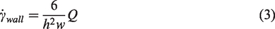
An in vitro blood flow platform was designed and fabricated out of polyether ether ketone (PEEK) as shown in Figure 2(a) and (b). Blood enters through the top PEEK piece inlet and flow develops guided by 500 μm thick silicone gasket 1. Developed blood flow enters through the slit in middle PEEK piece, and the test substrate is exposed to the blood flow. Blood then leaves out the slit in middle PEEK piece and out the outlet of the top PEEK piece. This system creates a blood flow channel height of 200 μm over the test substrate. As shown in Figure 2(c), a peristaltic pump drives the blood flow at a rate of 4 mL/min, yielding a shear rate of 1000/s. For this setup, no reservoir was used in order to avoid stagnant blood. Rather 100 cm of Masterflex, Tygon E-LFL L/S 14 Tubing (1.6 mm inner diameter tubing from Cole-Parmer, Vernon Hills, IL, USA) was used to connect the inlet and outlet of the blood flow platform. Prior to blood experiment, the tubing and experimental setup was circulated with D-PBS containing 50 USP/mL heparin for 30 min. The setup was then washed out with D-PBS and circulated with D-PBS for 30 min to remove residual heparin. The blood flow experiment was then conducted in a 37°C, 5% CO2 incubator for 2 h.
In vitro blood flow platform (a) exploded view, showing the top PEEK piece containing the inlet and outlet, silicone gasket 1, developing uniform blood flow and directing the blood flow through the slits in middle PEEK piece. Substrate surface is exposed to laminar blood flow with a channel height of 200 μm, and blood is directed out of the slit in middle PEEK piece and out the outlet on top PEEK piece. Silicone gasket 2 and bottom PEEK piece supports the substrate. (b) Cross-section showing blood flow path through device. (c) Setup of 2-h fresh human blood flow study in a 37°C incubator.
Post analysis following blood flow experiments
Following blood experiments, the setup was run with D-PBS until no trace of blood was detected in the tubing. The in vitro platforms were then taken apart, and the blood channels were analyzed for gross clots. The 1 × 6.5 cm2 substrate was rinsed three times with D-PBS and broken into ∼1 cm long pieces for surface analysis for cellular adhesion using SEM and platelet adhesion and activation using IHC.
SEM: Cellular adhesion was visualized using SEM. Samples were fixed by being placed in 3% glutaraldehyde (Sigma-Aldrich), 0.1 M sodium cacodylate (VWR) and 0.1 M sucrose solution (Sigma-Aldrich) for 48 h. Then, the substrates were removed and washed two times with deionized water and dehydrated by incubating in ethanol/water (v/v) solution for 10 min in each: 35%, 50%, 70%, 95% and two times in 100%. Samples were allowed to dry overnight and mounted on aluminum stubs for imaging using a Carl Zeiss Ultra 55 Field Emission Scanning Electron Microscope (Zeiss, Dublin, CA, USA). Prior to imaging, samples were sputter coated with gold-palladium.
IHC: IHC samples were fixed with 4% paraformaldehyde (Thermo Fisher Scientific) at room temperature for 30 min and allowed to remain in 1% paraformaldehyde at 4°C until they were to be imaged (∼1 week). Before imaging, the samples were marked for human platelets and platelet activation using FITC-labeled anti-CD41 (bs-2636R-fitc, Bioss Inc., Woburn, MA, USA) and Cy3-labeled anti-CD62p (bs-0561R-cy3, Bioss Inc.) markers, respectively. Samples were rinsed three times in D-PBS and imaging was conducted using a Nikon TI-E Microscope (Nikon Instruments, Melville, NY, USA).
Extracorporeal study
The methods used were approved by the University of California, San Francisco Institutional Animal Care and Use Committee, and conducted at the UCSF Laboratory Animal Resource Center. Tunneled catheters were placed in the carotid artery and jugular vein of a 55 kg Yorkshire female pig, and a closed external circuitry was set up. A custom designed, flow path optimized titanium housing containing separated blood flow chambers, 58 each holding 18 1 × 1 cm2 solid pSBMA-silicon chips was utilized for ex vivo blood flow experiments. Blood flowed from the carotid artery through Masterfelx, Tygon E-LFL Tubing (L/S 25, 4.8 mm inner diameter from Cole-Parmer) to the extracorporeal device, then back through Tygon tubing to the catheter connected to the jugular vein. The pig was given 200 U/kg heparin, and a bolus of heparin was given throughout the experiment in order to maintain a targeted activated clotting time of ∼300 s. The pig was also given aspirin and clopidogrel prior to the experiment. Before connecting the device to the pig, the whole system was disinfected with chlorohexadine. The system was then flushed and primed with heparinized saline. The flow experiment was allowed to run for 6 h, at flow rate generated by the heart, which was measured to be ∼60 mL/min. After 6 h, saline was flown through the device to remove the blood. The device was taken apart, and analyzed for gross blood clots. Single 1 cm × 1 cm substrates were removed and prepared for SEM and IHC imaging to analysis of platelet adhesion and activation as described above. For IHC, markers sensitive to porcine platelets were used: FITC-labeled anti-CD41 (orb181793, Biorbyt, Berkeley, CA, USA) and Cy3-labeled anti-CD62p (bs-0561R-cy3, Bioss Inc.). Samples were rinsed three times in D-PBS, and imaging was conducted using a Nikon TI-E Microscope.
Implant studies
Two implant studies were conducted in porcine model. The implant (Figure 3) holds four pSBMA coated 6.5 cm × 1 cm solid silicon chips. The device was implanted intravascularly in two juvenile Yucatan minipigs for durations of 7-, and 26-days, respectively in order to allow continuous exposure to flowing blood. Specifically, the device was implanted subcutaneously in a dorsal–lateral cervical position. Six-millimeter expanded polytetrafluoroethylene (ePTFE) vascular grafts were anastomosed to the external jugular vein and common carotid artery, then tunneled and attached to the device, establishing an arteriovenous shunt across the device.
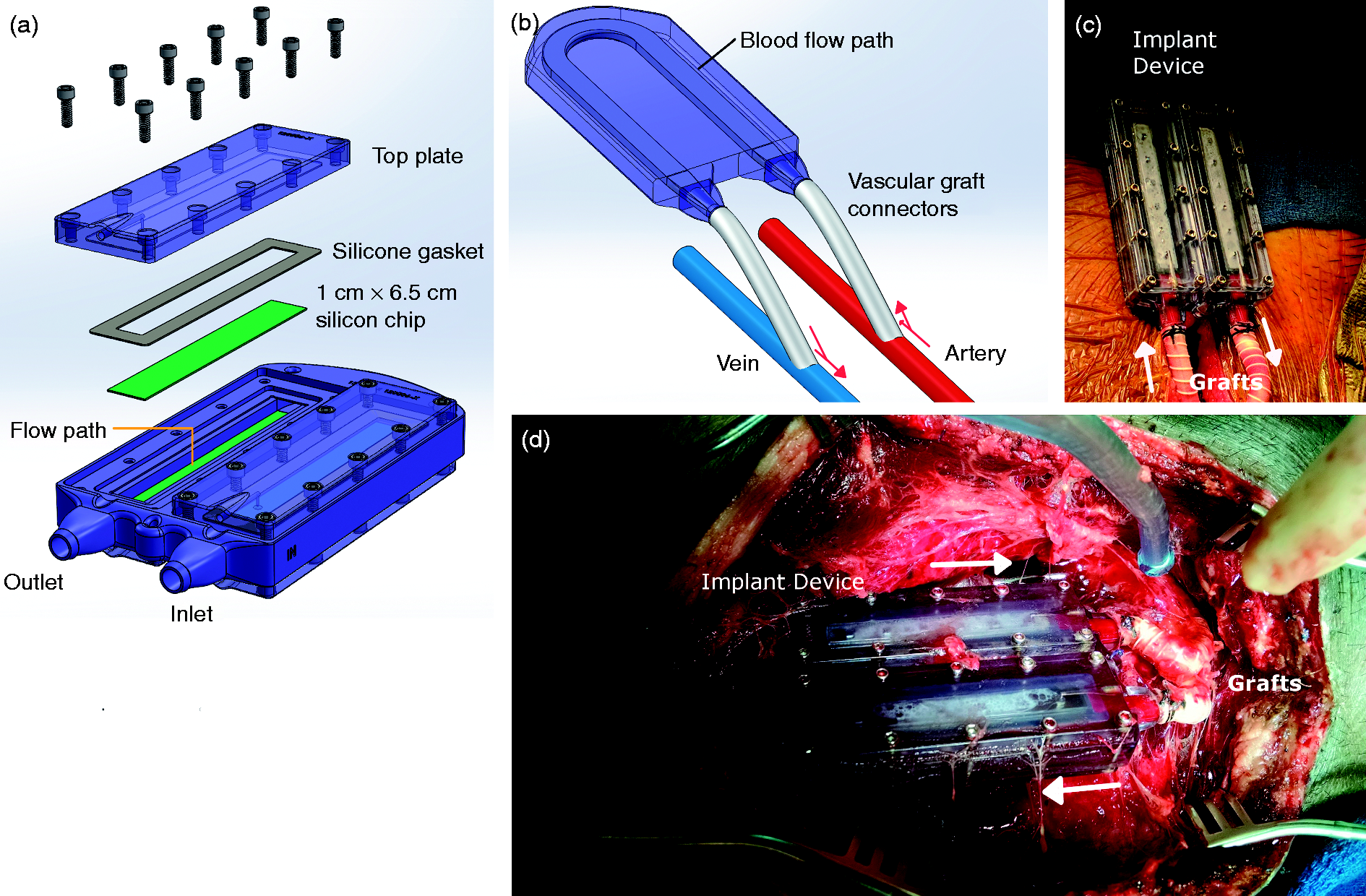
Implant study conducted with porcine model: (a) Exploded view of implant, showing the top and bottom of the flow channel is created by 1 cm x 6.5 cm silicon chips. A silicone gasket is used to form a watertight seal around the chip, which is held down by the top plate. (b) Blood flow path and vascular graft connection to the artery (red) and vein (blue). Red arrows indicate the direction of blood flow. (c) Implant device (day 0) connected to the carotid artery and external jugular vein via 6 mm ePTFE grafts. (d) Patent implant device (day 7) during explantation. Blood flow direction is indicated using white arrows.
After surgical implantation and acute recovery, swine were monitored for behavior, appetite, activity, and responsiveness twice daily. Daily hematologic assessment included complete blood count, serum C-reactive protein (CRP), serum lactate dehydrogenase, and plasma free hemoglobin. Blood flow patency was assessed daily through evaluation for a thrill and/or bruit. Additionally, Doppler ultrasonography was performed once weekly and fluoroscopic angiography performed at 7-days and prior to device removal. The device was recovered after 7- and 26-days via a terminal re-look procedure. After device removal, animals were euthanized. All animal procedures were performed at a contract research organization (PMI, San Carlos, CA), and underwent IACUC review and approval.
Results and discussion
Surface coating characterization
XPS, goniometry, and ellipsometry results (Figure 4) demonstrates the successful polymerization of pSBMA on the silicon surface. With BrTMOS coupled to silicon, C 1s (∼285 eV) concentration increased 3.1% and Si 2p (100 eV) concentration decreased 3.5%. There was also 1.1% and 0.6% increase in N 1s (∼400 eV) and Br 3d (∼69 eV), respectively, which are signature elements of BrTMOS, indicating presence of BrTMOS on the silicon surface. Based on stoichiometric ratio, 1:1 ratio of Br to N is expected. XPS data, however, show a ratio of 1.8:1. This trend has been seen previously and may be explained by the instability of C-Br bond under XPS conditions. 25
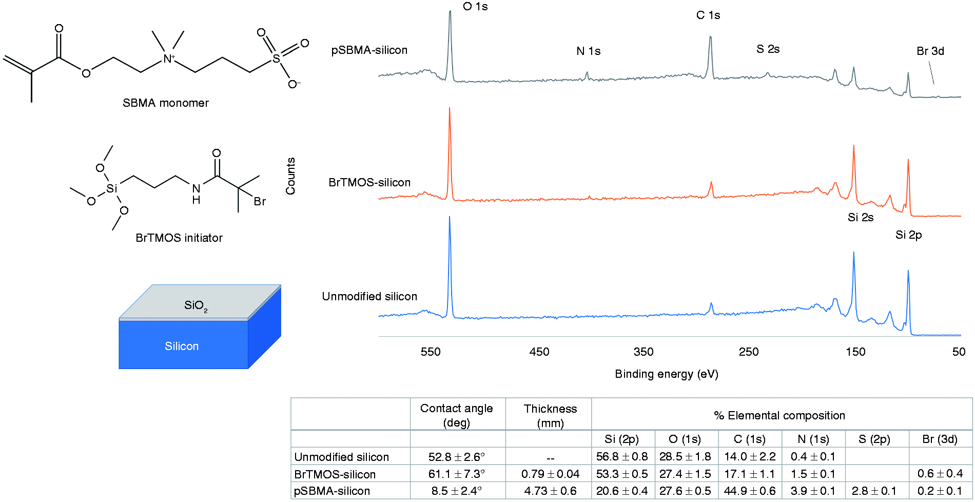
Chemical structure of SBMA monomer and BrTMOS initiator and XPS survey spectra of silicon, BrTMOS-treated silicon, and pSBMA grafted on silicon. Table below reports the contact angle, coating thickness, and surface elemental composition of unmodified silicon, BrTMOS-silicon, and pSBMA-silicon.
With pSBMA polymerized on silicon, there was a 27.8% increase in C 1s and 32.7% decrease in Si 2p, demonstrating presentation of carbon-based polymer in the top 10 nm of the substrate. Additionally, presence of signature elements such as N 1s (2.4% increase) and S 2p (2.8% increase) in a ratio of ∼1:1, matches the stoichiometric ratio in the monomer SBMA.
Other surface characteristics, such as water contact angle and coating thickness, also reaffirm the presence of pSBMA. Average contact angle of pSBMA-silicon was 8.5°, which is in agreement with previous literature.24,44 Total coating thickness of BrTMOS and pSBMA on the surface was 4.73 nm, which is sub-5 nm, as desired.
Topography is a critical factor in surface characteristics and non-fouling properties. Because higher surface roughness has been previously linked to increased protein adsorption, 59 lower surface roughness is preferred. Figure 5 shows a representative 1 µm2 area of silicon substrates in two-dimensional (2D) and three-dimensional (3D) images. Unmodified silicon, BrTMOS-silicon, and pSBMA-silicon have a surface roughness, Rq, of 0.54 ± 0.03 nm, 0.52 ± 0.02 nm, and 0.54 ± 0.10 nm, respectively. There are key visual differences among the three surfaces: Unmodified silicon is fairly smooth, with uniformly distributed nanocavities, and its roughness is on the same scale as noted in previous literature. 55 With unmodified silicon as the baseline, BrTMOS-silicon features are slightly smaller and seems to reduce the size of nanocavities found on bare silicon. Additionally, there are also an uneven distribution of tight peaks, typically on the scale of sub-10 nm. However, on average, surface roughness did not change significantly.
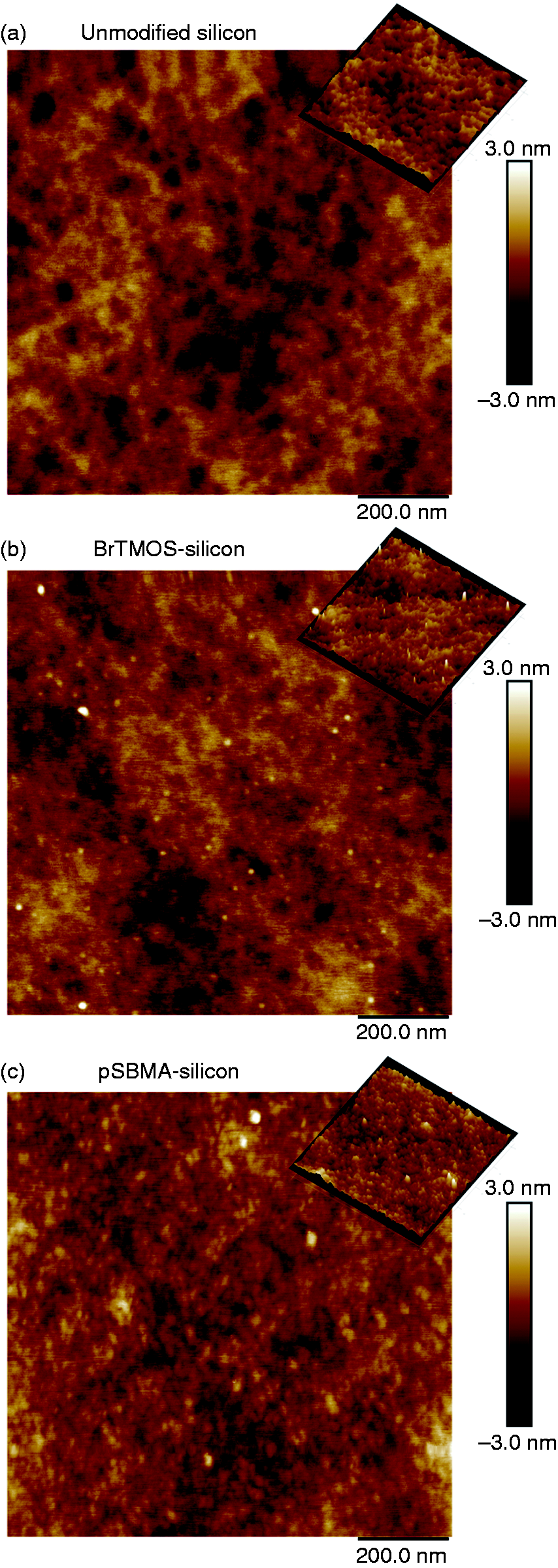
Two-dimensional AFM images (3D image shown in inset): (a) unmodified silicon surface (Rq = 0.54 ± 0.03 nm); (b) BrTMOS-silicon (Rq = 0.52 ± 0.02 nm); and (c) pSBMA-silicon (Rq = 0.54 ± 0.10 nm).
Based on known surface features and experimentally measured hydraulic permeability, the change in pore width was calculated and found to match with ellipsometry data. Ellipsometry of pSBMA surface showed a thickness of 4.37 nm.
With pSBMA modification, the tight peaks present on BrTMOS-silicon surfaces are no longer visible. Rather, they are replaced by nanoscale broad peaks on the scale of 25 nm. Since AFM was conducted in dry, ambient conditions, these mushroom-like features could be formed by the interaction between adjacent polymer chains or polymer chains curling in on themselves. Nonetheless, overall surface roughness did not significantly change from unmodified silicon, indicating that the surfaces remain smooth after pSBMA modification. Moreover, the standard deviation in Rq is low. These properties suggest that the uniformity of SNMs is maintained following surface modification.
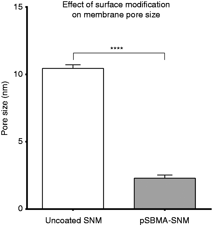
As presented in Figure 6, The change in effective pore size due to surface modification was calculated based on the hydraulic permeability. Unmodified SNMs have a pore size of ∼10.5 nm. Following pSBMA-coupling, pore size of membranes decreased by 8.2 nm. Assuming conformal coating, this implies a coating thickness of ∼4.1 nm inside the pores. Under the same experimental conditions, pSBMA thickness on solid silicon chips was measured to be ∼4.4 nm, and the discrepancy of ∼0.3 nm is within experimental error.
In vitro protein adsorption (fibrinogen and albumin)
Preliminary protein fouling from single protein solutions of HSA and human fibrinogen was assessed. HSA was chosen as model protein for non-specific surface binding, while fibrinogen was chosen for its role in thrombus formation.19,22,60 All data were normalized to TCPS and presented in Figure 7. Both HSA and fibrinogen adsorption data showed similar trend: silicon adsorbed ∼37–38% of TCPS, while pSBMA reduced protein adsorption by >80% compared to unmodified silicon. This demonstrates that at <5 nm thickness, the surface coatings can maintain their non-fouling properties.
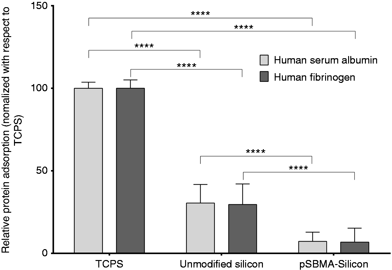
Relative protein adsorption from human serum albumin (HSA) and human fibrinogen. Tissue culture polystyrene (TCPS) was used as a positive control. Coating with pSBMA significantly reduced protein adsorption.
Interestingly, although albumin and fibrinogen consist of different levels of polar and nonpolar groups that influence their interaction with the surface, 61 the results indicate their adsorption trend to be similar for both silicon and modified silicon. This may be due to saturation of proteins adsorbed on the surface, given the long of incubation time and concentration of the protein solutions. 62 It should also be noted that behavior of proteins from a single protein solution may not be reflective of protein adsorption from a cocktail of proteins, such as it is in blood or plasma. However, it gives a general sense of protein fouling when compared side by side to other substrates.
Effect of shear stress on surface coating
Artificial implants in blood flow are exposed to shear stress. Therefore, surface modifications designed for such implants must be able to withstand shear stresses under biological conditions. Shear rates in the human body can be as high as 1650/s, 57 which may degrade polymers. Previous studies have observed coating degradation under static conditions,44,63 and found pSBMA to be stable over 30 days. 44 Here, we explored the effect of biological shear rates of 500–2000/s on ultrathin pSBMA surface modifications under physiological conditions over 24 h using D-PBS flow. Using D-PBS has its drawbacks compared to blood or serum: there are no enzymes, proteins, or cells present in D-PBS. Hence, the effect of these biological components could not be assessed. Nonetheless, PBS is often used in previous literature in place of blood due to its similarity in pH and saline content,44,63 and it allowed us to do sensitive surface analysis that would be compromised by proteins and cells attached on the surface.
Results from XPS (online Supplementary Table S1), contact angle, coating thickness, and protein resistance properties (online Supplementary Figure S3) demonstrate that pSBMA can survive shear rates up to 2000/s for 24 h. Aside from expected Si 2p and O 1s, control unmodified silicon also contains 9.8% and 0.4% adventitious carbon and nitrogen, respectively. When exposed to D-PBS over 24 h, there is at least a 6.3% elevation in carbon concentration, coupled with an increase in O 1s and N 1s. High resolution and spectral data show elevated oxygen and nitrogen is primarily bound to carbon, implying that shear stress with D-PBS leads to organic contamination on uncoated silicon. Experiments were conducted in a clean, but not sterile, environment, which may have led to bacterial growth on the surface over time. The highest level of contamination was found with 1500/s shear rate, with the maximum of 13.9% increase in C 1s.
Control substrates (unmodified silicon) had a contact angle of 27.3° and exposure to shear did not result in any significant alteration in silicon contact angle. The maximum difference in the uncoated silicon subset from control was due to 1500/s shear rate, yielding a difference of 6° from control unmodified silicon. Following exposure to shear stress, coating functionality was characterized by measuring their protein resistance to HSA. For unmodified silicon, despite additional contamination with shear, protein adsorption did not significantly change compared to control.
XPS, contact angle and ellipsometry showed significant changes in pSBMA characteristics when exposed to shear. As expected, control pSBMA-silicon had a 45.7% decrease in silicon and a 47.1% increase in carbon from baseline unmodified silicon. There is also a presence of N 1s and S 2s in a ratio of 1:1 when adventitious nitrogen is accounted for, which matches stoichiometric ratio. Regardless of shear stress, exposure to D-PBS led to ∼18–20% increase in silicon, ∼20–23% decrease in carbon, and ∼0.5% or less decrease in sulfur.
Control pSBMA-silicon has a contact angle of 10.9° and coating thickness of 4.7 nm. Regardless of the level of shear, exposure to D-PBS significantly increased contact angle of pSBMA-silicon, settling around ∼30°, and decreased the coating thickness to ∼2.5–2.8 nm, a ∼40% decrease. When combined with elemental sulfur data from XPS, the results indicate that approximately 20% of the thickness change is from removal of SBMA from the surface while the other 20% is due to conformational change in pSBMA chains. Solely based on the experiments conducted, it is difficult to discern if removal of SBMA is originating from shearing away uncrosslinked monomers or breakdown of pSBMA chains. If it was the latter, we would expect to see higher shear rates accelerate chain scission. Instead, we find all shear rates led to similar surface characteristics after 24 h, indicating it is likely that uncrosslinked monomers are being removed. Additionally, pSBMA coating performance was not affected by the tested shear stress. Control pSBMA-coated silicon improved protein resistance by ∼71% compared to uncoated silicon. With exposure to shear in D-PBS, there was no increase in protein adsorption, and functionality of the coating was preserved.
In vitro fresh human blood experiment
While animal testing is critical for any novel biomaterial or implant, preliminary in vitro testing can significantly reduce sample size, lowering the number of animal sacrifices required as well as cost. Additionally, it allows us to test against fresh human blood, which is critical since reaction to blood may vary from one species to another. 64 For testing the pSBMA-silicon in an in vitro setup that closely matches the conditions of the bioartificial kidney hemofilter, we designed a platform with optimized flow path and channel height of ∼200 µm. Flow experiments were conducted at a wall shear rate of 1000/s, which is within physiologic range.
After 2 h of blood flow, the PEEK flow cells were disassembled, and silicon and pSBMA-silicon surfaces were analyzed using SEM and IHC (Figure 8). SEM and IHC imaging shows increased amount of adhered, activated platelets, and platelet aggregates for unmodified silicon compared to pSBMA-silicon. Using representative images from IHC, the number of platelets adhered and activated were quantified (Figure 8(e)), which confirms the trend: pSBMA-silicon significantly lowers platelet adhesion and activation compared to unmodified silicon. For unmodified silicon, there are non-homogeneity in platelet adhesion to the surfaces causing large standard deviations. It should also be noted that the number of platelets were calculated based on 2D images. Therefore, overlapping platelets, for example, in a platelet aggregate, were under-counted using this method. However, the quantified data agrees trend-wise with the visualized images, and we found that at <5 nm thickness, pSBMA-silicon outperformed unmodified silicon. Overall, pSBMA consistently reduces protein adsorption, and resists platelet and cellular adhesion and activation.
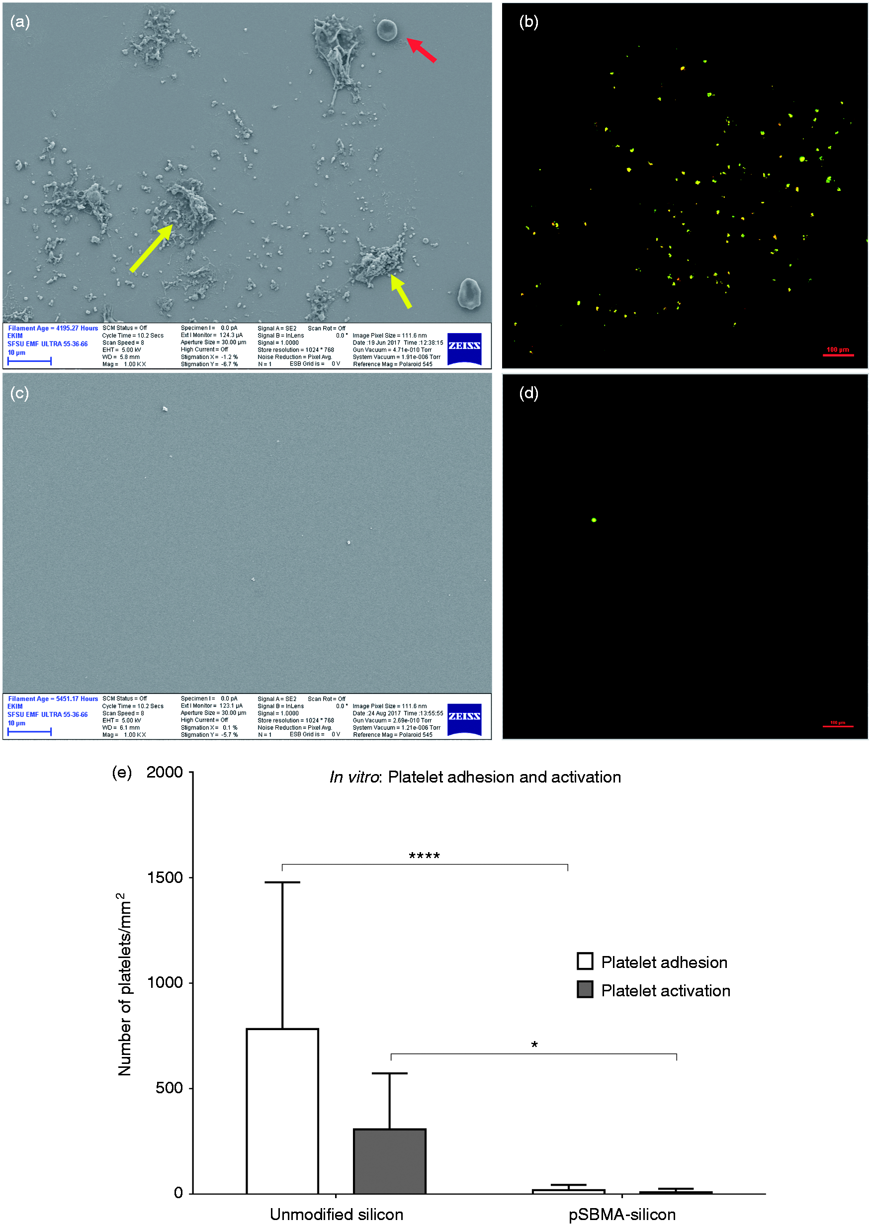
Surface analysis of unmodified silicon (a,b) and pSBMA-silicon (c,d) following exposure to fresh human blood for 2 h in vitro. SEM images are presented in (a) and (c) and IHC images are presented in (b) and (d). IHC images show overlap of two marked proteins, CD41, and CD62. CD41 is present on platelet surface, while CD62 is present on activated platelets. (a) SEM of unmodified silicon showing adhered platelets and platelet aggregates (yellow arrow), and red blood cells (red arrow). (b) IHC imaging of unmodified silicon shows platelet activation. (c) SEM image and (d) IHC imaging of pSBMA-silicon show minimal cellular and platelet adhesion and activation. (e) IHC platelet adhesion and activation data quantified, demonstrating significantly lower platelet adhesion and activation compared to unmodified silicon.
In vivo study
Although pSBMA-silicon surfaces reduced platelet adhesion and aggregation in vitro, fouling and activation in vivo may differ. Therefore, we have exposed the surfaces to blood flow in vivo extracorporeally for 6 h, and as an implant over 7 and 26 days in a porcine model. Porcine model was chosen because it has demonstrated very similar platelet behavior compared to human platelets in vitro. 64 These set of experiments better mimic the final implant conditions and enables us to test longevity of the coating under blood flow.
Initially, we conducted an extracorporeal study lasting 6 h and found that the pSBMA surfaces to be resistant to platelet adhesion and activation. As demonstrated by representative images of SEM and IHC fluorescence microscopy (Figure 10(a) and (d)), there is minimal platelet adhesion and activation on the pSBMA-silicon surfaces. However, there is presence of residual protein attachment as shown in SEM image in Figure 10(a). Given the structure of the residues, they may be attempted cell attachments that were removed by the blood flow (measured at 60 mL/min).
For the two implant studies, both animals underwent successful device implantation and acute recovery without incident. Throughout the post-implantation period, behavior, appetite, activity, and responsiveness remained appropriate. Serum and hematologic parameters demonstrated an initial acute inflammatory response to surgery, characterized by elevated white blood cell count and CRP, which resolved within seven to ten days. Following acute surgical blood loss, red blood cell counts and hemoglobin levels improved to pre-surgical levels by five days after surgery. Serum plasma free hemoglobin remained at pre-surgical baseline, indicating negligible if any, hemolysis. Throughout the implantation period, both devices remained patent based on physical exam, Doppler ultrasonography, fluoroscopic angiography (Figure 9), and visual examination upon device retrieval. Devices were removed intact.
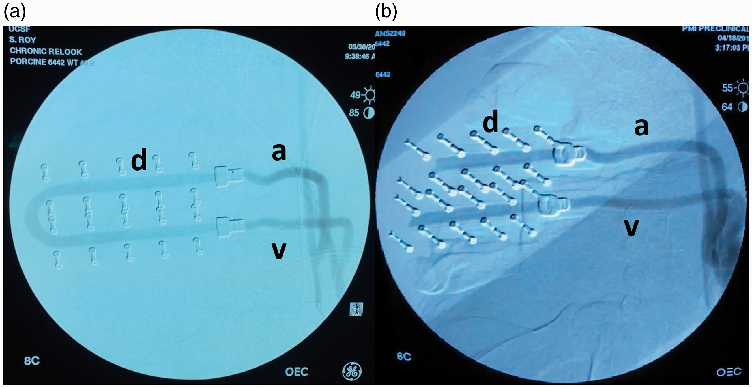
Fluoroscopic angiography of the implanted device at (a) 7 days and (b) 26 days. The U-shaped blood flow path of the device labeled “d”, arterial inflow “a”, and venous outflow “v” grafts are widely patent at both time points.
Representative images of SEM and IHC surface analysis post-7-day and 26-day implant study is shown in Figure 10((b) and (e)) and 10((c) and (f)), respectively. SEM (Figure 10(b) and (c)), in both cases, shows slight granularity at the surfaces, which may be due to protein adsorption. However, most of the pSBMA-silicon surfaces are clear of cellular adhesion, and there was no evidence of clots. IHC images were quantified in a similar manner as was done for in vitro study. After 2-h flow under in vitro conditions, pSBMA-modified silicon had adhered ∼19 platelets/mm2, of which ∼9 platelets/mm2 were activated. Following exposure to blood for 6 h in the extracorporeal study, platelet adhesion increased to ∼168/mm2 with only ∼0.9% (1.5 platelets/mm2) of the platelets activated. With implants, there was a positive trend in platelet adhesion. Following 7-day implant, the adhesion increased to ∼74 platelets/mm2. However, only ∼14% of the platelets were activated. After 26 days under blood flow, the number of adhered platelets increased to ∼159/mm2 and ∼36% of them were activated.
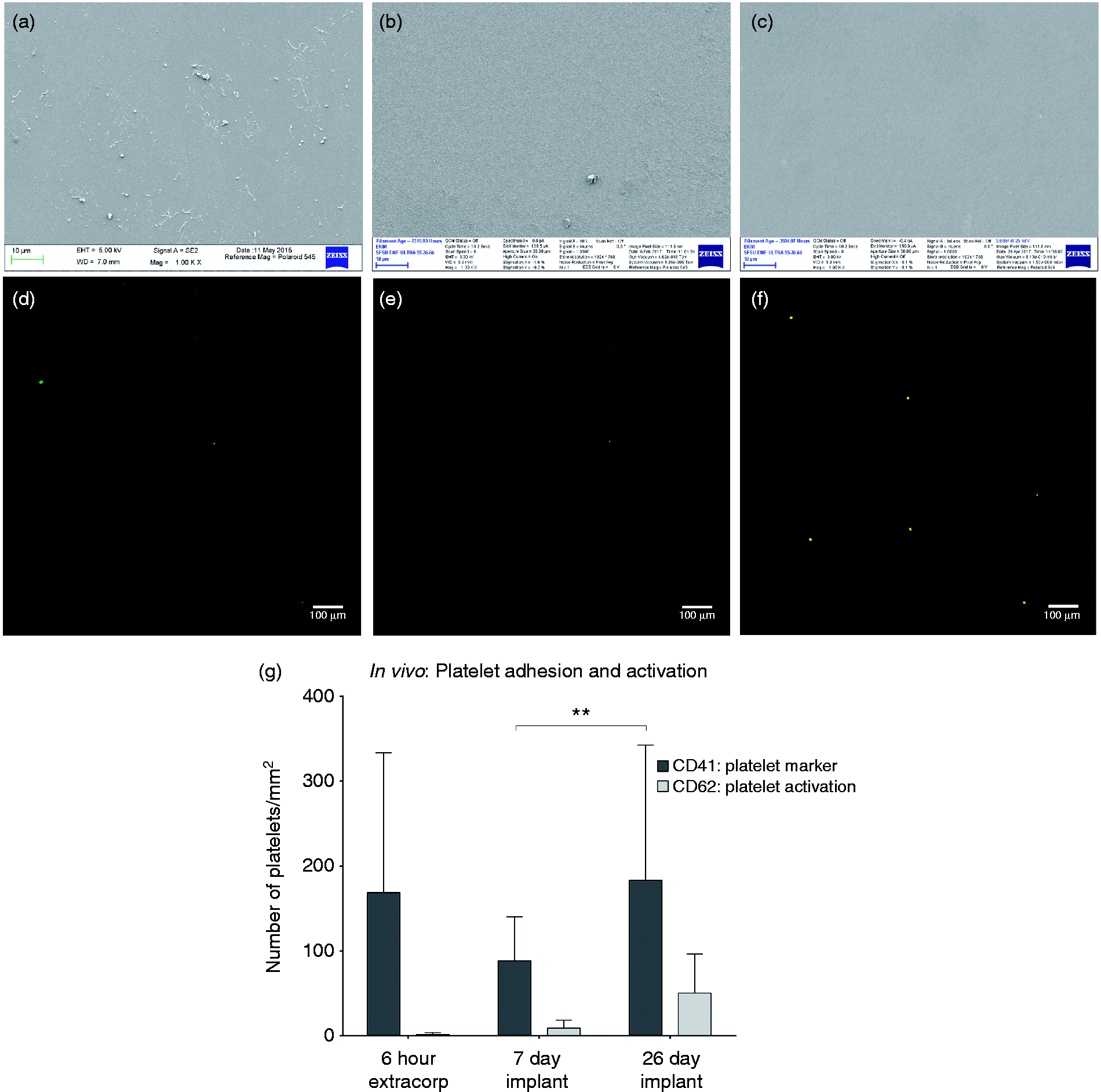
Surface analysis of pSBMA-silicon following in vivo experiments. SEM imaging following (a) 6-h, (b) 7-day, and (c) 26-day exposure to blood flow. IHC imaging showing overlap of platelet marker, CD41 and platelet activation marker CD62 following (d) 6-h, (e) 7-day, (f) 26-day exposure to blood flow. (g) Quantification of platelet adhesion and activation on pSBMA-silicon substrates in porcine model demonstrating minimal activation up to 26 days.
For the purposes of this study, no control unmodified silicon was used under implant conditions. Previous experiments done by our group show that silicon can induce clotting. 65 Therefore, for ethical reasons, unmodified silicon was not implanted. With pSBMA modification on silicon, patency of device after 26 days of blood flow without formation of clots on the surface is promising. Future experiments will incorporate membranes and monitor filtration as well as hemocompatibility.
Conclusion
Blood-contacting implants need to resist activating blood to hinder thrombus formation and retain functionality. Because of the precision and versatility of micro- and nanoscale features it offers, silicon has become a prevailing substrate material for novel biomedical devices. We found that for such devices, where critical nanoscale features are exposed to blood, ultrathin pSBMA coating under 5 nm thickness is a promising option. pSBMA coatings was able to survive biological shear over 24 h. Moreover, it reduced platelet adhesion and activation in vitro against fresh human blood for 2 h, and in vivo for up to 26 days in porcine model. These results are highly encouraging as we move into filtration and immunoisolation applications for SNMs.
Supplemental Material
Supplemental material for In vitro and in vivo hemocompatibility assessment of ultrathin sulfobetaine polymer coatings for silicon-based implants
Supplemental Material for In vitro and in vivo hemocompatibility assessment of ultrathin sulfobetaine polymer coatings for silicon-based implants by Zohora Iqbal, Steven Kim, Jarrett Moyer, Willieford Moses, Emily Abada, Nathan Wright, Eun Jung Kim, Jaehyun Park, William H Fissell and Shuvo Roy in Journal of Biomaterials Applications
Footnotes
Acknowledgement
We would like to thank Jimmy Ly, Ana Santandreu, Alonso Torres, and Deepika Sarode for their assistance in fresh blood experiments. We would also like to thank Brooke Benner for general assistance in experimentation and Alex Heller for assistance in flow cell design. We thank Gerry Hammer at the Molecular Analysis Facility, University of Washington for conducting XPS. We also acknowledge the Small Molecule Discovery Center at University of California, San Francisco, and in particular, Mark Burlingame, for his advice and access to equipment. Assistance from Illya Gordon on coordinating various aspects of this study is greatly appreciated. Shuvo Roy and William Fissell are founders, with ownership stake, of Silicon Kidney, LLC, a start-up company that is advancing the commercialization of silicon membrane technology. Nathan Wright has worked as an employee of Silicon Kidney, LLC.
Declaration of Conflicting Interests
The author(s) declared no potential conflicts of interest with respect to the research, authorship, and/or publication of this article.
Funding
The author(s) disclosed receipt of the following financial support for the research, authorship, and/or publication of this article: This research work was funded by the National Institutes of Health (NIH) R01EB04315, U01EB021214, and U01EB025136.
Supplemental Material
Supplemental material for this article is available online.
References
Supplementary Material
Please find the following supplemental material available below.
For Open Access articles published under a Creative Commons License, all supplemental material carries the same license as the article it is associated with.
For non-Open Access articles published, all supplemental material carries a non-exclusive license, and permission requests for re-use of supplemental material or any part of supplemental material shall be sent directly to the copyright owner as specified in the copyright notice associated with the article.