
Abstract
Biomaterials are often used in orthopedic surgery like cavity fillings. However, related complications often require long-term systemic antibiotics, device removal, and extended rehabilitation. Hydroxyapatite/silver (HA/Ag) composites have been proposed as implantation biomaterials owing to the osteogenic properties of hydroxyapatite and to the antimicrobial efficiency of silver. Nevertheless, higher silver concentrations induce cytotoxic effects. The aim of this study was to synthesize and characterize HA/Ag nanocomposites that will allow us to use lower concentrations of silver nanoparticles with better antimicrobial efficiency and anti-inflammatory properties. The characterization of HA/Ag was performed by scanning electron microscopy, energy dispersive spectroscopy, X-ray diffraction, Fourier-transform infrared spectra, X-ray photoelectron spectroscopy, and laser diffraction. Bioactivity was evaluated under a simulated body fluid. The viability of osteoblast like-cells (MG-63) was determined by MTT (3-(4,5-dimethylthiazol-2-yl)-2,5-diphenyl-2H-tetrazolium bromide) and the antimicrobial activity was evaluated by the standard McFarland method. The detection of nitric oxide was measured by a colorimetric assay and the inflammatory cytokines by flow cytometry. We obtained particulate composites of calcium phosphates identified as hydroxyapatite and silver nanoparticles. The bioactivity of the HA/Ag nanocomposites on SFB was confirmed by apatite formations. The viability of MG-63 cells was not affected. We also found antimicrobial activity against Escherichia coli, Staphylococcus aureus, and Candida albicans owing to the presence of silver nanoparticles at non-cytotoxic concentrations. HA/Ag reduced the release of nitric oxide and decreased the secretion of IL-1 and TNF-α in cells stimulated with Lipopolysaccharide (LPS). In conclusion, the inflammatory and antimicrobial capacity of the HA/Ag nanocomposites, as well as its bioactivity and low cytotoxicity make it a candidate as an implantation biomaterial for bone tissues engineering and clinical practices in orthopedic, oral and maxillofacial surgery.
Keywords
Introduction
Hydroxyapatite (HA) is the principal component of bone substitutes like powders, films and hydrogels and has long been used for the repair and reconstruction of bone tissues.1,2 HA (Ca10(OH)2(PO4)6) is chemically and structurally similar to the inorganic component of bone matrix, which is a very complex tissue. HA is also biocompatible with soft tissues such as skin, muscle and gums. Nevertheless, postoperative infections of the grafted area inevitably lead to the reabsorption of a large part of the graft or its complete loss by microbial colonization and biofilm formation on the surface of the biomaterial.3–5 Antibiotics are the most common way to treat infected bone, but increase in the antimicrobial resistance and poor penetration of antibiotics into bone tissues are some of the disadvantages.6–8
During normal bone formation osteoblasts decrease reactive oxygen species (ROS), promoting the expression of osteogenic markers such as collagen (COL I), and osteocalcin (OCN). Bone injury and inflammation caused by surgery and infections generate ROS that can lead to oxidative stress. Excessive and prolonged ROS activity inhibits osteogenesis and lowers bone regeneration, disturbing the balance between osteoclasts and osteoblasts. 9 Cell stress induces pro-inflammatory cytokines such as TNF-α, IL-1 and IL-6 that increase tissue damage and osteoclast activity, generating bone loss.10–13
The development of biomaterials with anti-inflammatory and antimicrobial properties will increase their functionality and efficiency mainly in tissues susceptible to infections.8,14 Silver nanoparticles have been incorporated into biomaterials for their antimicrobial properties against a broad spectrum of microorganisms.15–18 But high concentrations of silver that provide antimicrobial efficiency are toxic for eukaryotic cells, affecting the healing process. 19 In this work, we report a modified synthesis method to obtain bioactive HA/Ag nanocomposites with low toxicity and antimicrobial and anti-inflammatory properties.
Materials and methods
Synthesis of HA/Ag nanocomposites
The synthesis of HA/Ag was based on the method described by Vukomanović et al. 20 with some modifications. Briefly, the following reagents were used: calcium-nitrate Ca(NO3)2.4H2O, ammonium-dihydrogenphosphate NH4H2PO4, urea (NH2)2CO, and silver nitrate (AgNO3) as the starting materials. All reagents were purchased from Jalmek Científica (México). First, Ca(NO3)2.4H2O and NH4H2PO4 were dissolved in distilled water, the molar Ca/P ratio was fixed at 1.67. AgNO3 was added at 0.01%. The solution was adjusted at a pH of 5.5 with ammonium hydroxide and then heated up to 90°C with constant agitation at 15,000 r/min for 30 min. When the solution reached the mentioned temperature, (NH2)2CO at a concentration of 0.180 mg/mL was added. Precipitates were then aged for 12 h before washing with distilled water (Figure 1).
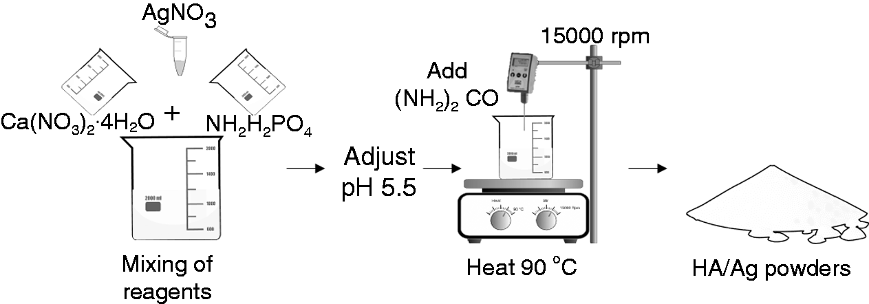
Reaction scheme of HA/Ag nanocomposites synthesis.
Characterization of HA/Ag nanocomposites
Scanning electron microscopy
Scanning electron microscopy (SEM) was used to reveal the morphology and size of the synthesized particles and the elemental contents of HA/Ag were determined by energy dispersive X-ray spectroscopy (EDS). The samples were dispersed in isopropanol using an ultrasound bath and examined on a JEOL JXA 840A scanning electron microscope.
Laser diffraction
The particle size distribution measurements of HA/Ag nanocomposite were done by diffraction laser (DLS) and examined on a HELOS (H3421) & RODOS using dispersing method with a pressure of 3.00 bar.
Infrared spectroscopy
To identify the nature of the chemical bonds between atoms, Fourier transformed infrared (FTIR) analysis was done. FTIR spectra were recorded on a Perkin Elmer Spectrum GX equipped with an attenuated total reflectance (ATR) unit. The ATR accessory contained a ZnSe crystal at a nominal incident angle of 45°. All spectra were obtained at the range of 650 to 4000 cm−1 and made using 24 scans with a resolution of 4 cm−1.
X-ray diffraction
X-ray diffraction (XRD) measurements of the HA/Ag nanocomposites were performed using a Dmax2100 (Rigaku, TX, USA) diffractometer operating at 30 kV and 20 mA with CuKα (λ = 1.5406 Å) radiation. The data were collected over the 2θ range from 4 to 60° with a step size of 0.02° intervals with a counting time of 0.6 s at each step. The software used to analyze the spectrum was MDI Jade 6.0.
X-ray photoelectron spectroscopy
X-ray photoelectron spectroscopy (XPS) was used to stablish the elementary composition of the samples in the surface and to identify the Ag presence and its chemical state. The spectra were obtained in an XPS equipment (Thermo Scientific model escalab 250Xi) with an Al-Kα (hν = 1486.7 eV) as excitation source. The spectra were collected at a pass energy of 20 eV.
Inductively coupled mass spectrometry
The quantitative analysis of silver concentration was performed by means of inductively coupled mass spectrometry. HA/Ag nanocomposite was carbonized at a temperature of 300°C for 1 h, the powder obtained was placed inside Teflon cups. To submit them to an acid digestion process via the brand’s microwave (CEM, MARS) to digest the samples, a mixture of nitric acid (HNO3-16 M and 68%) and hydrochloric acid (12 M HCl and 36%) were used, which were added to the vessels containing the sample, at a temperature of 250°C for 15 min (using a heating lamp).
Mineralization in simulated body fluid
To evaluate the bioactivity process, the simulated body fluid (SBF) was prepared according to Kokubo’s method. 21 Briefly, reagents: NaCl (8.035 g), NaHCO3 (0.355 g), KCl (0.225 g), K2HPO4·3H2O (0.231 g), MgCl2·6H2O (0.311 g), 1.0 M HCl (39 mL), CaCl2 (0.292 g) and Na2SO4 (0.072 g) were added to 1 L of distilled water in the order listed above. The pH of the solution was adjusted to 7.4 by the addition of Tris/HCL. HA/Ag powder was immersed in 1× SBF and maintained at room temperature for 1, 7, 14, 21 days. SBF was refreshed every 2 days to preserve ion concentration. The sample was removed from the fluid, washed in distilled water, and stored at room temperature. After that, the SEM and XRD measurements were carried out to study the morphological and structural changes, respectively.
Estimation of endotoxin levels
To exclude the possibility of pro-inflammatory effects due to bacterial contamination on the HA/Ag, nanocomposites were sterilized by autoclave and UV irradiation (CL-1000 ultraviolet crosslinker) and endotoxin levels were measured using Limulus Amoebocyte Lysate (LAL). The amount of endotoxin was detected by PYROGENT TM kit using the manual instructions.
Viability testing
Cell culture
To carry out the viability testing, the osteoblast-like MG-63 (osteosarcoma) cell lines were used because these cells are recognized as a model of study for osteoblasts.22–24 The MG-63 cells were cultured in Dulbecco’s Modified Eagle’s medium (DMEM, Gibco,) supplemented with 1% antibiotic-antimycotic and 10% fetal bovine serum (FBS) at 37°C (5% CO2 and 95% humidity). A total of 2.5 × 10⁴ cells were grown in a 25 cm2 flask until confluence.
Assessment of cell viability
The MTT (3-(4,5-dimethylthiazol-2-yl)-2,5-diphenyltetrazolium bromide) assay 25 was used to assess and compare the viability of MG-63 cells exposed to different concentration of HA/Ag (250–7 μg/mL). MG-63 cells were seeded at a density of 1 × 10⁴ cells per well in 96-well culture plates for 24–72 h. After culture, the medium in each well was replaced with medium containing 10% MTT (5 mg/mL stock solution) (Sigma-Aldrich, Steinheim, Germany). After 4 h of incubation at 37°C, the medium was removed and replaced with dimethyl sulfoxide to dissolve purple formazan crystals; optical density was read at 570 nm wavelength by the Elisa Reader (Biorad).
Nitrite oxide determination
To determine nitric oxide (NO) release, the MG-63 cells were cultured with a density of 2.5 × 10⁴ per well in 24-well plates; when there was 80% confluence the cells were stimulated with either Lipopolysaccharide (LPS) (E. coli 0127:B8; Sigma chemical Co., St Louis, MO, USA) 1 μg/mL. Several concentrations of HA/Ag (250-7 ug/ml), LPS (1 ug/mL) and HA/Ag (250-7 ug/ml) blended with LPS (1 ug/mL) (previously incubated for 1 h) and incubated at 37°C in a 95% O2 and 5% CO2 atmosphere for 24 and 48 h. The supernatants were collected and used for nitric oxide detection (Cayman Chemical). The concentration of nitrite (NO2) produced by MG-63 cells was used as a measure of the production of NO. The concentration of NO2 was determined spectrophotometrically at 540 nm using the NO colorimetric assay kit (Cayman Chemical, Michigan, USA) following the manufacturer’s instructions. Briefly, 80 μL of the supernatant was incubated with 10 μl of enzyme cofactor and nitrate reductase to 2 h after adding 50 μL of Griess reagent (1% sulfanilamide, 0.1% N-1-napthylethylenediamine dihydrochloride, 2.5% H3PO4) at room temperature for 10 min, and the absorbance read at 540 nm using sodium nitrite as standard. Concentration of NO2 in the sample was calculated using the following equation.
Quantification of inflammatory cytokines
To detect inflammatory cytokines, the MG-63 cells were cultured with a density of 2.5 × 10⁴ per well in 24-well plates; when there was 80% confluence the cells were stimulated with LPS 1 μg/mL, 15 μg/mL of HA/Ag and LPS (1 μg/mL) combinated with 15 μg/mL of HA/Ag (previously incubated for 1 h) at 37°C in a 95% O2 and 5% CO2 atmosphere for 24 and 48 h 37°C. After that the supernatant was stored at –80°C. The production of IL-8, IL-1β, IL-6, IL-10, TNF, and IL-12p70 was detected by BD Accuri flow cytometer (Becton Dickinson Holdings Pte Ltd, Singapore) using commercial Human inflammatory cytokines kit (Cat. No. 551811, BD Biosciences, USA) according to the manufacturer’s specifications. In brief, cytokine standards were prepared using a vial of lyophilized human inflammatory cytokines and assay diluent by the method of serial dilutions. Captured beads were added into each tube (sample, standards, and negative control) and incubated for 3 h at room temperature in the absence of light. The flow cytometer was calibrated using cytometer setup beads and the assays were performed.
Antimicrobial activity
Antimicrobial properties of the sample were tested against two bacterial strains: Escherichia coli DH5α (
Statistical analysis
Statistical analyses were performed using the GraphPad Prism 7.0 software (GraphPad Software, Inc., San Diego, CA, USA). The results were presented as the mean SD. Comparisons were made using one-way ANOVA based on a Tukey test contrast, considering a statistical significance p < 0.05.
Results
Characterization of HA/Ag nanocomposites
The HA/Ag powders showed an irregular shape (Figure 2(a) and (b)). The majority of the particles show a size of 1.37 μm, although HA/Ag particles of 340 nm were also found (Figure 2(c)).
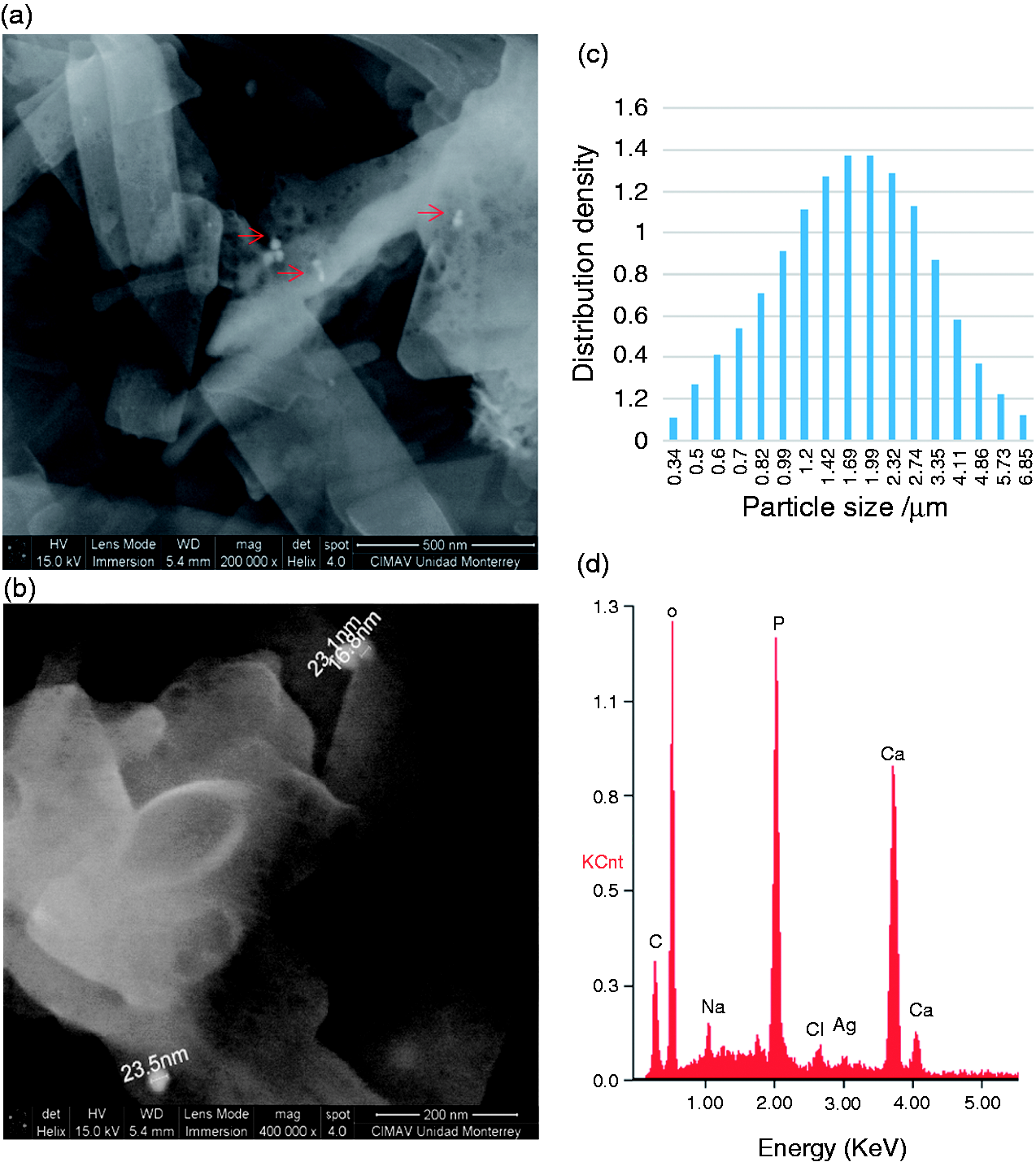
Characterization of HA/Ag powders. (a,b) scanning electron microscopy (SEM) (c) laser diffraction. The red arrows point to the silver nanoparticles. (d) Dispersive Energy Spectroscopy (EDS).
The size of the Ag nanoparticles estimated directly from the SEM image reveals the formation of Ag nanoparticles with an average size of 23 nm (Figure 2(b)).
EDS analysis confirmed the presence of Ca, P, O, C and Ag elements (Figure 2(d)). We further examined the Ag distribution in HA/Ag using EDS mapping (Figure 3). The main elements of Ca and P were uniformly distributed across the entire HA/Ag and particularly Ag were identified as nanoparticles on the surface on HA/Ag nanocomposite. The ICP analysis showed that the amount of silver was 16,640.0 mg/kg which corresponds to 0.01% (Table 1).
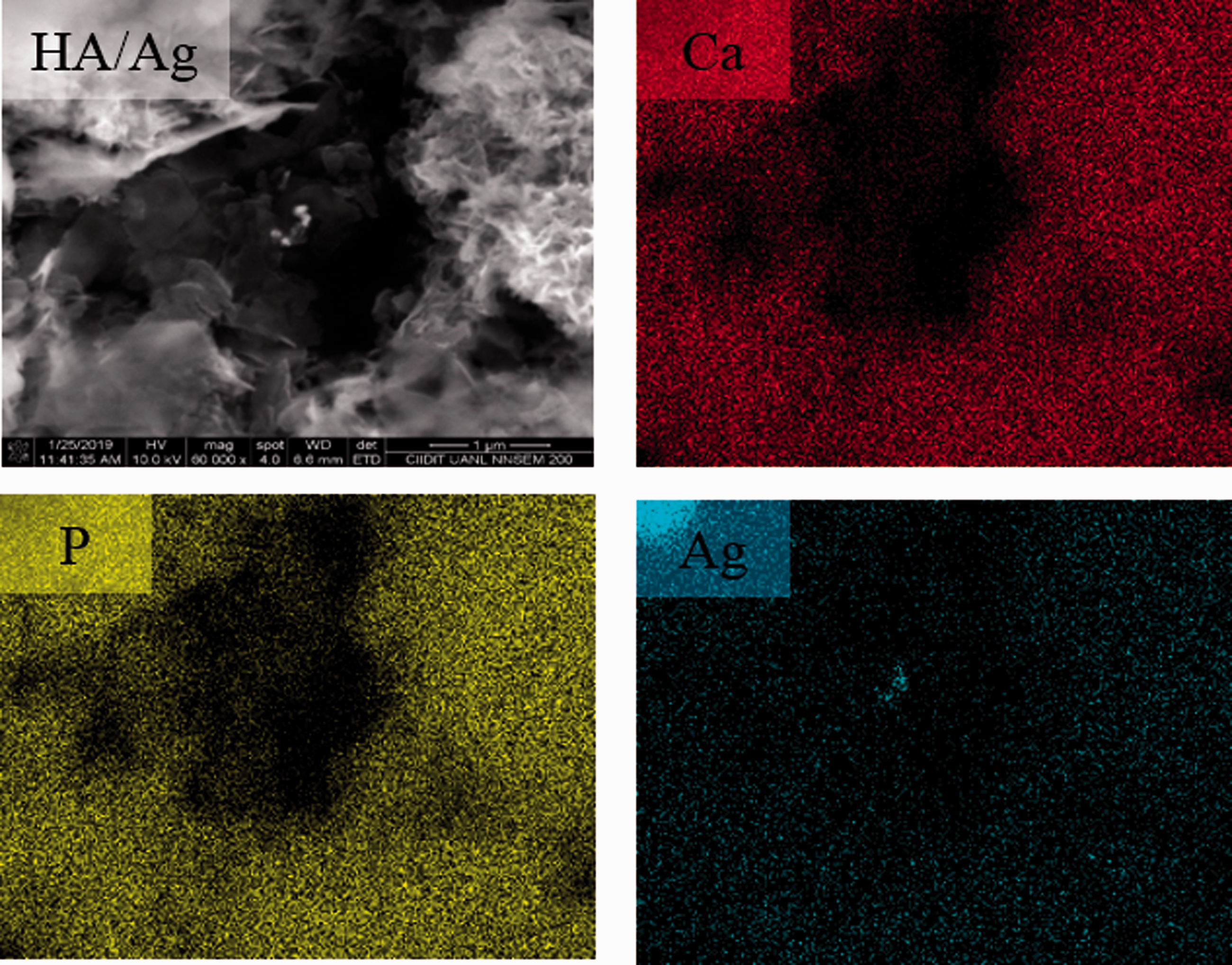
EDS mapping of the HA/Ag nanocomposite.
Quantitative analysis by ICP of the Ag within the HA/Ag nanocomposites.
HA/Ag: hydroxyapatite/silver.
The FTIR spectra for HA and HA/Ag composite are shown in (Figure 4(a)). The main characteristic bands for pure HA can be observed at 3456 and 3569 cm−1 related with vibration modes of OH– group, PO43 vibrations can be observed at 471, 561, 600, 962, 1027 and 1088 cm−1 and the vibration bands of CO32– appeared at 876, 1342, 1461, 1629 and 1666 cm−1. When Ag was added, the band that related with –OH group at 3456 cm−1 was decreased. Moreover, the bands that correspond to vibration modes of the CO32– group were decreased in composite with silver, which suggest an association with the increased interchange of the ions into the apatite structure.
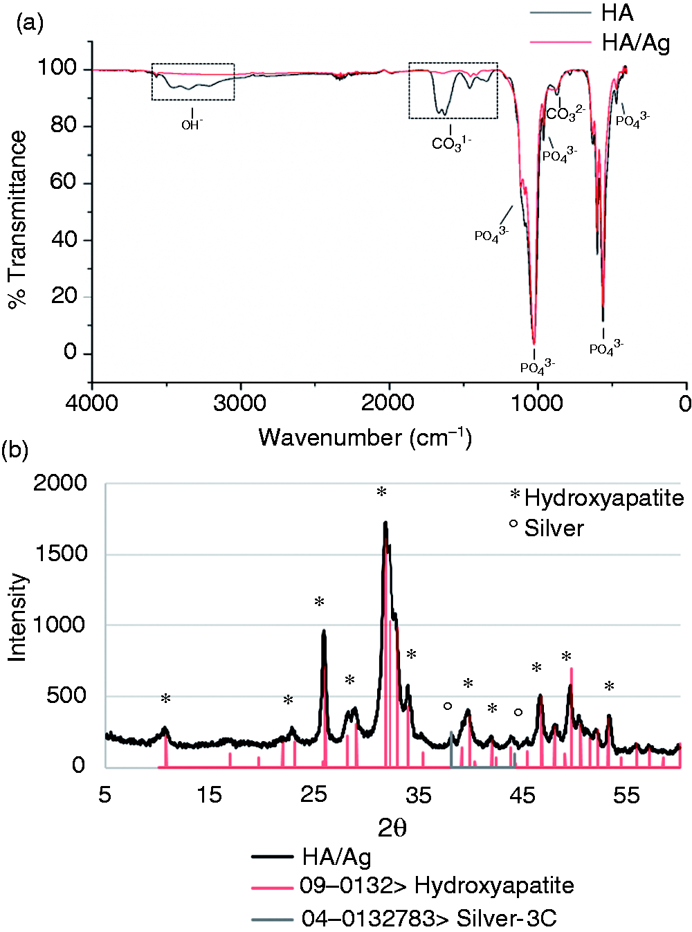
FTIR spectrums (a) and XRD patterns (b) of HA/Ag powders.
The phase identification of the HA/Ag composites revealed typical diffraction peaks at 38.16° and 44.28° that correspond to the cubic structure of Ag (JCPDS No. 4–0783) together with the characteristic HA diffraction peaks at 25.8°,31.7°, 32.1°, 32.9°, 34.04°, 46.7°, and 49.5° (Ca5(PO4)3OH) (JCPDS No. 9–0432) (Figure 4(b)).
The incorporation of Ag in the HA was corroborated using the XPS measurements. In Figure 5, HA and HA/Ag spectra are shown. The binding energy (BE) for the peaks found in both HA and HA/Ag samples are summarized in Table 2. In the HA/Ag spectrum it is also possible to see a contribution at around 373.02 and 368.69 eV, which might correspond to the Ag3d5/2 and Ag3d3/2, respectively (Figure 5).
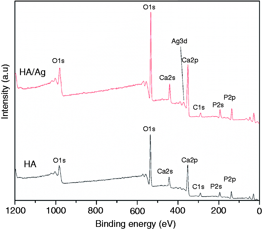
Full XPS spectra of the HA and HA/Ag samples.
Binding energy (BE) of the elements present in the full XPS spectra for undoped and Ag-doped HA samples.
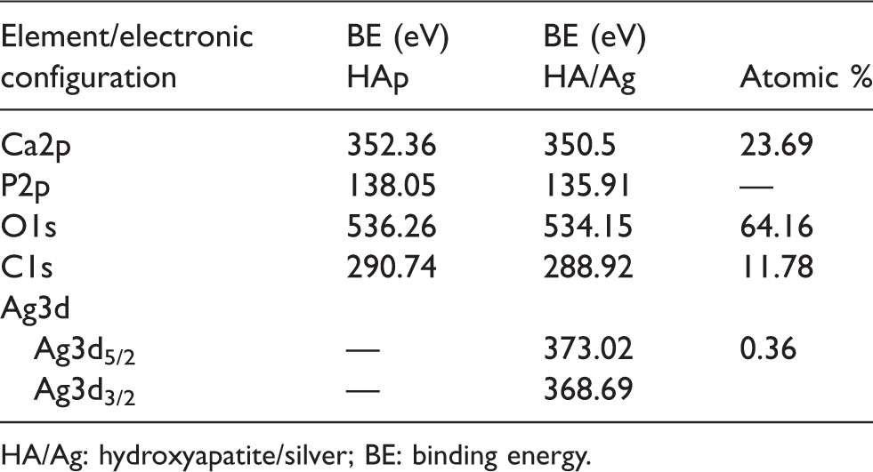
HA/Ag: hydroxyapatite/silver; BE: binding energy.
Bioactivity process
Figure 6 shows the SEM of the HA/Ag surface and XRD patterns after soaking in SBF for several periods. The SEM image for each soaking time is representative of those observations on several sites of the HA/Ag surface. Ca/P ratio was calculated from the EDS results and compared with the HA stoichiometric ratio (Ca/P 1.67) (Table 3).
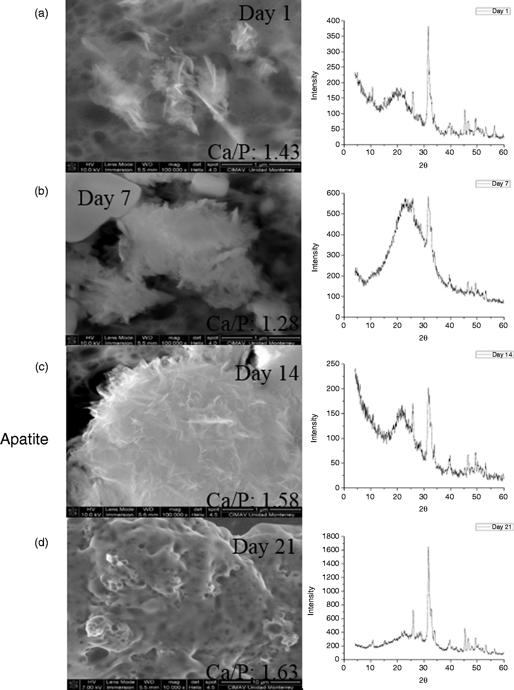
Scanning electron microscopy and X-ray diffraction of the materials after the bioactivity process on SBF. (a) 1 day, (b) 7 days, (c) 14 days and (d) 21 days.
Atomic percentage and Ca/P ratio at sample surface of HA/Ag by EDS.
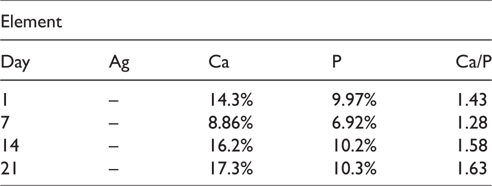
After 1 and 7 days soaking HA/Ag in SBF the formation of amorphous agglomerates was observed by SEM images with Ca/P ratio of 1.43 and 1.28, respectively (Figure 6(a) and (b)). After 14 days of soaking, the SEM images showed several tiny flake-like precipitates on the HA/Ag surface top where the Ca/P ratio increased to 1.58. The flake-like precipitates and Ca/P ratio of 1.58 correlate with the apatite layer formation on the HA/Ag surface (Figure 6(c)). The XRD patterns of HA/Ag soaking for 1–14 days reveal diffraction peaks which might be attributed to the presence of two phases of the calcium phosphate: Tricalcium phosphate (TCP) and octacalcium phosphate (OCP) but the peak in the pattern is not well resolved (Figure 6(a) to (c)).
In the pattern after soaking for 21 days, the most characteristic diffraction peaks of HA were observed (Figure 6(d)). The Ag element was not detected because of the formation of surface apatite, which is related with ionic exchange between the medium and the material after 3 h of soaking, 27 the increased presence of silver ions in the sample, and the ionic potential of medium stimulating the bioactivity. 28 Other elements, such as Na, Mg, K, Cl were also identified in this sample, which were also found in bone apatite structures and may be associated with the ion exchange during soaking time.
Cell viability
The viability was evaluated in osteoblast-like cells (MG-63) exposed to different concentrations of HA/Ag during 24, 48 and 72 h (Figure 7). At 24 h results indicate that 81% of cells were viable at the highest concentration evaluated (250 μg/mL), this suggests the absence of toxic effects on osteoblast-like cells. Additionally, long time exposure experiments were carried out. Our results showed a viability of 75% after 72 h of treatment. However, the concentrations below 31 μg/mL after 72 h showed a viability greater than 87% (Figure 7).
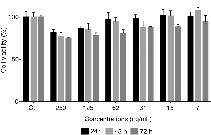
MG-63 cells viability exposed to different concentrations of HA/Ag.
Estimation of endotoxin levels
The detection of endotoxin levels assays shows that HA/Ag powders do not form a gel in the tube, for all dilutions (1:2, 1:4, 1:8, 1:16), This indicates that the endotoxin concentration is below 0.06 UE/mL (Table 4).
Endotoxin detection: HA/Ag powders were analyzed by PYROGENT™. Endotoxin of E. coli 0111B4 was used as positive control and saline solution as negative control.
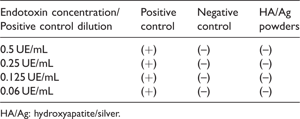
HA/Ag: hydroxyapatite/silver.
Detection of nitric oxide
The anti-inflammatory activity of HA/Ag powder has been quantified by observing the NO production of MG-63 stimulated by LPS. Figure 8 shows that the amount of NO2 increases considerably with treatment of LPS at 24 h (0.157 μM/mL) and 48 h (0.231 μM/mL). When HA/Ag treatment was added in combination with LPS treatment, all concentrations (250–7 μg/mL) exhibited relatively higher inhibition activity against NO release.
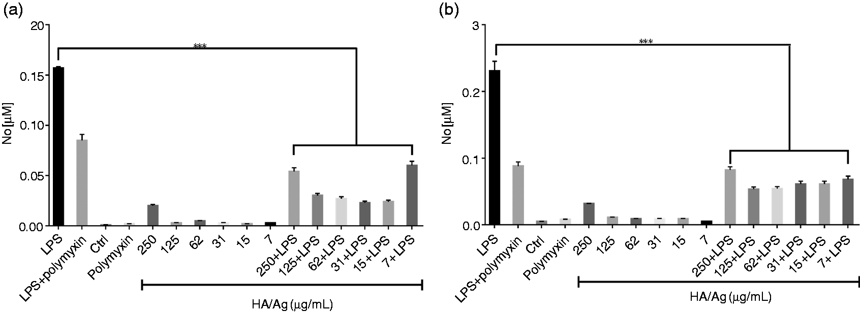
Effect of HA/Ag on inhibition of nitric oxide on osteoblast like-cells MG-63. (a) 24 h (b) 48 h. ***p < 0.001.
Detection of pro-inflammatory cytokines
LPS stimulation significantly increased the production of inflammatory cytokines in MG-63 cells, IL-1 (1200 pg/mL), IL-6 (600 pg/mL), IL-8 (9295 pg/mL), IL-10 (0.2 pg/mL), IL-12p70 (0.5 pg/mL), TNF-α (313.69 pg/mL). When the LPS was combined with HA/Ag, it decreased the production of IL-1 (968.2 pg/mL), IL-8 (4516.03 pg/mL) and TNF-α (207 pg/mL), although only HA/Ag induced the production of IL-8 (2624 pg/mL), IL-10 (12.54 pg/mL) and TNF-α (128 pg/mL) at 24 h.
However, after 48 h of treatment with LPS and HA/Ag IL-6 (110 pg/mL) and IL-8 (2065.04 pg/mL) decreases (Figure 9).
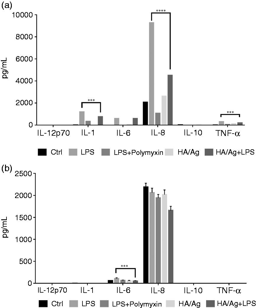
HA/Ag modulates the production of inflammatory cytokines in MG-63 cells stimulated by LPS. ***p < 0.001, ****p < 0.0001.
Antimicrobial properties
Figure 10 exhibits a significant difference in antibacterial effects between the HA and HA/Ag powders. After 24 h of incubation, all the concentrations (250–7 μg/mL) tested of HA/Ag had antimicrobial activity against E. coli. The lowest concentration of 7 μg/mL decreased growth by more than 78% compared to MRSA which had an 84% inhibition. However, the same concentration has an antifungal effect against C. albicans of 63% (Figure 10).
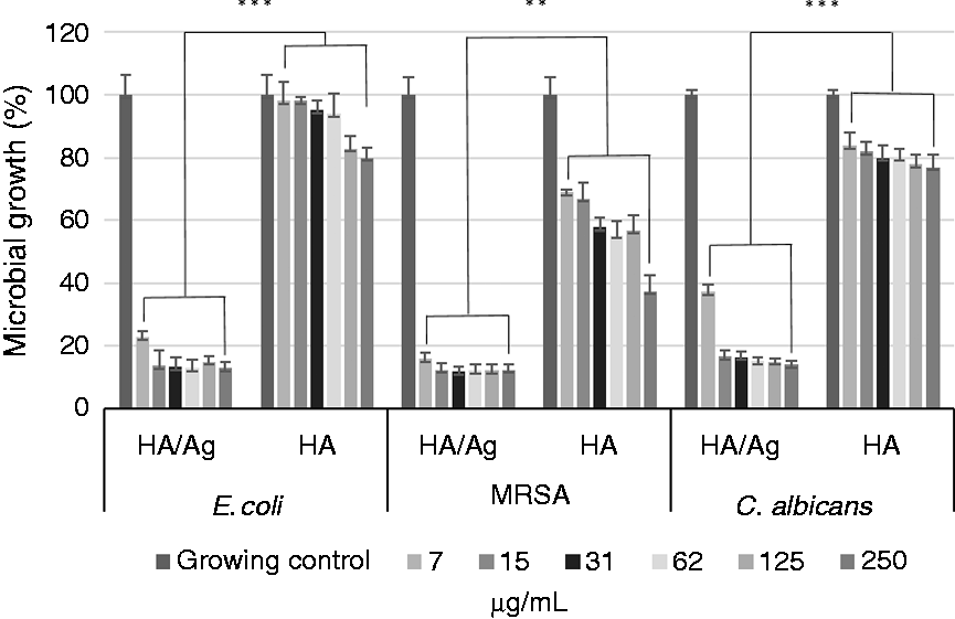
Antimicrobial activity of several concentrations of HA/Ag on E. coli, MRSA and C. albicans. **p < 0.01 and ***p < 0.001.
Discussion
The HA powder reveals irregular morphologies similar to those synthesized by Vukomanović et al. The diffraction angles at 25.8°, 31.7°, 32.1° and 32.7° are of higher intensity than those of the Joint Committee on Powder Diffraction Standards (JCPDS). 9–0432; in addition, 23 nm silver nanoparticles adhered to the surface of the HA were observed. The formation of metallic silver was co-tested by the angles 38.1° and 44.3°, corresponding to cubic silver according to the JCPDS. 04–0783. 20 The FTIR spectrum showed the HA characteristic bands as reported on literature. 29 The incorporation of Ag revealed a significant decreases of bands related with OH– (3456, 3222 cm−1) and the main functional groups of PO43– (1088 cm−1) CO32– (1666, 1629, 1461, 1343 cm−1); the observed changes suggest an interaction of silver ions and this radicals of HA.20,30
The XPS results showed that the presence of characteristic elements from the HA phase was confirmed in both samples (Table 2) and the elements present agree with the XRD, FTIR and EDS analysis. According to BE found for Ca, P, O and C for both samples, the slight shift to lower BE in the Ag-doped HA sample might be due to the Ag+incorporation into the structure. It can be explained considering the difference in radius sizes between Ca2+ and Ag+.31,32 Moreover, the incorporation of the Ag ions in the structure is also evident in the spectrum for HA/Ag (Figure 5), with the contribution at 373.02 and 368.69 eV, which corresponds to Ag3d5/2 and Ag3d3/2, respectively, and those, agree with the reported values.31–33
Bioactivity is a desired property in the case of biomaterials designed for bone tissues regeneration. Bioactivity test in SBF solution demonstrated that our HA/Ag powders are able to induce the nucleation and precipitation of Ca and P with the typical morphology of HA. The results of SEM-EDS indicate that HA/Ag after immersion in SBF undergoes characteristic structural changes in the process of apatite formation. The first change is the growth of a Ca-rich layer. The second structural change is based on interaction of the Ca-rich surface with negative ions such as phosphate to form a Ca-poor layer. Then, the layer crystallizes to form apatite. This process leads to changes in the Ca/P ratio due to ion exchange. 27 This process happens in a cyclical way, starts with the formation of amorphous calcium phosphate which is then modified forming intermediate phases like TCP (1.5), OCP (1.33) and at the end transforming into HA.34,35 The change in the surface structure of HA/Ag reveals the electrostatic interaction of the HA/Ag with calcium and phosphate ions in the SBF; these elements are the main constituents of the mineral phase in natural bone. In the case of our HA/Ag powders, the Ca/P ratio was closer to 1.67, which is the value of the stoichiometric HA. This study confirms the ability of HA/Ag to promote mineralization/apatite nucleation in vitro. On the other hand, the addition of Ag does not affect the formation of the HA structure; moreover, studies reported that silver nanoparticles of 20 nm have osteogenic activity on urine-derived stem cells, increasing the production of osteogenic proteins such as osteocalcin, alkaline phosphatase, morphogenic bone protein and the RUNX2 transcription factor. 36
Silver is known for its strong antimicrobial properties against Gram-positive and Gram-negative bacteria, viruses, and fungi.37,38 Therefore, it is widely used to limit the growth of microorganisms in medicine, dentistry16,37,39 and in other fields. 40 The interactions between HA/Ag and human cells have been studied and confirm that lower concentrations are highly compatible with osteosarcoma (U-2 OS) and fetal lung fibroblasts (IMR-90), but higher concentrations (5,10 wt %) of Ag into the HA showed a toxic effect capable of inducing morphological changes similar to those observed in bacterial infections. 19 In our study, the obtained HA/Ag nanocomposites with 0.01% of nanometric silver showed 80% of antimicrobial activity against E. coli, 85% against MRSA and 63% against C. albicans at concentrations of 7 μg/mL, in contrast with reports of Costescu et al. 41 where HA doped with silver ions inhibited the growth of Klebsiella pneumoniae, Candida krusei, Escherichia coli and Bacillus subtilis but at higher concentrations (5 mg/mL).42,43
The viability effect of HA/Ag against different cell lines has been evaluated in vitro on human and mouse osteoblasts,18,44–47 human stem cells, 48 and mouse fibroblasts 49 as well as in vivo studies in rats involving the implantation of HA/Ag. We obtained 100% of viability on MG-63 cells treated with 7 μg/mL of HA and 0.01% of nanometric silver. The increase of content and the rate of release of silver ions produce a negative impact on tissues, inhibiting their growth and development. Choi et al. showed that HA powders at 0.15% ionic Ag-doped were mildly toxic, whereas concentrations of 1.5 and 4.3% Ag-doped HA were moderately toxic. 44 Other experiments on human osteoblasts (hFOB) have shown that HA with silver content of 6% weight with particles of 150–212 μm used as a coating on pure titanium significantly inhibits the growth of osteoblasts and leads to the death of some of them, while 2% and 4% weights represent contents that enable a good balance between effective antimicrobial and cytotoxic activity. 45
The induction of bone resorption is partly related to the presence of bacteria and LPS. 50 Microbial infection produces a variety of products such as NO and cytokines that elicit a host response consisting of the expression of various signaling molecules and the recruitment of inflammatory cells. This process may culminate in tissue destruction and interfere with tissue regeneration and repair.51–53 NO production is associated with bone loss in some inflammatory conditions, leading to osteoclastogenesis, thereby favoring bone resorption.8,14 High circulating levels of TNF-α and IL-1 are directly linked to bone destruction. TNF-α and IL-1 have also been shown to inhibit collagen synthesis, and bone formation via inhibition of osteoblast differentiation and suppression of osteoblast function. IL-6 has been shown to increase in osteoclastogenesis by regulating the differentiation of progenitor cells into osteoclast.53,54
Due to the risks associated with endotoxins (LPS) in biomaterials and medical devices, the amount of endotoxin should not exceed 0.5 EU/mL, according to the guidelines of the US Food and Drug Administration. The LPS determination in the HA/Ag obtained for this study shows that the concentration of LPS is below 0.06 EU/mL by LAL assay55,56 rejecting the pro-inflammatory effect by endotoxin contamination.
When our HA/Ag powders were incubated in combination with LPS, the production of NO and pro-inflammatory cytokines such as TNF-α and IL-1 induced by LPS at 24 h and IL-6 at 48 h was attenuated significantly. This effect can be explained by structural modifications of LPS due to the interaction of HA and its molecular components, inhibiting the formation of links with reactive groups of LPS and its inflammatory activity. Ansari et al. reported the interaction of bacterial biomolecules such as LPS, a major component of Gram-negative bacteria cell wall, and L-a-phosphatidyl-ethanolamine, the principal bacterial outer-membrane phospholipid, with silver nanoparticles (AgNP) by ATR-FTIR. The IR spectrum of LPS showed changes in hydrogen bonding on O antigen of the LPS after exposure to AgNP. The stability of LPS decreased by the addition of Ca2+ ions binding to PO-4 in the lipid A region. It is likely that the LPS lost amphiphilic properties due to the structural changes induced by the AgNP and Ca2+ of the compound HA/Ag.57,58 Bone biomaterials with capacity to modulate the local immune environment that allows a significant effect on cell operation, determines the final outcome of bone regeneration and osteointegration.
Conclusion
Experimental data suggest that our modified method of synthesis allowed to incorporate silver nanoparticles on HA surface. The HA/Ag nanocomposite demonstrated bioactive capacity, and the antimicrobial activity against the tested microbial strains improved at low concentrations of silver without affecting the cell viability. In addition, HA/Ag attenuates the inflammatory effect, decreasing the production of cytokines such as IL-1 and TNF-α induced by LPS. Altogether, HA/Ag composites obtained could be an interesting alternative for the repair and reconstruction of bone tissues.
Footnotes
Acknowledgements
We thank the Laboratory of Immunology and Virology, Faculty of Biological Sciences, Universidad Autónoma de Nuevo León, San Nicolas de los Garza, Mexico. MSc. Alejandra Arreola for participating in the review of the article. We thank Natalia Angel and Silvia Santillana for their support. We also thank CENAPROT and LIDTRA national laboratories at Cinvestav-Querétaro.
Declaration of Conflicting Interests
The author(s) declared no potential conflicts of interest with respect to the research, authorship, and/or publication of this article.
Funding
The author(s) disclosed receipt of the following financial support for the research, authorship, and/or publication of this article: This study was supported by the funding of Laboratory of Immunology and Virology, scholarship no. 592735 and by basic research science project (251372) granted by CONACYT in México.