
Abstract
Combination of chemotherapy and small interfering RNA (siRNA)-based nanotherapeutics could cooperatively and effectively suppress multiple drug resistance in cancer development. Particularly, nano-delivery systems that efficiently encapsulate two or more therapeutic payloads to the tumor-targeted sites have been proven significantly. Here, we prepared a micellar nanoparticle formed by monomethoxy poly (ethylene glycol)–block–poly(ε-caprolactone)-block-poly(L-lysine)/cholesterol (mPEG-b-PCL-b-PLL/Chol, further abbr. as “P”). In this work, PCL as the hydrophobic core to encase doxorubicin and PLL as the cationic moiety to bind with negatively charged siRNA are used to achieve the co-delivery of therapies in one platform. The results showed that the micellar nanoparticle is capable of delivering siRNA and doxorubicin simultaneously to the same tumor cells, and consequently displays enhanced inhibition efficiency on MCF-7/ADR cells. Moreover, real-time polymerase chain reaction (RT-PCR) and Western blot experiments indicated that the co-delivery of micelleplex-delivering B-cell lymphoma 2 specific siRNA (siBcl) successfully down-regulated the expression of Bcl-2 protein, which triggers chemotherapy to be more sensitive to the cancer cells. Therefore, the strategy of co-delivering anticancer drug and siRNA showed promising potential in reversing drug resistance of tumor cells.
Introduction
Cancer treatments have been significantly refined due to an increased awareness of the molecular, cellular, and physiological mechanisms involved in the initiation and progression of the disease. 1 Chemotherapy is one of the most commonly utilized cancer treatment methods; however, it is often accompanied by systemic side effects and multidrug resistance. 2 In recent years, numerous studies have revealed that the combination of siRNA and chemotherapeutic drugs may be a new promising strategy to enhance therapeutic efficacy and decrease drug resistance by cooperatively prohibiting cancer development with different mechanisms.3,4 On the one hand, drug efflux can be bypassed, as the primary mechanism of cellular uptake would be endocytosis. 5 On the other hand, down-regulation of apoptotic proteins such as Bcl-2 expression has shown to increase the cytotoxicity of chemotherapy drugs in MDR cells. 6 However, naked small interfering RNA (siRNA) is easily degraded by RNases in vivo and is difficult to pass through cell membranes due to its electronegativity.7,8 Highly efficient and safe delivery vectors, which are suitable for systemic delivery of siRNA, remain a major hurdle for RNA-based cancer therapy.9,10
Nanomaterials-related technology can facilitate the advanced transport systems with dual capacity for siRNA and chemotherapeutic drugs.11,12 Li et al. used the low-molecular-weight branch PEI to conjugate hydroxypropyl-β-cyclodextrin (HP-β-CD) and folic acid (FA), forming the co-delivery nanocarrier (FA-HP-β-CD-PEI) to encapsulate DOX with the cavity HP-β-CD and bind siRNA with the positive charge of PEI for tumor-targeting co-delivering drugs. 13 Their study indicated that combined RNAi therapy and chemotherapy using functional co-delivery nanocarriers could produce synergistic effects to overcome MDR and enhance apoptosis in MDR cancer cells. 13 However, the polymers they used are not biodegradable and toxic.
The optimal delivery systems for combination of drug and gene delivery should have several characteristics. Firstly, the materials used as the carrier should be less toxic or non-toxic and biodegradable. Secondly, the delivery systems should have high transfection capabilities. 14 Thirdly, synergistic payload interactions should be achievable. Last but not least, the systems should have capabilities to target tumor cells. 15 Thus, biodegradable amphiphilic cationic copolymers have been employed as a promising platform for drug delivery. 16 Their amphiphilic feature is beneficial to micelle formation with drugs, and their cationic segments can combine with anionic RNA chains. Compared to simple polyplexes, micelle-complexes display unique advantages such as nanoscale dimension, segregated core/shell structure, protective effect of the hydrophobic core on encapsulated drugs, and immune-sheltering function of the hydrophilic shell. 17 We have recently developed a micelle system based on the assembly of a biodegradable triblock copolymer poly(ethylene glycol)-block-poly(ε-caprolactone)-block-poly(L-lysine) (mPEG-bPCL-b-PLL). We have demonstrated that mPEG-b-PCL-b-PLL vectors can efficiently deliver siRNA into cancer cells and induce significant gene silencing. 18
This study aims to develop an mPEG-b-PCL-b-PLL vector for delivery of siRNA and chemotherapeutics in one platform. The triblock copolymer is amphiphilic and can self-assemble into micellar nanoparticles, PEG as the hydrophilic corona to enhance circulation time, PCL as the hydrophobic core to encase DOX and PLL as the cationic shell to carry negatively charged siRNA (Figure 1). Moreover, cholesterol (Chol) is attached to the PLL block of mPEG-b-PCL-b-PLL to further increase the efficiency of cellular internalization and to speed up the endosomal escape. It is believed that the low transfection efficiency and poor capacity of endosome/lysosome escaping of the complex also are responsible for the low transfection efficiency at low N/P ratio. 19 We confirmed the performances of our micelleplex system including complex architecture and stability, drug release, cellular uptake and transfection efficiency. We also examined the ability of this micelleplex to simultaneously deliver Bcl-2-specific siRNA (siBcl) and DOX into the same tumor cells, and further demonstrated the synergistic tumor suppression effect.
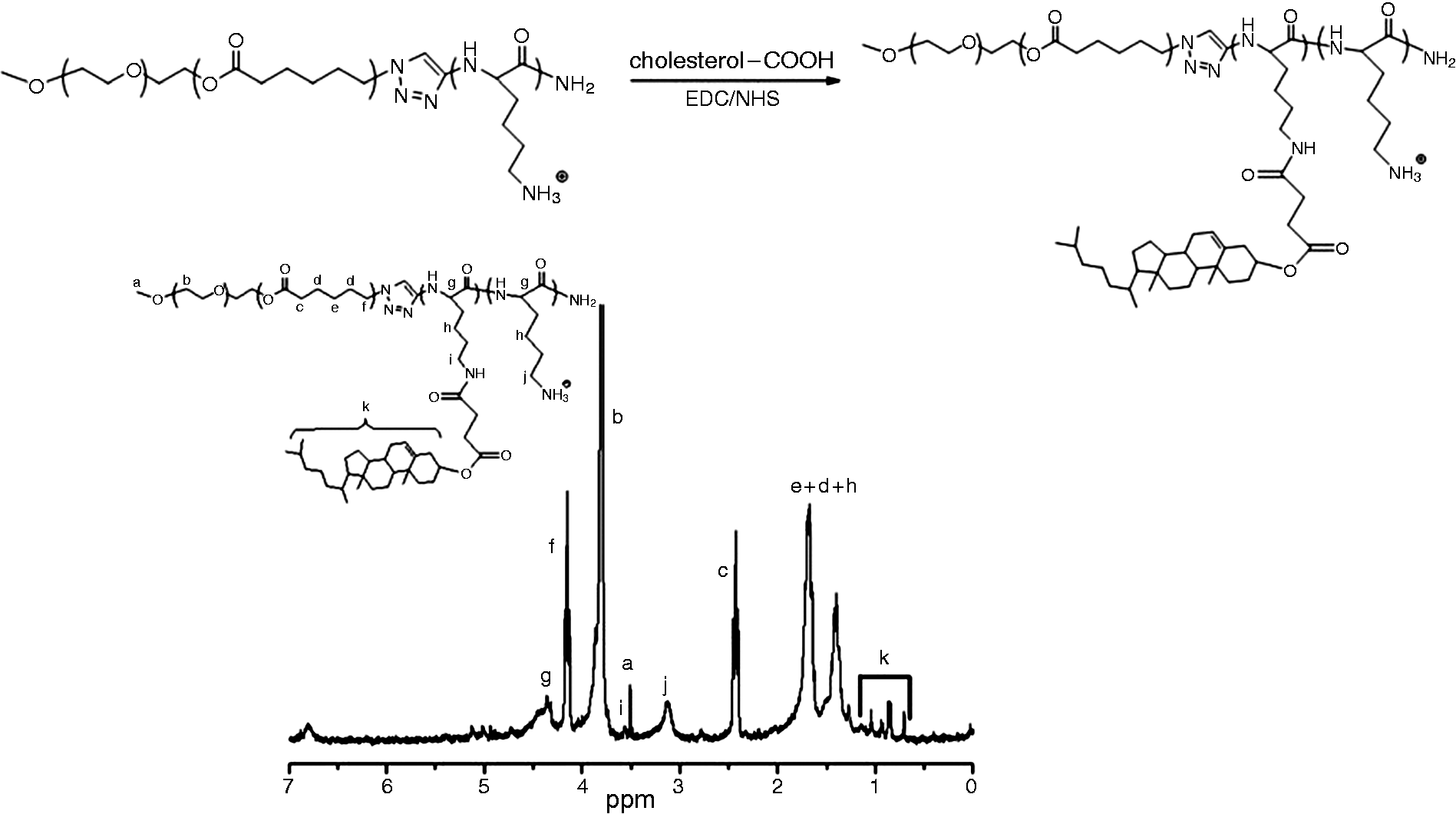
Synthesis of mPEG-b-PCL-b-PLL/Chol cationic triblock copolymer and the 1H-NMR spectrum of mPEG-b-PCL-b-PLL/Chol.
Experimental
Materials
Poly(ethylene glycol)-block-poly(ε-caprolactone)-block-poly(L-lysine) triblock copolymers (mPEG-bPCL-b-PLL) were synthesized as previously described in Sun et al.
16
Cholesterol (Chol), 4-dimethylaminopyridine (DMAP), diisopropylethylamine (DIEA), 1–(3-dimethylaminopropyl)-3-ethylcarbodiimide hydrochloride (EDC·HCl), and N-hydroxysuccinimide (NHS) were purchased from Sigma-Aldrich. 2–(4-Amidinophenyl)-6-indolecarbamidinedihydrochloride (DAPI) and LysoTracker Red TM were supplied by Beyotime Co. (Beijing, China). Control siRNA that targets the sequence 5′-
Preparation and characterization of mPEG-b-PCL-b-PLL/Chol
mPEG-b-PCL-b-PLL/Chol were synthesized as previously described. 11 In brief, the cholesterol-modified copolymer mPEG-b-PCL-b-PLL/Chol was synthesized as Figure 1: 100 mg of copolymer was dissolved in 50 mL of Milli-Q water, and the pH value was adjusted to 9.0; then, the water and THF mixture solution of EDC·HCl, NHS, and cholesterol-COOH of 1.1 equiv. of the amino groups to be converted were added dropwise; the mixture was stirred overnight, dialyzed against THF for 48 h to remove the unreacted cholesterol-COOH.
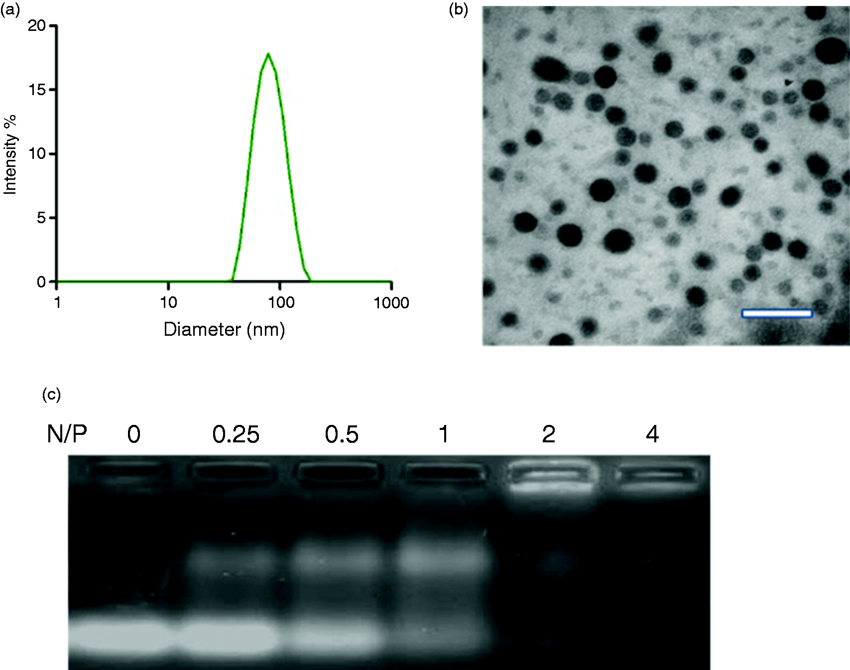
Characterization and optimization of the micellar nanoparticle. (a, b) Dynamic light scattering analysis and transmission electronic microscopic image (scale bar: 300 nm) of micellar nanoparticle; (c) binding ability of micellar nanoparticles to siRNA at different ratios of nitrogen in carrier to phosphate in siRNA (N/P ratio) demonstrated by the gel retardation assay.
1H-NMR spectrum was recorded on a Bruker AV300 NMR spectrometer. Deuterated chloroform containing 0.03 vol% tetramethylsilane and deuterated trifluoroacetic acid were used as the solvent for NMR measurements.
Fabrication and characterization of micellar nanoparticles
To prepare complexes from mPEG-b-PCL-b-PLL/Chol (Abbr. as “P” hereafter) with DOX and siRNAs, corresponding copolymer with DOX was dissolved in 1 mL of tetrahydrofuran and stirred at room temperature for at least 2 h (Abbr. as “PD” hereafter). Then, RNase-free water was added dropwise into the stirring solution. The mixture was stirred for an additional 1 h, followed by removal of tetrahydrofuran. Then, the solutions were diluted to desired concentrations and mixed with an siRNA solution (Abbr. as “PD/siRNA” hereafter).
Zeta potential and particle size measurements were performed on a Zeta sizer Nano ZS particle size analyzer. JEOL-1000 transmission electron microscope (TEM) was used with an accelerating voltage of 100 kV. The samples were prepared by pipetting a drop of the micelle solution (1 g/L) onto a 230 mesh copper grid coated with carbon and allowing the sample to dry in air before measurements.
The siRNA binding ability of the cationic amphiphilic copolymers was evaluated by agarose gel electrophoresis. The micelleplexes were prepared at N/P molar ratios of 0, 0.25 0.5, 1, 2 and 4. Electrophoresis was carried out on 2% agarose gel at 100 V for 15 min in TAE buffer solution (40 mmol/L Tris-HCl, 1 vol% acetic acid, and1 mmol/L EDTA). The retardation of the siRNA was visualized by staining with ethidium bromide, and by imaging under a UV lamp using a UVP EC3 bioimaging system (CA, USA).
For in vitro drug release, 1 mL of PD and PD/siRNA micelle solutions (0.5 mg/mL) were transferred to a dialysis tube (3500 D) and dialyzed in 10 mL of different release mediums: pH 7.4 PBS and pH 5.5 PBS. All samples were stirred at 37°C in a water-bath at 100 r/min. At defined time intervals, 1 mL of release medium was collected and replaced with same volume fresh PBS. The concentration of DOX in the release medium was performed by using a microplate reader fluorescence spectrophotometer (Bio-Rad Laboratories Ltd, Hertfordshire, UK). All assays were performed in three replicates.
Evaluation of toxicity and anticancer efficacy of PD/siRNA
The cytotoxicity of the copolymers was assessed with MTT viability assay against drug-sensitive human breast cancer (MCF-7) and adriamycin-resistant MCF-7 cells (MCF-7/ADR). The cells were seeded in 96-well plates (5000/well) in 100 μL of complete RPMI 1640 supplemented with 10% FBS, and incubated at 37°C in 5% CO2 atmosphere for 24 h. For the cytotoxicity of copolymers, 100 μL of copolymer solutions in RPMI 1640 at a series of concentration was added to the culture medium for 48 h. Then, MTT solution (5.0 mg/mL) was added to each well (10 μL), and the cells were incubated for an additional 4 h. After incubation, the reaction was stopped by the addition of 100 μL of dissolving solution, and the absorbance was measured at 570 nm using a microplate reader (Bio-Rad Laboratories Ltd, Hertfordshire, UK). All assays were performed in three replicates.
Cellular uptake of the PD/siRNA delivery system
Flow cytometry and confocal laser scanning microscopy (CLSM) were used to quantitate the uptake behavior of different formulations in MCF-7/ADR cells. PD containing both DOX and FAM-labeled siRNA was prepared as described above (PD/FAM-siRNA). Cells were grown as a monolayer and incubated with PD/FAM-siRNA (DOX concentration, 5 μg/mL) and diluted in culture medium for 4 h and 8 h at 37°C. The cells were then washed three times with cold PBS and fixed with 4% paraformaldehyde solution followed by 5 μg/mL of DAPI staining in PBS and observed using CLSM (NikonA1 HR Confocal, Tokyo, Japan). Meanwhile, the cells were then washed three times with cold PBS and digested with trypsin-EDTA solution (0.25%, 3 min), collected by centrifugation (1500 r/min, 5 min). The mean fluorescence intensity of DOX in cells was analyzed by an FACS Sort flow cytometry (Beckman Coulter, CytoFLEX, CA, USA).
In vitro siRNA transfection and gene silencing
The cellular levels of Bcl-2 mRNA and protein were assessed using quantitative real-time PCR (qRT-PCR) and Western blot, respectively. Cells were incubated with various formulations and diluted in culture medium for 24 h at 37°C. In qRT-PCR analysis, total RNA from transfected cells was isolated using the RNeasy mini-kits (Qiagen, Germantown, MD) according to the protocol of the manufacturer. Two micrograms of total RNA were transcribed into cDNA using the Prime Script First Strand cDNA Synthesis Kit (Takara, Japan). Thereafter, 2 μL of cDNA was subjected to qRT-PCR analysis targeting Bcl-2 and glyceraldehyde 3-phosphate dehydrogenase (GAPDH) using the SYBR Premix Ex Taq (Perfect Real Time) (Takara). Analysis was performed using the Applied Biosystems Step One Real Time PCR Systems. Relative gene expression values were determined by the ΔCT method using Step One Software v2.1 (Applied Biosystems). Data are presented as the fold difference in Bcl-2 expression normalized to the housekeeping gene GAPDH as the endogenous reference, and relative to the untreated control cells. Primers used in qRT-PCR for Bcl-2 and GAPDH are: Bcl-2-forward
In Western blot analysis, transfected cells were first washed twice with cold PBS, and then resuspended in 50 μL of lysis buffer freshly supplemented with Roche's Complete Protease Inhibitor Cocktail Tablets. The cell lysates were incubated on ice for 30 min and vortexed every 5 min. The lysates were then clarified by centrifugation for 10 min at 12,000 × g. The protein concentration was determined using the BCA Protein Assay Kit (Thermo, Madison, WI). Total protein (50 μg) was separated on 12% Bis-Tris-polyacrylamide gels and then transferred (at 300 mA for 45 min) to Immobilon-P membranes (Millipore, Bedford, MA). After incubation with 5% bovine serum albumin (BSA, Sigma-Aldrich) in phosphate buffered saline with Tween-20 (PBST, pH 7.2) for 1 h, the membranes were incubated in 1% BSA in PBS with monoclonal antibodies against Bcl-2 (1:500) overnight. After incubation in 1% BSA with goat anti-mouse IgG-HRP antibody (1:10,000) for 30 min, bands were visualized using the ECL system (Pierce).
Cell apoptosis analyses post-siBcl transfection
Cell apoptosis was assessed with flow cytometry using the annexin V-PE apoptosis detection kit I (BD Biosciences, San Jose, CA, USA) according to the manufacturer’s instructions. MCF-7/ADR cells cultured in six-well plates were treated with the abovementioned formulations at a DOX dose of 5 μg/mL. After 72 h of treatment, apoptotic cells were detected on flow cytometry using the annexin V-FITC Apoptosis Detection Kit I, and the results were analyzed using WinMDI 2.9 software.
Statistical analysis
All data represent mean values ± standard deviation of independent measurements. Statistical analysis was performed with a Student's t-test (two-tailed). Statistical significance was assigned at p < 0.05 (95% confidence level).
Results and discussion
Characteristics of copolymer for micellar nanoparticles
In this work, the cationic copolymer was further improved by attaching cholesterol onto the ε-terminal amino groups of the PLL segments through the EDC/NHS condensation (Scheme 1). The chemical structure and 1H-NMRspectrum of mPEG-bPCL-b-PLL/chol are given in Figure 1. mPEG-b-PCL-b-PLL/Chol contained all signals related to the mPEG, PCL, PLL blocks and the cholesteryl hemisuccinate (peak k). A new peak appeared at δ = 3.52, which was assigned to the methylene protons adjacent to the amide group formed between the cholesteryl hemisuccinate and the ε-terminal amino group of the lysine residue.
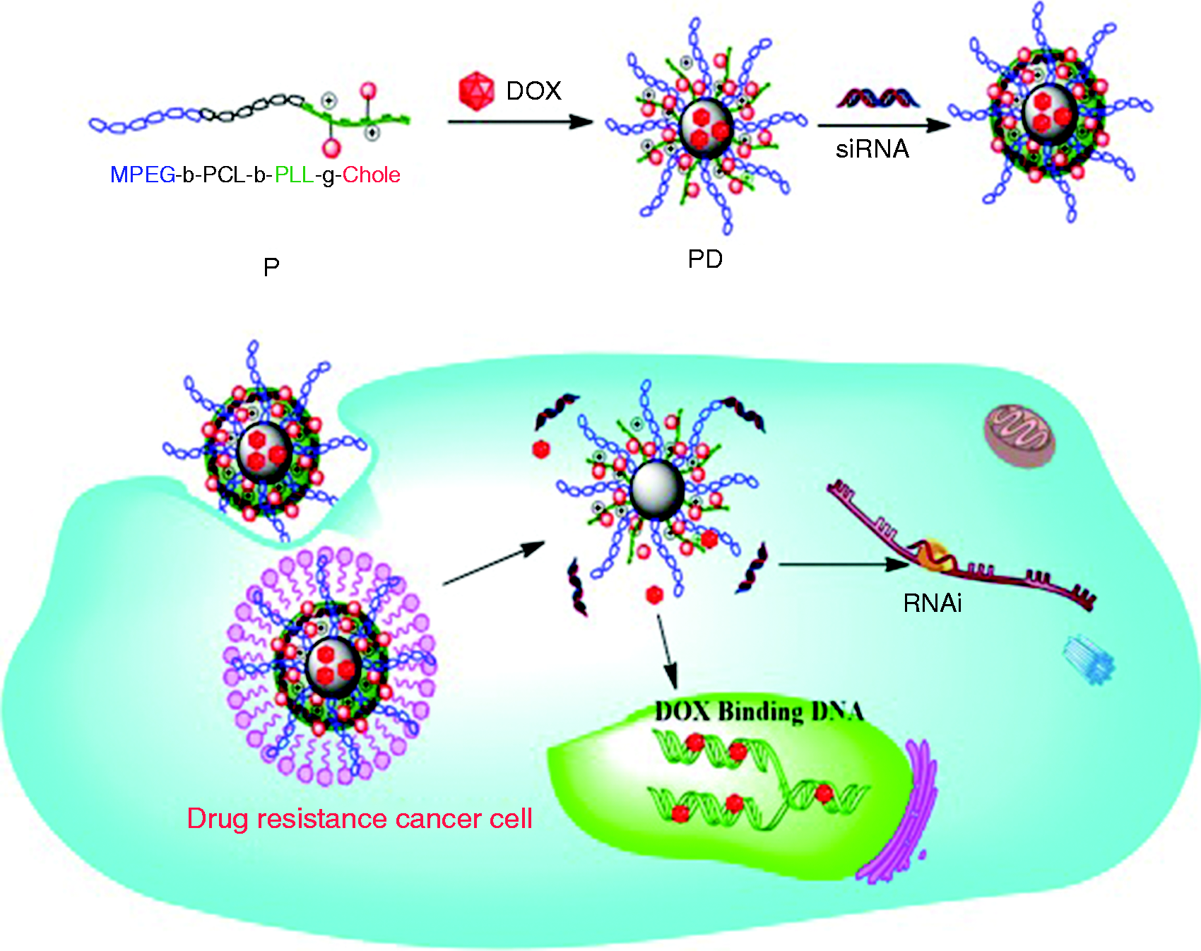
Chemical structure of mPEG-b-PCL-b-PLL/Chol cationic triblock copolymer (P). Doxorubicin was encapsulated in the P (PD) through the self-assembly of the triblock copolymer, the positive charge moiety of PLL for further binding with small interference RNA (siRNA) via electrostatic interaction. Schematic illustration of micellar nanoparticle formation is designed as a co-delivery platform for drug resistance cancer cell.
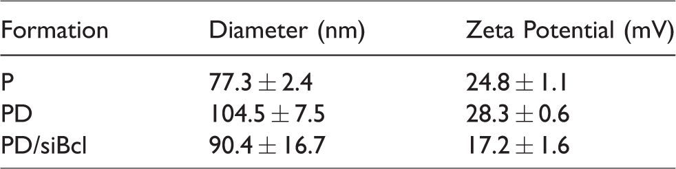
The polymer formed a micellar structure in aqueous solution and exhibited the ability of simultaneous loading of siRNA and DOX. DOX was entrapped in the hydrophobic PCL core via a hydrophobic–hydrophobic interaction. SiRNA was subsequently absorbed to the assembly through a charge interaction with the PLL block (Scheme 1). The micellar nanoparticles formed were spherical (Figure 2(a)) with a size of (77.3 ± 2.4) nm (P), (104.5 ± 7.5) nm (PD) and (90.4 ± 16.7 nm) (PD/siBcl), which were measured via TEM (Figure 2(b)) and DLS (Table 1). The zeta potentials of P, PD and PD/siBcl were 24.8 ± 1.1 mV, 28.3 ± 0.6 mV and 17.2 ± 1.6 mV, respectively. DOX with positive charge led to increase in the zeta potential. The zeta potential of PD/siBcl was the lowest due to the siRNA complexing with PLL. The zeta potential near 17 mV may benefit its affinity to the cells and internalization by the cells. 17 Efficient siRNA binding occurred at a molar ratio of nitrogen in the carrier/phosphate in siRNA at different N/P ratios as demonstrated by a gel retardation assay (Figure 2(c)). As expected, the micellar nanoparticles were capable of effectively binding siRNA when the N/P is above 1, resulting in disappearance of siRNA bands in agarose gel.
Particle size and zeta potential of micellar complexes of P, PD and PD/siBcl.
DOX released from micelles was studied under various conditions to verify the sensitivity of the micelles to acidic conditions. Different pH conditions were simulated to investigate the effects on micellar release, such as lysosomes (pH 5.5) and normal tissues (pH 7.4) (Figure 3). At pH 7.4, there was no significant difference in the release rate between PD and PD/siRNA (almost 50% during 96 h), and the rest 51.5% may be still incorporated in the hydrophobic core of the PCL. At pH 5.5, the release rates of DOX were faster and higher as compared to pH 7.4 (more than 75% during 96 h). The release rate of DOX from PD was slightly greater than PD/siRNA, which may be attributed to the electrostatic interaction between DOX and siRNA. The released DOX of PD and PD/siRNA with a pH-sensitive behavior could be attributed to the large amount of protonated amino groups in PLL that resulted in the charge repulsion. Additionally, low pH environment effectively accelerates the release proliferation of acidified DOX.19,20

The release proliferation of DOX from micellar nanoparticle at different pH conditions. Results are presented as the mean of three measurements ± standard deviation (n = 3).
Cellular uptake and internalization of the micellar nanoparticles
A key factor for achieving effective therapeutic is to successfully deliver the drugs into the targeted cells and tissues. 21 To demonstrate the simultaneous delivery, we first analyzed the cellular uptake and intracellular distribution of PD/FAM-siRNA micelleplex in MCF-7/ADR cells. Confocal microscopy was used to monitor the intercellular accumulation of particles at different time points (4 and 8 h) by tracking the red fluorescent DOX and green fluorescent siRNA, while the nucleus was stained with DAPI (Figure 4(a)). The PD/FAM-siRNA was rapidly absorbed by the cells after 4 h incubation and both DOX and siRNA successfully reached the cytoplasm as indicated by the yellow dots. At 8 h, most of DOX was present in nucleus indicated as purple dots. Flow cytometer was used to measure cellular uptake of FAM-siRNA and DOX at various time points after incubation (0, 4 and 8 h). The percentages of cells with internalized PD/FAM-siRNA were 0% (0 h), 74.6% (4 h) and 95.3% (8 h) (Figure 4(b)). These results indicated that the platform could effectively deliver the siRNA and DOX into the tumor cells. This platform overcomes the drawbacks of the siRNA with negative charge and DOX with the hydrophobic properties, which result in unsuccessfully crossing the cell membrane. 22 This may be due to the strong interaction of cholesterol with the cell membrane and the endosomal membrane. 23
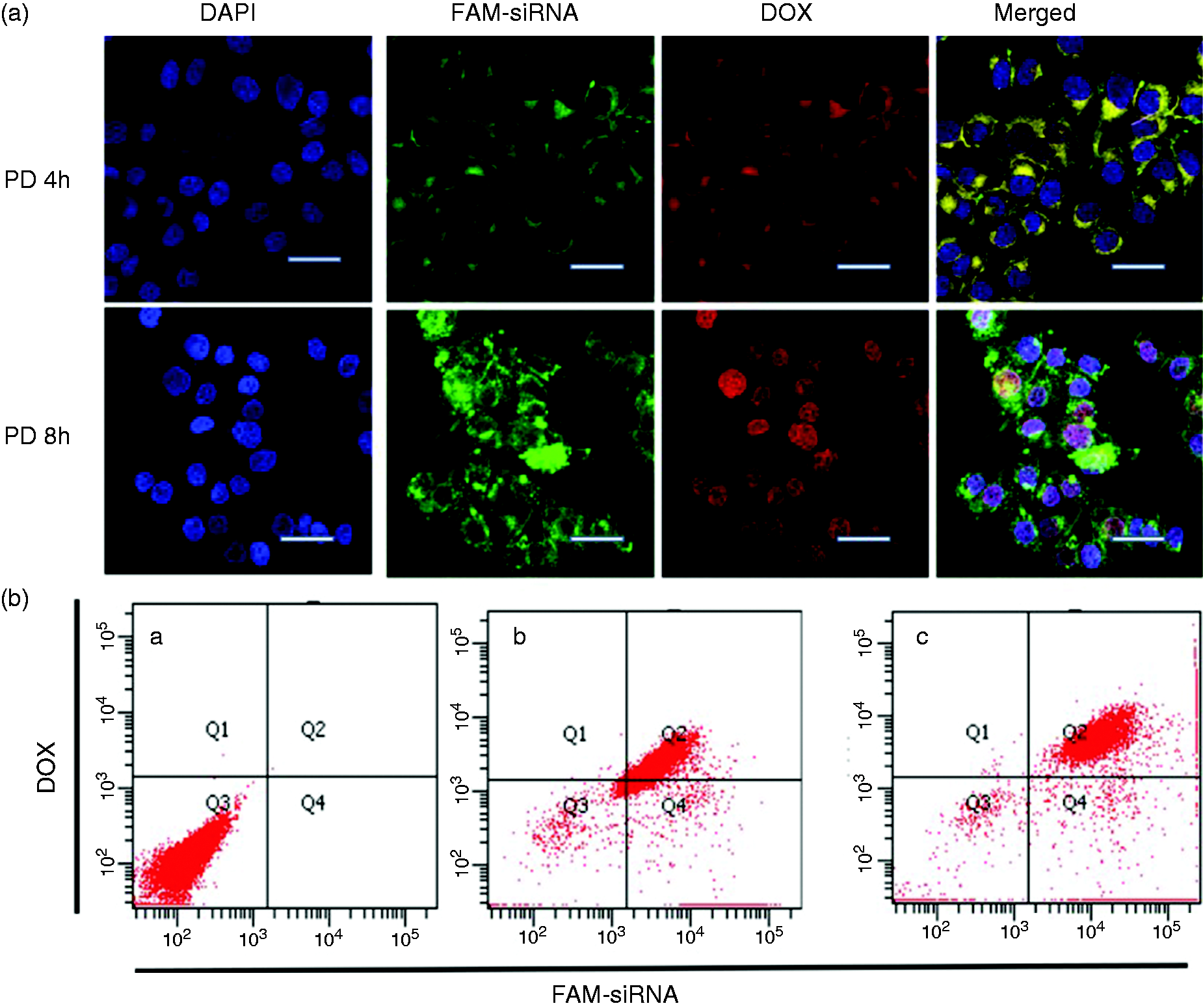
Cellular uptake and internalization efficiency of micellar nanoparticles. (a) Confocal laser scanning microscope (CLSM) image of intracellular distribution of DOX (red) and FAM-siRNA (green) in MCF-7/ADR cells. The nuclei were stained with DAPI (blue), the scale bar is 10 μm and (b) Cellular uptake of PD/FAM-siRNA over time as analyzed by flow cytometer.
Evaluation of cytotoxicity and cell apoptosis in vitro
A tetrazolium-based colorimetric assay (MTT) was used to monitor the in vitro anticancer activity of micelles in drug resistance of breast cancer MCF-7/ADR cells. It has been reported that DOX could not enter nucleus of drug-resistant MCF-7/ADR cells, while it could enter nucleus of drug-sensitive MCF-7 cells significantly. 24 Therefore, DOX exhibited lower cytotoxicity in MCF-7/ADR than that in MCF-7 cells (Figure 5(a)). Blank siRNA-loaded micelles (P/siNC) did not affect the cell viability. PD/siNC and P/siNC with free DOX showed the same cytotoxicity in a dose-dependent manner (Figure 5(b)). However, the cytotoxicity of PD/siBcl was significantly higher than the other groups. This result indicated the synergistic effects between siBcl and DOX to suppress the MCF-7/ADR cell growth. As reported previously, inhibition of Bcl-2 showed increasing metabolic activity, which correlates with chemotherapeutic drugs to be more sensitive to the cancer cells. 25
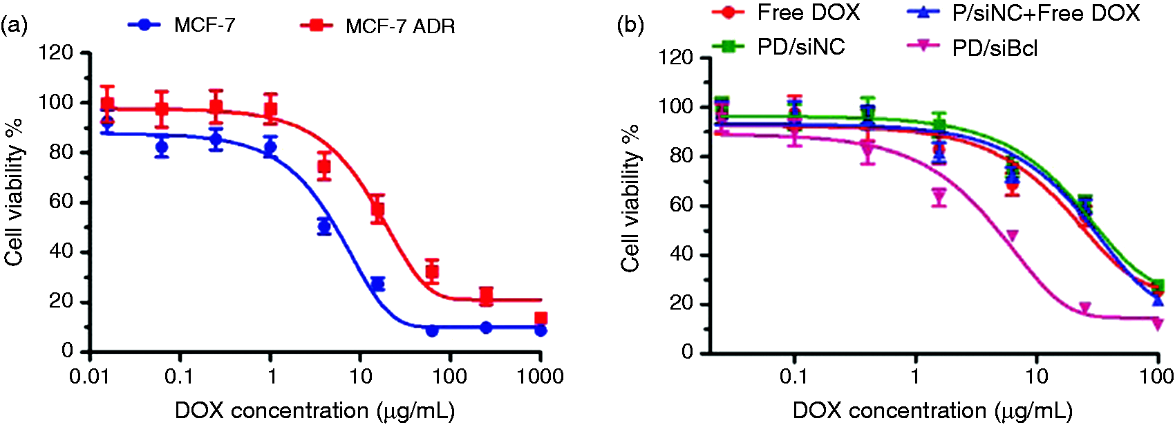
Evaluation of the cell viability. (a) Effect of co-delivery of siBcl and DOX by micellar nanoparticles on the cell viability of MCF-7 and MCF-7/ADR cells. The concentration of DOX varied from 0.01 to 1000 μg/ml and (b) In vitro cytotoxicity of DOX solution, P/siNC with free DOX, PD/siNC and PD/siBcl inMCF-7/ADR cells. Results are presented as the mean of three measurements ± standard deviation (n = 3).
The capacity of PD/siBcl to induce apoptosis was further studied by staining with SYTOX Blue and annexin V-Alexa Fluor 647. Early apoptotic cells are annexin V-Alexa Fluor®647 positive but SYTOX Blue negative because plasmic membrane is intact while phosphatidylserine is externalized. Double positive cells could be late apoptotic and necrotic. As shown in Figure 6, MCF-7/ADR cells were treated with PBS, P/siNC, P/siNC with free DOX, PD/siNC and PD/siBcl; the percentages of apoptotic cells are 11.3%, 18.9%, 52.9%, 50.3% and 75.1%, respectively. The ability of PD/siBcl to apoptosis is in accordance with their antitumor activity.
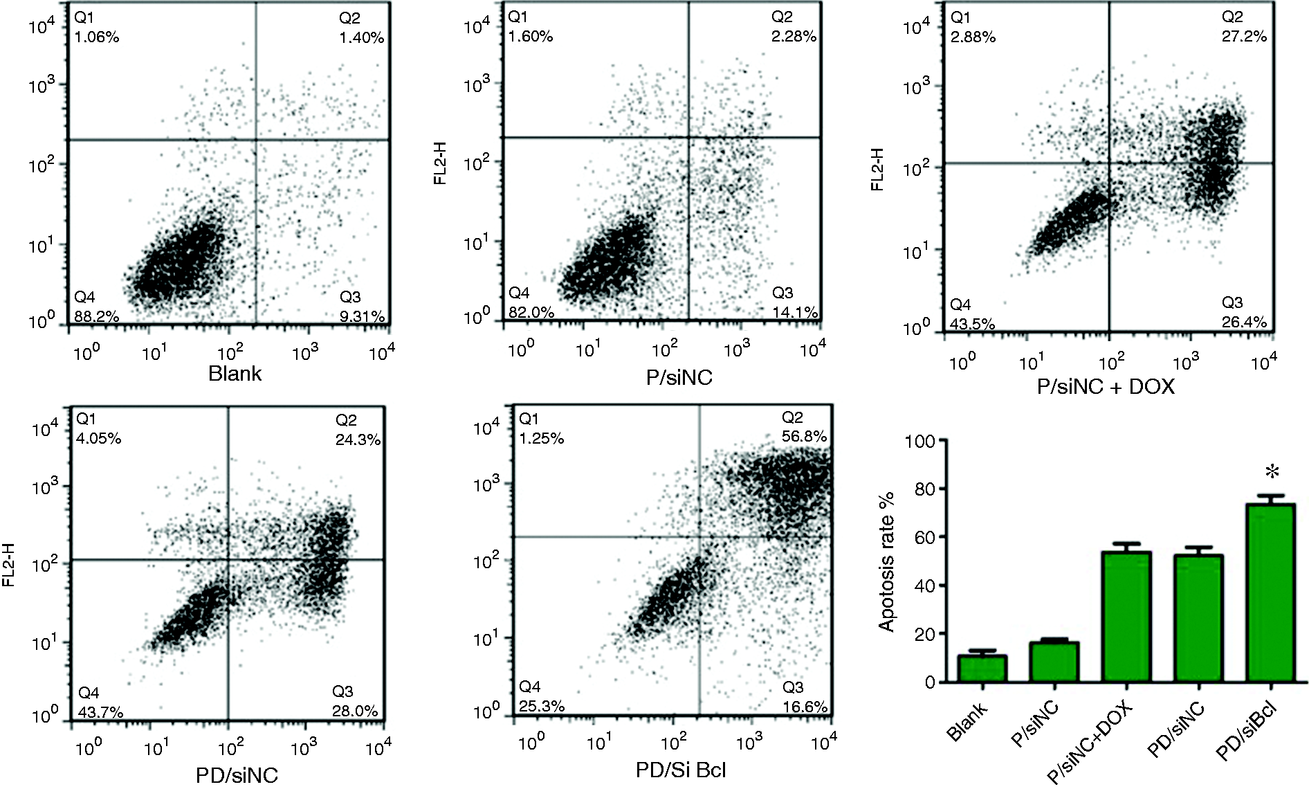
Evaluation of cellular apoptosis in MCF-7/ADR cells in response to PBS (Blank), P/siNC, P/siNC+DOX, PD/siNC and PD/siBcl. The early apoptotic cells are presented in the lower right quadrant, and fully apoptotic cells are presented in the upper right quadrant. Experiments were performed in triplicate and data are presented as mean + standard deviation. *p < 0.05.
Gene silencing of the micellar nanoparticles
Herein, we verified whether the micelleplex could efficiently knockdown the expression of the therapeutic target gene Bcl-2. As previously reported, Bcl-2 plays an important role in promoting cellular survival and inhibiting the actions of pro-apoptotic proteins. 26 Bcl-2 family proteins also dictate the ultimate sensitivity or resistance of cells to various apoptotic stimuli, including hypoxia, radiation, anticancer drugs, oxidants, and so on. 27 We incubated MCF-7/ADR cells with varous micelles for 24 h and then detected Bcl-2 mRNA expression using real-time PCR. As shown in Figure 7(a), DOX could induce the Bcl-2 gene expression to increase the drug resistance. 25 The micelle carrying blank siRNA (P/siNC or PD/siNC) showed no knockdown efficiency. However, the PD/siBcl and P/siBcl exhibit a high silencing efficiency of 46.8% and 62.7%, respectively. A reduction in Bcl-2 mRNA was subsequently accompanied by decreased Bcl-2 protein expression in a similar manner (Figure 7(b)) following transfection with variety micelles, as determined by Western blot analyses of Bcl-2 protein in the cell lysates 48 h after transfection. This result suggested that PD/siBcl would be a promising potential in reversing multidrug resistance of tumor cells.
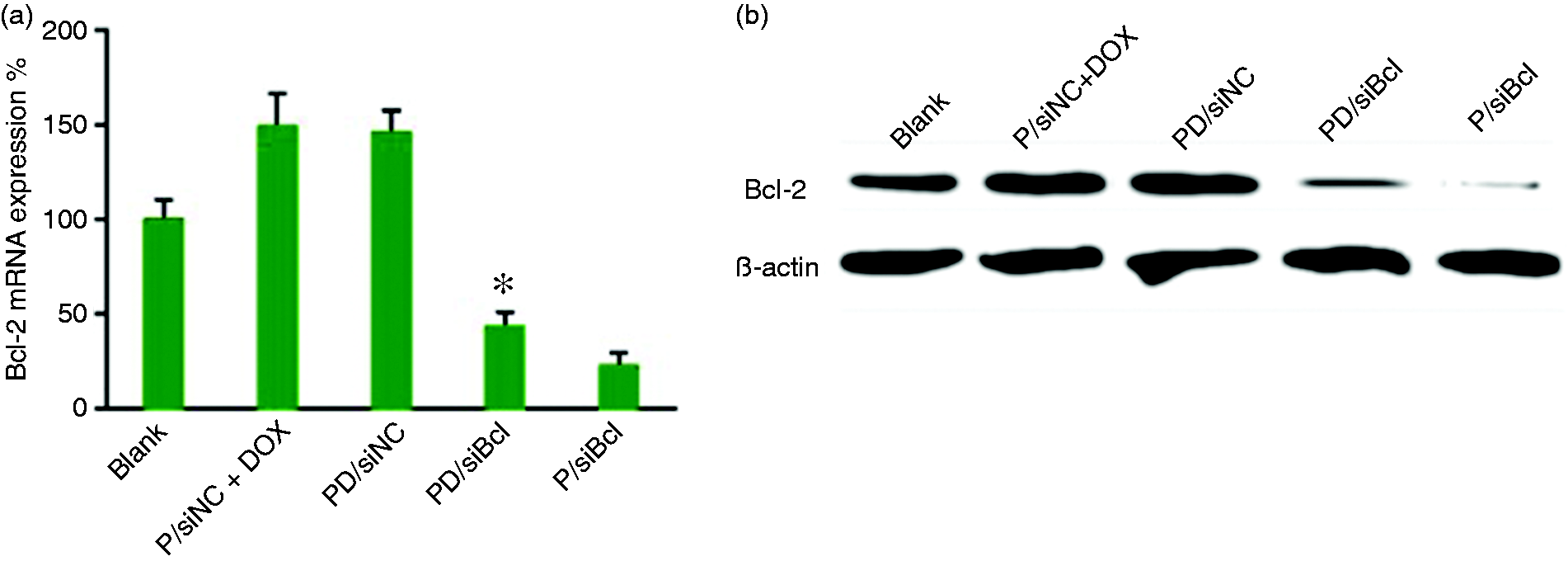
Evaluation of gene silencing on the mRNA and protein level expression in MCF-7/ADR cells in response to PBS (blank), P/siNC+DOX, PD/siNC, PD/siBcl and P/siBcl. Representative Bcl-2 expression determined by real-time PCR (a) and Western blot analysis (b). Experiments were performed in triplicate, and data are presented as mean + standard deviation. *p < 0.05.
Conclusions
In summary, this study investigated the potential of co-delivery of DOX and siBcl-2 in cationic copolymer formed micellar nanoparticles. Briefly, we synthesized triblock copolymers mPEG-b-PCL-b-PLL/Chol nanoparticles, which could load DOX via a hydrophobic interaction and bind siRNA by electrostatic interaction. The results indicated that the cholesterol-modified cationic copolymer exhibited the enhanced ability to escape from acidic environment like endosomes due to the strong interaction of cholesterol with the cell membrane and the endosomal membrane. According to the use of siBcl-2, it can be seen that PD/siBcl can down-regulate the expression of Bcl-2, which can increase the DOX sensibility of multidrug resistance of tumor cells. Therefore, the present study provides a useful strategy for constructing efficient DOX/siRNA co-delivery vehicles, which can be a promising approach for cancer therapy.
Footnotes
Authors' Contribution
Jianguo Gao and Lingxiao Chen contributed equally to the manuscript.
Declaration of Conflicting Interests
The author(s) declared no potential conflicts of interest with respect to the research, authorship, and/or publication of this article.
Funding
The author(s) disclosed receipt of the following financial support for the research, authorship, and/or publication of this article: This work was financially supported by the National Natural Science Foundation of China (31800833), the Wenzhou Medical University and Wenzhou Institute of Biomaterials & Engineering (WIBEZD2017001-03), Visiting Scholar Foundation of Key Laboratory of Biorheological Science and Technology (Chongqing University), Ministry of Education (No. CQKLBST-2018–005), the Wu Jie-ping medical foundation project (320.6750.15225), and the Zhejiang pharmaceutical health research fund project (2017KY638).