
Abstract
The irreversible correlation between the transfection efficiency and cytotoxicity of polycations has always been the huge obstacle that severely limited the gene delivery efficiency. The undesired inconsistency could be mainly attributed to the molecular weight (MW) of the polycations, that is, polymers with high MW and positive charge densities exhibited enhanced transfection efficiency but with associated cytotoxicity. To address such critical challenge, we developed the degradable high MW polymers strategy which could condense DNA for the internalization and release DNA upon the trigger-responsive degradation. In this work, two kinds of degradable PEI-based polymers were prepared via the polyaddition of PEI600 and dienes with ester groups or thiol ketal groups. The PEI-based degradable polymers could efficiently condense plasmid DNA to form the nano-complexes with the size around 180 nm. They also exhibited improved gene delivery efficiency compared with commercial available transfection reagent, PEI25k, and were 380–520 folds higher than PEI600 in HeLa cells. The toxicity of the polymers could be reduced by the rapid degradation upon acid or ROS triggering as well as intracellular release of DNA and the cell viability could reach higher than 80% even at high doses. ROS-responsive PEI-based polymers also demonstrated its potential applications especially in cancer cells, which were proved by the enhanced in vivo gene expression in cancer cells. Our strategy therefore allows an effective tool to manipulate the relationship between transfection efficiency and cytotoxicity of polycations, and thus provides an effective insight into the rational design of non-viral gene delivery vectors.
Keywords
Introduction
Gene therapy offers tremendous promise for the treatment of various genetic diseases, such as cancer, genetic disorders, neurodegenerative diseases, cardiovascular diseases and immunodeficiency by delivering genetic cargos into target cells.1–5 The key challenge toward successful clinical gene therapy depends on the development of safe, efficient and targeted nucleic acids carriers because the anionic gene itself cannot enter cells and suffer from rapid degradation during circulation.6–8 Successful gene delivery examples have been extensively achieved based on the viral vectors due to their excellent transfection efficiencies.9–11 However, the viral vectors often suffer from severe immunogenicity, insertional mutagenesis and oncogenicity which were associated with serious safety concerns.10,12,13 Recently, synthetic non-viral vectors, mainly exemplified by cationic polymers (polycations) and cationic lipids, have attracted great attentions as safer and more promising alternatives to conventional viral vectors owing to their low immunogenicity and oncogenicity.14–18 However, the clinical application of non-viral vectors is often limited by the relatively lower transfection efficiencies compared to viral vectors due to various extra- and intracellular barriers. Nowadays, the gene delivery efficiency of non-viral vectors could be dramatically improved by designing diverse and multi-functionalized synthetic materials.19–21
Polycations, capable of condensing negatively charged nucleic acids for intracellular delivery, are one of the most important categories of non-viral vectors. It has been widely known that the higher molecular weight (MW) polycations exhibited both stronger gene condensation ability and more efficient transfection efficiency than their lower MW analogues. Despite the desired properties of the high MW polycations, the associated high cationic charge density leads to the severe and undesired cytotoxicity.22–24 Therefore, the irreversible correlation between the transfection efficiency and cytotoxicity significantly hampers the further applications of polycations. 16 In addition, extra positive charges will also restrict the intracellular gene unpacking due to the tight binding ability. To address such critical challenges of polycations, it is important to develop the non-viral vectors which hold enough MW to condense genes and the ability to release genes intracellularly with self-diminished cytotoxicity, simultaneously.
Polyethyleneimine (PEI), as the typical polycations, has been extensively utilized in non-viral gene delivery fields in the past decade. PEI could mediate the effective endosomal escape via the “proton sponge” effect due to its excellent pH buffering capability.25–29 However, as a kind of non-degradable polycation, PEI also suffers from the inconsistency between the transfection efficiency and the cytotoxicity. For example, the PEI 25 kDa (PEI25k) is a commercial available transfection reagent for excellent transfection efficiency, while the low MW PEI (PEI 600 Da) exhibits much lower efficiency. 30 However, PEI25k shows remarkably higher cytotoxicity compared to PEI 600 Da (PEI600). Considering the drawback of such MW-determined irreversible efficiency-cytotoxicity correlation, it is reasonable and of great interest to design the degradable PEI-based polymers with high MW while allowing the self-diminished cytotoxicity after specific extra- or intracellular triggers.31–37
Recently, various types of degradable polymers responsive to the specific extra- or intracellular stimuli, such as the low extracellular tumour pH value and matrix metalloproteinases (MMPs), low intracellular endosome pH value and high level of cytoplasmic glutathione (GSH), have been developed for the “on-demand” gene delivery.38–43 For example, the PEI-based polymers containing disulfide groups or the ketal groups could undergo the trigger-responsive cleavage due to the difference between the extra- and intracellular environment of most kinds of normal cells.30,31,44–47 However, only a few of them have been developed with the response to the intracellular stimuli of the cancer cells. As a result, it is highly required to develop a kind of polycation which has enough MW and sufficient charge density during the delivery process to overcome the cellular barriers while it can be degraded into small segments at the post-transfection procedure to diminish the cytotoxicity.
To realize this goal, two kinds of trigger-responsive high MW PEI (H-PEI-A and H-PEI-B) were prepared via the polyaddition reaction between the amine groups from PEI600 and the dienes from the newly designed degradable linkers containing pH-labile ester groups or reactive oxygen species (ROS)-responsive thiol ketal groups (Scheme 1). Although the acid-triggered hydrolysis of the ester linkers in the H-PEI-A would lead to the degradation of the polymers and the intracellular release of the gene cargos,48–51 the hydrolysis of the ester groups could occur in any cells and fail to distinguish specific cell types (such as cancer cells) efficiently.49,52,53 As a result, the location of gene release could not be controlled precisely. Since higher levels of intracellular ROS are generated constantly in cancer cells compared to normal cells, thiol ketal linkers which could be specifically cleaved by ROS was subsequently introduced to H-PEI-B.17,54–56 As a control, high MW PEI linked by the non-degradable linkers (H-PEI-C) were also developed. We hypothesized that the trigger-responsive H-PEI with high MW and cationic charge density could facilitate successful gene condensing and intracellular internalization. Upon the specific triggers, such as the acidic environment in the endosome or the high levels of intracellular ROS in the cancer cells, the H-PEI can be rapidly degraded into the original non-degradable fragments (PEI600) via the cleavage of the ester or thiol ketal linkers. Consequently, gene could be released intracellularly and the cytotoxicity of the materials is reduced upon the degradation (Scheme 2). To demonstrate the above-mentioned hypothesis, trigger-responsive gene delivery properties including the polymer degradation profiles, DNA release, in vitro and in vivo transfection, intracellular internalization and cytotoxicity were systemically evaluated.
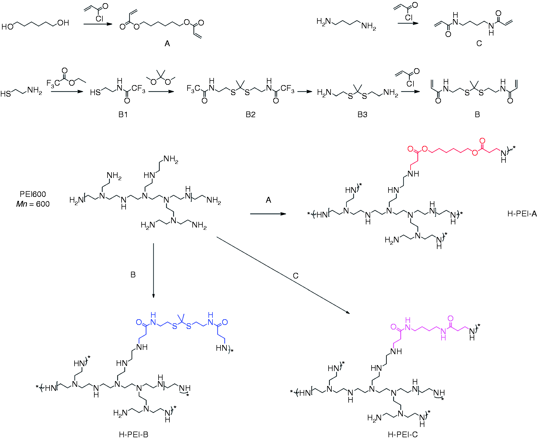
Synthetic routes of degradable linkers (a, b and c) and high MW PEI (H-PEI-A, H-PEI-B and H-PEI-C). [CrossRef]
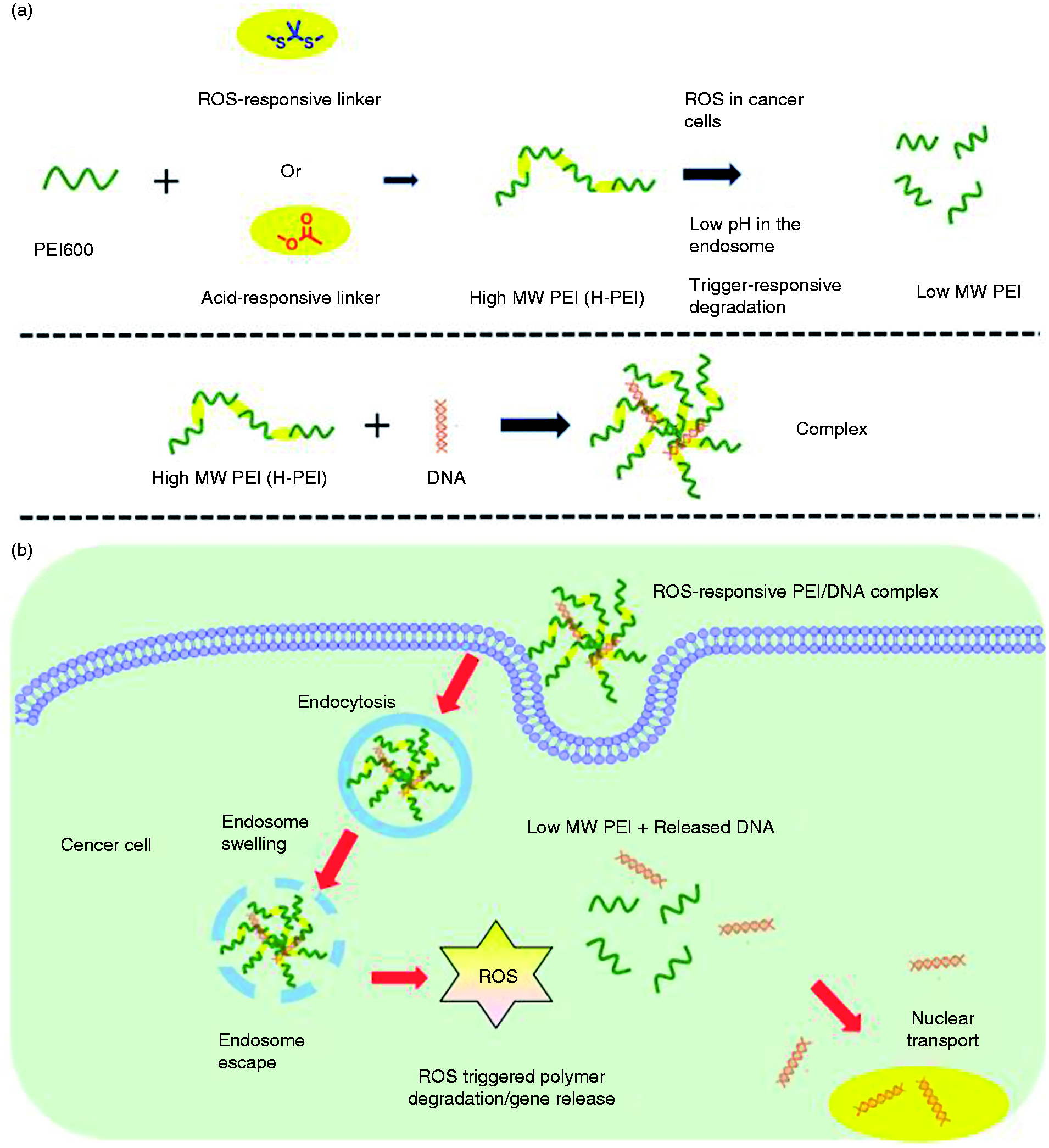
(a) Formation of pH and ROS-responsive high MW PEI/DNA complexes. (b) Intracellular delivery of plasmid DNA to the cancer cells using ROS-degradable high MW PEI. After the complexes have been internalized by the cancer cells and escaped from the endosome, ROS-triggered degradation of the high MW PEI facilitates the release of DNA, leading to the self-diminished cytotoxicity and enhanced gene transfection.
Experimental
Study of the acid and ROS-triggered degradation of H-PEI-A, H-PEI-B and H-PEI-C
H-PEI-A was dissolved in the mixture of DCl and D2O (pH = 4) and incubated at 37°C for 0 h, 4 h and 24 h before being subjected to 1H-NMR analysis to study the polymer degradation.
H-PEI-B was dissolved in the mixture of H2O2 and D2O (H2O2 = 100 mM) and incubated at 37°C for 0 h, 2 h, 4 h and 24 h before being subjected to 1H-NMR analysis to study the polymer degradation.
H-PEI-C was dissolved in the mixture of DCl, H2O2 and D2O (pH = 4, H2O2 = 100 mM) and incubated at 37°C for 0 h and 24 h before being subjected to 1H-NMR analysis to study the polymer degradation.
Formulation and characterization of polymer/DNA complexes
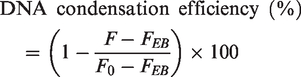
H-PEI-A, H-PEI-B and H-PEI-C and plasmid DNA were firstly dissolved in DI water at 0.2 mg/mL. The polymer solutions were added to DNA solutions at the weight ratios of 0.5, 1, 2 and 5 and incubated at 37°C for 20 min to allow the formation of the polymer/DNA complexes. Gel retardation assay was used to evaluate the DNA condensation. The complexes at various weight ratios were loaded on a 1% agarose gel at 200 ng DNA/well followed by electrophoresis at 100 V for 45 min. To quantitatively determine the level of DNA condensation, ethidium bromide (EB) exclusion assay was performed as follows. 18 Plasmid DNA was stained with EB at the DNA/EB weight ratio of 10 at RT for 1 h. Then the polymers (0.2 mg/mL) were added into the DNA/EB solutions at the weight ratios of 0.5, 1, 2 and 5. The mixture was incubated at RT for another 30 min to allow the DNA condensation before evaluation by fluorescence intensity (λex = 510 nm, λem = 590 nm). EB exclusion assay was also used to evaluate the DNA condensation ability of polymers and the DNA release profile upon acid and ROS treatment.
The DNA condensation efficiency (%) was calculated according to the following equation
Particle sizes and zeta potentials of polymer/DNA complexes at various weight ratios were evaluated by dynamic laser scanning (DLS) on a Malvern Zetasizer. Particle sizes of freshly prepared complexes after acid and ROS treatment were also monitored for different time to evaluate the responsiveness of complexes.
In vitro transfection
HeLa and 293T cells were seeded on 96-well plates at 1 × 10⁴ cells/well and incubated for another 24 h until the cells reached 70% confluence. The cell culture medium was replaced by serum-free DMEM (100 µL/well), into which polymer/DNA complexes at the weight ratios of 1, 2, 3, 4 and 5 were added at 0.2 µg DNA/well. After incubation for 4 h, the medium was replaced by fresh medium containing 10% FBS. Cells were further cultured for another 20 h before quantification of luciferase expression level using a Bright-Glo Luciferase assay kit and cellular protein level using a BCA kit. The result of PEI600 expressed as relative luminescence unit (RLU) associated with 1 mg of cellular protein was normalized as 1. The results of the other complexes were expressed as normalized RLU/mg protein. The transfection efficiency was also evaluated using plasmid DNA encoding EGFP and the EGFP expression was observed by fluorescence microscopy.
Cell uptake of polymer/DNA complexes
Plasmid DNA was labeled with YOYO-1 at one dye molecule per 50 bp DNA. 57 The obtained YOYO-1-DNA was allowed to form complexes with polymers as described above. HeLa cells were seeded on 96-well plates at 1 × 10⁴ cells/well and cultured for another 24 h until the cells reached 70% confluence. The cell culture medium was changed by serum-free medium (100 µL/well) into which the complexes were added at 0.1 µg DNA/well. After incubation at 37°C for 4 h, cells were washed with cold PBS for three times to remove the surface-coated complexes and subsequently lysed by the RIPA lysis buffer (100 µL/well) at RT for 20 min. The YOYO-1-DNA level was evaluated by spectrofluorimetry (λex = 485 nm, λem = 530 nm) and the protein level was evaluated by the BCA kit. The cell uptake level was expressed as ng YOYO-1-DNA associated with 1 mg of cellular protein. To explore the involvement of the energy-dependent endocytosis pathway, HeLa cells were cultured on 96-well plates and incubated with polymer/YOYO-1-DNA complexes for 4 h at 37°C or 4°C as described above. Results were expressed as percentage uptake of control cells that were treated with complexes at 37°C for 4 h.
In vitro cytotoxicity
HeLa cells were seeded on 96-well plates at 1 × 10⁴ cells/well and cultured for another 24 h or 48 h until the cells reached 90% confluence. The medium was replaced by serum-free medium (100 µL/well) into which the polymers were added at the final concentrations of 0.01, 0.02, 0.05 and 0.1 µg/µL. After incubation at 37°C for 12 h, the medium was refreshed with serum-containing medium and further incubated for 20 h before the assessment of cell viability using the MTT assay. To further study the cytotoxicity of polymers upon acid and ROS treatment, cells were incubated with acid-pretreated H-PEI-A, ROS-pretreated H-PEI-B and acid/ROS pretreated H-PEI-C at the same final concentrations as described above at 37°C for 12 h. The medium was replaced by serum-containing medium and cells were further incubated for another 20 h. Cell viability was evaluated by the MTT assay as described above. Cells with PEI600 and PEI25k treatment served as the positive controls. Cells without polymer treatment served as the negative control. The results were presented as percentage viability of control cells.
In vivo transfection
Female BALB/c mice (five to six week old, 20–25g) were obtained from Dalian Medical University and all animal experimental procedures were approved by the Institutional Animal Care and Use Committee (IACUC) of Dalian Medical University. H22-bearing mice were obtained by subcutaneous injection of H22 cells collected from the peritoneal cavity of the BALB/c mouse after six days; 100 µL of the ascites containing H22 cells (1 × 10⁷ cells in total) were subcutaneously injected to the BALB/c mice at the back near right flank. After the tumor volume of mice reached around 100 mm3, the mice were randomly divided into six groups (four mice per group), anesthetized, and intra-tumorally injected with H-PEI-A/DNA, H-PEI-B/DNA, H-PEI-C/DNA, PEI600 and PEI25k complexes in PBS at 20 µg DNA/mouse (50 µL/injection). PBS group was used as the negative control group. The weight ratios of polymer to DNA were fixed at 3 and the mixtures were vortexed before injection. Twenty-four hours post-injection, mice were sacrificed and tumors were harvested, washed three times with PBS, and homogenized with tissue lysis buffer. Luciferase expression level was evaluated using a Bright-Glo Luciferase assay kit and cellular protein level was evaluated using a BCA kit. The result of in vivo transfection efficiency was expressed as RLU associated with 1 mg of cellular protein.
Statistical analysis
Statistical analysis was performed using Student’s t-test and differences were judged to be significant at *p < 0.05 and very significant at **p < 0.01.
Results and discussion
Synthesis and characterization of high MW PEI
For the efficient and controlled gene delivery, it is ideal that the cationic vectors could tightly bind gene cargos extracellularly and response to the specific intracellular triggers to facilitate the precisely release of genetic materials with limited cytotoxicity. Herein, we designed two kinds of high MW PEI-based polymers which contained acid-responsive ester linkers and the ROS-responsive thiol ketal linkers, respectively.43,58,59 Ester-containing linkers were firstly selected for polymerization due to their relative stability at the neutral physiological pH in the extracellular environment while could be rapidly cleaved at the acidic endosomal. As a result, H-PEI can be rapidly degraded into small pieces (PEI600) to trigger DNA release and self-diminish the cytotoxicity. Besides ester groups, thiol ketal groups responsive to the high level of the ROS in cancer cells were subsequently selected in the design of the polymerization linkers since the degradation of ester groups lacked the cell specificity and would happen in most of the cells. H-PEI-A (ester linkers) and H-PEI-B (thiol ketal linkers) were synthesized via the poly-Michael-addition reaction between PEI600 and the corresponding linkers containing ester groups (Compound A) or thiol ketal groups (Compound B). 60 Compound A and Compound B were afforded from diols or diamines. 58 Compound C, the non-degradable analogue of compound B, was obtained in a similar manner (Scheme 1, Supplementary Figure S1 to S6) and used as the linker to synthesize the H-PEI-C as a control. 1H-NMR spectrum indicated the polymerization occurring smoothly and the MALTI-TOF results revealed the MW of H-PEI-A, H-PEI-B and H-PEI-C were at the range of 6000–8000 Da after the polymerization (MW were 6371, 7846, 6219, respectively for H-PEI-A, H-PEI-B and H-PEI-C.) Although high MW could be obtained at the exactly the same molar ratio, previous studies have demonstrated that amine-terminated polycations exhibited higher transfection efficiencies. 61 As a result, we fixed the amine/acrylate molar ratio at 1.1 in this study so that H-PEI with amine end groups were obtained. (Scheme 1, Supplementary Figure S7 to S9). All the obtained polymers were white solid or colorless liquid and were soluble in DI water.
Acid and ROS triggered degradation of H-PEI
The acid-triggered and ROS-triggered polymer degradation were investigated and monitored by 1H-NMR. H-PEI-A was incubated in the mixture of DCl and D2O at 37°C for different time and the 1H-NMR spectrum was used to monitor the degradation. As illustrated in Figure 1(a), the typical proton peak in compound A gradually disappeared upon acid treatment as incubation time elongated. The proton peak of H-PEI-A after 24 h acid treatment shifted to the similar ppm with original PEI600, indicating that H-PEI-A degraded into PEI600 analogues upon acid triggers. As shown in Figure 1(b), the proton peak of the dimethyl groups in thiol ketal moiety at around 1.6 ppm also gradually disappeared after the H-PEI-B was incubated with H2O2 (100 mM). As the incubation time increased, the newly generated proton peak of acetone groups at the ppm of 2.0 obviously increased, indicating the ROS-triggered degradation of H-PEI-B. In comparison, both acid and ROS cannot trigger the degradation of non-degradable H-PEI-C within 24 h (Figure 1(c)). Thioketal was a group sensitive to ROS, while insensitive to acid-, base-, protease and reducing environments. 59 Since higher levels of intracellular ROS are generated constantly in cancer cells compared to normal cells, thioketal linkers could be specifically cleaved by ROS and respond to the tumor ROS microenvironment instead of tumor pH microenvironment.
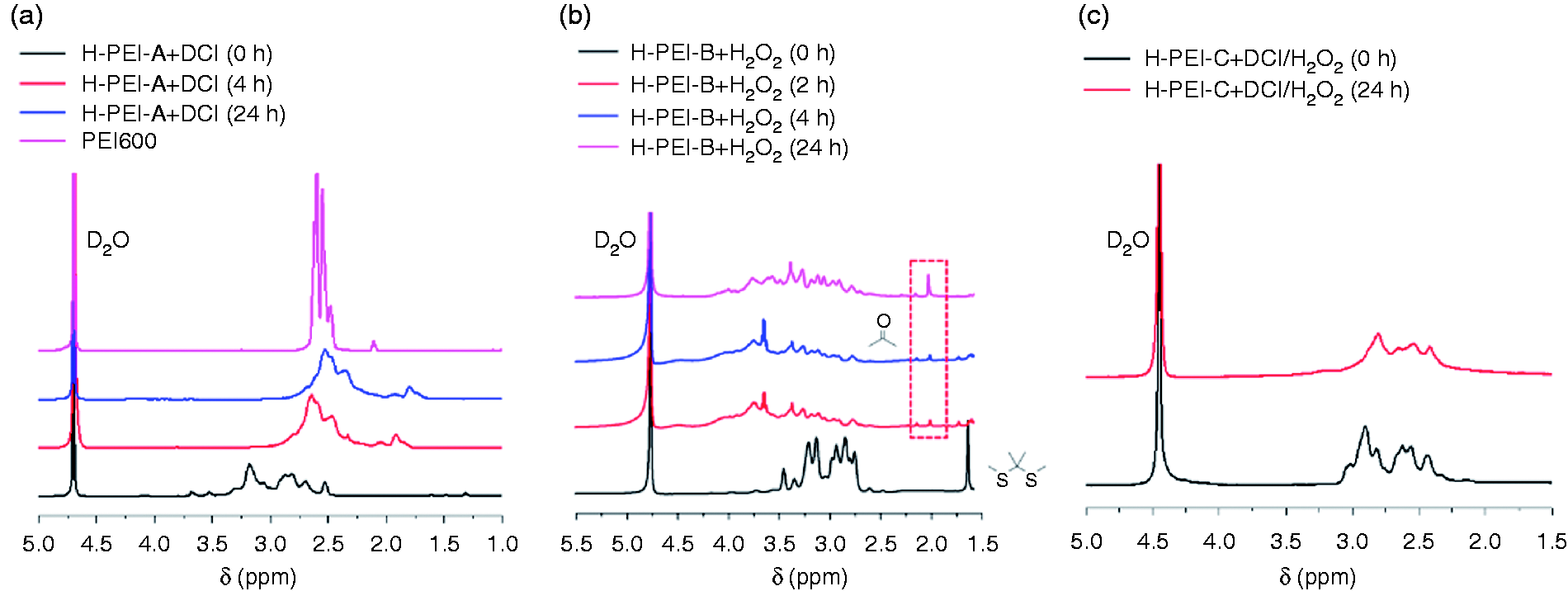
1H NMR spectrum of H-PEI-A, H-PEI-B and H-PEI-C in D2O following acid treatment (a), ROS treatment (b) and acid/ROS treatment (c) for different time.
Formulation and characterization of polymer/DNA complexes
The capabilities of H-PEI to mediate gene delivery were evaluated with plasmid DNA encoding luciferase as the reporter gene. Both H-PEI-A and H-PPEI-B were able to condense the plasmid DNA at the polymer/DNA weight ratios higher than 1 (Supplementary Figure S10(a)) as evidenced by the gel retardation assay. Such results were further verified by a quantitative EB exclusion assay that more than 90% of the plasmid DNA could be condensed at the polymer/DNA weight ratio higher than 1 (Supplementary Figure S10(b)). In consistent with the condensation results, DLS measurement revealed that stable nano-scale complexes (∼180 nm) were formed at the polymer/DNA weight ratios higher than 1 with positive surface charge of ∼30 mV (Figure 2(a) and (b)). SEM image also indicated that H-PEI-B/DNA complexes had a spherical morphology with an average particle size of ∼150 nm (Supplementary Figure S11), which is slightly smaller than that measured by DLS owing to the existence of hydration layer in DLS analysis.
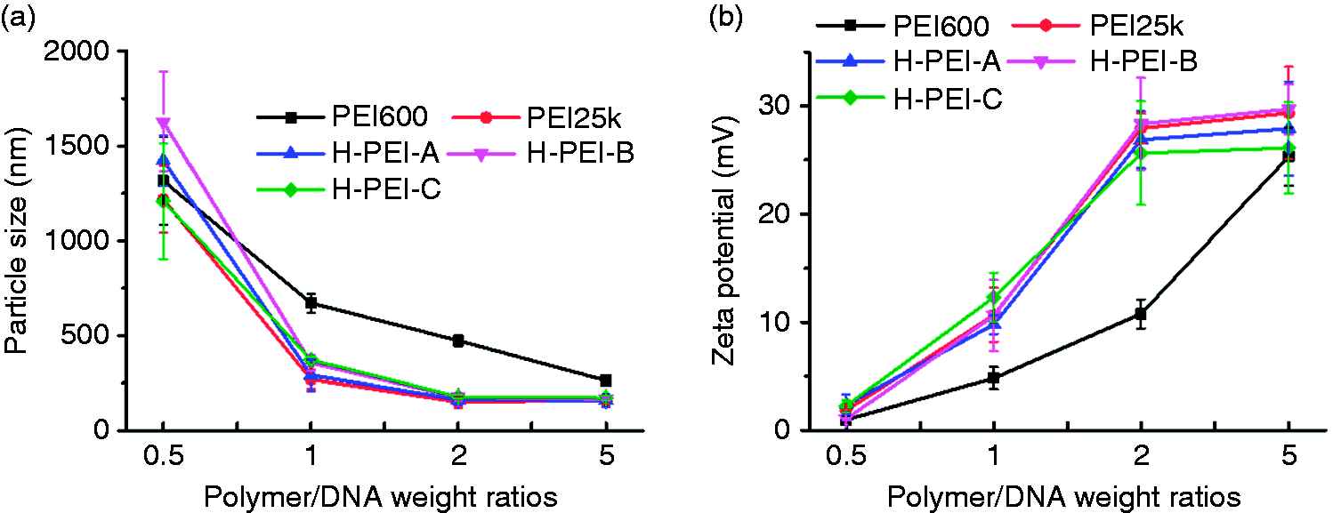
Particle sizes (a) and zeta potential (b) of H-PEI/DNA complexes at various polymer/DNA weight ratios.
Acid and ROS-triggered DNA release
Trigger-induced DNA unpacking was then monitored using the DLS and EB exclusion assay. As shown in Figure 3(a), particle sizes of the H-PEI-A/DNA and H-PEI-B complexes were remarkably augmented from 180 nm to 1500 nm upon acid or ROS treatment, which revealed that the degraded polymers lost the binding ability and led to the transformation of the particles from the tight complexes to the loose complexes for the DNA release. In consistent with the DLS results, DNA was gradually released from the H-PEI-A/DNA and H-PEI-B complexes from 0 h to 4 h upon acid or ROS treatment (Figure 3(b)). As a control, the non-degradable H-PEI-C exhibited unappreciable alteration of DNA release profiles upon both acid and ROS treatment, which indicated that the degradable linkers in the polymer structures contributed to the DNA unpacking.
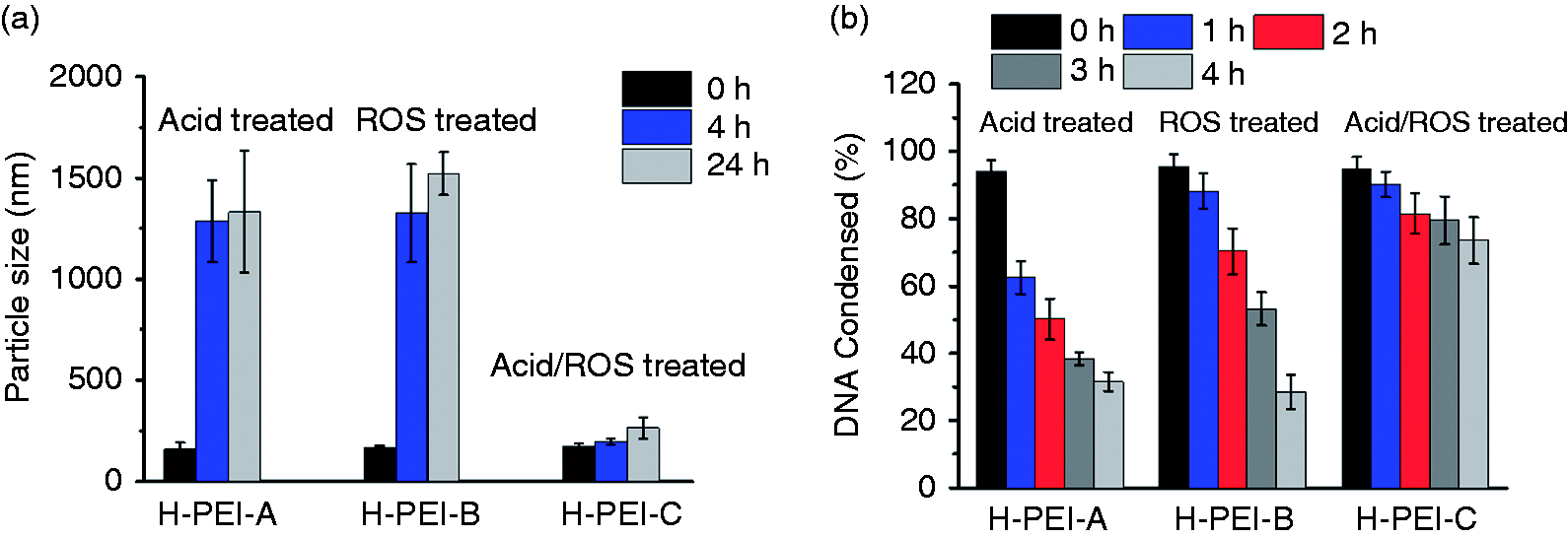
(a) Acid/ROS-trigged particle sizes change of H-PEI/DNA complexes (polymer/DNA weight ratio was fixed at 5) following the treatment for different time. (b) Acid/ROS-trigged DNA release from H-PEI/DNA complexes (n = 3).
Degradable H-PEI-mediated gene delivery in vitro
We then investigated the in vitro gene transfection efficiencies in HeLa cells (cancer cell type) and 293 T cells (normal cell type). Under serum-free conditions, enhanced luciferase expression levels were noted for all the H-PEI including H-PEI-A, H-PEI-B and H-PEI-C compared with PEI600. H-PEI-A and H-PEI-B which afforded the degradable linkers enabled the higher transfection efficiency in HeLa cells, which were 380-fold and 520-fold higher than PEI600 (normalized as 1) and even higher than the commercial available transfection reagent, PEI25k. The transfection efficiency of PEI600 and PEI25k was firstly optimized by varying the weight ratios (data not shown). In direct comparison with PEI600 and PEI25k as the control, the weight ratios of PEI600/DNA and PEI25k/DNA were fixed at 3 and 1, respectively, which were the optimized weight ratios with highest transfection efficiencies. H-PEI-C, which held similar MW while with the non-degradable linkers, exhibited limited increased folds and not as well as PEI25k, which was mainly due to the fact that the degradation of the polymers would greatly promote the intracellular DNA release and potentiate the improved transfection efficiency (Figure 4(a)).Such result was comparable to the PEI-derivatives modified to improve the efficiency when used as the transfection reagent.30,58 Fluorescent images of HeLa cells also revealed higher percentage of GFP-positive cells and higher green fluorescence intensities for H-PEI-A and H-PEI-B compared to other complexes (Figure 4(c)).
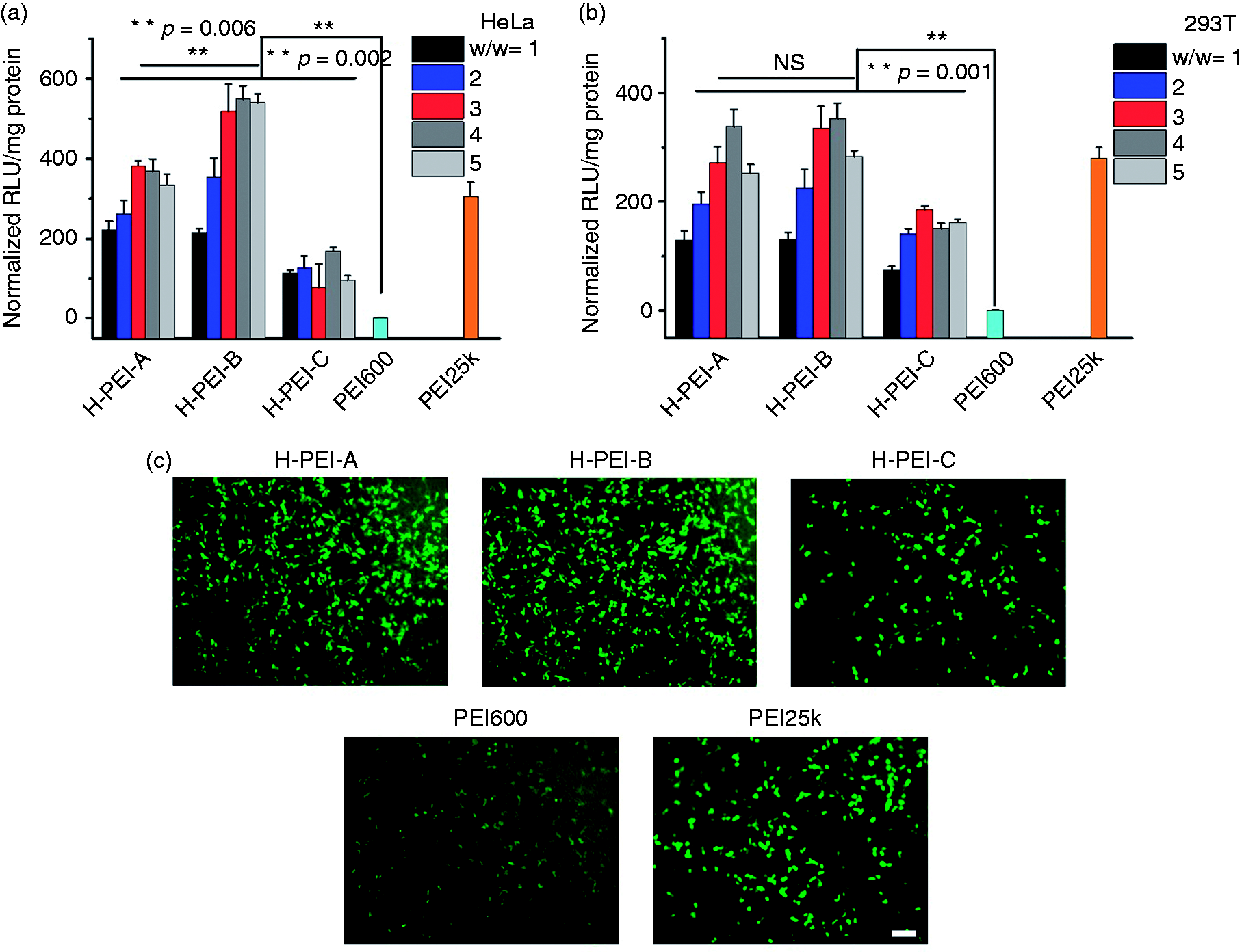
Transfection efficiencies of H-PEI-A, H-PEI-B, H-PEI-C, PEI600 and PEI25k in HeLa cells (a) and 293T cells (b) in the absence of serum (n = 3). Transfection efficiency of PEI600 was normalized as 1. (c) Fluorescent images of HeLa cells transfected with H-PEI-A, H-PEI-B, H-PEI-C, PEI600 and PEI25k (polymer/DNA weight ratio = 3). Bar represents 100 µm.
To further explore whether the enhanced gene transfection efficiency by H-PEI-B was specifically in response to high levels of ROS in cancer cells, 293T cells, which is a kind of normal cell and produce notably lower levels of ROS, were selected to investigate the transfection as a control. H-PEI-B exhibited significantly higher increased folds of transfection efficiency in HeLa cells than in non-cancerous 293T cells, whereas H-PEI-A and non-degradable H-PEI-C revealed similar transfection efficiencies for both cells (Figure 4(b)). Considering the different linkers of polymers and the different properties of cells, the noticeably enhanced gene transfection by H-PEI-B in HeLa cells can be attributed to more efficient disassembly of the DNA only in cancer cells triggered by ROS.
In vitro intracellular internalization
The gene transfection efficiencies of synthetic polycations as non-viral vectors are closely related to their intracellular internalization. Thus, the capability of polymers to mediate the intracellular DNA delivery and release was investigated in HeLa cells following 4 h incubation. Plasmid DNA was labeled with YOYO-1 dye and was allowed to form polymer/DNA complexes. As shown in Figure 5(a), all the H-PEI including H-PEI-A, H-PEI-B and H-PEI-C exhibited excellent cell uptake level at the polymer/DNA weight ratios around 3–5. Cell uptake level for all the H-PEI were similar while the notably higher transfection efficiencies for H-PEI-A and H-PEI-B exhibited notably higher transfection efficiencies, which would be attributed to the more efficient disassembly of complexes upon the intracellular triggers. We then probed the internalization mechanism by performing the cell uptake study at lower temperature (4°C), the energy-dependent endocytosis of which could be completely blocked. As shown in Figure 5(b), the cell internalization level was significantly decreased by more than 80% at low temperature, indicating that the energy-dependent endocytosis was the major internalization mechanism for all the H-PEI.
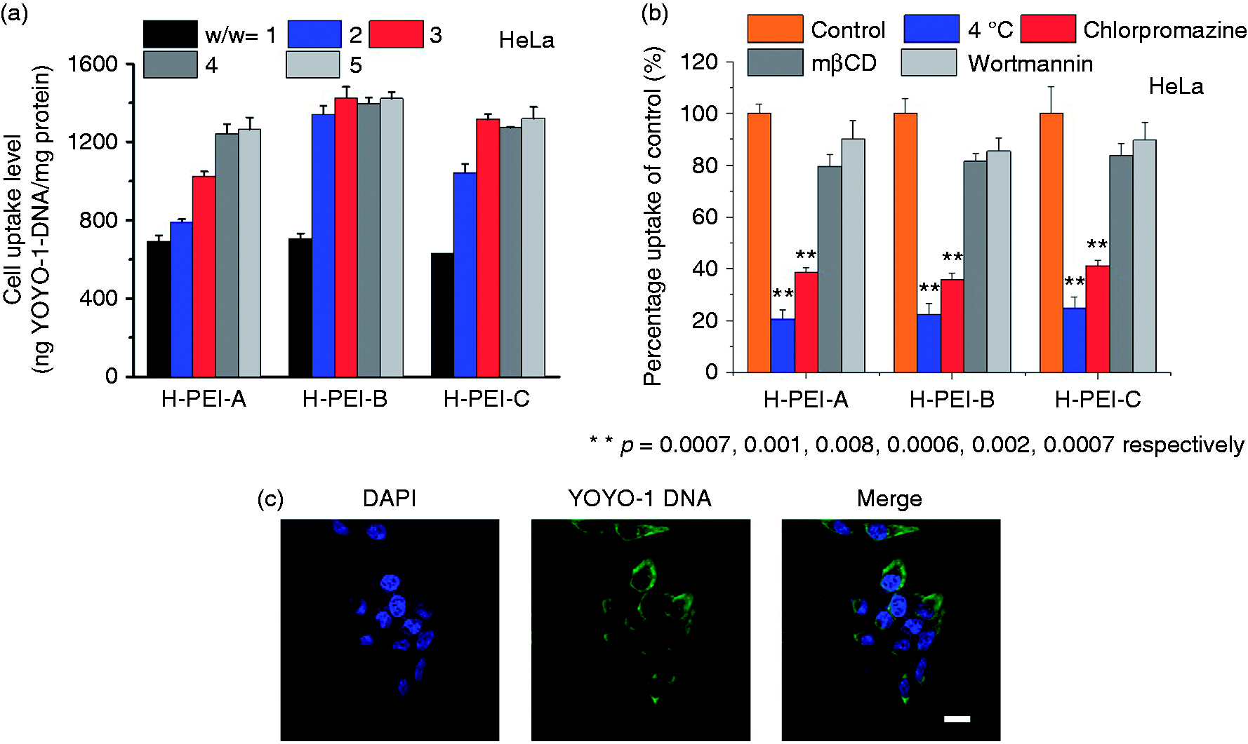
Intracellular kinetics of H-PEI/DNA complexes in HeLa cells. (a) Uptake level of H-PEI/YOYO-1-DNA complexes following incubation at 37°C for 4 h (n = 3). (b) Uptake level of complexes (H-PEI/DNA weight ratio was fixed at 5) at 4°C or in the presence of various endocytic inhibitors for 2 h (n = 3). (c) CLSM images of HeLa cells following incubation with H-PEI-B/YOYO-1-DNA complexes at 37°C for 4 h. Cell nuclei were stained with DAPI. Bar represents 20 µm.
Cytotoxicity
High MW polycations exhibited stronger DNA binding and higher transfection efficiencies than the low MW polycations. However, due to the irreversible correlation between the transfection efficiency and the cytotoxicity, high MW polycations often generate undesired cytotoxicity since low MW polycations are much easier to be expelled from the cell membranes. The undesired cytotoxicity would ultimately damage the cell conditions and hampered the gene transfection. As a result, we then investigate whether the triggered degradation of the synthetic high MW polymers would allow the self-diminish of the cytotoxicity and alleviate the damage to cells during the post-transfection process. As shown in Figure 6(a), all the H-PEI exhibited higher cytotoxicity than the original PEI600, but lower toxicity than PEI25k, which is because of the MW difference. H-PEI-A exhibited notable lower cytotoxicity after the acid pre-treatment and the cell viability level was similar with the PEI600, which revealed that the acid-triggered degradation of the H-PEI-A would lead to the diminish of the cytotoxicity. In consistent with H-PEI-A, ROS-pretreated H-PEI-B also exhibited decreased cytotoxicity. However, as a comparison, non-degradable H-PEI-C showed a totally different phenomenon. Neither acid nor ROS treatment would lead to the diminish of the cytotoxicity of H-PEI-C because H-PEI-C was not able to degrade into low MW PEI600 upon triggers. All the H-PEI/DNA complexes exhibited low cytotoxicity at the transfection doses in HeLa cells and less toxic than the PEI25k complexes. Such observations thus validated our proposed design strategy.
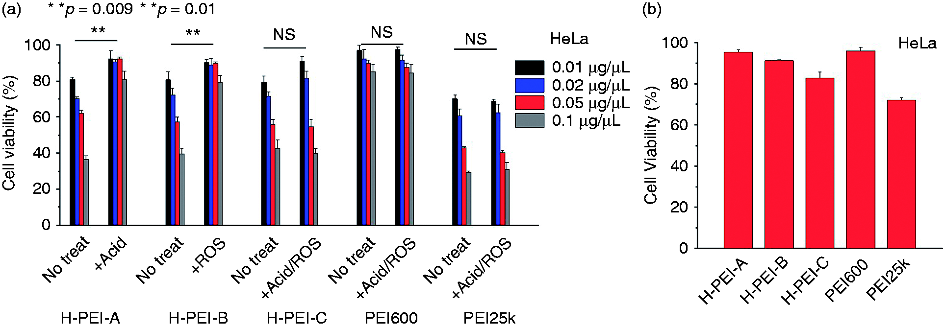
(a) Cytotoxicity of acid or ROS pre-treated free H-PEI following 4 + 20 h incubation (n = 3). (b) Cytotoxicity of polyplexes at the transfection doses and conditions in HeLa cells (n = 3).
In vivo gene expression
High MW polycations, identified above in terms of the in vitro transfection efficiency, were further explored for their potential to mediate in vivo transfection in H22-bearing mice following intra-tumoral injection. In consistent with previous conclusions, all the high MW PEI led to significantly higher transfection efficiencies than PEI600 in terms of enhanced luciferase expression levels 24-h post intra-tumoral injection at 20 µg pCMV-Luc/mouse (Figure 7), and even higher than the commercial available transfection reagent, PEI25k. H-PEI-C with the non-degradable linkers exhibited limited increased folds. That was mainly because the degradation of the polymers would greatly promote the intracellular DNA release and potentiate the improved transfection efficiency. The incorporation of the biodegradable linkers in response to the internal triggers such as the acidic environment (endosome) or the ROS environment (cancer cells) indicated the significant potentials toward topical gene delivery for cancer therapy.
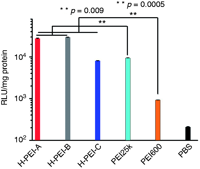
In vivo transfection efficiencies of different PEI/DNA complexes following intra-tumoral injection in tumor-bearing mice (n = 4).
Conclusion
In conclusion, we have developed a strategy to improve the gene transfection efficiency as well as self-diminished cytotoxicity by designing the high MW PEI containing the degradable linkers. With this principle, two kinds of H-PEI with different trigger-responsive linkers were synthesized and the triggered release of DNA, intracellular internalization, transfection efficiency and cytotoxicity was systemically evaluated. Transfection efficiencies of all the synthetic degradable H-PEI significantly enhanced compared to both PEI600 and PEI25k. ROS-degradable H-PEI-B exhibited excellent transfection efficiency especially in cancer cells instead of normal cells. Such strategy not only provides an effective tool in overcoming the irreversible correlation between transfection and cytotoxicity of polycation-mediated gene delivery, but also furnishes the insights into the design of non-viral gene delivery vectors for the cancer gene therapy.
Supplemental Material
Supplemental material for Thioketal-crosslinked: ROS-degradable polycations for enhanced in vitro and in vivo gene delivery with self-diminished cytotoxicity
Supplemental Material for Thioketal-crosslinked: ROS-degradable polycations for enhanced in vitro and in vivo gene delivery with self-diminished cytotoxicity by Nan Zheng Dan Xie, Zhiyi Zhang, Jia Kuang, Yubin Zheng, Qing Wang and Yang Li in Journal of Biomaterials Applications
Footnotes
Declaration of conflicting interests
The author(s) declared no potential conflicts of interest with respect to the research, authorship, and/or publication of this article.
Funding
The author(s) disclosed receipt of the following financial support for the research, authorship, and/or publication of this article: This work was supported by grants from the National Natural Science Foundation of China (Nos. 51703018, 21702025). Doctoral Program Foundation of Liaoning Province (Grant Number 20170520378, 20170520274), the Fundamental Research Funds for the Central Universities (Grant Number DUT16RC(3)114).
Supplemental material
Supplemental material for this article is available online.
References
Supplementary Material
Please find the following supplemental material available below.
For Open Access articles published under a Creative Commons License, all supplemental material carries the same license as the article it is associated with.
For non-Open Access articles published, all supplemental material carries a non-exclusive license, and permission requests for re-use of supplemental material or any part of supplemental material shall be sent directly to the copyright owner as specified in the copyright notice associated with the article.