
Abstract
Current photodynamic therapy faces two major challenges, which are the limited tissue penetration of excitation light and poor tumor selectivity of the photosensitizer. To address these issues, in the present work, a photosensitizer protoporphyrin IX was delivered by aptamer-coated upconversion nanoparticles. The aptamer, namely, AS1411, could fold to G-quadruplex structures with the bi-functions of loading protoporphyrin IX and targeting the cancerous cells. The protoporphyrin IX aptamer-coated upconversion nanoparticle nanospheres exhibited good biocompatibility, high nuclease resistance, and targeted cellular internalization. The receptor blocking experiment indicated that the cellular uptake of protoporphyrin IX aptamer-coated upconversion nanoparticle mainly attributed to the binding of AS1411 to the nucleolin overexpressed on the cancerous cell membrane. Moreover, high-efficient photodynamic therapy of the as-prepared protoporphyrin IX aptamer-coated upconversion nanoparticle nanospheres was achieved to cancerous cells under the irradiation of near-infrared light, which was induced by the upconversion property of the upconversion nanoparticle. These results indicate that the fabricated protoporphyrin IX aptamer-coated upconversion nanoparticle nanoplatform has higher efficiency, lower toxicity, and less side effects compared to the free photosensitizer protoporphyrin IX, and thus is a new promising photodynamic therapy agent especially for the deep-seated tumors.
Introduction
In recent decades, photodynamic therapy (PDT) has emerged as an effective, noninvasive, and economical treatment for cancers.1,2 Although PDT has gained wide interest among current superficial cancer treatment options such as surgery, radiotherapy, and chemotherapy, its clinical applications in deep-seated tumors are restricted by the limitation of the insufficient tissue penetration ability of the excitation lights for the photosensitizer (PS). Generally, most PS molecules used in PDT are activated by visible or UV light, which exhibit rather limited penetration depth in biological tissues, thus hampering the application of PDT in the treatment of deep-seated tumors.3,4 However, it is believed that near-infrared (NIR) light (typically ranging from 700 nm to 1000 nm) can not only afford penetration depths of an order of magnitude greater than that of visible light, but also with a low phototoxicity to normal cells and tissues.5–7 Thus, recently the methodology of introducing NIR to PDT has captured many researchers’ attention. An important new approach to deliver NIR light into deeper tissues for PDT treatment is using NIR-excitable upconversion nanoparticles (UCNP) as an energy donor.8–10 UCNP can not only emit shorter wavelength photons under excitation by NIR light, but also have improved tissue penetration depth, higher photochemical stability, and free of autofluorescence background.11,12 Among various UCNP, Ln3+-doped particles were widely employed in PDT because of their superior upconversion luminescence efficiency, cheap excitation source, low power densities, no photobleaching, and low toxicity.13–15 Owing to these unique advantages, Ln3+-doped, UCNP-based PDT has been intensively researched in the past few years.16,17 Huang et al. 18 and Wang et al. 19 combined UCNP with Si-based, hollowed nanomaterials and drugs were loaded in the pores of the nanospheres. Loh’s group synthesized Ln3+-doped UCNP with low polydispersity, which were then covalently conjugated with the PS chlorin e6 (Ce6). Promising efficacy was achieved with low concentration and mild dosage of this nanocomplex.
Given the great success in the vitro and vivo studies of UCNP-based PDT strategy, the poor tumor selectivity of UCNP is still a challenge for its application. The lack of tumor targeting results in poor location of drugs, high toxicity to normal cells, and low anticancer activity. Several research groups have developed selective PDT agents, such as peptide or protein conjugates, and PS-encapsulated nanocarriers.11,12 In recent years, single-stranded (ss) oligonucleotides, namely aptamers, have emerged as a novel class of molecules which rival antibodies in both therapeutic and diagnostic applications.20–22 Compared to antibodies, aptamers offer significant advantages, such as flexible design, synthetic accessibility, easy modification, chemical stability, and rapid tissue penetration.23–26 Thus, aptamers are expected to endow traditional PDT with high selectivity and accurate localization.27,28
Especially, an aptamer named AS1411, which was selected by Aptamera Inc. (Louisville, KY) using exponential enrichment (SELEX), is the most advanced aptamer and the first to enter clinic study. AS1411 can form a dimeric G-quadruplex structure to target nucleolin, which is over-expressed in most cancer cell membranes and functions as a cell surface receptor where it is associated with the actin cytoskeleton.29–31 The nucleolin can also act as a shuttling protein between cytoplasm and nucleus. On the other hand, the quadruplex structures have been previously proved to be able to load porphyrin PS, such as N-methylmesoporphyrin IX (NMM) and 5, 10, 15, 20-tetrakis (1-methylpyridinium-4-yl) porphyrin (TMPyP).32–37 Since the AS1411 aptamer is an excellent selector for the higher nucleolin-expressing cancer cells and a good candidate for carrying porphyrin ligands because of its quadruplex structure, it has been frequently employed as a tether for the drug nanocarrier.
Herein, a widely employed PS, protoporphyrin IX (PpIX) was delivered by aptamer-coated UCNPs, namely, PpIX-Apt-UCNP. The G-rich aptamer AS1411 in the PpIX-Apt-UCNP nanoplatform could form G-quadruplex structure to load the PpIX molecules and preferentially bind with nucleolin overpressed on the cell membrane of the tumors. Furthermore, for the as-prepared PpIX-Apt-UCNP nanoplatform, in vitro studies indicated that NIR could efficiently trigger its PDT inhibition to tumors by inducing the apoptosis of cancerous cells (see the schematic process for preparing PpIX-Apt-UCNP for PDT in Figure 1). The intracellular reactive oxygen species and singlet oxygen generation of the PpIX-Apt-UCNP under NIR were also evaluated. This functional nanoplatform is expected to provide new insights into the design of nanocarriers to overcome the penetrating and targeting limitations of traditional PDT.
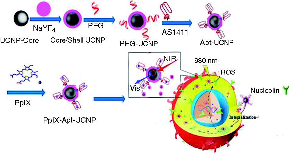
Schematic showing the process for preparing PpIX-Apt-UCNP for photodynamic therapy.
Experimental section
Materials
Unless specified otherwise, all of the chemicals used were of analytical grade and used without further purification. Yttrium(III) chloride hexahydrate (YCl3·6H2O), ytterbium(III) chloridehexahydrate (YbCl3·6H2O), erbium(III) chloride hexahydrate (ErCl3·6H2O), ammonium fluoride, oleic acid, N-(3-dimethylaminopropyl)-N-ethylcarbodiimide hydrochloride (EDC), N-hydroxysuccinimide (NHS), (3-aminopropyl) triethoxysilane (APTES), 2, 2'-azino-bis(3-ethylbenzothiazoline-6-sulfonic acid) (ABTS), 3-mercaptopropionic acid, and acetone were all purchased from Aladdin. Aminated nucleic acid sequence (5′–NH2–GG
Instruments
The scanning electron microscopy (SEM) analyses were performed with a JSM-6330F field emission scanning electron microscope. The transmission electron microscope (TEM) analyses were performed with a JEM-2010HR TEM. Magnetic measurements were performed by using a XL-7 magnetic property measurement system. UV–Vis and Fourier transform infrared (FTIR) spectra were recorded on a Shimadzu UV-3150 spectrophotometer and an Equinox 55 Fourier transformation infrared spectrometer, respectively. Fluorescence microscopy of apoptosis assays was performed with an OX31 fluorescence microscope (Olympus, Japan). Cell cycle analysis was performed with a FACS flow cytometer (BD, USA). Confocal laser scanning fluorescence microscopy images were obtained with an Olympus FV1100 (Japan) microscope.
Preparation of PpIX-Apt-UCNP nanoparticles
Surface PEG modification of UCNP
NaYF4:Yb(20%), Er (2%) nanocluster (UCNP) was synthesized by thermal decomposition of rare-earth/sodium trifluoroacetate precursors in oleic acid (OA) and octadecene (ODE) as reported previously. 38 Polyethylene glycol (PEG) diacid 3000 (20 mg), NHS (2 mg), Dicyclohexylcarbodiimide (DCC) (3 mg), and dopamine hydrochloride (1.27 mg) were dissolved in a mixture solvent containing CHCl3 (2 mL), N,N-Dimethylformamide (DMF) (1 mL), and anhydrous Na2CO3 (10 mg) and the mixture was stirred for 2 h. Then, the as-prepared UCNP (5 mg) were added, and the system was stirred overnight under argon. The modified UCNP were precipitated by adding hexane and the particles were dispersed in water or PBS. The extra surfactants and other salts were removed by dialysis using a dialysis bag (molecular weight cut off [MWCO] = 10,000) for 24 h in water. PEG-modified UCNP were thus obtained, namely, PEG–UCNP.
Conjugation of AS1411–aptamer to PEG–UCNP
A solution of PEG–UCNP (50 mL, 1.5 mg) was treated with EDC (25 mL) and NHS (25 mL) and gently shaken for 15 min. Ss N-terminated AS1411 (5′-amino-C6-GGTGGTGG TGGTTGTGGTGGTGGTGG-3′, 1 mg in 100 mL nuclease-free water) was then added, and the solution gently shaken for a further 4 h. Unreacted aptamer was removed using centrifugal filtration to get aptamer-conjugated UCNP (Apt-UCNP).
Aptamer AS1411 oligonucleotides annealing and ABTS test
The Apt-UCNP and the complementary sequence of AS1411 were dispersed in annealing buffer (200 mM KCl, 4 mM MgCl2, and 28 mM Tris-HCl, pH 7.5), annealed at 95°C for 10 min, and then slowly cooled to room temperature to form the G-quadruplex structure of the aptamer AS1411. The annealed Apt-UCNP was thus obtained.
ABTS chromogenic experiment was employed to confirm the formation of G4-quadruplex for the annealed Apt-UCNP. Low concentrations of hemin, unannealed ss Apt-UCNP, annealed Apt-UCNP were suspended in 200 µL ABTS/H2O2 substrate (1 and 2 mM). After reaction at 37°C for 20 min, the color of the mixture was observed and the absorbance was read at 405 nm on a microplate reader.
PpIX loading into Apt-UCNP
PpIX loading into the annealed Apt-UCNP was carried out by mixing PpIX (20–400 µM) with annealed Apt-UCNP (0.5 mg/mL) and shaking at room temperature. Free PpIX was removed by centrifugation at 7500 r/min for 5 min and washing three times to give PpIX-loaded Apt-UCNP (PpIX-Apt-UCNP).
Hemolysis testing experiment
Four milliliters of fresh anticoagulation human blood was diluted with 5 mL physiological saline. One milliliter Apt-UCNP suspension (concentration is 1.0 mg/mL, drug free) was incubated in the 37°C water bath for 30 min, and then 1 mL diluted blood was added to the nano suspensions, following by shocking for 60 min in the water bath. The mixture was then centrifuged at 7500 r/min centrifugal for 5 min, and the absorbance of the supernatant at 545 nm was measured. Physiological saline and distilled water were used as negative control and positive control, respectively. The hemolysis rates of materials were calculated using the following equation:
Here, H% refers to the hemolysis rates of materials, Am, An, Ap refer to the absorbance of the supernatant for the studied material, the negative control and the positive control at 545 nm, respectively.
Biocompatibility research
To investigate the biocompatibility of the UCNP and Apt-UCNP, HeLa and A549 cells were placed in 96-well plates at a cell density of 1 × 10⁵ cells per well, respectively. After incubation for 24 h, the medium was replaced with UCNP or Apt-UCNP solutions at different concentrations. After incubation for a further 24 h, standard 3-(4, 5Dimethylthiazol- yl)-2, 5Dimethylthiazol-2-yl)-2, 5diphenyltetrazolium bromide (MTT) assay was carried out to determine the cell viabilities relative to the control untreated cells.
For the biocompatibility assessment for the 980 nm NIR illumination, MCF-7 cells were seeded for 24 h in standard 96-well plates at 1 × 10⁴ cells per well. Then the cells were irradiated with 980 nm laser for 2, 4, 6, 8, 10, 12 min. The cells were rinsed thrice with Hank’s buffered salt solution (HBSS, pH 7.4) and incubated for 4 h in 1 mL of medium containing 0.5 g/L of a tetrazolium dye (MTT). Absorbance was measured at 490 nm using a multiwell plate reader.
Nuclease resistance
The solutions of AS1411 oligomer (AT26, 5′-GGTGGTGGTGGTTGTGGTGGTGGT GG-3′), annealed AS1411, annealed Apt-UCNP and PpIX-Apt-UCNP were separately incubated with 400 U DNaseI at 37°C for 1 h and 24 h, respectively. The mixtures were then run on a 1% agarose gel and stained with gel green. The band intensity was quantitated by Gel imaging system (Tanon, 2500).
Cell migration by scratch wound assay
To determine if quantifiable cell migration occurred after PpIX-Apt-UCNP treatment, in vitro scratch assays were performed. HeLa cells were seeded in 24-well plates and when cells reached a confluence of 95%, wounds were made in cell culture using a tip. Culture medium was changed to remove loose cell debris, and a defined area of the wound was photographed under an inverted microscope for 24 h and 48 h.
Cell invasion inhibition study by transwell experiment
HeLa cells were incubated in the serum starvation for 24 h to remove the impact of serum before the preparation of cell suspension. The cells were then digested, and were resuspended in Bovine Serum Albumin (BSA)-free serum-free medium. One-hundred microliters of cell suspension containing 0.5 mg/mL PpIX-Apt-UCNP or same amount of PpIX were added to the transwell chamber and 600 µL medium containing 10% fetal bovine serum (FBS) was generally add to 24-well plate room. After incubated for 24 h in CO2 incubator, the transwell chamber was removed and the culture medium in the wells was discarded. The cells were washed twice with PBS and then were fixed with methanol for 30 min. The cells was stained with 0.1% crystal violet for 20 min, and counted by microscope.
Cell cytotoxicity
For cytotoxicity assays, cells were seeded for 24 h in standard 96-well plates at 1 × 10⁴ cells per well. Then the culture medium was discarded and the cells were treated for 24 h with 500 µL of medium containing different PpIX concentrations (0.001–30 µM), either as PpIX solutions or as PpIX-Apt-UCNP suspensions. For PDT group, the mixture was irradiated with 980 nm laser for 10 min. The cells were rinsed thrice with HBSS and incubated for 4 h in 1 mL of medium containing 0.5 g/L of a tetrazolium dye (MTT). Absorbance was measured at 490 nm using a multiwell plate reader.
Cellular uptake and nucleolin blocking experiments
To investigate the tumor targeting drug delivery of PpIX-Apt-UCNP to the cells, intracellular uptake of PpIX-Apt-UCNP and the receptor-blocking experiments were performed with the nucleolin overexpressed MCF-7 and HeLa cells as well as the nucleolin-negative MCF-10A cells. Three cell lines were in the confocal dishes at a density of 3 × 10⁵ cells/dish. After 24 h of cell attachment, 200 µL of PpIX-Apt-UCNP (containing 57 µg/mL PpIX) was added in the culture medium and incubated for 24 h. Cell nuclei were stained with Hoechst 33258 for 30 min. After washing three times with PBS, the cells were imaged by a laser confocal microscope equipped with an external 980 nm laser.
In the nucleolin-blocking experiment, MCF-7 cancer cells (3 × 10⁵ cells/dish) were first incubated with endostatin (1 mM) for 30 min and then 200 µL of PpIX-Apt-UCNP (containing 57 µg/mL PpIX) was added in the medium. As a control, the cells were directly incubated with the PpIX-Apt-UCNP without endostatin blocking. After 2 h incubation, cell nuclei were stained with Hoechst 33258 for 30 min and then the cells were washed three times with PBS for fluorescence confocal imaging by a laser confocal microscope equipped with an external 980-nm laser.
Detection of reactive oxygen species in deep-seated tumor cells
HeLa cells were seeded into six-well plates and incubated for 24 h. The medium was removed and replaced with medium containing PpIX-Apt-UCNP nanospheres (containing 57 µg/mL PpIX) for 24 h. The cells were washed with Roswell Park Memorial Institute (RPMI) 1640 medium and then incubated with 10 µM 2’, 7’-Dichlorodihydrofluorescein diacetate (DCFH-DA) at 37°C for 20 min. For PDT group, after washing twice with RPMI 1640 medium, cell dishes were covered with 1.2 cm adipose tissues and then irradiated with 660 nm and 980 nm laser for 10 min, respectively. The cells were then harvested and the cell pellets were suspended in PBS–EDTA, which were then imaged by fluorescence microscope.
Detection of Species of reactive oxygen
Singlet oxygen in aqueous was determined following the Kraljic procedure.39 Solution of p-nitrosodimethylaniline (RNO, 30 µM) and imidazole (0.5 mM) in PBS (10 mM, pH = 5.86 or 7.40) was added into PpIX-Apt-UCNP (0.1 mg/mL) and then irradiated with 980-nm laser light. The absorbance spectra of the solution were recorded at 30 s intervals.
Fluorescence microscopy of apoptosis assays
HeLa cells in the logarithmic growth phase were seeded in 24-well plates and allowed to attach overnight. Cells were incubated with PpIX-Apt-UCNP for 24 h under dark or exposed to 980-nm light for 10 min, then stained with an annexin V-Fluorescein isothiocyanate (V-FITC) apoptosis detection kit (Beyotime Institute of Biotechnology, China) as described in the manufacturer’s instructions, and observed by fluorescence microscopy.
Statistical analysis
Significant differences were determined using the Student’s t-test where differences were considered significant (*P < 0.05). All data are expressed as mean (standard error of the mean).
Results
Synthesis and characterization of PpIX-Apt-UCNP
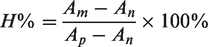
Herein, we designed an aptamer-coated NaYF4:Yb,Er@NaYF4 core/shell structure to deliver PpIX. TEM image in Figure 2(a) of the PpIX-Apt-UCNP nanospheres exhibits the uniform shape with an average diameter of 120 ± 4 nm. Since devices ranged in 10–500 nm are proper to evade reticuloendothelial system and achieve tumor passive targeting by enhanced permeability and retention (EPR) effect, these PpIX-Apt-UCNP nanospheres may demonstrate prolonged blood circulation and offer the most effective distribution in certain tissues.
The characterization of nanospheres: TEM (a) images of PpIX-Apt-UCN; (b) solid UV–Vis spectra of PpIX (insert), UCN (
The conformational G-quadruplex structure of DNA was confirmed by ABTS experiment and the results were given in Figure S1. Both hemin and the ss Apt-UCNP complex have negligible colors when incubated with ABTS. In contrast, after annealing, the absorbance of Apt-UCNP–hemin complex at 405 nm was significantly increased and green color was observed visibly. The strong absorption at 405 nm and obvious green color are widely accepted as the verification of the formation of G-quadruplex structure. The formation of G-quadruplex structure for AS1411 aptamer was thus confirmed.33,34,40,41
The presence of porphyrin in the PpIX-Apt-UCNP nanospheres could be further confirmed by the results of solid state UV–Vis spectra (Figure 2(b)). It was found that UCNP has no substantial absorption while the PpIX-Apt-UCNP has the characteristic absorption peak (Soret band around 410 nm) of porphyrin. The chemical composition of the organic layers was confirmed by FTIR spectra in Figure 2(c). From Figure 2(c), UCNP exhibits intense vibration band at around 2900 cm−1, which could be attributed to the large amount of CH2 groups on PEG chains. The peaks in the region from 1000 cm−1 to 1400 cm−1 are mainly related to the stretching vibration of C–O groups of PEG on UCNP. Compared with that of UCNP, the FTIR spectra of PpIX-Apt-UCNP exhibit increased intensity of the pyrrole ring’s vibration bands in the region from 800 cm−1 to 1000 cm−1, which are the characteristic FTIR signals of porphyrin.
The existence of PpIX was further confirmed by the florescence emission spectra of the nanospheres, shown in Figure 2(d). The UCNP emission at 980-nm excitation was recorded as initial reference. After PpIX loading, significant quenching of the UCNP emission peak excited at 980 nm was observed in the range of 400–410 nm, which corresponds to the absorbance range of PpIX (see Figure 2(b)). This indicated that the emission energy from UCNP was absorbed by PpIX, which evidenced the successful loading of porphyrin to UCNP in a close proximity where the energy transfer could happen efficiently.
Biocompatibility and nuclease resistance of UCNP
The biocompatibility of the as-prepared NaYF4:Yb,Er@NaYF4 UCNP was evaluated by hemolysis rate and the cell toxicity. The interaction of devices or biomaterials with blood is an immediate and serious concern during safety assessment. Hemolysis of red blood cells (RBCs) would result in leakage of free hemoglobin into the plasma, potentially leading to severe hepatic and renal injury among other effects.
Fresh human blood was used in the hemolysis assessment of the as-fabricated drug delivery vectors. From the OD value at 545 nm of the treated RBCs, the hemolysis rate for the UCNP nanospheres was calculated to be 0.51%, which is far lower than the maximum hemolysis rate (5%) for the medical devise requested by Food and Drug Administration (FDA) This result suggests that the studied material is biocompatible for the circulating blood.
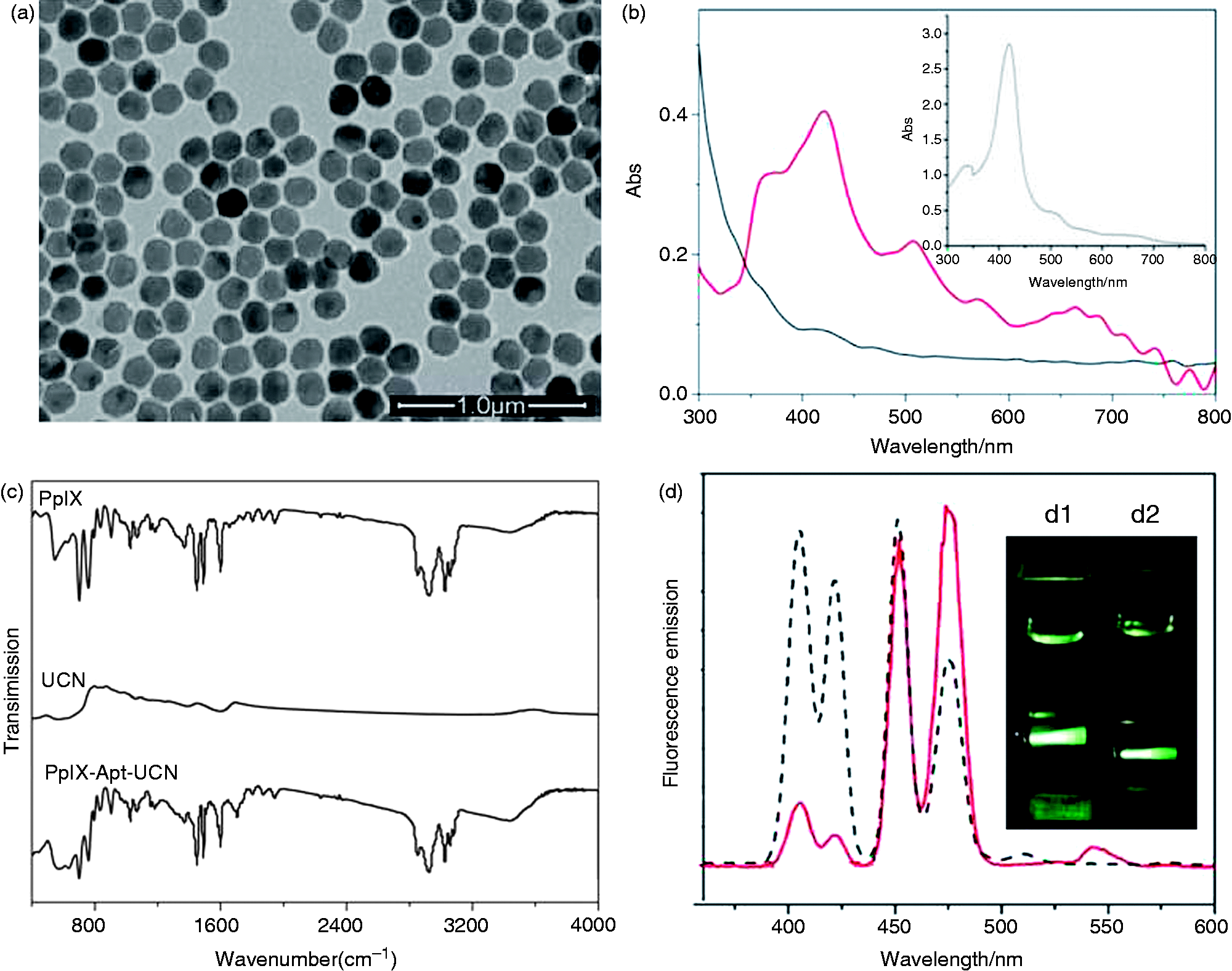
Meanwhile, the in vitro tests for cytotoxicity could assess the response of cells in culture to direct contact with devices or to their extracts. According to the requirements of FDA, cellular cytotoxicity test is strongly recommended for the approval submission of medical devices. The presence of cytotoxicity is indicated by loss of cell viability. To more deeply assess the biosafety of UCNP and Apt-UCN nanospheres (drug-free), HeLa and A549 cells were incubated with the nanospheres for 24 h. The cell viabilities of the HeLa and A549 cells with Nps at concentrations ranged from 0.03 to 1.0 mg/mL were given in Figure 3(a) and 3(b). It was found that no appreciable deduction in cell viabilities were observed, indicating that the UCNP and Apt-UCN nanospheres were highly biocompatible for regulatory bodies. Moreover, the biosafety of NIR illumination was assessed by illuminating MCF-7 cells by 980-nm light for different time. It was found from Figure 3(c) that the cell viability of MCF-7 was not significantly affected under the illumination by NIR light for up to 12 min, confirming its biosafety to the cells.
Biocompatibility of UCNP (a) and Apt-UCN (b) accessed by HeLa and A549 cell viability under different concentrations, respectively. The biosafety of 980 nm laser was accessed by MCF-7 cells under different illumination time (c). Resistant digestion of annealed AS1411 aptamer (lane 1), oligonucleotide AT26 (lane 2), annealed Apt-UCNP (lane 3), PpIX-Apt-UCNP (lane 4) treated with 400 U DNase I for 1 h (c) and 24 h (d), respectively.
The stability of the prepared PpIX-Apt-UCNP against degradation and digestion of DNase I was evaluated by using the agarose gel electrophoresis. From Figure 3(d), after treated with enzyme DNase I for 1 h, the 26 base pair oligonucleotides for AS1411 without G-quadruplex structure formation, namely AT 26, were significantly digested and the DNA band was negligibly observed. In contrast, when incubated with DNase I for 24 h, the oligonucleotides AT26 were completely digested and could not be observed on the gel. However, as to the AS1411 aptamer, Apt-UCNP and the PpIX-Apt-UCNP complex, no obvious digested DNA bands could be observed on the gel when treated them with the enzyme DNase I for even 24 h. The drag phenomenon of the AS1411aptamer band was assumed to be due to the special G-quadruplex structure of AS1411 aptamer.42,43 Furthermore, no DNA fragments could be detected on the gel after the Apt-UCNP and the PpIX-Apt-UCNP was digested with DNase I, indicating the stability of AS1411 after conjugating on the UCNP surface. The small size of the PpIX-Apt-UCNP nanoparticles and the high stability against DNase I digestion may facilitate the PpIX-Apt-UCNP with a rapid vascular extra vacation and intratumoral penetration and high stability in blood, thereby making it a therapeutically effective drug system for in vivo applications.
Inhibition of cell migration and invasion
In order to determine if quantifiable cell migration occurred after treatment, we also analyzed cell migration by scratch wound assay. As is shown in Figure 4, without PpIX-Apt-UCNP nanospheres, significant cell migration was observed after 24 h and 48 h. However, in the presence of the studied nanospheres, only a negligible fraction of HeLa cells shows the capacity of closing wounds. Our results showed that the minimal fraction of HeLa cells not committed to cell death did not display any capacity of closing wounds after incubation with the nanospheres. The scratch wound assays corroborate that HeLa cells’ migration was completely inhibited at 48 h after PpIX-Apt-UCNP treatment. Meanwhile, the inhibition capability of PpIX-Apt-UCNP to the invasion of HeLa cells was accessed by the transwell experiment (Figure S2 in supplemental materials). It is found that, compared with the control, the invasion of HeLa cells was dramatically decreased in the presence of PpIX-Apt-UCNP. The combination of scratch wound assays and transwell experiment confirmed the efficiency of PpIX-Apt-UCNP nanospheres to inhibit the migration and invasion for cancerous cells.
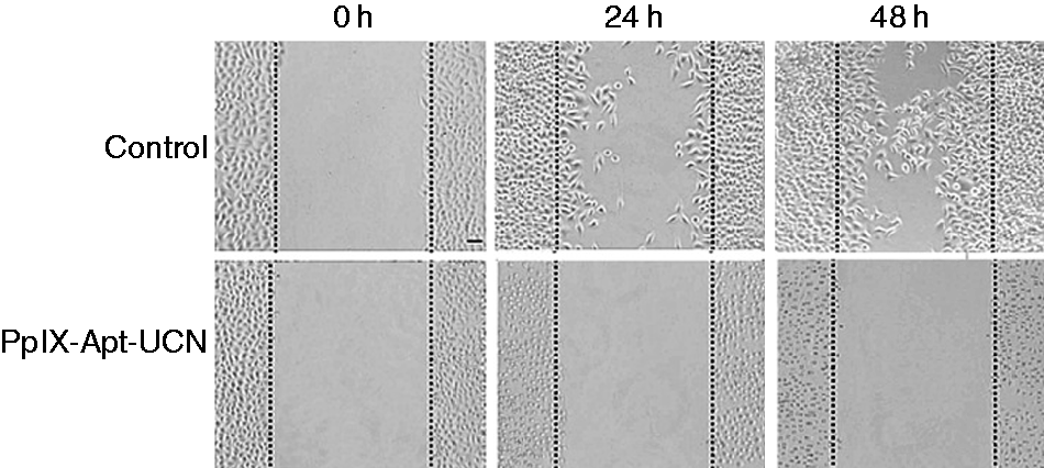
Analysis of cell migration for control and cells incubated with PpIX-Apt-UCNP for 0 h, 24 h, and 48 h by scratch wound assay. Wounds were made when cells reached a confluence of 95% in cell culture. A defined area of the wound was photographed under an inverted microscope.
Tumor-targeting cellular uptake of the PpIX-Apt-UCNP nanospheres
Nucleolin, a multifunctional protein involved in RNA transcription, DNA replication, rRNA processing, and bcl-2 stabilization, is highly expressed in continuously proliferating cells, such as cancer cells, and it has been reported to shuttle between the nucleus, the cytoplasm, and the cell surface. 44 AS1411 in the studied PpIX-Apt-UCNP nanospheres was widely accepted as an aptamer which is able to target the nucleolin on the cancer cell membrane and then internalize into the tumor with the nucleolin. 45 To verify the tumor-targeting ability of the PpIX-Apt-UCNP nanospheres, we compared the in vitro cellular uptake efficiency of PpIX-Apt-UCNP on cancer and normal cells. Nucleolin overexpressed cancer cells MCF-7 cells (human breast cancer) and HeLa cells (human cervical cancer), nucleolin-negative expressed normal human epithelial cells MCF-10A, were employed in this study. The cellular uptake was evaluated by the red fluorescence of PpIX in the cells under a confocal microscope.
The cellular uptake results of PpIX-Apt-UCNP by the cancerous and normal cells were given in Figure 5(a). As shown in Figure 5(a), after cells were fed with PpIX-Apt-UCNP nanospheres for 4 h, red fluorescence of PpIX was clearly observed in MCF-7 and HeLa cells, suggesting the efficient cellular uptake of PpIX-Apt-UCNP by the nucleolin overexpressed tumor cells. However, in the case of MCF-10A normal cells, which have negligible nucleolin expression in their membrane, the intracellular florescence of PpIX was relatively weak. The fluorescence experiments unambiguously evidenced the tumor-targeting ability of the studied nanospheres.
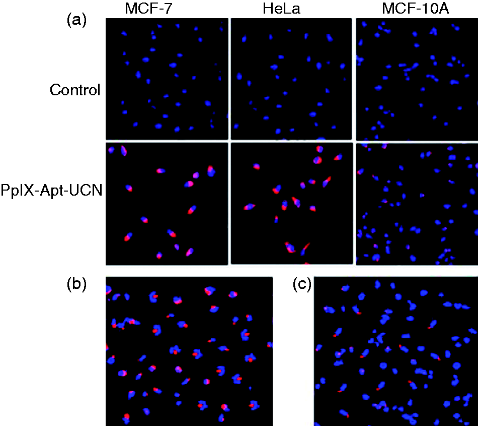
Tumor-targeting capability of PpIX-Apt-UCNP. Confocal microscopy images of (a) MCF-7, HeLa (nucleolin overexpressed) and MCF-10A (nucleolin negative) cells, (b) nonblocked, and (c) endostatin-blocked MCF-7 cells incubated with 200 µL of PpIX-Apt-UCNP (containing 57 µg/mL PpIX) and incubated for 24 h. Cells were labeled with Hoechst 33258 and imaged by a laser confocal microscope equipped with an external 980-nm laser.
Since it is widely accepted that the interaction between nucleolin and aptamer plays an important role in the selective tumor binding of the aptamer AS1411, receptor-blocking experiments were carried out to study the uptake mechanism of the aptamer-modified nanospheres. Because nucleolin had been reported as a receptor of endostatin, 46 here we employed endostatin as a nucleolin blocker in MCF-7 cells.
Two groups of cell experiments were established to elucidate the cellular uptake mechanism for PpIX-Apt-UCNP across the membrane of MCF-7 and the results were given in Figure 5(b) and 5(c). One was incubated with PBS for 1 h before being cultured with PpIX-Apt-UCNP (PBS group). The other was incubated with endostatin in advance (endostatin group). In the PBS group, significant red fluorescence of PpIX was observed (Figure 5(b)), indicating the efficient cellular uptake for the PpIX-Apt-UCNP by the unblocked MCF-7 cells. However, in the endostatin group, the fluorescence was not very apparent in the endostatin-blocked MCF-7 cells (Figure 5(c)). The presence of endostatin dramatically reduced the cellular fluorescence of PpIX and thus decrease the cellular uptake of the blocked cells treated with PpIX-Apt-UCNP. The endostation-blocking experiment indicated that the cellular uptake of PpIX-Apt-UCNP mainly attributes to the binding of aptamer to the nucleolin receptor which is over expressed on the cancerous cell membrane.
Cell photocytotoxicity and singlet oxygen generation of PpIX-Apt-UCNP
PpIX is a well-known PS and frequently used in PDT. However, it can only be activated by visible light, whose penetration depth in biological tissues is very limited. Thus, the application of PpIX in PDT has been seriously hampered in the treatment of deep-seated tumors. Here we loaded the PpIX on UCNP nanospheres, which are able to emit shorter wavelength photons under excitation by NIR light.8–10 Since NIR can afford penetration depths of an order of magnitude greater than that of the visible light, it is supposed that the PpIX-Apt-UCNP could exhibit photocytotoxicity under the irradiation of NIR.
The photo-cytotoxicity and IC50 of these PpIX-Apt-UCNP nanospheres on three cancer lines, namely A549, HeLa and MCF-7 cancer cells, were determined by MTT assay (Figure 6). From Figure 6(a), under the irradiation of 980 nm light, PpIX-Apt-UCNP nanospheres exhibit significant inhibition on all the cancer cell viability with very close IC50 values (Figure 6(b)). Since it is demonstrated in the biocompatibility experiment above that drug-free UCNP produce negligible cytotoxicity on cancer cells, this dramatic reducing of cell viability can mainly attribute to the photo-cytotoxicity of PpIX. The NIR light was successfully upconverted to Vis light by the UCNP system and thus activated PpIX molecules. However, in the dark, the PpIX-Apt-UCNP nanospheres have no cell inhibition even to promote the cancerous cell proliferation. This distinct behaviors of the PpIX-Apt-UCNP nanospheres in the dark and NIR light convinced their potential as PSs.
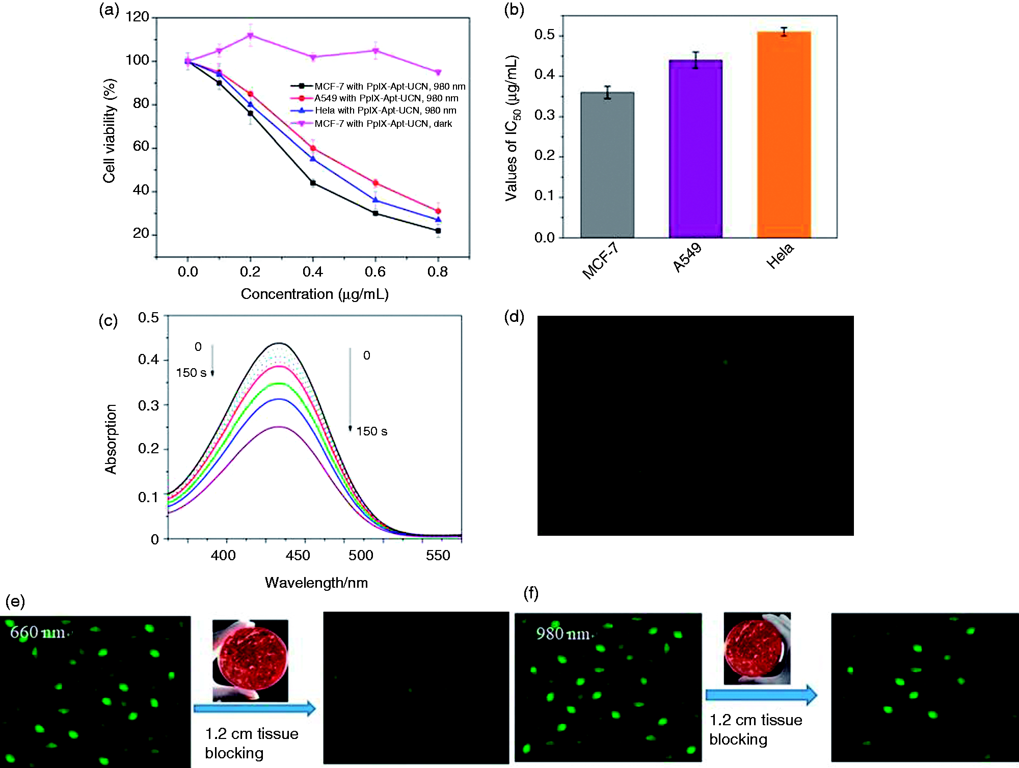
Cell viability (a) and IC50 values (b) of PpIX-Apt-UCNP toward MCF-7, A549, and HeLa cell lines. HeLa cells were incubated with the nanospheres for 24 h and the cell viability of the HeLa cells with PpIXApt-UCNP at concentrations ranged from 0 to 0.8 µg/mL. Detection of intracellular reactive oxygen production (ROS) by DCFH-DA staining in HeLa cells incubated with PpIX-Apt-UCNP. Cell dishes were covered with 1.2-cm tissue and then were exposed to (c) 660 nm or (d) 980 nm light for 10 min, respectively.
It is widely accepted that the PS induce apoptosis through reactive oxygen species (ROS) generation. Intracellular ROS levels of these PpIX-Apt-UCNP nanospheres were investigated with a fluorescence microscope and microplate analyzer using H2DCFDA as a probe. H2DCFDA is a fluorescent dye that diffuses through cell membranes and is hydrolyzed by intracellular esterases to DCFH. In the presence of ROS, DCFH could be oxidized to DCF, whose fluorescent level corresponds to the level of generated ROS.
In control experiments without light irradiation, no obvious fluorescent spots of DCFH were detected (Figure S3). However, as shown in Figure 6(c) and 6(d), when was directly exposed to the excitation light at 660 nm or 980 nm, the PpIX-Apt-UCNP produced significant intracellular ROS with very similar production. This indicates that the UCNP-based nanoconstruct can efficiently upconvert NIR to visible light and excite the PS successfully.
To investigate the penetration advantage of the NIR light, thick pork tissue was used to mimic living tissue for deep tissue PDT treatment. From Figure 6(c) and 6(d), it is found that, after blocking the excitation light with 1.2 cm pork tissue, fluorescence signals of DCFH in 660-nm light-treated cells decreased much more significantly compared with that in 980 nm light-treated cells. This implies that less ROS was produced by 660 nm light in tissue because of the limited tissue penetration of this wavelength. However, NIR light exhibited much better tissue penetration and thus the PpIX-Apt-UCNP nanoplatform could remain high ROS generation in deep tissues.
Meanwhile, our previous research indicates that singlet oxygen (1O2) is the main species produced by porphyrin compounds under irradiation.32–34,47 To further understand the mechanism of the photodynamic cell cytotoxicity of the PpIX-Apt-UCNP nanospheres, we evaluated the 1O2 generation by the absorption change of RNO following the Kraljic procedure. 39 PS PpIX in the light irradiation could react with O2 to produce 1O2 and the intermediates of 1O2 and could cause RNO oxidation, resulting in the absorbance reduction of RNO at 440 nm. 47 We detected the absorption spectral change of RNO in the presence of PpIX-Apt-UCNP in the dark and under 980 nm light irradiation (Figure S4). The absorbance of RNO at 440 nm decreased remarkably owning to the photo-oxidation bleaching effect after NIR irradiation for 150 s with PpIX-Apt-UCNP, which indicated the significant production of 1O2 from PpIX-Apt-UCNP when exposed to NIR light. However, there are no substantial absorption changes of RNO when incubated with the PpIX-Apt-UCNP nanospheres in the dark. It is confirmed that, the PpIX-Apt-UCNP can produce toxic reactive oxygen species under NIR irradiation but are relatively safe to the tissues in the dark, and thus are promising PS with high biosafety.
Cell apoptosis detection
Flow cytometry experiments were carried out for quantitatively evaluated the cell apoptotic induced by PpIX-Apt-UCNP after incubation for 24 h. Figure 7 gives the scatterplots of the bivariate flow cytometry for the cells incubated with the studied nanospheres. For the scatterplots, the left inferior quadrant (Annexin V−/Propidium Iodide [PI]−) represents living cells; the lower right quadrant (Annexin V+/PI−) represents early apoptotic cells; the upper right quadrant (Annexin V+/PI+) represents apoptotic and necrotic cells; Annexin V−/PI+) is considered to be within the permissible detection error.
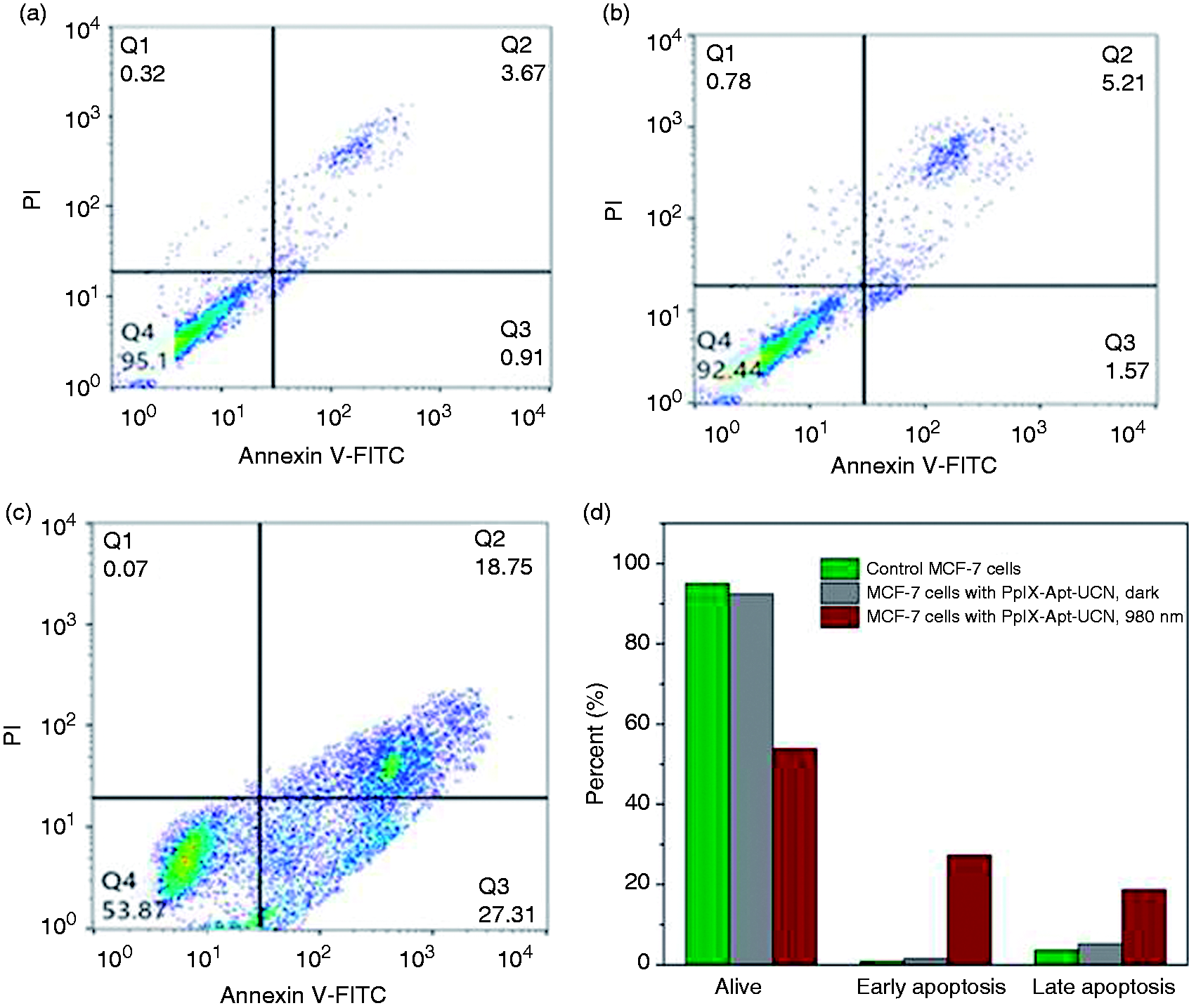
Scatter plots of Annexin V-FITC-PI apoptosis detection for (a) controlled MCF-7 cells, (b) MCF-7 cells treated with PpIX-Apt-UCNP for 24 h in dark, or (c) followed by irradiation with 980 nm light for 10 min. (d) The histogram of apoptosis detection results.
From Figure 7, it is found that the ratio of the alive cells decreased from 95.1% of the controlled MCF-7 cells to 53.87% for cells treated with PpIX-Apt-UCNP under NIR irradiation. However, the percentage of alive cells was not significantly influenced in the case of dark, confirming the biocompatibility of the PpIX-Apt-UCNP in the dark. PDT treatment dramatically increased the ratios for the apoptotic cells, from 1.57% to 27.31% for early apoptosis and from 5.21% to 18.75% for late apoptosis, respectively. These results clearly indicate that the PpIX-Apt-UCNP could significantly induce the apoptosis of cancerous cells under the irradiation of NIR light, exhibiting potent PDT anticancer capability to tumor cells.
Discussion
The limited tissue penetration of excitation light and poor tumor selectivity of the PS seriously restricts the development of PDT in deep-seated tumors. The aim of this work is to overcome these drawbacks. Upconversion nanospheres have captured the attention of researchers for their advantage that converting the excitation light of PS (normally, visible light) to NIR light with deeper tissue penetration while aptamers were well known as targeted ligands of drug delivery vectors. We herein reported an aptamer-coated upconversion nanoplatform to deliver PpIX to cancerous cells and deep-seated tumors.
In the present work, we firstly synthesized NaYF4:Yb(20%),Er (2%) nanoclusters using a one-pot synthesis method to load drugs. Unfortunately, these nanoclusters lacked sufficient colloidal stability in organic solvents to enable further functionalization. Thus, we further fabricated a NaYF4:Yb,Er@NaYF4 core/shell structure that exhibited much better colloidal stability. The increase in colloidal stability may be attributed to a higher ratio of OA to ODE in the core/shell reaction mixtures. 38
The mechanism of UCNP prolonging the excitation range of PS is that UCNP could upconvert NIR light to visible light which were then successfully transferred to PS. In our research system, the fluorescence of PpIX-Apt-UCNP has a significant quenching in the absorption range of PpIX compared with UCNP, indicating that the energy could be efficiently transferred from UCNP to PS. The as-fabricated PpIX-Apt-UCNP was thus endowed with the capability to be triggered by NIR in deep-seated tumors.
Previous research has demonstrated that guanine nucleotides within the core of a G-quadruplex are relatively protected from chemical attack, and the loops in G-quadruplex structure give the chance for nuclease cleavage.42,43 In this study, the well-defined tumor-specific ligand aptamer AS1411 was covalently conjugated to the surface of UCNP through the EDC reaction. AS1411 could fold to a stable G-quadruplex structure in the presence of K+ and well resist nuclease cleavage for a long incubation time with high nuclease concentration. This super anti-cleavage ability of PpIX-Apt-UCNP ensured the safety and biocompatibility of the drug-delivery vectors.
Tumor-targeted UCNP-based PDT agents can effectively increase the accumulation of the nanoparticles and improve PDT efficiency in deep-seated tumors. AS1411 was widely accepted to target the nucleolin on the cancer cell membrane and then internalize into the tumor with the nucleolin. The enhanced intracellular uptakes of PpIX-Apt-UCNP by nucleolin-positive tumor cells (HeLa and MCF-7) were significantly higher than that in nucleolin-negative MCF-10A normal cells. This increased uptake could be mainly attributed to nucleolin-mediated endocytosis, which is evidenced by endostatin-blocking experiment in nucleolin-positive cancer cells. This result suggests that conjugating AS1411 aptamer to the UCNP-based nanoplatform could significantly improve the active tumor targeting of the drug vector to nucleolin-overexpressed tumors (MCF-7 and HeLa).
It is well known that PDT mainly kills cancer cells through the activation of PS by light irradiation, which will generate cytotoxic ROS that directly destroy the tumor cells. In general, there are two ways for PDT to kill cells, defined as Type I and Type II mechanisms, respectively. For the Type I mechanism, hydrogen atom abstraction or electron transfer reactions occurs between the PS and a substrate to yield free radicals and radical ions, which are able to interact with molecular oxygen to either generate ROS (such as superoxide anions and hydroxyl radicals) or cause irreparable biological damage. The Type II mechanism mainly refers to an energy transfer between the PS and the molecular oxygen to produce singlet oxygen. DCFH-DA probes have been frequently employed to report the potential PDT efficacy of drugs, resulting from their capabilities of detecting the lethal cytotoxic ROS (not only singlet oxygen) production inside cells and in solution. It is well known that, unlike visible or UV light, PDT triggered by NIR light enables the feasibility of deep-tissue PDT treatment. Herein, we investigate the PDT efficiency of the constructed UCNP-based nanospheres in deep-seated tumors by covering the cell dishes with 1.2-cm pork tissue. Significant fluorescence was observed through adipose tissue when excited with 980-nm light while no obvious fluorescent spots were monitored in the case of 660-nm excitation. It is suggested that UCNP-based PDT induced by deep-penetrating 980 nm light produces more ROS than 660-nm light in the deep-seated cancer cells.
Conclusion
In summary, we present herein an easy and efficient strategy to coadministrate UCNP and PS PpIX with a single drug-delivery system by taking advantages of the G-quadruplex structure of AS1411 aptamer. The as-established PpIX-Apt-UCNP nanospheres could have efficient cellular uptake to cancerous cells, which could be mainly attributed to the tight binding between the AS1411 aptamer and overexpressed nucleolin on the cell membrane. Moreover, NIR light, which has improved penetration depth and high biosafety to tissues, successfully triggered the PDT of the as-prepared PpIX-Apt-UCNP nanospheres because of the upconversion properties of UCNP. In vitro experiment suggested that the cellular ROS level was remarkably elevated and the cells were induced to early or late apoptosis after incubation with PpIX-Apt-UCNP for 24 h following by 980 nm light irradiation. In conclusion, our study provided a novel solution to construct Apt-UCNP-based nanotherapeutic that combines the precise tumor targeting and PDT functions into one single platform, and achieved a new promising and safe anticancer PS delivery system. This simple solution can be easily expanded to other PS, cancer-targeting treatment, and drug-delivery vectors.
Measurement of ROS production under deep tissue both in the solution and in cancer cells reveals that NIR light triggered PDT by using the nanoconstructs generates more ROS in contrast to direct irradiation of PpIX upon 660-nm light. In comparison with conventional PDT depending on red light irradiation, the NIR-induced PDT based on the nanoconstructs possesses higher tumor inhibition ratio for the treatment of simulated deep-seated tumors. Together, our results demonstrate the considerable advantages of tumor-selective UCNP-based PDT induced by NIR light over traditional PDT for internal tumors and prompt further explorations of these nanoconstructs for targeted drug delivery and deep-tissue PDT of other related diseases.
Supplemental Material
JBA882152 Supplemental Material - Supplemental material for Aptamer-guided upconversion nanoplatform for targeted drug delivery and near-infrared light-triggered photodynamic therapy
Supplemental material, JBA882152 Supplemental Material for Aptamer-guided upconversion nanoplatform for targeted drug delivery and near-infrared light-triggered photodynamic therapy by Hui-Chao Lin, Wen-Tian Li, Thushara W Madanayake, Can Tao, Qiang Niu, Si-Qi Yan, Bao-An Gao and Zhao Ping in Journal of Biomaterials Applications
Footnotes
Availability of data and materials
All data generated or analyzed during this study are included in this published article.
Author contributors
Hui-Chao Lin and Wen-Tian Li contributed equally to this work. Thushara W Madanayake gave valuable suggestions and kindly read this paper.
Declaration of conflicting interests
The author(s) declared no potential conflicts of interest with respect to the research, authorship, and/or publication of this article.
Funding
The author(s) disclosed receipt of the following financial support for the research, authorship, and/or publication of this article: The research was funded by Natural Science Foundation of Guangdong (2016A030313807), Guangzhou key laboratory of construction and application of new drug screening model systems (No. 201805010006) and Key Laboratory of New Drug Discovery and Evaluation of ordinary universities of Guangdong province (No. 2017KSYS002).
Supplemental material
Supplemental material for this article is available online.
References
Supplementary Material
Please find the following supplemental material available below.
For Open Access articles published under a Creative Commons License, all supplemental material carries the same license as the article it is associated with.
For non-Open Access articles published, all supplemental material carries a non-exclusive license, and permission requests for re-use of supplemental material or any part of supplemental material shall be sent directly to the copyright owner as specified in the copyright notice associated with the article.