
Abstract
Postoperative infections caused by bacteria are now thought to be the main cause of guided tissue regeneration (GTR) failure. In this study, six groups of PLGA/wool keratin composite GTR membranes loaded with the antibacterial agent ornidazole (0% ORN, 1% ORN, 5% ORN, 10% ORN, 20% ORN, and 30% ORN composite membranes) were prepared by an electrospinning method. The surface morphology and physicochemical properties of the composite membranes were determined, and the in vitro drug release behavior, bacteriostatic properties and in vitro cytotoxicity were assessed to optimize the amount of drug loaded into the composite membranes. The composite membrane with 1% ORN showed strong water absorption and mechanical properties as well as suitable in vitro degradation and drug release characteristics; the 5% ORN composite membrane showed good water absorption and degradation characteristics; and the 10% ORN composite membrane showed good mechanical and degradation properties. The five PLGA/wool keratin/ornidazole composite membranes could release ornidazole continuously for 7 d. When the content of ornidazole in the composite membranes was greater than or equal to 10%, the growth of human periodontal ligament fibroblasts (hPDLFs) was inhibited, while composite membranes with an ornidazole content of less than 10% facilitated the growth and proliferation of hPDLFs and could promote their osteogenic differentiation. The five PLGA/wool keratin/ornidazole composite membranes all inhibited the growth of Porphyromonas gingivalis (Pg), Fusobacterium nucleatum (Fn) and Peptostreptococcus anaerobius (Pa), and higher drug contents resulted in stronger bacteriostatic effects. In summary, the 1% ORN composite membrane had good physicochemical properties, biocompatibility and bacteriostatic properties, making this membrane applicable for use as an antibacterial GTR membrane for periodontal tissue repair.
Introduction
Periodontal disease is a common oral disease in clinical settings. It is a chronic inflammatory disease caused by a mixture of periodontal pathogens (mainly anaerobic bacteria) that occurs in periodontal supporting tissues. Its clinicopathological features are the progressive and irreversible destruction of soft and hard periodontal tissues, and it is the main cause of tooth loss in adults and an important oral disease that endangers the human oral cavity and overall health. 1
The goals of periodontal disease treatment are not only to control inflammation and prevent the further development of periodontal disease but also to restore the structure and function of the lost periodontal tissue. The traditional method used to treat periodontal disease can control inflammation, but achieving periodontal tissue regeneration is difficult. Developing methods to regenerate lost periodontal tissue is a long-standing challenge and is key to the treatment of periodontal disease.
The emergence and development of guided tissue regeneration (GTR) technology have inspired new treatment concepts and achieved clear clinical results for the treatment of periodontal disease and the repair of periodontal defects. Moreover, GTR membranes play a key role in periodontal GTR.
In our previous study, a PLGA/wool keratin composite GTR membrane was prepared by electrospinning. 2 The composite membrane had desirable mechanical properties, thermal stability, biological activity and cell compatibility and could effectively promote the regeneration of periodontal tissues such as the alveolar bone and the periodontal ligament in beagle dogs as effectively as a commercial collagen membrane. 2 However, although postoperative infection caused by bacteria is currently considered the main cause of GTR failure, this composite membrane did not have an antibacterial function. 3 Generally, oral antibiotics can effectively inhibit bacteria and reduce the incidence of inflammatory reactions. However, the long-term use of antibiotics can lead to a variety of side effects, such as undesirable side reactions, allergic reactions, and drug resistance. 3 To solve this problem, antibacterial drugs can be loaded onto a GTR membrane to achieve targeted, continuous and controllable release, thereby inhibiting local inflammatory reactions.
Ornidazole is a new third-generation nitroimidazole derivative that shows good tolerance, a low incidence of adverse reactions and lasting efficacy and has strong inhibitory and killing effects against most anaerobes. 4 It can effectively eliminate bacterial infections in wounds in the oral cavity and maintain a desirable healing environment. 4
In this study, ornidazole-loaded PLGA/wool keratin composite membranes were prepared by electrospinning. The composite membrane can not only meet the general requirements of GTR films but also exert a specific antibacterial effect. This study will serve as a foundation for further studies on GTR membranes with antibacterial functions and on regenerative treatments for periodontal disease.
Materials and methods
Materials
PLGA (Mw: 5 × 10⁴ g/mol) with a lactide/glycolide ratio of 75:25 w/w was purchased from Shandong Daigang Co., Ltd (Jinan, China). Wool was purchased from Kunshan Sanli Wool Carbonization Co., Ltd (Jiangsu, China). Wool keratin was extracted from clean wool. 2 Dimethylformamide (DMF), trichloromethane (TCM), and other analytical reagents were purchased from Tianhong Chemical Reagent Co., Ltd (Jiangsu, China). Dulbecco's modified Eagle's medium (DMEM), 0.25% trypsin and phosphate-buffered saline (PBS) were obtained from HyClone (Utah, USA). A 3–(4,5-dimethylthiazol-2-yl)-2,5-diphenyltetrazolium bromide (MTT) cell proliferation kit and an alkaline phosphatase (ALP) test kit were obtained from Kaiji Biotechnology Co., Ltd (Jiangsu, China).
Preparation of ornidazole-loaded PLGA/wool keratin composite membranes
The ornidazole-loaded PLGA/wool keratin composite membranes were prepared by electrospinning using an in-house solution spinning device. 2 A 20 wt % PLGA solution was prepared by dissolving PLGA in a mixture of TCM and DMF (7:3 v/v). Wool keratin (1 wt %) was added to the PLGA solution, and then ornidazole (at six different levels 0, 1, 5, 10, 20, or 30 wt%) was added to the PLGA solution. The solutions were sonicated for 1 h to ensure homogeneous dispersion of the wool keratin and ornidazole. The blended solutions were used for spinning with the electrospinning methods and parameters reported previously. 2 After electrospinning, six groups of ornidazole-loaded PLGA/wool keratin composite membranes were prepared: PLGA/wool keratin, PLGA/wool keratin/1% ornidazole, PLGA/wool keratin/5% ornidazole, PLGA/wool keratin/10% ornidazole, PLGA/wool keratin/20% ornidazole and PLGA/wool keratin/30% ornidazole composite membranes. For simplicity, these membranes are referred to below as 0% ORN, 1% ORN, 5% ORN, 10% ORN, 20% ORN, and 30% ORN composite membranes, respectively.
Characterization of the ornidazole-loaded PLGA/wool keratin composite membranes
Surface morphology
The surface morphology of the six groups of ornidazole-loaded PLGA/wool keratin composite membranes was examined by scanning electron microscopy (SEM; JSM-5900LV, JEOL, Tokyo, Japan).
X-ray diffraction (XRD)
The crystal structures of the composite membranes were observed by an X-ray diffractometer (SA-HF3, Rigaku, Tokyo, Japan). The tube pressure was 40 kV, the tube flow was 30 mA, the scanning rate was 8°/min, and the scanning range was 3° to 60°.
Fourier transform infrared spectroscopy
Infrared spectra of the composite membranes were acquired in the range from 4000 cm−1 to 400 cm−1 by an FTIR spectrophotometer (Nicolet 560, Nicolet Instrument Co., WI, USA).
Mechanical properties
The tensile strength, modulus of elasticity and elongation at break of the composite membranes were measured using a universal testing machine (CTM8050S, Xie Qiang Instrument Manufacturing (Shanghai) Co. Ltd, Shanghai, China). The thickness of the composite membranes was measured using a spiral micrometer, and the sample composite membranes were made into long strips (10 mm × 70 mm) for tensile testing. Five samples from each set of membranes were tested, and the tensile speed of the tensile machine was 5 mm/min.
Encapsulation efficiency
Ornidazole (10 mg) was completely dissolved in 1 ml of dimethyl sulfoxide (DMSO), and methanol was added to bring the volume to 10 ml for extraction. The mixture was ultrasonicated for 2 h, and the supernatant was then separated by centrifugation. Afterwards, the supernatant was filtered through a 0.22 μm filter and analyzed by high-performance liquid chromatography (HPLC; Primaide, Hitachi, Tokyo, Japan) at 318 nm, and the ornidazole absorption peak (C 18 column, water: methanol = 40:60) was acquired. The encapsulation efficiency of the composite membranes was calculated using the following formula.
Encapsulation efficiency (%) = actual amount of drug in the composite membrane (mg)/theoretical amount (mg) of drug in the composite membrane × 100% (n = 3)
Water absorption rate
A 10 mg sample of each ornidazole-loaded composite membrane (M0) was immersed in 5 ml of PBS at 37°C. After 24 h, the surfaces of the composite membranes were carefully dried with filter paper, and then the membranes were weighed (M1). The composite membranes were then dried in a constant-temperature drying box for 24 h and weighed again (M2). The apparent water absorption rate and the actual water absorption rate of the composite membranes were calculated using the following formulas.


Degradation performance in vitro
Ornidazole-loaded composite membranes from the six groups were cut into 18 mm × 18 mm pieces, weighed (M0) and numbered. The samples were placed in PBS in a 37°C water bath for a total of 16 weeks. The PBS solution was changed every other day. At predetermined times (2, 4, 6, 8, 12 and 16 weeks), the samples were removed, washed with distilled water, dried, and weighed (M1), and the weight loss rates of the composite membranes at the different time points were calculated by the formula below. The surface morphologies of the composite membranes during degradation were observed by SEM at 2 and 8 weeks.

Drug release
Ten-milligram samples of composite membranes from each group (n = 3) were placed in 5 ml of PBS and shaken in an oscillator (HZS-H, Donglian, Beijing, China) at 37°C. At 4 h, 12 h, 24 h, 2 d, and 7 d, 1 ml of the soaking solution was removed and analyzed by HPLC. Next, the remaining liquid was aspirated, 5 ml of fresh PBS solution was added, and the mixture was then further shaken in the oscillator. The quantity of drug released by different samples at different times was calculated by comparison with the standard curve, and the drug release curve was drawn.
Cytocompatibility of ornidazole-loaded PLGA/wool keratin composite membranes
Composite membranes from the six groups (n = 3) were cut into circles with a diameter of 10 mm, sterilized, and then placed in a 24-well culture plate (Corning, USA). Human periodontal ligament fibroblasts (hPDLFs) at passage 4 were seeded on the surface of the composite membranes at a density of 1 × 10⁵/ml, and 1 ml of the cell suspension was placed in each well. Samples were taken from each group at 4 h and 7 d, and the samples were fixed, dehydrated, dried, sprayed with gold, and observed by SEM.
In the experimental groups, cells at passage 4 were seeded on composite membranes from each group at a density of 1 × 10⁵/ml in a 24-well culture plate (n = 3). In the control group, cells were seeded in a 24-well plate at the same density, but no composite membrane was added. The culture plates were placed in a cell culture incubator, and the medium was changed every other day. The culture plates were removed at 1, 3, 5, and 7 d. The culture medium was aspirated, 500 μl of MTT (1 mg/ml) was added to each well, and the plates were incubated at 37°C for 4 h. Then, the supernatant was aspirated, 1000 μl of DMSO was added to each well, and the plates were shaken in an oscillator. The absorbance value (OD) of each well was detected at 570 nm by a microplate reader (Infinite M200 Pro, Tecan, Zürich, Switzerland).
Composite membranes from each of the six groups (n = 3) were cut into circles with a diameter of 10 mm, and the disks were sterilized and then placed in a 24-well culture plate. Cells at passage 4 were seeded on the surface of the composite membranes at a density of 1 × 10⁵/ml. After 24 h, the culture medium was replaced with osteogenic induction medium, and the induction was continued for 10 d. At 4, 7 and 10 d, the culture medium was discarded, and 1% TritonX-100 cell lysate was added into each well. After 40 min, the fluid was removed and centrifuged at 1000 r/min for 5 min. The supernatant was then taken for analysis. Next, 50 μl of buffer solution and 50 μl of a basic solution were added to each well, and the mixtures were incubated at 37°C for 15 min. Next, 150 μl of developing solution was added to each well. The mixture was gently shaken, and the absorbance value (OD) of each well was measured at a wavelength of 520 nm by a microplate reader.
Antibacterial performance test
The antibacterial properties of the composite membranes were tested against Porphyromonas gingivalis (Pg, ATCC33277), Fusobacterium nucleatum (Fn, ATCC25586) and Peptostreptococcus anaerobius (Pa, ATCC27337). The Pg, Fn and Pa were anaerobically cultured on a blood plate at 37°C. The ornidazole-loaded composite membranes (n = 3) were cut into circles with a diameter of 4 mm, which were sterilized before use. The concentrations of the three bacterial solutions were adjusted to 0.5 Mei, and then the prepared bacterial solutions were evenly smeared on the surface of the flat plates with aseptic cotton swabs. After 5 min, the sterilized composite membranes were evenly attached to the surface of the flat plates, and the plates were incubated at 37°C in an anaerobic environment. At 3, 7, 14 and 21 d, the diameter of the zone of inhibition was observed and measured, and photographs were taken at 21 d.
Statistical analysis
Statistical Package for Social Science 23.0 (SPSS 23.0, USA) was used to analyze the data. The results are reported as the mean ± standard deviation (SD). A t-test was used to identify the significant differences between the two groups. One-way ANOVA was used for comparisons among groups, and the least significant difference (LSD) was used for further comparisons between two groups. A value of p < 0.05 was considered statistically significant.
Results
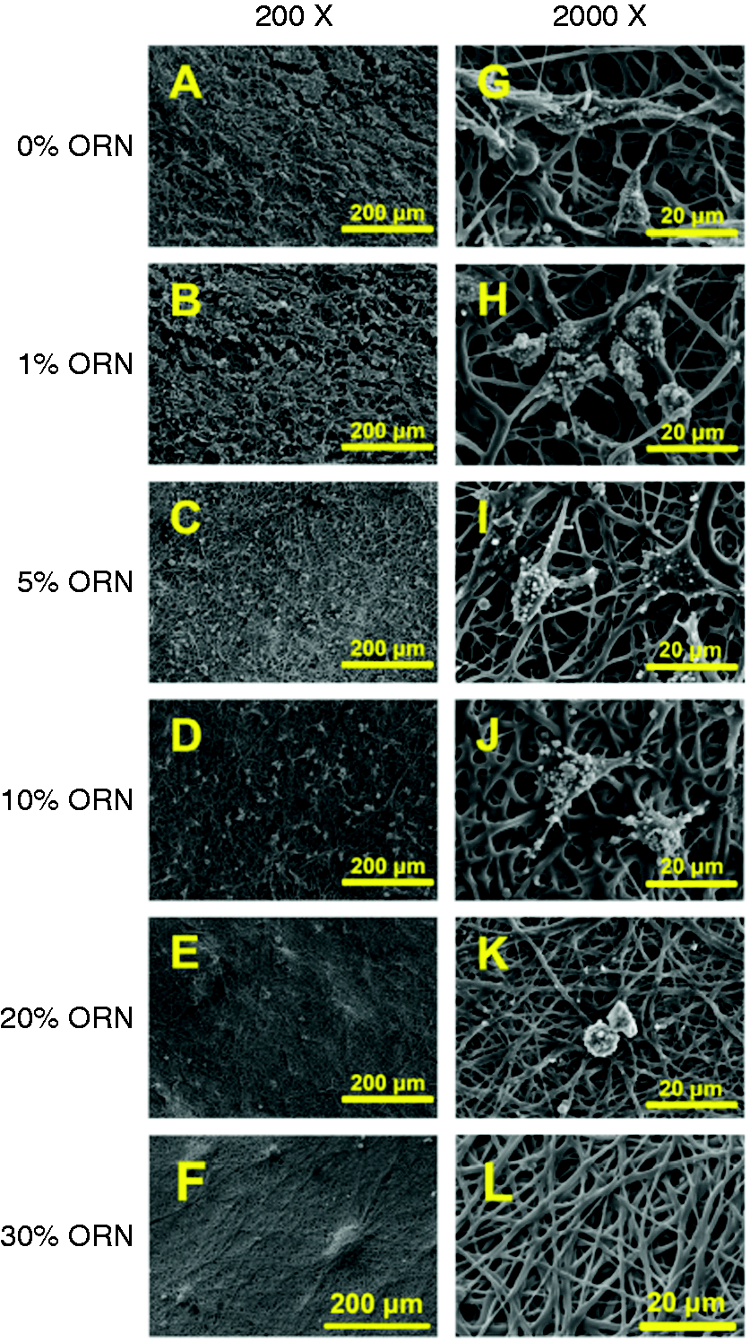
Surface morphology
SEM images of the six groups of ornidazole-loaded PLGA/wool keratin composite membranes are shown in Figure 1. The fibers of the 0% ORN composite membranes were uniform and smooth, a porous structure was formed between the fibers, and no bead-like structures were observed (Figure 1(a)). In the 1% ORN composite membrane, the fiber diameter was uniform, and small ornidazole crystals were attached to the surface of the fibers (Figure 1(b)). As the content of ornidazole in the composite membranes increased, the number and volume of ornidazole crystals coated on the fiber surfaces also gradually increased. Bulk crystals were observed on the surfaces of the 5% ORN and 10% ORN composite membranes (Figure 1(c) and (d)), while the surfaces of the fibers in the 20% ORN and 30% ORN composite membranes were completely covered with ornidazole crystals (Figure 1(e) and (f)).

SEM images of the six groups of ornidazole-loaded PLGA/wool keratin composite membranes: (A) 0% ORN composite membrane; (B) 1% ORN composite membrane; (C) 5% ORN composite membrane; (D) 10% ORN composite membrane; (E) 20% ORN composite membrane; and (F) 30% ORN composite membrane. The yellow arrows indicate ornidazole crystals on the surface of the composite membranes.
X-ray diffraction (XRD)
XRD curves of the six groups of ornidazole-loaded PLGA/wool keratin composite membranes and ornidazole powder are shown in Figure 2. Ornidazole showed intense and sharp characteristic peaks at 2θ = 13.3°, 15.8°, 20.0°, 20.8°, 24.4°, 25.2°, 27.4° and 30.2°. 5 By contrast, the 0% ORN composite membrane was amorphous and showed only a broad peak at approximately 2θ = 18°. The XRD curves of the 1% ORN, 5% ORN and 10% ORN composite membranes were all similar to that of the 0% ORN composite membrane, and none of them showed the characteristic peak of ornidazole. This peak may be absent due to the low content of ornidazole in these composite membranes and the encapsulation of the ornidazole by the fibers. The 20% ORN and 30% ORN composite membranes showed the characteristic peaks of ornidazole at 2θ = 13.3°, 15.8°, 20.0°, 20.8°, 24.4°, 25.2°, 27.4° and 30.2°, and the intensities of the characteristic peaks of ornidazole in the curve of the 30% ORN composite membrane were higher than those in the curve of the 20% ORN composite membrane.
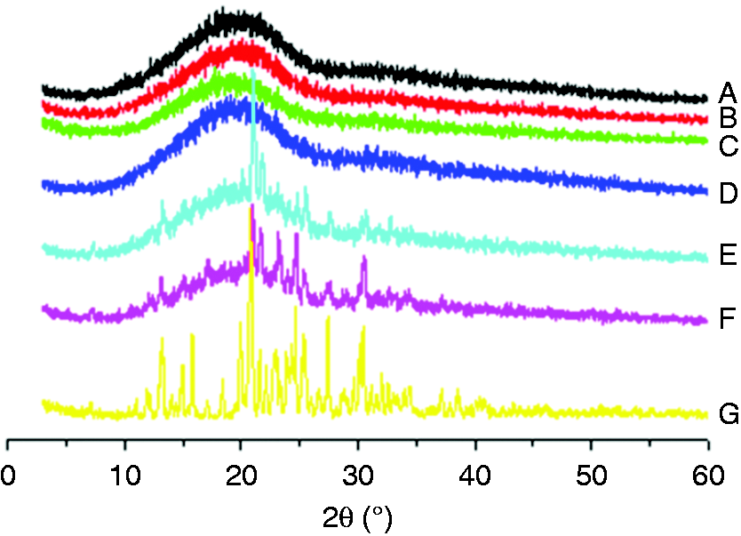
XRD curves of the six groups of ornidazole-loaded PLGA/wool keratin composite membranes and ornidazole powder: (A) 0% ORN composite membrane; (B) 1% ORN composite membrane; (C) 5% ORN composite membrane; (D) 10% ORN composite membrane; (E) 20% ORN composite membrane; (F) 30% ORN composite membrane; and (G) Ornidazole powder.
Fourier transform infrared (FTIR) spectroscopy
Figure 3 shows the FTIR spectra of the six groups of ornidazole-loaded PLGA/wool keratin composite membranes and ornidazole powder. Ornidazole powder showed a C-Cl stretching vibration peak at 655 cm−1, C = N stretching vibration peaks at 1191 cm−1 and 1537 cm−1, a C-N stretching vibration peak at 1269 cm−1, a -NO2 stretching vibration peak at approximately 1363 cm−1, C = C stretching vibration peaks in the range from 1470 to 1490 cm−1, and a C-H stretching vibration peak at 3126 cm−1. 6 The 0% ORN composite membrane exhibited a C = O stretching vibration peak at approximately 1752 cm−1, a C-C stretching vibration peak at 866 cm−1, a C-O-C stretching vibration peak at 1089 cm−1, a C-O stretching vibration peak at 1130 cm−1, an A-type C-O-C stretching vibration peak at 1186 cm−1, an O-H stretching vibration peak at 1267 cm−1, a C-H stretching vibration peak at 1451 cm−1, and a C-H2 stretching vibration peak at 2941 cm−1. 6 The FTIR spectra of the 1% ORN, 5% ORN and 10% ORN composite membranes were all similar to that of the 0% ORN composite membrane, and no characteristic peaks of ornidazole were visible. These peaks may have been absent due to the low content of ornidazole in these composite membranes and the encapsulation of the ornidazole by the fibers. In contrast, the spectra of the 20% ORN and 30% ORN composite membranes showed the characteristic peaks of ornidazole.
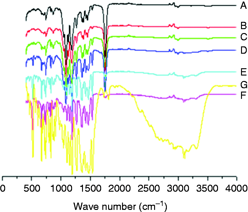
FTIR spectra of the six groups of ornidazole-loaded PLGA/wool keratin composite membranes and ornidazole powder: (A) 0% ORN composite membrane; (B) 1% ORN composite membrane; (C) 5% ORN composite membrane; (D) 10% ORN composite membrane; (E) 20% ORN composite membrane; (F) 30% ORN composite membrane; and (G) Ornidazole powder.
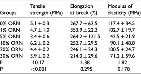
Mechanical properties
The tensile properties of the six groups of ornidazole-loaded PLGA/wool keratin composite membranes are shown in Table 1. There were no significant differences in the elastic modulus and elongation at break among the six groups of composite membranes, but there were significant differences in tensile strength among the six groups. The addition of 1% ornidazole had no significant effect on the tensile properties of the PLGA/wool keratin composite membrane. However, when the content of ornidazole was increased from 1% to 5%, the tensile strength of the composite membrane decreased. Among the six groups of composite membranes, the 10% ORN composite membrane had the highest tensile strength (6.2 ± 0.2 MPa), while the tensile strengths of the 20% ORN and 30% ORN composite membranes were lower than that of the 10% ORN composite membrane. This trend may be due to the weakness of the clusters of ornidazole in the polymer matrix, which are prone to rupture when stress is applied, thereby reducing the strength of the composite film.7,8 In general, a GTR membrane should have sufficient mechanical properties to withstand the force applied during the operation, maintain enough space for cell physiological activity and tissue growth, and preserve the appropriate barrier effect after partial degradation of the membrane to facilitate the regeneration of periodontal tissues.9,10 The results showed that compared with the other three groups of composite membranes, the 0% ORN, 1% ORN and 10% ORN composite membranes had better tensile properties, and their mechanical properties met the requirements of GTR membranes.
Tensile properties of the six groups of ornidazole-loaded PLGA/wool keratin composite membranes (x ± s, n = 3).
Encapsulation efficiency
The standard curve of ornidazole is shown in Figure 4. Ornidazole had a good linear fitting range from 0.01 mg/ml to 0.2 mg/ml.
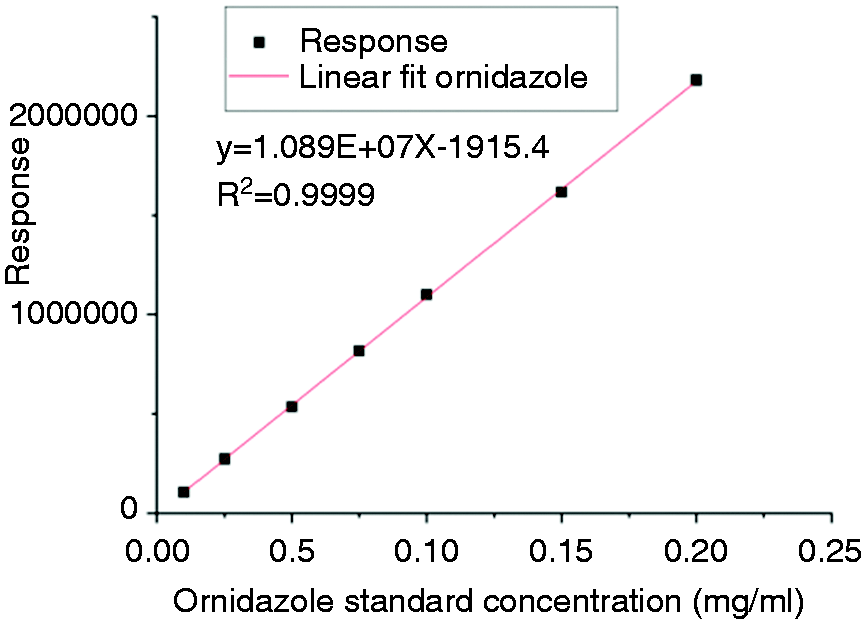
Ornidazole standard curve.
Table 2 shows the encapsulation efficiencies of the five groups of drug-loaded composite membranes, all of which were all above 42%. There were no significant differences in encapsulation efficiency among the five groups (P = 0.079, P > 0.05).
Encapsulation efficiencies of the five groups of PLGA/wool keratin/ornidazole composite membranes (x ± s, n = 3, %).

Water absorption rate
The detection of water absorption properties is an important step in studies on barrier membranes. Higher water absorption performance results in better nutritional uptake ability and exudate absorption by a GTR membrane, which will promote cell growth and the absorption of inflammatory exudates by the GTR membrane. 11 Table 3 shows the water absorption rates of the six groups of ornidazole-loaded PLGA/wool keratin composite membranes. The apparent water absorption rate of the 0% ORN composite membrane was the highest (205.0 ± 27.1%) and that of the 30% ORN composite membrane was the smallest (−37.0 ± 4.6%). The apparent water absorption rates of the 1% ORN and 5% ORN composite membranes were greater than those of the 10% ORN and 20% ORN composite membranes but less than that of the 0% ORN composite membrane. Moreover, the apparent water absorption rate of the 10% ORN composite membrane was greater than those of the 20% and 30% ORN composite membranes, and the differences were significant (P10%20%<0.001/P10%30%<0.001, P < 0.05). The results showed that the addition of ornidazole reduced the apparent water absorption capacity of the composite membranes.
Water absorption rates of the six groups of ornidazole-loaded PLGA/wool keratin composite membranes (x ± s, n = 3, %).
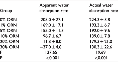
The actual water absorption rates of the six groups of composite membranes were all greater than 130%, and that of the 0% ORN composite membrane was the largest (224.3 ± 3.8%). The results showed that the addition of ornidazole could reduce the actual water absorption performance of PLGA/wool keratin composite membranes.
The water absorption properties of a composite membrane depend mainly on its porosity, which involves the pore size and density of the fibers in the membrane. 12 In this study, the water absorption performance of the composite membranes decreased upon the addition of ornidazole, possibly because the porosity of the composite membranes decreased due to the lattice formation of ornidazole during the electrospinning process, leading to a decrease in the water absorption performance of the composite membranes.12,13 When the content of ornidazole in the composite membrane was increased, the extent of lattice formation increased, the internal fibers became denser, and the water absorption performance gradually decreased. Therefore, the water absorption properties of the 0% ORN, 1% ORN and 5% ORN composite membranes were superior to those of the 10% ORN, 20% ORN and 30% ORN composite membranes.
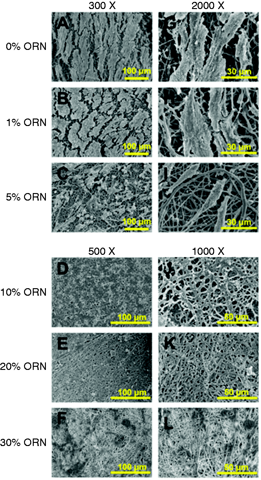
Degradation performance in vitro
Figure 5 shows the gross observations of the degradation of the six groups of ornidazole-loaded PLGA/wool keratin composite membranes at different time points. During the degradation process, the composite membranes gradually became brittle and thin. At 2 weeks, the samples of the six groups of the composite membranes only shrank; at 8 weeks, the composite membranes began to break down into small pieces; at 12 weeks, the composite membranes were shredded; and at 16 weeks, the samples of the six groups of composite membranes were essentially fully degraded.
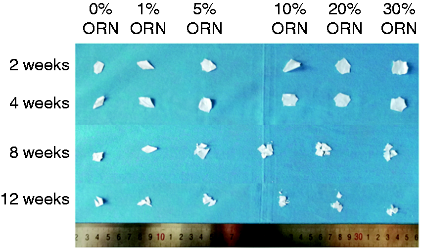
Gross observations of the degradation of the six groups of ornidazole-loaded PLGA/wool keratin composite membranes at different time points.
Figure 6 shows SEM images of the six groups of ornidazole-loaded PLGA/wool keratin composite membranes after 2 and 8 weeks of in vitro degradation. At 2 weeks, the fibers in the composite membranes had changed from straight to curved, the diameter of the fibers was different, the surface of the fibers became rough, some of the fibers showed swelling, and micropores appeared on the surface of the fibers. For the 0% ORN and 1% ORN composite membranes, the fibers were significantly thicker and more tortuous at 8 weeks than at 2 weeks. Moreover, the fibers of the 5% ORN, 10% ORN, 20% ORN, and 30% ORN composite membranes showed a higher number of pores at 8 weeks than at 2 weeks, and some of the fibers were broken. These changes may have been caused by the initial water absorption and swelling of the fibers in the composite membranes, which increased the contact area between the fibers and the buffer. After swelling, pores began to appear in the fibers; then, the fibers broke, and the weight loss rate of the composite membranes increased.14,15
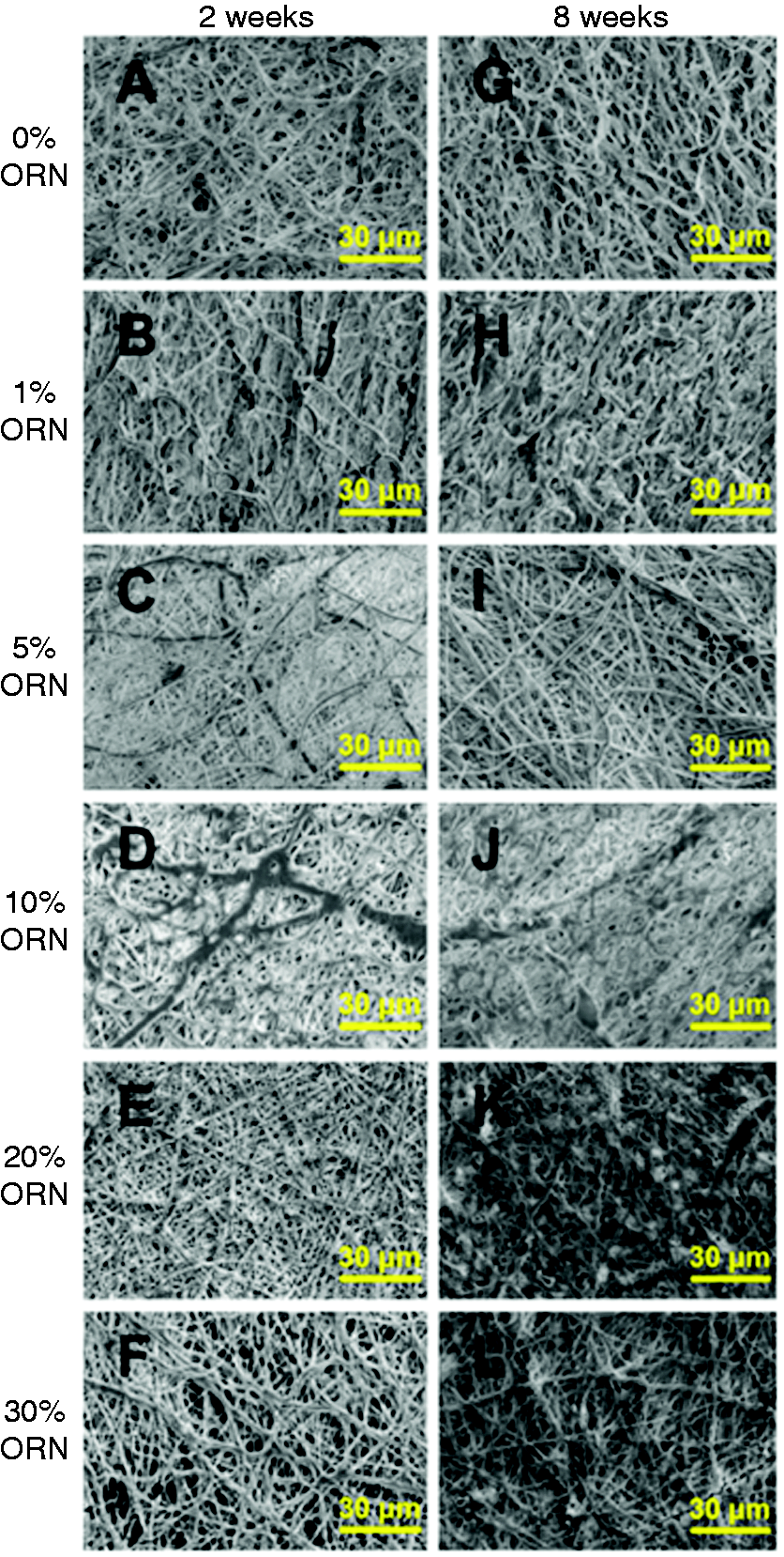
SEM images of the six groups of ornidazole-loaded PLGA/wool keratin composite membranes degraded in vitro for 2 and 8 weeks.
For GTR membranes, it is very important to synchronize the biodegradation rate of the membrane with the regeneration rate of the tissue. 16 The weight loss rates of the six groups of ornidazole-loaded PLGA/wool keratin composite membranes during in vitro degradation are shown in Figure 7. Over 12 weeks, the weight loss rates of the six groups gradually increased with increasing degradation time. At 2 weeks, the 30% ORN and 0% ORN composite membranes showed the largest and smallest weight loss rates, respectively, and the weight loss rates of the five groups of PLGA/wool keratin/ornidazole composite membranes were all higher than that of the 0% ORN composite membrane. This trend indicated that the addition of ornidazole accelerated the degradation of the PLGA/wool keratin composite membrane. During the electrospinning process, a portion of the ornidazole solidified on the surface of the composite membrane. When the composite membrane was in contact with the buffer, the drug could rapidly diffuse directly into the buffer, which increased the weight loss rate of the composite membrane. 12 At 2–8 weeks, except for the 20% ORN and 30% ORN composite membranes, there were no significant differences in weight loss rate among the groups (P > 0.05). At 8 weeks, the weight loss rates of the 20% ORN and 30% ORN composite membranes were still higher than those of the other four groups, and higher ornidazole contents resulted in greater weight loss rates. There were no significant differences in the degradation rates among the 0% ORN, 1% ORN, 5% ORN, and 10% ORN composite membranes from the 2nd week to the 8th week, but the 20% ORN and 30% ORN composite membranes degraded faster than the other composite membranes. At 12 weeks, the weight loss rates of the 20% ORN and 30% ORN composite membranes were both above 85%. At 16 weeks, the 0% ORN, 20% ORN and 30% ORN composite membranes were completely degraded, and the weight loss rates of the 1% ORN, 5% ORN, and 10% ORN composite membranes were greater than 95%. The degradation of the 0% ORN, 1% ORN, 5% ORN, and 10% ORN composite membranes accelerated from 8 to 16 weeks, possibly because the continuous degradation of the composite membranes increased the contact area between the fibers and the buffer due to the swelling and fracture of the fibers. Therefore, the 0% ORN, 1% ORN, 5% ORN, and 10% ORN composite membranes met the requirement that the GTR membrane should maintain a certain space at 4–6 weeks after GTR surgery. 14
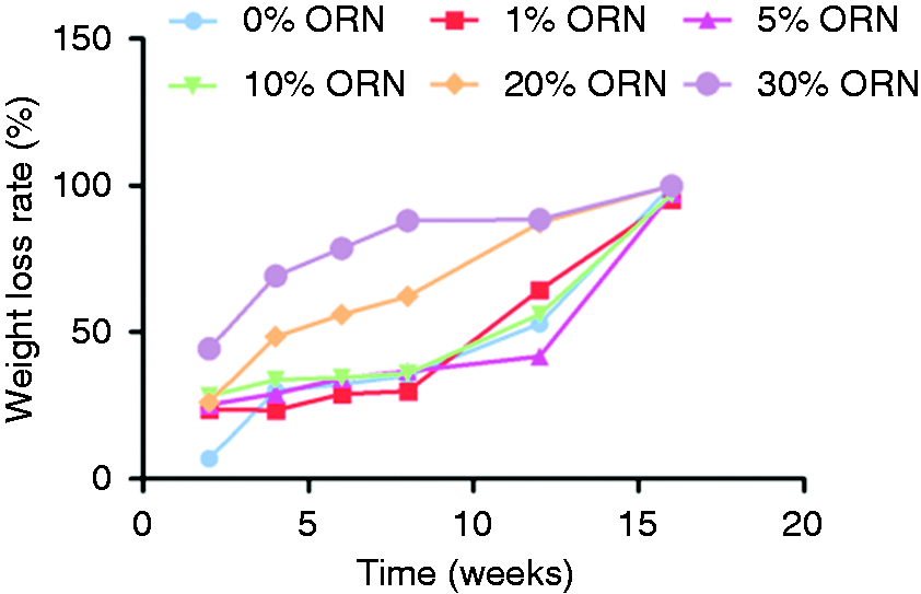
Weight loss rates of the six groups of ornidazole-loaded PLGA/wool keratin composite membranes during in vitro degradation.
Drug release in vitro
The drug release curves of the five groups of PLGA/wool keratin/ornidazole composite membranes are shown in Figure 8. The drug release behavior of the five groups of composite membranes could be divided into two stages: an initial burst release and subsequent sustained release. At 4 h, the cumulative ornidazole release percentages of the 1% ORN, 5% ORN, 10% ORN, 20% ORN and 30% ORN composite membranes were 82.5%, 23.4%, 20.6%, 39.5% and 59.4%, respectively; at 12 h, the cumulative ornidazole release percentages reached 85.9%, 30.0%, 23.3%, 40.8% and 74.2%, respectively; and at 24 h, the cumulative ornidazole release percentages reached 88.2%, 31.5%, 25.1%, 42.2% and 76.1%, respectively. At 4 h, the five groups of PLGA/wool keratin/ornidazole composite membranes showed different degrees of burst drug release. This result was consistent with the findings of a study by He et al., in which physically blended drug-loaded composite membranes showed a burst drug release phenomenon in the early stage. 15 This behavior may be caused by the fact that when the PLGA/wool keratin/ornidazole composite membranes were immersed in PBS, ornidazole entered the buffer from the surface of the membranes, resulting in a sharp increase in the drug concentration in the buffer.
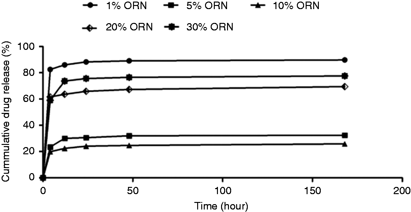
Drug release curves of the five groups of PLGA/wool keratin/ornidazole composite membranes.
After the initial release, the five groups of drug-loaded composite membranes entered a sustained release period. This change occurred because in the later stage, as the buffer gradually permeated the composite membranes, the ornidazole in the composite membranes continued to spread from the inside to the outside of the membrane. At the same time, as the fibers in the drug-loaded composite membranes degraded, the ornidazole was slowly released into the buffer until all of the drug was released. 8
At 7 d, the cumulative release rate of the 1% ORN composite membrane reached 89.6%; the 30% ORN composite membranes released 78.1% of the drug, while the 5% ORN, 10% ORN, and 20% ORN composite membranes had cumulative drug release rates of less than 50%. Because infection and inflammation are most likely to occur during the first week after GTR, the ornidazole-loaded composite membranes should release more drug in the first 7 d to eliminate invading bacteria while still having the ability to continuously release the drug to inhibit potential subsequent bacterial infections. 14 Therefore, the drug release properties of the 1% ORN and 30% ORN composite membranes were more in line with the requirements for the antibacterial properties of GTR membranes.
Cytocompatibility of human periodontal ligament fibroblast cells
SEM images of cells grown on the surface of the six groups of ornidazole-loaded PLGA/wool keratin composite membranes for 4 h and 7 d are shown in Figures 9 and 10, respectively. At 4 h, a large number of spherical or fusiform cells were attached to the surface of the 0% ORN and 1% ORN composite membranes, and some of the cells began to stretch; fewer cells adhered to the 5% ORN composite membrane than to the 0% ORN and 1% ORN composite membranes; little cell adhesion was observed with the 10% ORN composite membrane; and no cells were found on the surfaces of the 20% ORN and 30% ORN composite membranes, indicating that when the content of ornidazole was 20% or higher, the composite membrane was cytotoxic and could inhibit the growth of cells. The results showed that hPDLFs could adhere to the surface of PLGA/wool keratin composite membranes with ornidazole contents less than or equal to 10% at 4 h and that a lower ornidazole content resulted in composite membranes with better cell compatibility.
SEM images of cells grown on the surfaces of six groups of ornidazole-loaded PLGA/wool keratin composite membranes at 4 h.
SEM images of cells grown on the surfaces of the six groups of ornidazole-loaded PLGA/wool keratin composite membranes at 7 d.
At 7 d, the surfaces of the 10% ORN, 20% ORN and 30% ORN composite membranes were free of cells, indicating that at 7 d, when the content of ornidazole was more than 10%, the drug-loaded composite membrane was toxic to the cells. The cells on the surface of the 5% ORN composite membrane did not show complete spreading, and some of the cells were still fusiform. In contrast, the cells on the surface of the 0% ORN and 1% ORN composite membranes were completely spread, inserting into the pores and covering more than 90% of the surface area, and significantly more cells were observed on these two groups at 7 d than at 4 h, indicating that the 0% ORN and 1% ORN composite membrane groups facilitated the adhesion and proliferation of cells more effectively than the other four groups of composite membranes.
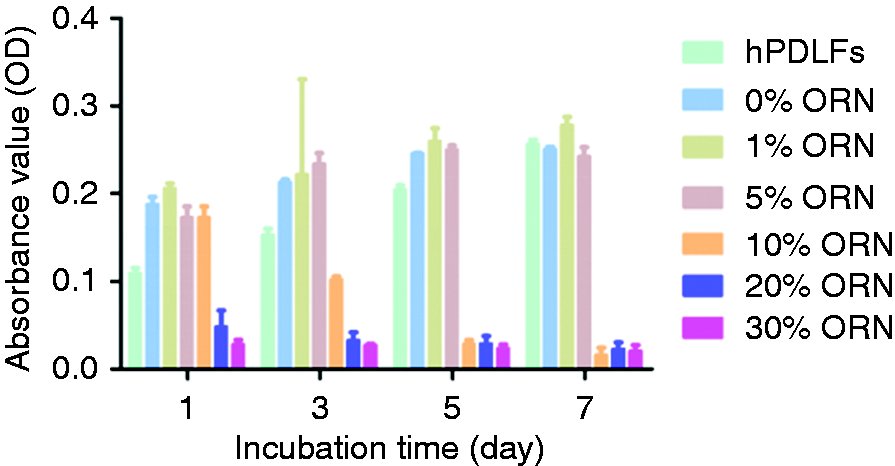
The MTT results for hPDLFs cultured on the six groups of ornidazole-loaded PLGA/wool keratin composite membranes for 7 d are shown in Figure 11. From the 1st day to the 7th day of growth, the number of cells on the ornidazole-loaded PLGA/wool keratin composite membranes with ornidazole contents less than or equal to 5% increased with increasing culture time, while the number of cells on the ornidazole-loaded PLGA/wool keratin composite membranes with ornidazole contents greater than 5% decreased with increasing culture time. At 1 d, the absorbance values of the 20% ORN and 30% ORN composite membrane groups were lower than that of the hPDLF control group, while the absorbance values of the other groups were all higher. At 3 d, the absorbance value of the 10% ORN composite membrane was lower than that of the hPDLF group, and the difference was statistically significant (P < 0.001, P < 0.05). The ornidazole-loaded PLGA/wool keratin composite membranes containing 10% or more ornidazole could inhibit the growth of hPDLFs. The absorbance values of the 0% ORN, 1% ORN and 5% ORN composite membrane groups were higher than that of the hPDLF group, and the differences were statistically significant (P0%hPDLFs<0.001/P1%hPDLFs<0.001/P5%hPDLFs<0.001, P < 0.05), indicating that these three groups could promote the growth of hPDLFs. At 7 d, the absorbance of the 1% ORN composite membrane was the highest and was significantly different from those of the other groups (P1%hPDLFs=0.004/P1%0%=0.001/P1%5%=0.003/P1%10%<0.001/P1%20%<0.001/P1%30%<0.001, P < 0.05), indicating that the cell number, adhesion and proliferation on this composite membrane were the best. Therefore, the 1% ORN composite membrane had the best cell proliferation performance.
Results of the MTT assay of hPDLFs cultured on the six groups of ornidazole-loaded PLGA/wool keratin composite membranes for 7 d.
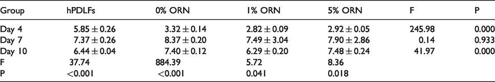
The composite membranes with ornidazole contents greater than or equal to 10% had inhibitory effects on cells, so the ALP activities of only three of the groups of composite membranes (those with ornidazole contents less than 10%) were assessed. The ALP activity results for the three groups of ornidazole-loaded PLGA/wool keratin composite membranes and for the hPDLF group at different time points are shown in Table 4. Between 4 and 7 d, the ALP activity values of each group increased significantly, indicating strong osteogenic differentiation ability of the hPDLFs in each group at 4 and 7 d. At 4 d, the ALP activity values of the three groups of ornidazole-loaded PLGA/wool keratin composite membranes were smaller than that of the hPDLF group; at 7 d, there were no significant differences in ALP activity between the three groups and the hPDLF group, indicating that the differentiation ability of the hPDLFs on the three composite membranes increased with time and was stronger at 7 d than at 4 d. At 10 d, there was no significant difference between the ALP activity values of the 1% ORN composite membrane group and the hPDLF group; however, the ALP activity values of these two groups were lower than those of the 0% ORN and 5% ORN composite membranes, and the differences were statistically significant. The results showed that the 0% ORN, 1% ORN and 5% ORN composite membranes all promoted the osteogenic differentiation of hPDLFs.
The ALP results of the three groups of ornidazole-loaded PLGA/wool keratin composite membranes and the hPDLF group at different time points (x ± s, n = 3, Gin units/gprot).
Antibacterial performance
The inhibition zones of the six groups of ornidazole-loaded PLGA/wool keratin composite membranes against Pg, Fn and Pa at 21 d are shown in Figure 12. The blood plate of the 0% ORN composite membrane was full of bacteria; however, the other five groups all had inhibition zones, and the membranes with higher ornidazole contents induced larger bacteriostatic circles. The 0% ORN composite membrane could not inhibit the growth of Pg, Fn and Pa, but the PLGA/wool keratin/ornidazole composite membranes could inhibit the growth of Pg, Fn and Pa; that is, the ornidazole-loaded PLGA/wool keratin composite membranes had good antibacterial performance against Pg, Fn and Pa, and higher ornidazole contents resulted in stronger antibacterial effects.
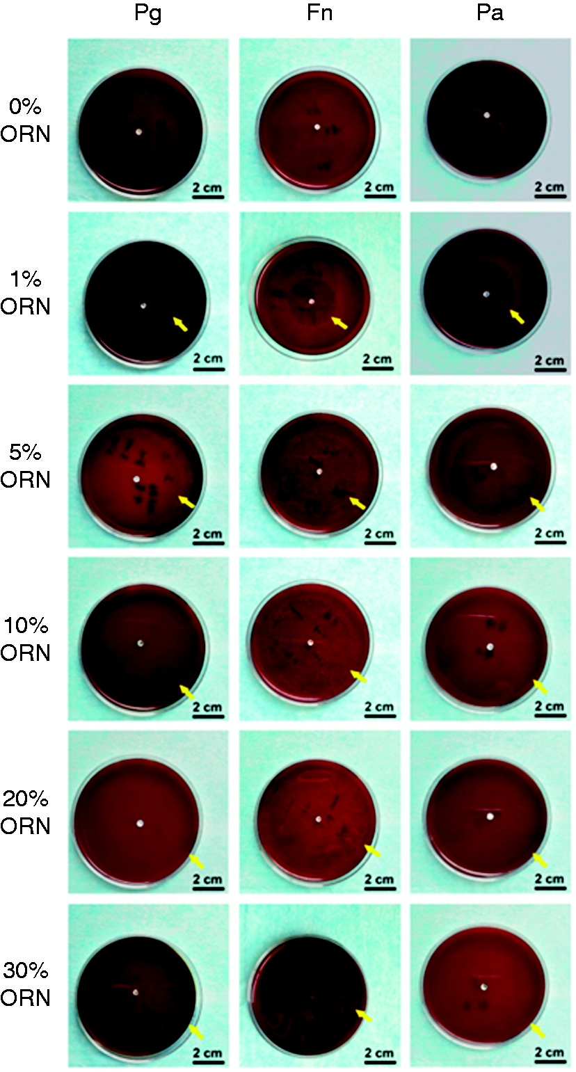
Pictures of the inhibition zones of the six groups of ornidazole-loaded PLGA/wool keratin composite membranes against Pg, Fn and Pa at 21 d. (a) 0% ORN composite membrane. (b) 1% ORN composite membrane. (c) 5% ORN composite membrane. (d) 10% ORN composite membrane. (e) 20% ORN composite membrane. (f) 30% ORN composite membrane. The yellow arrows indicate the inhibition zones.
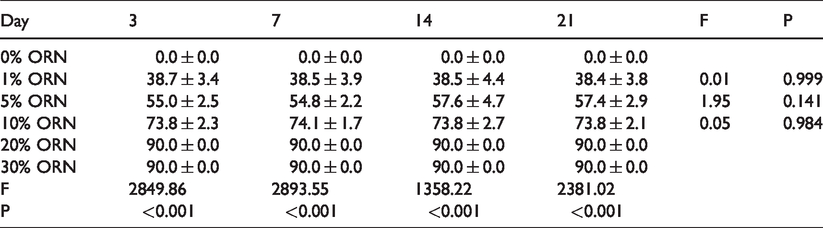
Tables 5 to 7 show the diameters of the inhibition zones of the six groups of ornidazole-loaded PLGA/wool keratin composite membranes against Pg, Fn and Pa for 21 d, respectively. After 21 d, the diameters of the inhibition zones of the 0% ORN composite membrane against the three bacteria were all 0 mm, indicating that the 0% ORN composite membrane had no antibacterial effect on Pg, Fn and Pa. In contrast, the other five groups all showed good antibacterial effects on the three types of bacteria. For composite membranes with ornidazole contents less than 20%, the diameters of the inhibition zones against the three types of tested bacteria increased with increasing ornidazole content. However, there were no significant differences in the diameters of the three inhibition zones between the 20% ORN composite membrane and the 30% ORN composite membrane. The diameters of the inhibition zones of the five groups of PLGA/wool keratin/ornidazole composite membranes did not change significantly from day 3 to day 21, and there were no significant differences among the diameters (P = 0.983/0.988/0.955/0.939/0.933, P > 0.05). The initial 3-day inhibition zones were controlled by the gradual release of the ornidazole from the composite membranes through diffusion effects; after this period, the rates of drug release from the composite membranes slowed and then stabilized, so the diameters of the inhibition zones remained constant, which was consistent with the in vitro drug release results. Through this process, the ornidazole-loaded composite membranes achieved antibacterial and bactericidal effects. Higher ornidazole contents in the composite membranes resulted in more drug being released and a larger inhibition zone. The inhibition zones of the 10% ORN, 20% ORN and 30% ORN composite membranes against Pg were larger than those against Fn and Pa, indicating that the inhibitory effects of the 10% ORN, 20% ORN and 30% ORN composite membranes against Pg were stronger than the inhibitory effects of these membranes against Fn and Pa.
Diameters of the inhibition zones of the six groups of ornidazole-loaded PLGA/wool keratin composite membranes against Pg (x ± s, n = 3, cm).
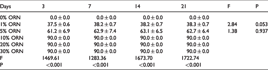
Diameters of the inhibition zones of the six groups of ornidazole-loaded PLGA/wool keratin composite membranes against Fn (x ± s, n = 3, cm).
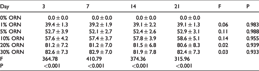
Diameters of the inhibition zones of the six groups of ornidazole-loaded PLGA/wool keratin composite membranes against Pa (x ± s, n = 3, cm).
Discussion
In this study, six groups of ornidazole-loaded PLGA/wool keratin composite membranes were successfully prepared by electrospinning. Among these membranes, the 1% ORN composite membrane showed strong water absorption and mechanical properties and had suitable in vitro degradation and drug release properties. The 5% ORN composite membrane showed good water absorption and degradation properties, and the 10% ORN composite membrane showed good mechanical and degradation properties. Xue et al. prepared PCL/gelatin/metronidazole composite films by electrospinning. 14 Their results showed that with increasing metronidazole content, the tensile strength of the composite films initially decreased (reaching a minimum at PGH10), then increased up to PGH30, and finally decreased at PGH40. This trend was similar to the mechanical property results observed in this study. Among the six groups of composite membranes, the tensile strength of the 5% ORN composite membrane was the smallest, and that of the 10% ORN composite membrane was the highest; the tensile strengths of the 20% ORN and 30% ORN composite membranes decreased with increasing ornidazole content.
The dosage of ornidazole recommended in adult patients for the clinical prevention and treatment of anaerobic infection should be 0.5 g/once and twice a day. 17 The maximum content of ornidazole in 10 g of the 30% ORN composite membrane prepared in this study was approximately 0.13 g. The dose of ornidazole was much lower than that used for systemic treatment and would not cause allergic or other reactions. In addition, the encapsulation efficiency of ornidazole in the composite membranes was approximately 40% to 70%, indicating that some of the ornidazole was encapsulated in the composite fibers and some was deposited on the surface of the composite membranes. Although there was an obvious initial burst release of ornidazole from both the 1% ORN and 30% ORN composite membranes, these composite membranes could continuously release the drug over 7 d, and the total release rate reached more than 78%.
hPDLFs are the main cells responsible for periodontal regeneration. In addition to promoting the formation of new attachments, they can also perform a variety of key cell activities, such as cell division, chemotaxis, differentiation and extracellular matrix synthesis, through the synthesis of structural and functional proteins, allowing the repair and regeneration of periodontal tissue. 18 The cells used in this study came from primary culture. There are currently two basic methods for the primary culture of hPDLFs: an enzyme digestion method and a tissue block method. The enzyme digestion method involves the digestion of tissues that have been cut into small pieces with trypsin and/or collagenase to directly obtain cells and culture cells. This method has a high yield of primary cells, but it requires a large amount of tissue. Furthermore, the steps are tedious and readily susceptible to contamination, and the experimental cost is high. The tissue block method is simple to perform, but the rate of tissue adhesion is low. In recent years, enzyme digestion-tissue culture methods, whole-tooth enzyme digestion methods, etc. have also been reported.19,20 The slide covering method was used in this study, as it avoids the floating of tissue blocks and facilities the migration of cells. Because epithelial-like cells grow in clusters and are not sensitive to trypsin, their digestion takes a long time, so in this study, the hPDLFs were purified by digestion and passage. 20
Xue et al. found that a PCL nanofibrous membrane containing 40% metronidazole had an inhibitory effect on the growth of L929 cells, while a PCL nanofibrous membrane containing 30% metronidazole promoted cell proliferation. 14 We reached a similar conclusion in this study. When the ornidazole content was greater than or equal to 10%, the composite membrane could inhibit the growth and proliferation of cells, while 0% ORN, 1% ORN and 5% ORN composite membranes could promote cell growth and proliferation.
Fn, a member of the normal oral flora, is closely related to gingivitis and periodontitis, which are especially active periodontal diseases. Pg has the highest detection rate in chronic periodontitis, and it can inhibit hPDLF repair and periodontal tissue reconstruction by directly inhibiting the proliferation of hPDLFs, downregulating vascular endothelial growth factor and upregulating matrix metalloproteinase-9. The detection rate of Pa in patients with chronic generalized periodontitis is approximately 50%. 21 Ornidazole is used to treat anaerobic bacterial infections via a mechanism that involves preactivation by reduction of the nitro group and the production of toxic derivatives and radicals, resulting in an antibacterial effect. 22 The cure rate of ornidazole in the treatment of oral anaerobic infection is higher than that of metronidazole, and the side effects of ornidazole are milder. The local use of ornidazole for the control of periodontal bacteria and the elimination of periodontal inflammation is more effective than the use of metronidazole or tinidazole.23,24
Bottino et al. found that an electrospun matrix containing ciprofloxacin could inhibit the growth of Fn and Actinobacillus actinomycetemcomitans (Aa). 25 Reise et al. prepared a metronidazole/PLA fiber that can be used in the treatment of local periodontitis. The agar test results showed that the antibacterial effect of metronidazole on Fn was slightly lower than that of Pg. 26 Da Silva et al. studied the application of a poly(3-hydroxybutyrate) composite membrane containing metronidazole in the treatment of periodontitis. 8 The results showed that the composite membrane containing metronidazole could inhibit the growth of actinomycetes, clostridium nucleatum and Pg, and the bacteriostatic effect could last 28 d. In this study, five groups of PLGA/wool keratin/ornidazole composite membranes (1% ORN, 5% ORN, 10% ORN, 20% ORN and 30% ORN composite membranes) showed good bacteriostasis against Fn, Pg, Pa, and higher contents of ornidazole resulted in stronger bacteriostasis. In addition, the inhibitory properties of the five groups of composite membranes on the three types of anaerobic bacteria could last for 21 d.
Although we prepared ornidazole-loaded PLGA/wool keratin composite membranes that could inhibit the growth of anaerobic bacteria, the composite membranes could not actively induce tissue regeneration and repair. Therefore, we will try to add basic fibroblast growth factor (bFGF) to the composite membranes and construct a GTR membrane loaded with both ornidazole and bFGF to reduce the incidence of postoperative infection and increase periodontal tissue regeneration. At present, a large amount of literature supports the application of enamel matrix protein in regenerative periodontal treatment, and it is believed to realize the real regeneration of periodontal tissue. 27 If enamel matrix protein can be used in combination with the PLGA/wool keratin composite membranes in the GTR procedure, the composite membranes may more effectively promote the regeneration of periodontal tissue. If the composite membranes prepared in this study are to be further used in humans, it is important to further explore the ability of the composite membranes to guide periodontal tissue regeneration and repair periodontal defects through animal experiments and human tests. Therefore, more comprehensive animal experiments and human trials should be performed to reduce the occurrence of clinical complications.
Conclusions
Six groups of ornidazole-loaded PLGA/wool keratin composite membranes were successfully prepared by electrospinning. Among these membranes, the 1% ORN composite membrane showed good physical and chemical properties, drug release performance, good cell compatibility and antibacterial effects, and it has potential for use as an antibacterial GTR membrane for periodontal tissue repair.
Footnotes
Declaration of conflicting interests
The author(s) declared no potential conflicts of interest with respect to the research, authorship, and/or publication of this article.
Funding
The author(s) disclosed receipt of the following financial support for the research, authorship, and/or publication of this article: This work was supported by the National Natural Science Foundation of China (no. 81560191 and no. 81760205) and the Program for Excellent Talents in Ningxia, China.