
Abstract
Investigating the effects of rhBMP2 (recombined human bone morphogenetic protein-2) release kinetics on bone formation is important to optimize the use of rhBMP2 in tissue-engineered bone. However, there is still no direct evidence regarding how the release kinetic of rhBMP2 has effects on mesenchymal stem cells homing and differentiation during bone regeneration. In this study, we construct control and non-control released rhBMP2 coatings within the pores of 3D printed hydroxyapatite scaffold, and investigate how the release kinetic of rhBMP2 affects bone inductivity of tissue-engineered bone. We show that control released rhBMP2 has better effects on enhancing mesenchymal stem cells proliferation and migration than non-control released rhBMP2. Control released rhBMP2 promotes mesenchymal stem cells differentiation to a better extent than non-control released rhBMP2. Those findings are in consistent with the results of quantitative rhBMP2 stimulation for mesenchymal stem cells proliferation, migration and differentiation. The histological results after in vivo implantation also reveal that control released rhBMP2 coating can induce more bone formation than non-control released rhBMP2. Our data provide a basis for optimizing the rhBMP2 delivery modes in tissue-engineered bone and improving the bone inductivity of rhBMP2.
Introduction
The manufacture of tissue-engineered bone (TEB) is important, as the repairing of bone defects is still a major challenge for orthopedics. Promising TEB materials require sufficient pores and interconnectivity to allow bone integration and nutriment transportation.1,2 With the advancement of 3D printing technique, we can easily control the porosity and structure of bone scaffolds nowadays. We have fabricated porous hydroxyapatite (HA) scaffolds by the micro-syringe extrusion system. Previous researches have proved that the HA scaffold has favorable biocompatibility and osteoconductivity.3,4 However, the scaffold alone does not usually induce sufficient bone regeneration in vivo due to the weak bone inductivity. The incorporation of osteoinductive factors into TEB is usually required to promote bone regeneration.5,6
Bone morphogenetic proteins (BMPs), members of transforming growth factor beta superfamily, have been widely investigated for their osteoinductive properties. BMP2, which has strong osteoinductive capacity, is commercially available. 7 However, the local clearance of BMP2 from the implantation site is rapid and the biologic half-life of BMP2 is short (The half-life is 0.3 days for ectopically injection).8,9 To overcome its rapid clearance, extensive high dose is used in vivo, which is associated with considerable side effects such as implantation failure, swelling and inflammation. 10 Delivering recombined human bone morphogenetic protein-2 (rhBMP2) to the scaffold and maintaining it in place are crucial issues. Investigating the effects of rhBMP2 release kinetics on bone formation is important to optimize the use of rhBMP2 in TEB. 9
The bone inductivity of BMP2 relays on the dynamic transcriptional regulation of osteoprogenitor cells or the bone marrow mesenchymal stem cells (BMSC) that produce these progenitors. Following BMP2 induction, both the Smads and p38 MAPK (mitogen-activated protein kinase) pathways converge at the Runx2 gene to control the differentiation of mesenchymal precursor cells.11,12 The maturity of osseous tissues needs the participation of sufficient osteoblasts. MSCs (mesenchymal stem cells) play a crucial role in the process of bone regeneration. 13 Bone regeneration within the scaffold can be divided into two stages, which are endogenous stem cell homing to the scaffold and subsequent osteogenic differentiation of the recruited stem cells.14,15 MSCs homing, including recruitment and migration to the bone defects sites, is the start of bone regeneration, followed by MSCs proliferation and differentiation induced by various cytokines at the bone defects sites. At last, calcifications of osteoblast cells differentiated from MSCs to form the mature osseous tissues. 10 Many researches have proved that slow and sustained release of rhBMP2 can improve the bone formation of TEB.16,17 However, there is still no direct evidence regarding how the release kinetic of rhBMP2 has effects on MSCs homing and differentiation during bone regeneration.
In this study, we investigate how the release kinetic of rhBMP2 affects the homing and proliferation of BMSCs in vitro, via two-dimensional and three-dimensional cell cultures. Furthermore, we construct control and non-control released rhBMP2 coatings within the pores of 3D printed hydroxyapatite scaffold to study how the release kinetic of rhBMP2 coatings affects the bone formation within the scaffold in vivo.
Materials and methods
Materials
RhBMP2 was obtained from Medtronic, Inc., USA. Human BMP2 ELISA Kit, Type I collagen(C9879), 1-ethyl-3-[3-dimethylaminopropyl] carbodiimide hydrochloride (EDC), Nhydroxysuccinimide (NHS) and chitosan were purchased from Sigma-Aldrich Co., USA.
Fabrication of rhBMP2 loaded chitosan microspheres
The fabrication of rhBMP2 loaded CMs followed the methods as described previously. 4 Briefly, span, tween and magnesium stearate were dissolved in liquid paraffin according to the ratio of 3%, 2.4% and 3% (W/V) respectively to obtain the oil phase. And then, the oil phase was stirred at 45°C water bath for 1 h; 450 mg chitosan was dissolved in 2% aqueous acetic acid solution. The pH of the chitosan solution was adjusted to pH 4–4.5 with alkali solution (0.1 M, NaOH), and then 4 mg rhBMP2 was added to the solution to obtain the chitosan/rhBMP2 solution (water phase). Thereafter, the chitosan/rhBMP2 solution was dripped into the oil phase slowly under stirring; 1 g 3-methoxy-4-hydroxybenzaldehyde (vanillin) was dissolved in 2 ml alcohol and added to the water/oil emulsion to crosslink the chitosan. The microspheres were obtained after the emulsion was stirred for 6 h. Petroleum ether and isopropanol were used for removing the remaining solvent, and then the microspheres were lyophilized for 18 h. To measure the total amount of rhBMP2 within the CMs, 1 mg microspheres were dissolved with 500 µl of acetic acid (0.2 M), and then diluted with 3.5 ml PBS. The dissolved rhBMP2 was assessed using Human BMP2 Elisa kit (Sigma-Aldrich Co., USA).
Preparation of HA scaffold
The elliptic cylindrical porous HA scaffold with size of 15 × 7 × 5 mm3 was designed and prepared as previously described. 3 30% (v/v) HA slurry was used to print the HA scaffold via extrusion deposition system (MAM-II, Fochif, China). After printed, the HA scaffold were dried for 24 h at room temperature. Then the scaffolds were sintered in a microwave furnace to improve the strength of the scaffold.
Synthesis of scaffolds coated with collagen/rhBMP2 and collagen/rhBMP2-CMs
Collagen I was dissolved in acetic acid (0.05 M) to obtain a concentration of 0.1 g/ml. And then the sterilized HA scaffolds were soaked in the above collagen solution under vacuum aspiration for 24 h. EDC and NHS were dissolved at MES-buffered solution (pH 5.5) at a ratio of 4:1(w/w). Then, 2.5 mg/ml EDC and 0.63 mg/ml NHS solution were used for the cross-link of collagen. After incubated for 4 h, the HA/collagen scaffold was dried at 37°C. Thereafter, the scaffold was washed with phosphate buffer solution (PBS, pH7.4).
The synthesis of rhBMP2/collagen-coated HA scaffold (BCH)
The rhBMP2 was dissolved in deionized water at a concentration of 50 µg/ml, and then 72 µl of the rhBMP2 solution was dripped on the collagen/HA scaffold evenly and then the scaffold was dried and stored at −20°C until use. The synthesis of rhBMP2-CMs-coated HA scaffold (MBCH) was using the post-seeding technique as described previously. 18 RhBMP2-CMs were suspended with deionized water at a concentration of 50 mg/ml; 50 µl of the CMs suspension was dripped on the collagen/HA scaffold evenly. The MBCH was dried at 37°C for 12 h and stored at −20°C until use. The coating process was performed under sterilized condition.
Scanning electron microscopy observation of scaffolds
The microstructures of MBCH and BCH scaffolds were observed by a scanning electron microscopy (SEM, S-2400; Hitachi, Japan).
In vitro release of rhBMP2
The soaking methods were used for the release of rhBMP2 from MBCH and BCH scaffold. Each MBCH and BCH scaffold was soaked in 5 ml PBS (phosphate buffer saline) at 37°C. At each tested time point, the release media was collected and renewed with fresh PBS. The release medium was collected at 1 h, 3 h, 8 h, 24 h, 3 d, 5d, 10d and 20d. The collected samples were stored at −20°C until testing. The experiments were run in triplicate per time point.
Elisa assay
The release medium from MBCH and BCH scaffolds was subjected to human BMP2 Elisa Assay. The Human BMP2 ELISA Kit was from Sigma-Aldrich Co., USA (RAB0028). The detailed protocols for the Elisa Assay from Sigma were followed.
Cell culture and differentiation
Mice mesenchymal stem cells were derived from bone marrows of C57/BL6 mice. Total bone marrow cells were extracted from C57/BL6 mice and cultured in stem cell cultures with 10% FBS from Cyagen. Different from the suspended hematopoietic cells, mesenchymal stem cells could attach to the culture dishes and grow. After 48 h, suspension cells were discarded. The adherent cells were continued to be cultured for two weeks to get the primary mice BMSC cells. All the BMSC cells for the differentiation assays were from passage 2 to passage 8. The cell differentiation assay was performed within the Corning cell culture insert using BMSC culture media with 10% FBS (fetal bovine serum). MBCH or BCH materials were placed on the upper layer of cell culture insert with polyester membrane (Transwell®, 0.4 µm pore size, Corning). The cells were placed below the permeable membrane. Additionally, to assess the impact of rhBMP2 stimulating patterns on BMSC osteogenic differentiation, mice BMSC cells were directly cultured in a serious of culture media with preset rhBMP2 concentration.
Alkaline phosphatase staining
Mice BMSC cells were cultured with MBCH or BCH materials for seven days. The culture media were renewed at 1d, 3d and 5d. ALP staining was conducted with BCIP/NBT Alkaline Phosphatase Color Development Kit (Beyotime).
Alizarin red staining
Mice BMSC cells were cultured with MBCH, BCH materials or media with BMP2 directly for 14–21 days. The culture media in material groups were renewed at 1d, 3d, 5d, 10d, 15d and 20d. After cells were fixed with 95% Ethano for 5 min, 1% Alizarin red (pH = 8.3) was stained for 15 min.19,20
Quantitative real-time PCR
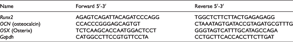
Mice BMSCs were co-cultured with MBCH or BCH in Corning cell culture insert for 1d, 2d, 5d, 10d and 14d. Total cell RNA of the sample collected at each time point was isolated with TRIzol (Invitrogen), and cDNA was synthesized with Revertra Ace (Promega, Madison, USA). Real-time PCR was performed with an ABI QuantStudio 5 system. The expression level of genes was measured using the comparative Ct method. Expression values were normalized to GAPDH (Glyceraldehyde-3-Phosphate Dehydrogenase) expression. The primer sequences are shown in Table 1.
Primer pair sequences for the osteogenic transcription factors.
Cell migration and viability assays
1 × 10⁴ BMSCs in BMSC culture media without FBS were placed on the upper layer of Corning cell culture insert with polycarbonate membrane (Transwell®, 8 µm pore size, Corning). The MBCH, BCH materials or culture media with specific rhBMP2 concentration were placed below the cell permeable membrane. Following an incubation period of 24 h in 37°C, 5% CO2, the cells that had migrated through the membrane were stained with 0.1% crystal violet and counted. To assess the cell viability, 1 × 10⁴ BMSCs in BMSC culture media with 2% FBS were placed on the lower layer of Corning cell culture insert; MBCH, BCH materials or culture media with specific rhBMP2 concentration were placed above the cell permeable membrane (Transwell®, 0.4 µm pore size, Corning). Following an incubation period of 2d and 4d, cell counting kit-8 (Dongjindo Molecular Technologies, Inc., Japan) was used for calculating the cell intensity.
Collagen 3D cultures and immunofluorescence assay
BMSC cells in 2D cultures were trypsinized, resuspended in media and plated 20,000 cells on each 100 µL 3 mg/mL Collagen-I (BD BioSciences, New Jersey, USA). 1 N NaOH was added to achieve a pH of 6.5–7.5. The collagen with cells was incubated at 37°C in humidified incubator until a firm gel was formed. BMSC media contained 10% FBS was added for cell growth. The MBCH or BCH materials were placed on the upper layer of cell culture insert (Transwell®, 0.4 µm pore size, Corning). The collagen gels with cells were placed below the permeable membrane. Immunofluorescence assay was performed after 14 days of culturing. After cells were fixed with 4% paraformaldehyde (Sigma) for 20 min, cells were permeated with 0.5% Triton X-100 for 5 min at room temperature. Then, 10% BSA was used to block the cells for 20 min at room temperature. After that, cells were incubated with anti-OCN antibody (Biorbyt, orb259644) overnight at 4°C. At the second day, cells were incubated with TRITC-conjugated anti-Rabbit antibody (Abclonal, AS040) for 1 h at room temperature. DAPI (Solarbio, C0060) was used to stain the nucleus. Images were taken by Zessis LSM 800 Laser scanning confocal microscope.
In vivo implantation and histological evaluations
Animal experiments were approved by the animal ethics committee of Fujian medical university, China. To exclude the interference of in situ osteoblast, only ectopic bone formation model was used to assess the bone inductivity of control versus non-control released rhBMP-2 coatings in 3D printed hydroxyapatite scaffold in vivo. New Zealand rabbits (2.5 to 3.0 kg), aged three to six months, were anesthetized with an intravenous injection of 3% pentobarbital sodium (1 ml/kg body weight, Sigma, USA). A 2 cm skin incision from the groin midpoint to the knee was taken, and the myolemma was longitudinally split. MBCH or BCH scaffolds were implanted into the intramuscular gap. Muscles and skins were sutured; 800,000IU penicillin sodium was injected intramuscularly to prevent infection. Animals were sacrificed after four weeks. Samples were collected and fixed with 4% paraformaldehyde. After dehydration with ethyl alcohol, the specimens were embedded in methyl methacrylate (MMA), and sectioned at a thickness of 50 µm with the Leica SP1600 saw microtome (Leica Biosystems, German). The slides were stained with modified VanGieson for histological evaluations.
Statistical analysis
Data were expressed as mean ± standard deviation (SD). One-way ANOVA was used to analyze the differences among groups. A P < 0.05 value was considered as statistically different.
Results
Characteristic of BCH and MBCH scaffolds and the release kinetics of rhBMP2 in vitro
The shape of the final printed scaffold was shown in Figure 1(a)-a. After sintered, the HA scaffold shrank for about 34.4% in the size. The SEM image of BCH revealed that a thin layer of rhBMP2/collagen coating was adhered to the surface of scaffold, with the pores not blocked by the collagen (Figure 1(a)-b. The final scaffolds had excellent interconnectivity with a pore size of about 460 µm (Figure 1(a)-c). In MBCH scaffold, several rhBMP2 CMs were evenly adhered to the wall of the pores via collagens, as shown in Figure 1(a)-d.
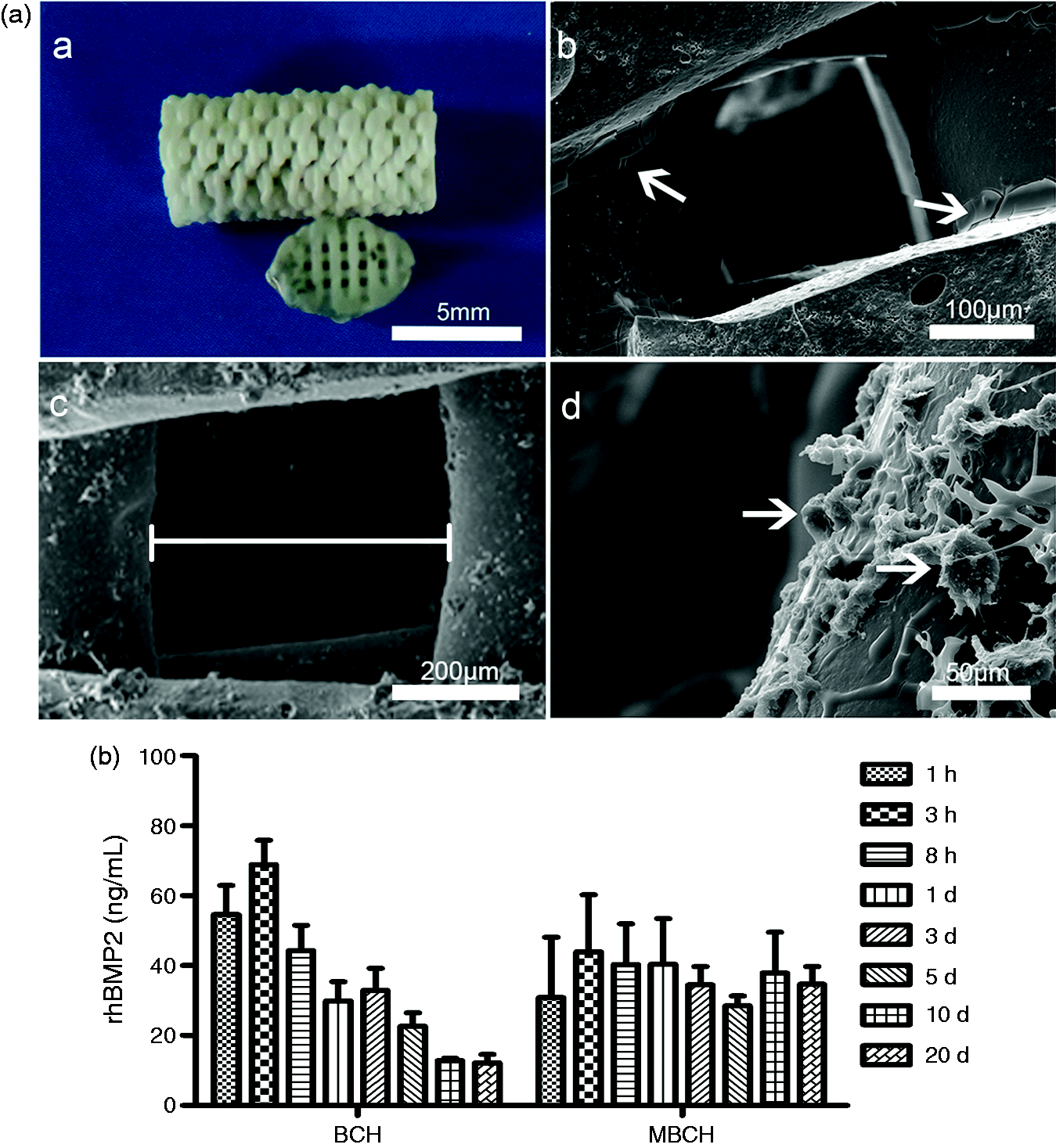
Characteristics of 3D printed HA scaffold with control released rhBMP2 and non-control released rhBMP2. (a) The digital image and SEM observation of the scaffold (a: The digital image showed the shape of 3D printed HA scaffold. b: SEM observation illustrated that a thin layer of rhBMP2 collagen coating was attached to the surface of BCH scaffold. The white arrows indicated the thin layer of rhBMP2 collagen. c: SEM observation illustrated the pore of the HA scaffold. The white line showed the width of the pore size. d: SEM observation illustrated rhBMP2 CMs were successfully adhered to the MBCH scaffold by collagen. The arrows showed the rhBMP2 CMs). (b) RhBMP2 release kinetics of BCH and MBCH scaffolds. The release media of BCH and MBCH scaffolds were collected at 1 h, 3 h, 8 h, 24 h, 3 d, 5d, 10d and 20d, and examined and calculated with rhBMP2 Elisa assay.
The average amount of rhBMP2 in MBCH scaffold was 3.61 ± 0.08 µg, which was comparable with the amount of rhBMP2 in BCH scaffold (3.6 µg). The release kinetics of rhBMP2 for BCH and MBCH were quite different from each other, as shown in Figure 1(b). The release of rhBMP2 in BCH behaved as a non-control pattern. The rhBMP2 concentration in BCH was much higher than that in MBCH at the early stage (<1d), but decreased fast within five days. The average rhBMP2 concentration at the 10th day and 20th day was below 13 ng/ml. A more control released kinetic of rhBMP2 was found in MBCH, in which the concentration of rhBMP2 was stable with a narrow range of fluctuation. The average concentration of rhBMP2 was all above 28 ng/ml at each testing time point. More importantly, the average amount of rhBMP2 released from MBCH at late stages (10d and 20d) was about two times higher than that from BCH. These results indicated that rhBMP2 CMs coating could efficiently avoid initial burst release and enhance long-term drug release in a controlled manner as compared with rhBMP2 collagen coatings.
Control released rhBMP2 had better effects on enhancing MSCs proliferation and migration than non-control released rhBMP2
Bone marrow mesenchymal stem cells were important cells that produce osteoprogenitors. The homing of MSCs played an important role in the repair of bone fractures. 21 Migration and proliferation of MSCs in the fracture site promoted the bone healing. Thus, we wondered whether the release kinetic of rhBMP2 coating affected the proliferation and migration of BMSCs in vitro. We found that control released rhBMP2 from MBCH could promote MSCs proliferation in contrast with the BCH group and the negative control (Figure 2(a)). The results of migration assay were shown in Figure 2(b). Compared with the negative control, both MBCH and BCH could enhance BMSC migration examined by Corning Transwell Assay. It should be noted that control released rhBMP2 from MBCH stimulated cell migration better than non-control released rhBMP2 from BCH. Furthermore, we cultured the cells in culture media with preset rhBMP2 concentration, which mimicked the release kinetics of BCH and MBCH. In mode I, BMSCs were cultured in media with 50 ng/ml rhBMP2 for 24 h like MBCH, while in mode II, rhBMP2 concentration within the culture media was decreased with time like BCH. Cell proliferation and migration of Mode I and Mode II rhBMP2 stimulation for MSCs were in line with the results of MBCH and BCH stimulation for MSCs (Figure 2(c) and (d)).
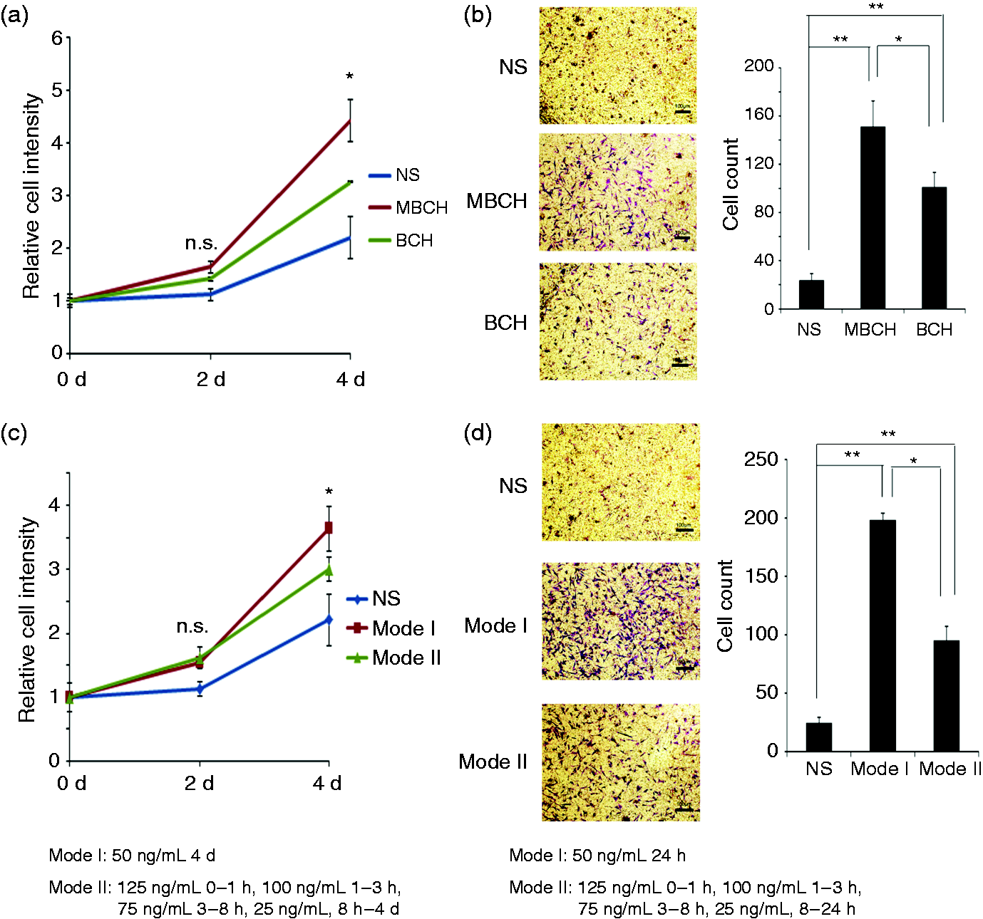
Control released rhBMP2 had better effects on enhancing MSCs proliferation and migration than non-control released rhBMP2. (a) The relative cell intensity of MSCs was examined and calculated with the CCK8 assay after two days and four days of MSCs cultured (MBCH vs. BCH and MBCH vs. NS, *P < 0.05). (b) MSCs were cultured in Transwell plates with BCH or MBCH for 24 h. The cells invading to the bottom were stained with crystal violet and counted. Average cell numbers of at least three fields were shown on the right (*P < 0.05). (c) The relative cell intensity of BMSCs was examined and calculated with CCK8 assay, after they were cultured in culture media with different rhBMP2 concentration modes for two and four days (Mode I vs. NS, *P < 0.05). (d) MSCs were cultured in Transwell plates with different rhBMP2 concentration modes for 24 h. The cells invading to the bottom were stained with crystal violet and counted. Average cell numbers of at least three fields were shown on the right (*P < 0.05; **P < 0.01).
Control released rhBMP2 induced more osteogenic differentiated MSCs than non-control released rhBMP2
Alkaline phosphatase was an early marker of osteoblast differentiation. As the release kinetics of rhBMP2 showed, rhBMP2 in BCH group behaved burst release, and the accumulated amount of rhBMP2 released from BCH was higher than that of MBCH at the first day. However, the rhBMP2 concentration in BCH group was all lower than that in MBCH group after the third day. To investigate whether the initial burst release had advantages in the early osteoblast differentiation of BMSCs, BMSCs were incubated with MBCH and BCH for seven days to examine their early differentiation. As the local clearance of BMP2 from the implantation site is rapid and the biologic half-life of BMP2 is short (The half-life is 0.3 days for ectopically injection),8,9 the culture media was renewed at 1d, 3d and 5d to imitate the degradation of rhBMP2 in vivo. We found that both BCH group and MBCH group had ALP+ Cells after seven days, but the MBCH group showed a more intense staining of ALP than the BCH group. And the percentage of ALP-positive cells in MBCH was significantly higher than that of the BCH group (P < 0.01) (Figure 3(a)). What’s more, rhBMP2 stimulation of mode I could induce more ALP+ cells than that of mode II as well (Figure 4(a)). Our results demonstrated that sustained and stable released rhBMP2 promoted more osteogenic differentiated MSCs than non-control released rhBMP2.
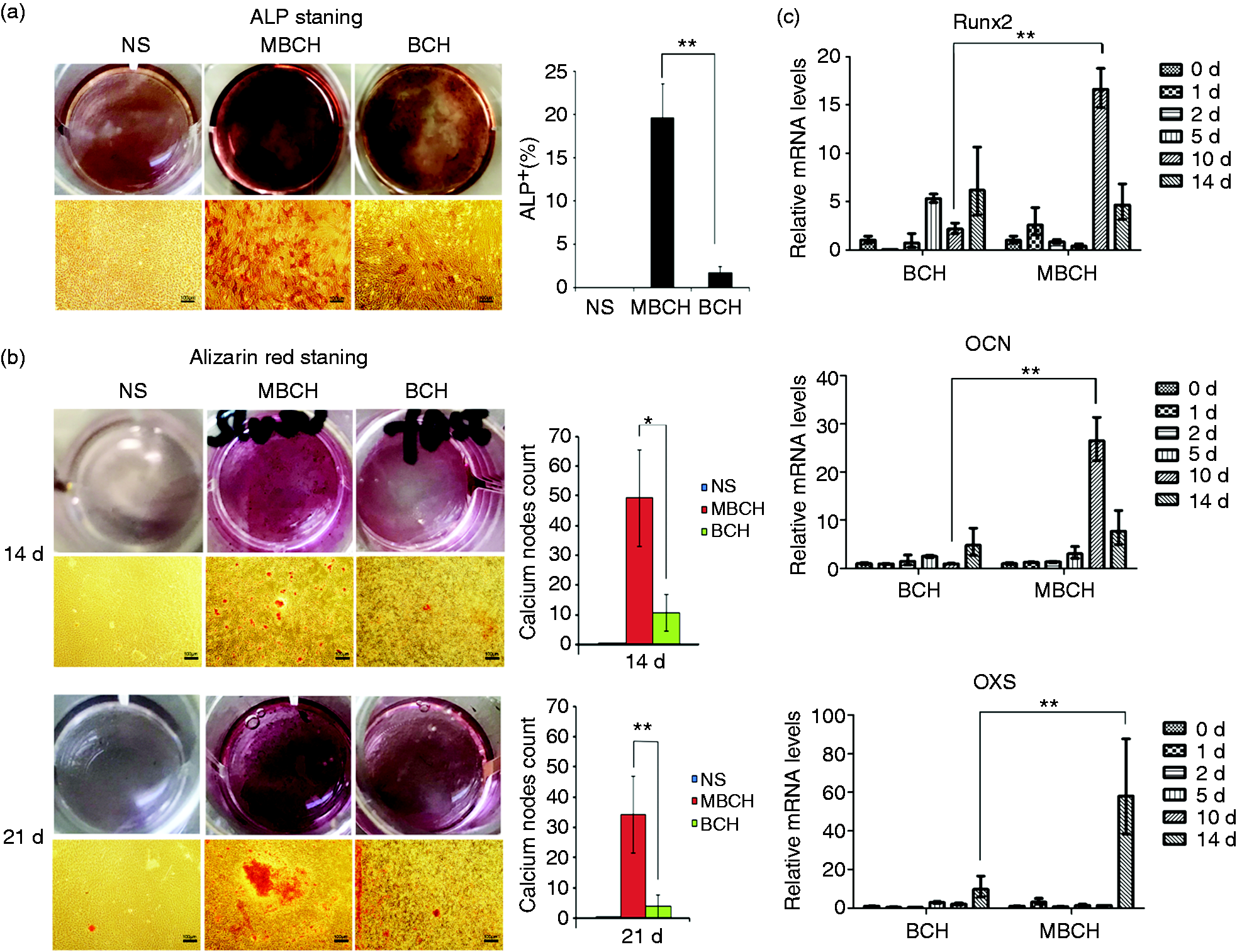
Control released rhBMP2 induced more osteogenic differentiated MSCs than non-control released rhBMP2. (a) Photos of ALP staining result after the MSCs were cultured for seven days with MBCH or BCH. The percentage of ALP-positive cells was on the right. Scale bars, 100 µm (**P < 0.01). (b) Alizarin red staining to show calcium nodes formation was taken at 14d or 21d after the MSCs were cultured with MBCH or BCH. Average cell numbers of calcium nodes under at least three fields were shown on the right (*P < 0.05; **P < 0.01). Scale bars, 100 µm. (c) Expression of osteogenesis-related genes after MSCs were cultured with MBCH or BCH for 0d, 1d, 2d, 5d, 10d, 14d. The relative mRNA levels of Runx2, OCN and OSX in MBCH were examined by qRT-PCR (**P < 0.01).
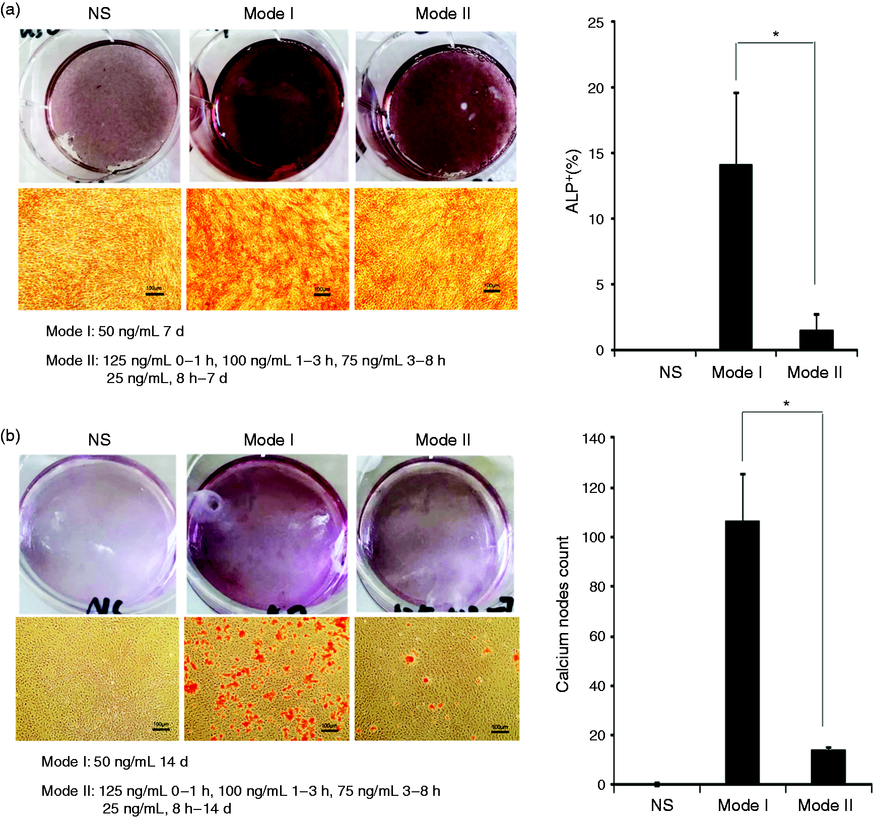
Sustained moderate dosage of rhBMP2 delivery induced more osteogenic differentiation of MSCs than the initial high dosage and subsequent low dosage of rhBMP2 delivery. (a) Photos of ALP staining result after the MSCs were cultured for seven days with different modes of rhBMP2 treatment. The percentage of ALP-positive cells was on the right. Scale bars, 100 µm (**P < 0.01). (b) Alizarin red staining to show calcium nodes formation was taken at 14d or 21d after the MSCs were cultured with different modes of rhBMP2 treatment. Average cell numbers of calcium nodes under at least three fields were shown on the right (*P < 0.05; **P < 0.01). Scale bars, 100 µm.
To examine the long-term differentiation efficiency, BMSCs were cultured with MBCH and BCH materials for 14 days and 21 days. The collected cells were stained with alizarin red to test the calcium nodes formed by differentiated osteoblast cells. More red stained calcium nodes could be found in MBCH group than that in BCH group at 14d (Figure 3(b)). After 21 days of the culture period, the areas of calcium nodes in MBCH group were increased and the nodes fused together, while in BCH groups we could not find many calcium nodes. In order to quantitatively compare the impact of rhBMP2 stimulating mode on the induction of BMSCs osteogenic differentiation, BMSCs were incubated in two concentration modes mentioned above, respectively. As shown in Figure 4(b), when BMSCs were cultured with 50 ng/ml rhBMP2 for 14 days consecutively, many calcium nodes were generated. On the contrary, initial high dose followed by low concentration of rhBMP2 stimulation for 14 days only induced very few calcium nodes formation.
Furthermore, we examined the expression of osteogenic transcription factors, Runx2, and osteogenic differentiation marker OSX and OCN by quantitative PCR. 22 Expressions of Runx2, OSX and OCN were increased at certain time points for both groups, and the peak mRNA levels for Runx2, OCN and OSX were all significantly higher in MBCH group than that in BCH group. The expression of Runx2 was increased at an early stage. There were two peaks in the expression of Runx2: in BCH group, the first peak appeared on the 5th day, and the second peak appeared on the 14th day. Differently, the peak levels of Runx2 expression came earlier in MBCH group, which appeared at the 1st day and the 10th day. The mRNA levels of OCN and OSX were all upregulated at the late stages of incubation (Figure 3(c)).
Control released rhBMP2 enhanced osteogenesis in vivo
The interaction between stem cells and the surrounding environment was critical for cell behaviors. 3D cultures could mimic a more physiological environment than 2 D cultures for stem cells. Thus, we used a 3D culture model to imitate the environment within the pores of scaffold in vivo. The experimental procedure is shown in Figure 5(a). MBCH and BCH materials were placed above the permeable membrane, and BMSCs were cultured below. After 14 days of the culture period, BMSCs were stained with DAPI and OCN-TRITC. The immunofluorescence image is shown in Figure 5(a). BMSCs were successfully seeded in the collagen gels. DAPI staining images showed that most of the cells situated at the upper layer of the collagen gels. OCN-TRITC staining revealed that the bone inductivity of MBCH and BCH was quite different. More cells in MBCH were stained red as compared with that in BCH. Moreover, the percentage of TRITC/DAPI in MBCH group was significantly higher than that of BCH. And the distribution of osteogenic differentiated BMSCs within the collagen gels was also varied between the two groups. In MBCH group, the osteogenic differentiated BMSCs were mainly concentrated at the upper layer of the collagen gels, which were closed to the materials, while in BCH, the location of differentiated cells was more diffusive in the gels.
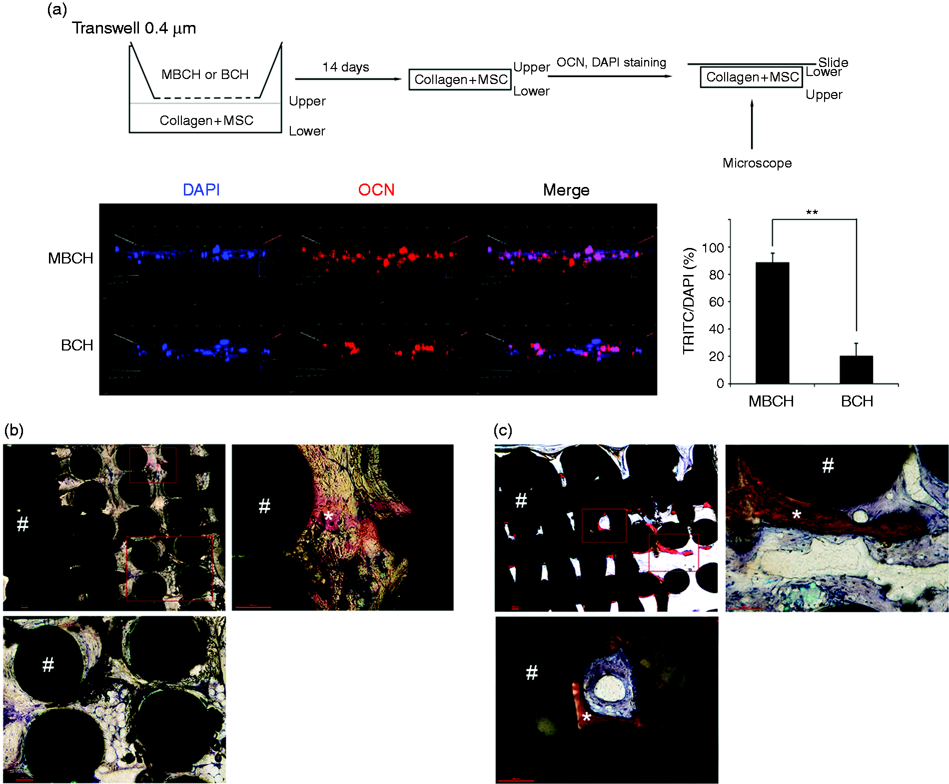
Control released rhBMP2 in HA scaffolds enhanced osteogenesis in vivo. (a) Immunofluorescence observation of MSCs in 3D collagen gels after MSCs were stained by DAPI and OCN-TRITC (blue area represents DAPI-stained nuclei, and red area represents TRITC-labeled OCN). And the percentages of TRITC-positive cells were calculated on the right (**P < 0.01). (b) Representative histological images of BCH at 4th week after implantation in vivo. (c) Histological images of MBCH at 4th week after implantation in vivo (# indicates HA scaffold; * represents new osseous tissue).
To exclude the influence of in situ osteogenesis mechanism, we utilized ectopic bone formation models rather than bone defect models to compare the bone inductivity of MBCH and BCH in vivo. The histological results are shown in Figure 5(b) and (c). Although the total amounts of rhBMP2 in the two scaffolds were comparable, the bone inductivity of the two coating methods was different. Within the MBCH scaffold, control released rhBMP2 coating induced extensive bone formation. The new bone majorly grew around the wall of pores, while the osteoid tissue grew in the center. On the contrary, the non-control released rhBMP2 coating in BCH group only induced the formation of very few immature osseous, and the osseous distributed unevenly within the scaffold. The histological results indicated that control released rhBMP2 coating had a stronger bone inductivity than non-control released group in vivo.
Discussion
Investigation on the TEB was still one of the most attractive aspects of biomedical engineering, as the requirement of artificial bone grafts in clinic was still increasing. TEB scaffolds consisted of scaffolds, growth factors and cells. RhBMP2 had attracted much attention in the field of bone tissue engineering for its bone inductivity. It could induce bone formation both orthotopically and ectopically. Direct printing of growth factor within the scaffold was still difficult due to the limitation of techniques. Thus, we used surface coating methods to incorporate rhBMP2 to the scaffold. Release kinetics of rhBMP2 in vitro revealed that a more control released kinetic of rhBMP2 was found in MBCH, while the release rate of rhBMP2 in BCH behaved burst release within one day and decreased fast later. MBCH could be a new valuable modification method for TEB.
There were unique advantages of animal models of ectopic bone formation over orthotopic environments as it could eliminate the effects of endogenous bone-forming mechanisms. 23 Thus, we evaluated the bone inductivity of rhBMP2 coated HA scaffolds using the ectopic bone formation model. Results of the muscle pouch implantation of the MBCH and BCH revealed that control released rhBMP2 coating induced more bone formation within the HA scaffold than non-control released rhBMP2 coating, indicating that the release kinetics of rhBMP2 coating were correlated with the bone formation in HA scaffolds. Control released rhBMP2 coating could induce more bone formation, which could be applied in the modification of TEB in the future.
MSCs played a crucial role in the process of bone generation. The sources of MSCs were widespread, including bone marrow, periosteum, vessel walls, adipose, muscle, tendon, peripheral circulation, etc. 21 Bone marrow was the major source of MSCs. Seong-Seo et al. 15 proposed that dual delivery of substance P, which could facilitate endogenous stem cell recruitment to bone defects, could enhance the bone inductivity of BMP-2. In addition, vascular endothelial growth factor(VEGF) and stroma cell-derived factor-1(SDF-1) had also been reported to improve the bone regeneration of BMP2 via promoting the recruitment of MSCs to TEB scaffolds.24,25 Yu et al. 26 revealed that direct infiltration of MSCs into Ti6Al4V scaffolds could improve bone formation properties of Ti6Al4V scaffolds in vivo. Those results demonstrated that promoting recruitment of endogenous stem cells or direct transplantation of ex vivo MSCs to the damage tissues were ways to improve bone regeneration. In this study, we found that control released rhBMP2 had better effects on enhancing the migration of MSCs at the early stage (24 h) than the non-control released rhBMP2, indicating that growth factors release kinetics could be another method to improve stem cells recruitment.
The role that rhBMP2 played in the proliferation of MSCs was still controversial. BMP2 had been reported to have few effects on the proliferation of MSCs derived from patients with osteoporosis within three days. 27 Others studies, however, found that BMP2 had positive effect on the growth of MSCs after 7 or 14 days.28–30 In the present study, we noticed that rhBMP2 released from control released rhBMP2 could accelerate the proliferation of MSCs, and the effect was especially significant on the fourth days, as compared with negative control and non-control released rhBMP2. To the best of our knowledge, there was no study investigating the role of the release kinetic of BMP2 played in MSCs proliferation. Previous reports and our study indicated that long-term treatment of BMP2 with efficient dosages could enhance the proliferation of MSCs. According to our results, it could be speculated that control released rhBMP2 could be a new mechanism for improving MSCs proliferation.
BMP signaling played an important role in inducing osteogenic differentiation of MSCs. MSCs within the HA construct should undergo several stages of maturation to become osteoblast, which subsequently secreted osteoid matrix to form bone tissue within the scaffolds. In the present study, we investigated how the release profiles of rhBMP2 coating affected the differentiation of MSCs. We found that control released rhBMP2 coatings in MBCH could induce more ALP-positive cells at the early stage and more calcium nodes at the late stage than non-control released rhBMP2 coatings in BCH. We went further to monitor the expression of bone differentiation markers after MSCs were cultured with bone materials, and found that MBCH could induce significant higher expression of Runx2, OCN and OXS than BCH.
Runx2 is a transcriptional factor which can be upregulated by BMP2 signaling at the early stage of osteogenesis. 11 However, in Figure 3(c), the expression of Runx2 was activated at the early stage but could still retain a high level with OCN and OSX at late stages of osteoblasts differentiation in the MBCH group. About 50 ng/mL rhBMP2 released from MBCH continuously from the early stage to the late stage. Thus, the expression of Runx2 could still be activated at the late stages of osteogenesis. Consistently, previous reports also found that continuous BMP2 stimulation could induce the upregulation of Runx2 within a short time but could also retain its expression for over seven days.31–33 However, the dosage of BMP2 decreased below 50 ng/mL within 8 h in the BCH group. The results shown in Figure 3(c) demonstrated that the short time treatment of BMP2 with enough dosage in the BCH group could not induce the expression of the master transcriptional factor, Runx2, at an early stage. Instead, the upregulation of Runx2 came late at the 5th day. Thus, continuous BMP2 stimulation with enough dosage was essential for the mature osteogenesis. Moreover, there were also other reports showing that a steady release of BMP2 over a period of time can induce better osteogenesis than the bulk release. 34
Usually, 50 ng/mL–100ng/mL BMP2 is used to treat stem cells to induce osteogenesis in vitro35–38; 50 ng/mL can be a relative low dosage. 39 The release of rhBMP2 in BCH behaved as a non-control pattern. The rhBMP2 concentration in BCH was over 50 ng/mL at the early stage (<1d), but decreased fast within five days. The average rhBMP2 concentration at the 10th day and 20th day was below 13 ng/ml. A more control released kinetic of rhBMP2 was found in MBCH, in which the concentration of rhBMP2 was stable with a narrow range of fluctuation around 50 ng/ml. Thus, MBCH could release BMP2 of efficient dosages for osteogenesis during the whole differentiation process of MSCs, but BCH could not.
Three-dimensional culture conditions could more closely mimic the environment within the pores of 3D printed HA scaffolds. Besides, previous reports had indicated that cellular behaviors in the 3D culture system might distinct from that in the 2D culture system.40–42 Thus, we constructed a 3D culture model to imitate the interaction between the stem cells in the extracellular matrix within the pores and the rhBMP2 coatings. Interestingly, the distribution of OCN-positive cells within the 3D culture system was quite similar to the distribution of newly generated bone tissues in the pores of HA scaffolds in vivo. In MBCH group, most of the differentiated OCN-positive cells migrated to the upper layer of the gels. Similarly, we found that the growing of new bones was majorly around the wall of pores rather than in the center in the ectopic bone formation models in vivo. Differently, in BCH group, OCN-positive cells were diffusive in the gels, and the osseous tissues were very few and diffusive within the pores after implanted in vivo. This cellular behavior would probably due to the chemotactic effects of rhBMP2. In MBCH group, control released rhBMP2 in the upper layer of the gels and the wall of pores in the scaffold could maintain the chemotactic effects, while the fast released rhBMP2 in BCH group only led to weak chemotactic effects. Based on those results, we speculated that control released rhBMP2 coatings had more advantages than non-control released rhBMP2 in chemoattractant, proliferation and differentiation of MSCs.
Conclusion
We showed that control released rhBMP2 coatings could favor the migration, proliferation and differentiation of MSCs in vitro. Although similar amount of rhBMP2 was used in the scaffold, control released rhBMP2 in MBCH could induce more bone formation in vivo than the non-control released group. Those findings were further confirmed by the results of quantitative rhBMP2 stimulation for MSCs, in which we found that the sustained moderate dosage of rhBMP2 delivery induced more osteogenic differentiation of MSCs than the initial high dosage and subsequent low dosage of rhBMP2 delivery. The results of this study provided a basis for optimizing the rhBMP2 delivery mode in tissue-engineered bones and improving the bone inductivity of rhBMP2.
Footnotes
Declaration of conflicting interests
The author(s) declared no potential conflicts of interest with respect to the research, authorship, and/or publication of this article.
Funding
The author(s) disclosed receipt of the following financial support for the research, authorship, and/or publication of this article: This work was supported in part by the Clinical Specialty Discipline Construction Program of Fujian, P.R.C (2012–149), Startup Fund for scientific research, Fujian Medical University (Grant number:2016QH051) and Joint Funds for the innovation of Science and Technology, Fujian province (Grant number: 2018Y9086).