
Abstract
Polyethylene glycol has been widely investigated for wound healing and dressing applications. Despite its advantages (i.e. great biocompatibility), polyethylene glycol lacks antibacterial activity. For this reason, semi-interpenetrated polymeric networks were prepared by combining a chemically cross-linked polyethylene glycol network with chitosan. The aim of this work was to identify the best amount of chitosan able to improve the antibacterial properties against Staphylococcus aureus. Briefly, the networks were synthesized by a sequential method, adding chitosan in different proportion to the polyethylene glycol. The antibacterial activity was tested following the MGA 0100 of the Pharmacopeia of the United States of Mexico. Fourier-transform infrared with attenuated total reflection spectroscopy, scanning electron microscopy and swelling behavior PBS at 37° C and room temperature were also performed to characterize the polymeric networks. The results showed that PC-2% was able to inhibit the bacterial growth of Staphylococcus aureus even more than Fosfomycin antibiotic. The networks showed cylindrical pores of different sizes (50–100 µm). The maximum swelling of all the networks was achieved in PBS at 37°C (>315%). Free hemoglobin and hemolysis assays were also evaluated to know the compatibility with erythrocytes. Human dermal fibroblasts were used to evaluate direct cytotoxicity. Therefore, the produced gels exerted interesting antibacterial activity and showed good biocompatibility properties.
Introduction
Chronic wounds are those ones that do not progress through the physiological healing process. In most of the cases, these wounds tend to be detained in the inflammatory phase. In addition, chronic wounds are hard to heal and the time span required for chronicity has been established in the range of 4 weeks up to more than 3 months. In developed countries it is estimated that 1 to 2% of the population are going to experience a chronic wound during their lifetime.1,2 Chronic wounds are clasified into four categories based on the Wound Healing Society: pressure ulcers, diabetic ulcers, venous ulcers and arterial insufficiency ulcers. 1
When a skin wound occurs, a part of the dermis (a skin layer that provides the elasticity and deformation resistance for skin) is dehydrated by exposure. 3 For this reason, a dressing for the treatment of wounds must keep them in wet conditions. 4 An ideal wound dressing material also should remove excess fluid, protect the wounds from bacteria infections and stop the wound desiccation. 4 Some wound dressings have been developed in the last years, such as alginates, foams, hydrocolloids and hydrogels. 5
Hydrogels are hydrophilic polymeric gels formed by a three-dimensional network that have the capacity to absorb a large amount of water, swelling and increasing their volume considerably without losing their shape, until reaching their maximum degree of hydration or index swelling. 6 Some other characteristics of the hydrogels are high exudate absorption capacity, easily removed from wound, accelerate the healing, pain and inflammatory reduction, costless, and can be produced by easy simple reaction methods.4,7 One of the biggest problem that comes with a chronic wound is the invasion and multiplication of pathogens, Staphylococcus aureus is the most common bacteria in chronic wound. The presence of this microorganism within the wound causes local tissue damage and impedes wound healing.8–10
One of the most studied polymers for wound dressing is polyethylene glycol (PEG), which is a synthetic polymer approved by the Food and Drug Administration (FDA) for intravenous, oral and dermal applications in humans.11,12 In addition, PEG has unique properties such as high hydration capacity, inert in biological environment with poor protein adsorption, low cell activation and adhesion, non-toxic and good biocompatibility. 11 Thanks to all these properties, PEG is a good candidate for the treatment of wounds. However, an ideal wound dressing must have antibacterial properties, which PEG itself does not have. 4 On the other hand, there are other polymers, as chitosan, that have antibacterial properties. Polymers are combined to mix their properties in a single one.5,13
Chitosan is a natural polymer with intrinsic antibacterial properties. It is the most studied biopolymer and was discovered in 1811 by the French chemist Henri Braconnot. 14 Chitosan is obtained from chitin through a deacetylation process in alkaline medium. 15 The degree of deacetylation determines the physicochemical and biological properties of chitosan.16,17 Some of these properties are biodegradability, biocompatibility, non-toxicity and antimicrobial effect. The antimicrobial activity of chitosan is attributed to its positively charged amino group at a pH less than 6.3. This positive charge interacts with the negative charges that bacteria have on their cell wall, causing its breakdown and, consequently, the loss of some protein compounds and other cellular constituents. 16
One single polymer sometimes is not enough to have an ideal biomaterial with the desirable properties. Therefore, the combination of polymers is a strategy to improve the properties of the polymeric networks performance. The combination of polymers results in interpenetrating polymeric networks, which can be designed to have synergistic effects. 18
Taking advantage of the properties of both PEG and chitosan, in this work we synthetized different compositions of PEG/chitosan interpenetrated hydrogels through a photo-polymerization process. The aim of this work was to find the best combination able to exert antibacterial properties while retaining good compatibility towards the human body.
Materials and methods
Acrylation of PEG
Polyethylene glycol di-acrylate (PEGDA) was obtained by microwave (DAEWO DE, Korea) synthesis (Figure 1) with the addition of anhydride methacrylate (0.35 mL; Sigma-Aldrich, USA) to 2.0 g of PEG (MW = 4000 g/mol; Polioles, México). To complete the reaction, two cycles of microwaves (800 watts) were necessary, one of 2.5 min and the second one of 3.5 min (the solution was cooled down before the second cycle). Within these cycles, every minute the solution was taken out of the microwave and mixed 30 s with a vortex to avoid overheating. Methylene chloride (Sigma Aldrich, USA) was used as solvent (1.5 mL), and ethyl ether (Fermont, México) for the precipitation of the final product. The obtained PEGDA was dried in a vacuum desiccator connected to a vacuum pump (10−3 mbar; room temperature) until a constant weight was achieved before being used.
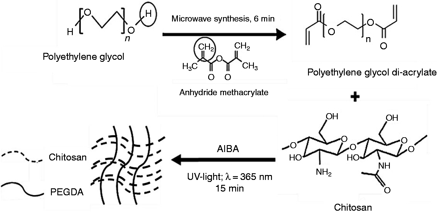
Chemical reaction of the acrylation of PEG and synthesis of PC network. PEG was acrylated with the addition of anhydride methacrylate by means of a microwave synthesis. To prepare PC networks, a solution of PEGDA diluted in deionized water (10% w/v) was mixed with different solutions of chitosan diluted in acetic acid at 1% (Table 1). The photoinitiator AIBA was added too (20% w/v). The polymerization was made by means of UV light (365 nm) for 15 min. PEG: polyethylene glycol; PC: polyethylene glycol-chitosan; PEGDA: polyethylene glycol di-acrylate.
Synthesis of PEG network
For the preparation of PEG network, PEGDA was solubilized in deionized water. Then, the photo-initiator 2,2´-azobis(2-methylpropionamidine) dihydrochloride (AIBA, Sigma Aldrich, USA; purity 97%) was added in a proportion of 10% w/v and mixed in a glass vial at room temperature (25 ± 2°C). The final solution was put in a 48-well plate (200 µL per well) which was closed and put under UV light (365 nm; UV lamp model GL-25 UVP, USA) for 15 min to initiate the polymerization process. The resulting sterilized polymeric networks were washed with sterile PBS for 3 days to remove any residue from the synthesis process, changing the solution everyday, and then stored until characterization.
Synthesis of PEG-chitosan network
The chitosan used for these networks was bought from Sigma Aldrich, USA. The chitosan had the following characteristics: Mv = 50,000–190,000 g/mol, viscosity =20–300 cP, 1 wt% in 1% acetic acid (25°C, Brookfield) and 75–85% deacetylated.
To obtain PEG-chitosan (PC) networks, a solution containing PEGDA (20 % w/v) and AIBA photoinitiator in deionized water was mixed with different solutions of chitosan (poly-1, 4-Dglucosamine) dissolved in acetic acid at 1%. The amount of chitosan used is shown in Table 1. The two solutions were mixed together (1:1) in a glass vial at room temperature (25 ± 2°C). The solution (200 µL per well) was put in a 48-well plate (Sigma Aldrich, USA) and was polymerized for 15 min under UV light (365 nm; UV lamp model GL-25 UVP, USA). The resulting sterilized polymeric networks were washed with PBS for 3 days, changing the solution everyday, and stored for subsequent analysis.
Amount of chitosan used for each polymeric network.
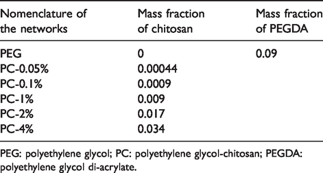
PEG: polyethylene glycol; PC: polyethylene glycol-chitosan; PEGDA: polyethylene glycol di-acrylate.
Fourier-transform infrared transmittance with attenuated total reflection spectroscopy analysis
The polymeric networks were completely dried before starting the Fourier-transform infrared transmittance with attenuated total reflection (FT-IR ATR) spectra analysis. Pure PEG and chitosan were analyzed too. The samples were put directly into the infrared beam. Infrared spectra of the materials were obtained using a Cary 600 Series FTIR spectrometer (Thermo Fisher Scientific, USA). FT-IR ATR spectra of the samples were studied at a scan range of 400–4000 cm−1.
Scanning electron microscopy
The polymeric networks were completely dried and coated with two layers of gold. Subsequently, the coated samples were attached to a metal support with a conductive tape and observed under high vacuum in an FEI Quanta 250 FEG scanning electron microscope (SEM; Thermo Fisher Scientific, USA).
Swelling behavior
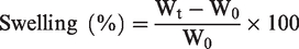
The swelling of the samples was characterized in PBS (pH = 7.4) at 37°C and at room temperature (25 ± 2°C) by weighting each sample with an analytical balance at selected time points (Boeco, Germany). For this purpose, the materials were completely dried and put in PBS solution. The weight of the samples was measured at different time points: 0.5, 1, 2, 3 and 4 h and then every 24 h until equilibrium was reached. The percentage of swelling was calculated according to the next equation
Antibacterial assay
The antibacterial activity of PC networks was tested using the method Microbiological evaluation of antibiotics of the Pharmacopeia of the United States of Mexico (11a edition, 2011, MGA 0100).
A reference strain S. aureus ATCC25922 provided by the Laboratory of Microbiology of the Faculty of Chemistry (UAEMéx) was used. This bacterium was placed in tubes with agar Mueller Hinton (Bioxon, Mexico) solidified in an inclined position and incubated for 24 h at 35°C. From a recent tube, an amount of bacteria was taken to prepare a suspension of 1.5 × 10⁸ CFU/mL in sterile saline solution at 0.9%. This suspension was prepared following the 0.5 mcfarland nephelometric standard. 19 From this solution, a volume of 0.7 mL was taken and added in 100 mL of culture medium number 3. 20 Briefly, the samples of each polymeric network previously sterilized and swollen in PBS (0.05 ± 0.02 g) were added in sterile glass tubes. Then, a volume of culture medium number 3 without bacterial inoculation and the bacterial suspension previously prepared were added in each tube in a dilution 1:10, respectively. For the positive control a vial containing Fosfomycin in a concentration of 10 µg/mL (CETUS laboratories, Mexico) and the bacterial suspension was used (1:10). Then, all the tubes were placed in a water bath with continuous agitation at 37 ± 2°C for 24 h. The absorbance was read using a plate reader (Awareness Technologinc, USA) at a wavelength of 492/630 nm. The lower the absorbance, the lower is the growth of bacteria in the media.
Direct viability assay
The effect of the addition of different proportions of chitosan to the polymeric networks on cell viability was analyzed using a direct viability assay performed with human dermal fibroblasts (HDF). Briefly. PC hydrogels were prepared in 48 culture plates. After gelification, cells were seeded on the surface of each sample at a concentration of 12,000 cells/well in a volume of 120 µL of DMEM media (Gibco, Invitrogen Corporation, Canada) supplemented with 10% fetal bovine serum (FBS, Gibco, Invitrogen Corporation, Burlington, Canada) and 1% penicillin/streptomycin (P/S, Gibco, Invitrogen Corporation, Canada). Subsequently the plates were incubated at 37°C, 5% CO2 and 85% humidity for 1 h, in order to let the cells reach and start to attach to the polymeric networks. Later, 380 µL of DMEM, supplemented as described before, were added to have a final volume of 500 µL/well. After 1, 3 and 7 days of incubation, the media was removed and replaced with a resazurin solution (1% in DMEM medium) and the plates were incubated for 4 h at 37°C, 5% CO2 and 85% humidity. After the incubation, the obtained product (resorufin) was collected and fluorescence intensity at 545 nmex/590 nmem wavelength was measured with a SpectraMax i3x Multi-Mode Plate Reader (Molecular Devices, USA).
Erythrocyte compatibility
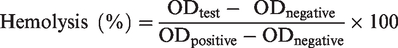
To perform this test, human erythrocytes were used. The entire procedure was performed under the criteria established in ISO 10993–4 (2017) of the biological evaluation of medical devices. The blood of three different healthy adult donors (over 18 years) was collected in EDTA tubes (n = 3 replicates per donor). Subsequently, the erythrocytes were isolated from the rest of the blood and diluted to a final solution concentration of 5% in PBS. Then, each gel was put in 1 mL of this solution in Eppendorf tubes (Fisher Scientific, USA); triton (Sigma Aldrich, USA) in a concentration of 2% and isotonic saline solution were used as positive and negative controls, respectively. All the tubes were incubated for 1 h at 37°C. After incubation, one centrifugation was performed at 3000 rpm/min for 2 min. Absorbance measurements of each sample were obtained with a plate reader (Awareness Technologinc, USA) at 450/492 nm in the UV-VIS range. The percentage of hemolysis was calculated with the following formula
Free hemoglobin test
Free hemoglobin test was performed using human blood of one donor and following the standard ISO 10993–4 (2017). This assay was performed to study the hemocompatibility of the PC networks. First the polymeric networks were prepared and put in 24-well plates (Thermo Fisher, USA). Citrated blood (50 µL) was put on the surface of each sample and a volume of 10 µL of 0.1 M CaCl2 (Sigma Aldrich, Oakville, Canada) was added to inhibit the anti-coagulant effect of the citrate. After 15, 30, 45 and 60 min, 1 mL of distilled water was added to each sample. Subsequently, the obtained solution was put in 96-well plate and absorbance was measured at 540 nm using a SpectraMax i3x Multi-Mode Plate Reader (Molecular Devices, San Jose, California, USA).
Statistical analysis
For each experiment a number of five replicates for each condition was assayed. Only in the erythrocyte compatibility, triplicate per donor (three donors) was analysed. The data shown are means ± standard deviation (SD). Statistical significance of the results was calculated using ANOVA non-parametric Fisher’s least significant difference method (LSD). The p values lower than 0.05 were considered statistically significant.
Results
FT-IR ATR analysis
FT-IR ATR spectroscopy was performed to get evidence of the characteristic functional groups of pure PEG, PEGDA, chitosan and PC networks. Pure PEG (Figure 2(a)) showed one peak at 1108 cm−1, which is attributed to the binding C-O-H. Another peak at 2894 cm−1 belongs to CH stretching, a band at 1463 cm−1 comes from CH-binding group. The PEGDA (Figure 2(b)) has the same bands as pure PEG but also a characteristic peak at 1724 cm−1 attributed to C = O group as a result of its acrylation.
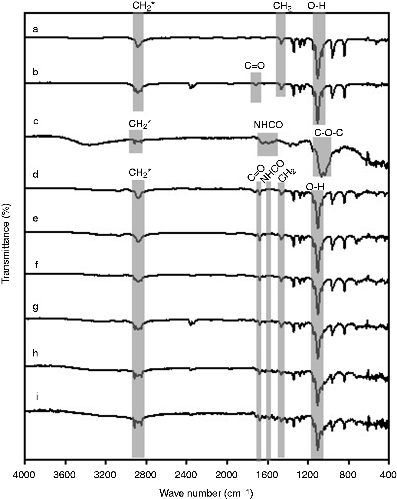
FT-IR ATR analysis. The materials were completely dried and then they were put in an infrared spectrometer. All the samples were studied at a scan range of 400–4000 cm−1. The figure shows the FT-IR spectra of (a) PEG, (b) PEGDA, (c) chitosan, (d) PEG network, (e) PC-0.05%, (f) PC-0.1%, (g) PC-1%, (h) PC-2% and (i) PC-4%. FT-IR ATR: Fourier-transform infrared transmittance with attenuated total reflection; PEG: polyethylene glycol; PC: polyethylene glycol-chitosan; PEGDA: polyethylene glycol di-acrylate.
The FT-IR of chitosan is shown in the Figure 2(c) with characteristic bands at 2848–2915 cm−1 (CH-stretching), 1656–1596 cm−1 (secondary amide NHCO-) and 1078–1027 cm−1 (C-O stretching characteristic of its saccharide structure). 15
Characteristic bands of PEG network were observed (Figure 2(d)). Five peaks appeared: 1108 cm−1 of C-O-H stretching, 1724 cm−1 of carboxylic acid (C = O) that comes from the acrylation of PEG, 2890 cm−1 of CH-stretching, 1469 cm−1 of CH-bending and at 962 cm−1 of alcoholic group (C-O-H). The spectrum of PC networks also showed the same bands.
The infrared spectra of PC-0.1% network (Figure 2(f)) starts to show two peaks at 2844 cm−1 and 2919 cm−1, which are characteristic of the C-H-stretching group of the chitosan. The intensity of these peaks is higher with the increase of the amount of chitosan in the networks, having more intensity in the PC-4% network. The C-H stretching in 2880 cm−1 of the PEG network disappears from the spectrum with the addition of chitosan. Moreover, the bands at 1656–1596 cm−1 (secondary amide NHCO-) from chitosan, appeared in all PC networks.
Morphological analysis
The morphology of the surfaces of PC networks was done to analyze their topographical features and porosity (Figure 3). Cylindrical interconnected pores of different sizes were found in all the samples. The micrographs of the PEG network (Figure 3(a) and (b)) show multiple cylindrical pores with different sizes. PC-4% network (Figure 3(k) and (l)) showed less and smaller interconnected pores and the surface is more irregular than the PEG network. The combination of PEG and chitosan polymers created irregular surfaces with smaller and less amounts of pores. The increase in the amount of chitosan increases the surface irregularity.
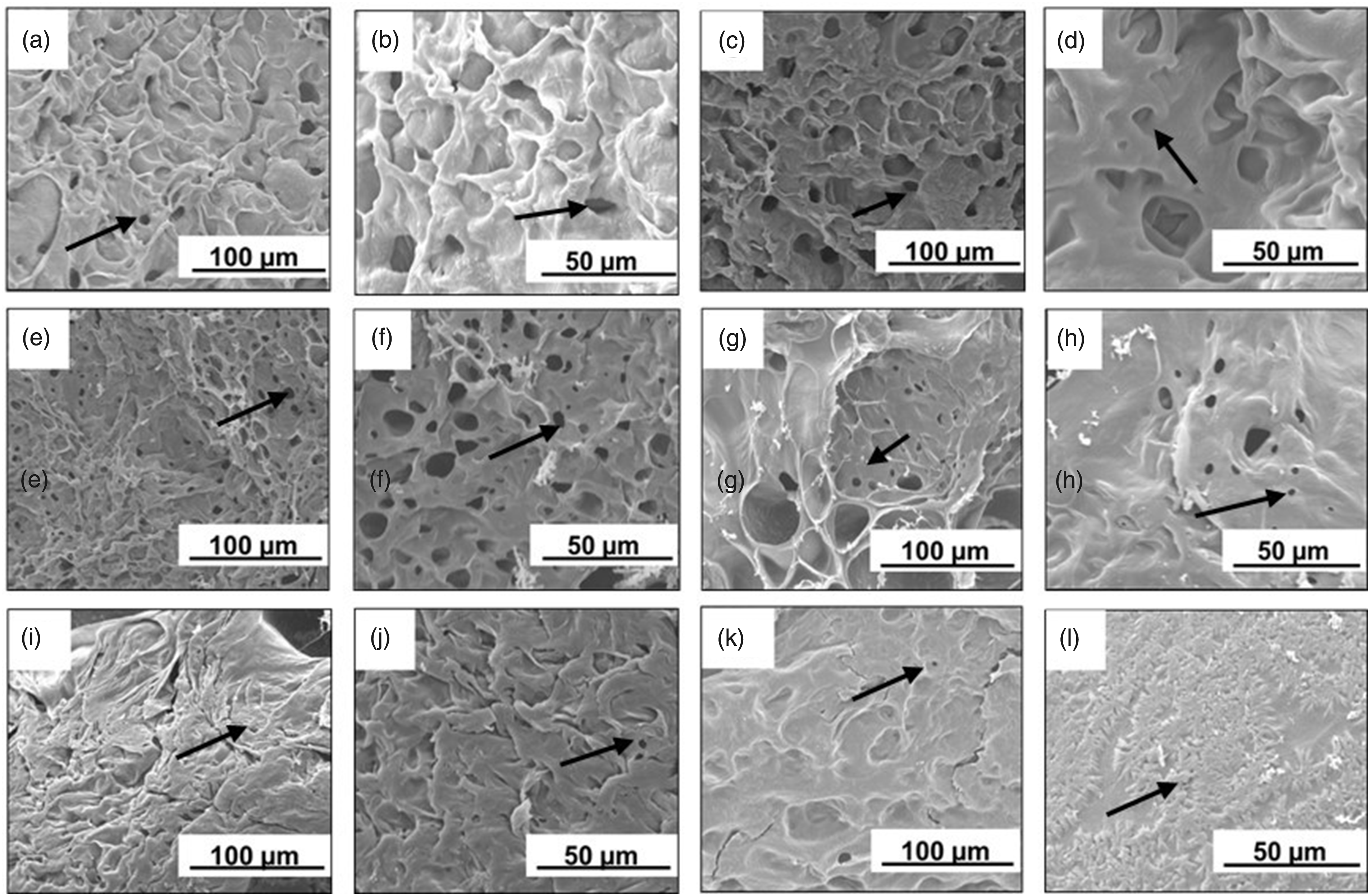
Scanning electron microscopy analysis. PC networks were completely dried and coated with gold. Then the samples were put in a scanning electron microscope to observe the surface. The figure shows the optical micrographs of the polymeric networks of PEG (a and b), PC-0.05% (c and d), PC-0.1% (e and f), PC-1% (g and h), PC-2% (i and j) and PC-4% (k and l). PEG: polyethylene glycol; PC: polyethylene glycol-chitosan; PEGDA: polyethylene glycol di-acrylate.
Swelling behavior
The swelling behavior of the polymeric networks was studied in PBS at 37°C and at room temperature (25 ± 2°C). In PBS at 37°C (Figure 4(a)) the percentage of swelling of all the networks was higher than at room temperature (Figure 4(b)). In PBS at 37°C and at room temperature, the PC-4% network exhibited the lowest swelling capability ((3.2 ± 0.3) × 102 at 37°C; (2.6 ± 0.5) × 102 at room temperature). The PEG network showed the highest percentage of swelling in PBS at 37°C ((1.1 ± 0.3) × 103) and at room temperature ((6.1 ± 0.6) × 102). The percentage of swelling appears to decrease with the increase of the amount of chitosan. In PBS at 37°C significant differences were found between PEG network and PC-0.05%, PC-0.1%, PC-1%, PC-2%, PC-4%; PC-0.05% and PC-0.1%, PC-1%, PC-2%, PC-4%; PC-0.1% and PC-1%, PC-2%, PC-4%; PC-4% and PC-1%, PC-2% (two-way ANOVA). In PBS at room temperature, there were statistically significant differences between: PEG network and PC-0.1%, PC-1%, PC-2%, PC-4%; PC-0.05% and PC-0.1%, PC-1%, PC-2%, PC-4%; PC-0.1% and PC-1%, PC-2%, PC-4% (two-way ANOVA, p < 0.05).
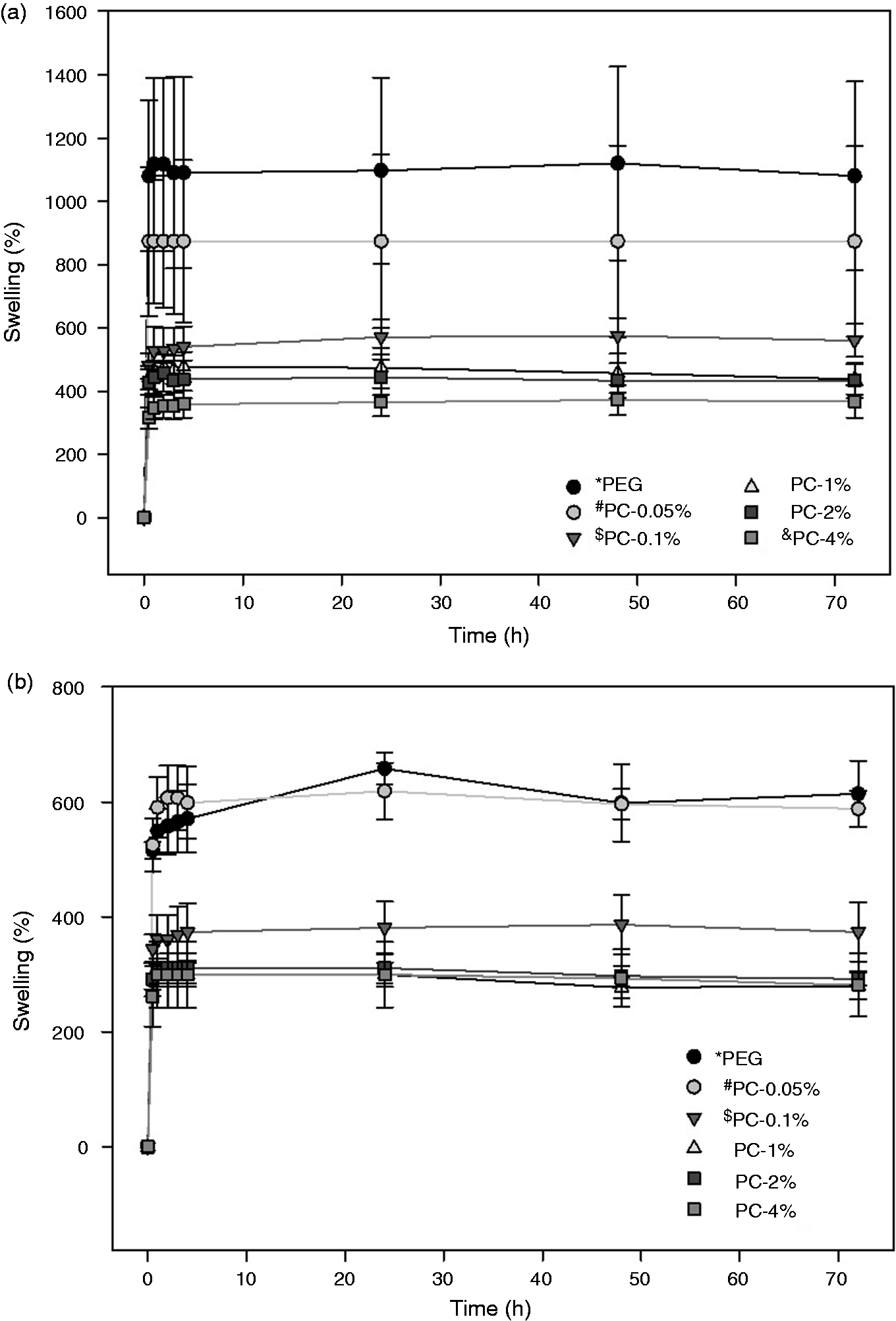
Swelling behavior of PC networks determined gravimetrically in (a) PBS at 37°C (pH = 7.4) and (b) PBS at room temperature (pH = 7.4). The graphics show the mean percentage of swelling of all the samples ± SD. ANOVA two-way. (a) *p < 0.05 vs. PC-0.05%, PC-0.1%, PC-1%, PC-2% and PC-4%; #p < 0.05 vs. PC-0.1%, PC-1%, PC-2%, PC-4%; $p < 0.05 vs. PC-1%, PC-2% and PC-4%; &p < 0.05 vs. PC-1% and PC-2% (two-way ANOVA). (b) *p < 0.05 vs. PC-0.1%, PC-1%, PC-2% and PC-4%; #p < 0.05 vs. PC-0.1%, PC-1%, PC-2% and PC-4%; $p < 0.05 vs. PC-1%, PC-2% and PC-4%. PC: polyethylene glycol-chitosan.
The maximum swelling in PBS at 37°C of PEG, PC-0.05% and PC-0.1% was approximately at 24 h. Samples with higher percentage of chitosan reached the maximum swelling in lower time. For example, while PC-1% and PC-2% reached maximum swelling in 4 h, PC-4% absorbed the highest quantity of PBS in 2 h. In PBS at room temperature the hydrogels of PEG, PC-0.05% and PC-0.1% had the highest swelling at 24 h, while the PC-1%, PC-2% and PC-4% reached maximum swelling at 2 h.
Antibacterial assay
To perform the antibacterial assay, the microbiological evaluation of the Pharmacopoeia of the United Mexican States was followed. 20
The antibacterial activity of the polymeric networks against S. aureus is shown in the Figure 5. PC-2% (0.020 ± 0.005) was able to significantly decrease the growth of S. aureus in the medium, compared with positive control (0.032 ± 0.001; p < 0.05 vs. PC-2%). PC-1% (0.29 ± 0.005) had a similar activity against S. aureus than the positive control (0.032 ± 0.001; p > 0.05 vs. PC-1%). PEG network (0.087 ± 0.009), PC-0.05% (0.078 ± 0.002) and PC-0.1% (0.078 ± 0.004) were not able to significantly decrease the amount of S. aureus in the medium compared with the positive control (0.032 ± 0.001; p < 0.05 vs. PEG network, PC-0.05% and PC-0.1%). No significant differences were found between positive control (0.032 ± 0.001) and the PC-4% (0.033 ± 0.001; p > 0.05 vs. C+) (Figure 5).
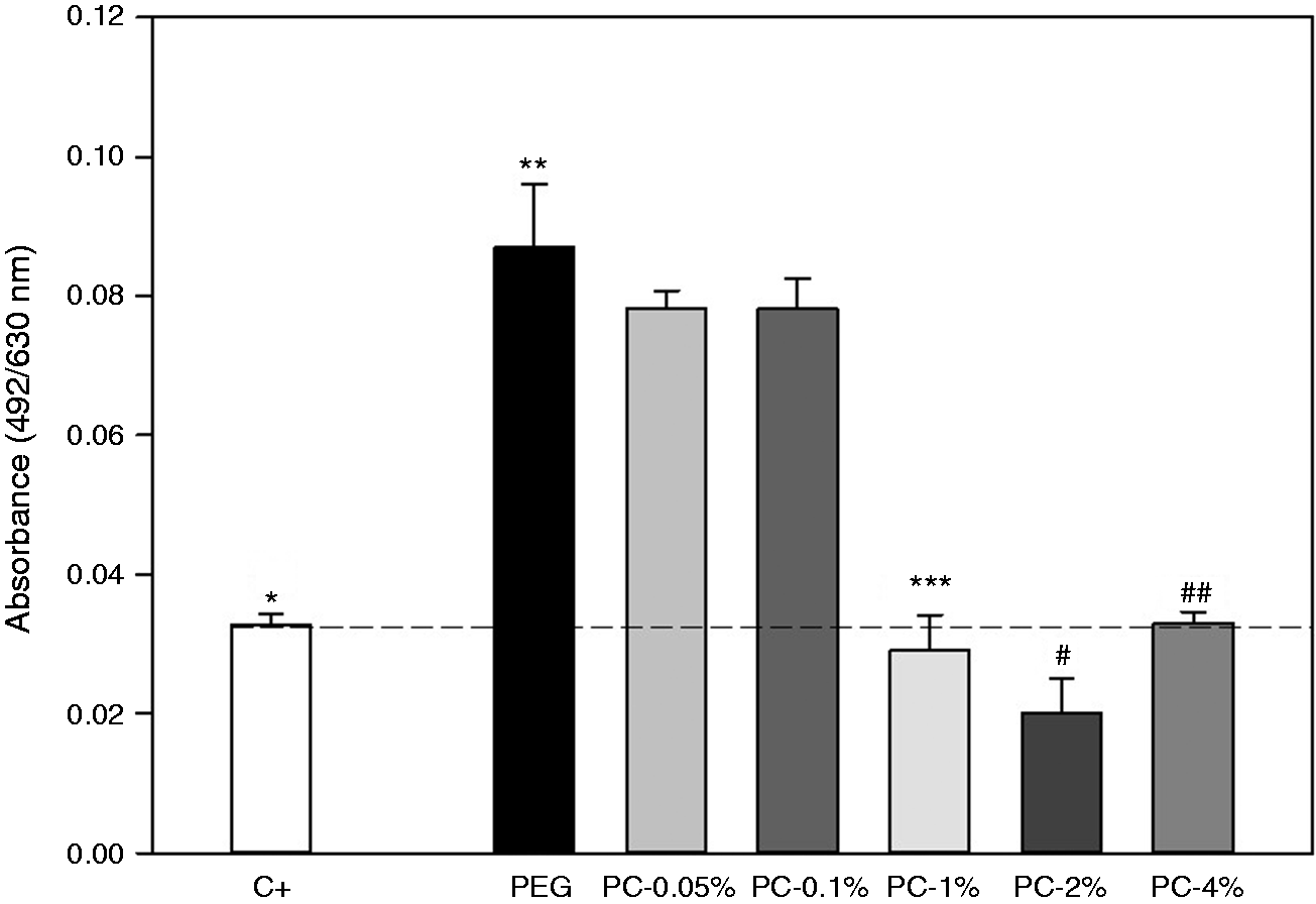
Antimicrobial activity. An amount of 7.2 × 10⁵ CFU/mL S. aureus was added in culture medium number 3 in glass tubes. The materials were put in this media for 24 h at 37 ± 2°C. After the absorbance of all the tubes was measured at a wavelength of 492/630 nm. The graphic shows the mean absorbance of all the samples ± SD. *p < 0.05 vs. PEG network, PC-0.05%, PC-0.1% and PC-2%; **p < 0.05 vs. PC-0.05%, PC-0.1%, PC-1%, PC-2% and PC-4%; ***p < 0.05% vs. PC-0.05% and PC-0.1%; #p < 0.05 vs. PC-0.05%, PC-0.1%, PC-1% and PC-4%; ##p < 0.05 vs. PC-0.05% and PC-0.1% (one-way ANOVA). PEG: polyethylene glycol; PC: polyethylene glycol-chitosan.
Direct viability assay
Direct viability assay was performed on HDF directly seeded on the different PC networks to evaluate the effect of the chitosan present in the gels. After 1 day of incubation, HDF seeded on PC-1% ((3.3 ± 0.2) ×107) showed a significant increased viability compared to the PEG network ((3.1 ± 0.2) × 107 p < 0.05 vs. PC-1%). However, a significant decrease in viability was observed with the PC-4% ((3 ± 0.2) ×107). After 3 days of incubation, PC-0.1% ((3.4 ± 0.2) × 107), PC-1% ((3.5 ± 0.2) × 107) and PC-4% (3.5 ±0.2) × 107) showed a significantly increased viability compared with PEG network (3.1 ± 0.2) × 107 p < 0.05 vs. PC-0.1%, PC-1% and PC-4%). No statistical differences were found between PEG network and PC-0.05%. Otherwise, significant differences were found between PC-0.1% ((3.4 ± 0.3) × 107), PC-1% ((3.5 ± 0.2) × 107) and PC-2% ((3.5 ± 0.2) × 107), comparing with PC-4% ((3.1 ± 0.3) × 107 p < 0.05 vs. PC-0.1%, PC-1% and PC-2%). After 7 days of incubation, PC-0.05% ((3.3 ± 0.2) × 107) and PC-0.1% ((3.3 ± 0.2) × 107) had a significantly increased viability compared with PEG network ((3 ± 0.1) × 107) p < 0.05 vs. PC-0.05% and PC-0.1%). On the other hand, PC-0.05% ((3.3 ± 0.2) × 107) and PC-0.1% ((3.3 ± 0.1) × 107) showed a higher viability compared with PC-1% ((2.9 ± 0.2) × 107), PC-2% ((3 ± 0.2) × 107) and PC-4% ((2.8 ± 0.2) × 107) (Figure 6).
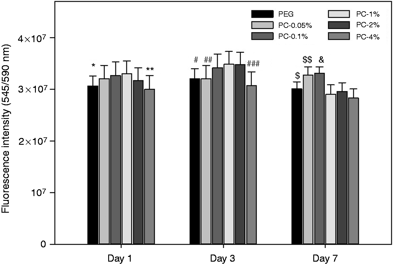
Direct viability assay. HDF were seeded directly on the surfaces of the polymeric networks previously placed in 48 culture plates. Cell viability was measured after 1, 3 and 7 days by means of Alamar Blue Cell Viability Assay with a solution of resazurin (1% in DMEM medium). The graphic shows the mean fluorescence intensity ± SD of each sample. *p < 0.05 vs. PC-1%; **p < 0.05 vs. PC-0.1% and PC-1%; #p < 0.05 vs. PC-0.1%, PC-1% and PC-2%; ##p < 0.05 vs. PC-0.1%, PC-1% and PC-2%; ###p < 0.05 vs. PC-0.1%, PC-1% and PC-2%; $p < 0.05 vs. PC-0.05%, PC-0.1% and PC-4%; $$p < 0.05 vs. PC-1%, PC-2% and PC-4%; &p < 0.05 vs. PC-1%, PC-2% and PC-4% (two-way ANOVA). HDF: human dermal fibroblast; PC: polyethylene glycol-chitosan.
Erythrocyte compatibility
The absorbance of the hemoglobin released, due to erythrocyte hemolysis, after the direct contact with the networks was measure and analyzed (Figure 7). No significant differences were found between the hemolysis induced by the polymeric networks. All the materials induced less than 5% of hemolysis, which means that they are non-hemolytic, hence hemocompatible.
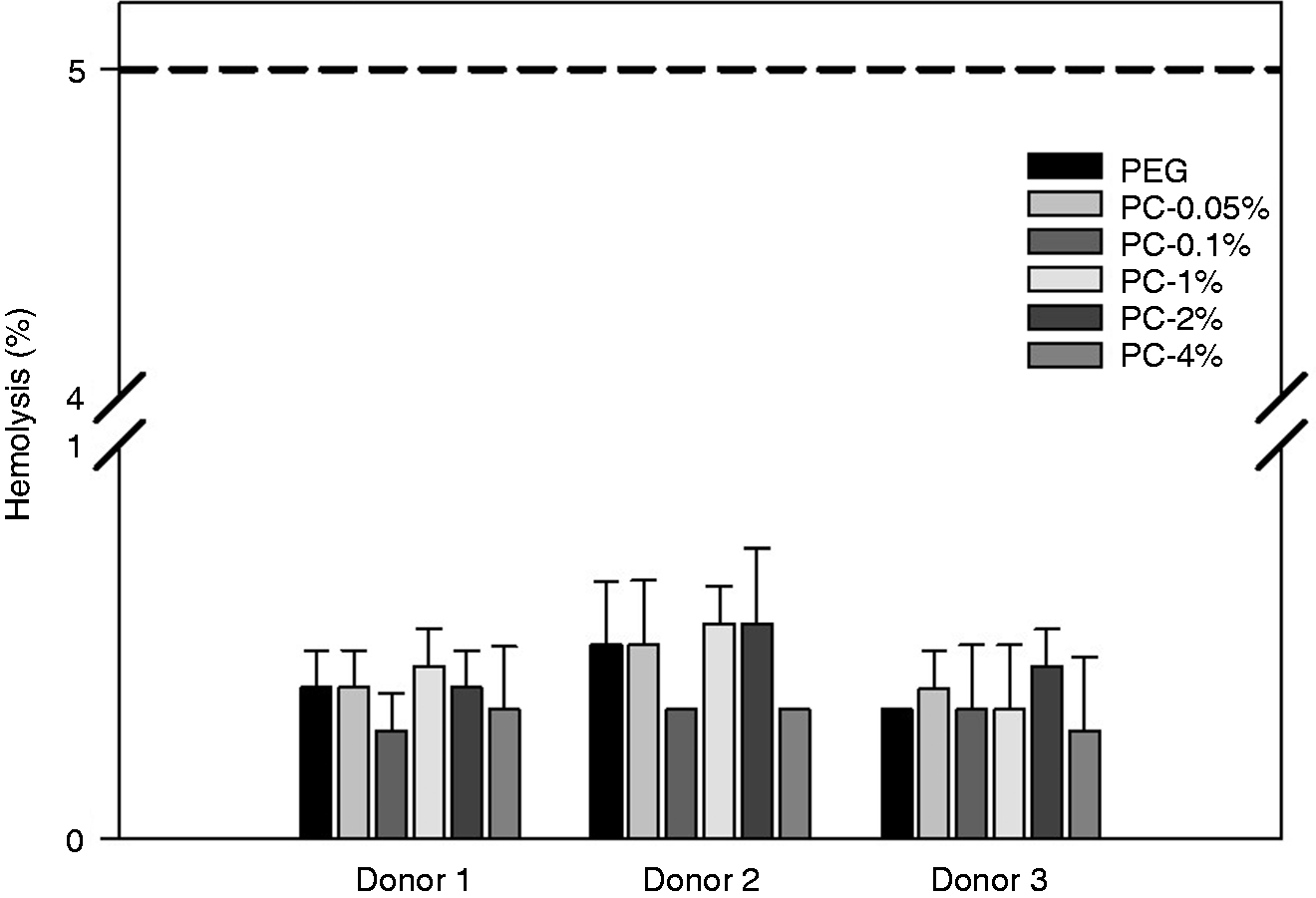
In vitro hemolysis induced by PC networks was performed following the ISO-10994–4 (2017). Human erythrocytes (5% in PBS) were incubated in contact with the networks for 1 h. The graphic shows the mean of absorbance ± SD of each sample. No significant differences were found between the samples (one-way ANOVA; p > 0.05). PC: polyethylene glycol-chitosan.
Free hemoglobin test
The hemocompatibility of PC networks was tested using the free-hemoglobin test (Figure 8). In this test, the higher the absorbance recorded, the higher is the amount of free hemoglobin, hence the lower the coagulation meaning a higher hemocompatibility. After 15 min of incubation a higher value of free hemoglobin (hence a higher hemocompatibility) was obtained with PC-2% (0.98 ± 0.08) compared with PEG network (0.70 ± 0.09; p < 0.05 vs. PC-2%). However, with PC-0.05% (0.25 ± 0.07), the value of free hemoglobin was lower compared with PEG network (p < 0.05 vs. PC-0.05%). A different behavior was observed after 30 min of incubation: PC-2% (0.50 ± 0.14) and PEG network (0.45 ± 0.15; p > 0.05 vs. PC-2%) had similar free hemoglobin, hence similar hemocompatibility. PC-1% (0.19 ± 0.04) had a lower free hemoglobin compared to both PEG network (0.45 ± 0.15; p < 0.05 vs. PC-1%) and PC-2% (0.50 ± 0.14; p < 0.05 vs. PC-1%). After 45 min of incubation, PEG network was more hemocompatible than the other materials (0.44 ± 0.12; p < 0.05 vs. PC-0.05%, PC-0.1%, PC-1%, PC-2% and PC-4%). PC-0.05% (0.18 ± 0.05) and PC-1% (0.18 ± 0.05) decreased the free hemoglobin compared with PEG network. Again, after 60 min of incubation, PEG network had a higher hemocompatibility than the other networks (0.45 ± 0.14; p < 0.05 vs. PC-0.05%, PC-0.1%, PC-1%, PC-2% and PC-4%). PC-0.1% (0.17 ± 0.04) and PC-1% (0.17 ± 0.07) had a lower hemoglobin and hemocompatibility compared with PEG network (0.45 ± 0.14; p < 0.05 vs. PC-0.1% and PC-1%).
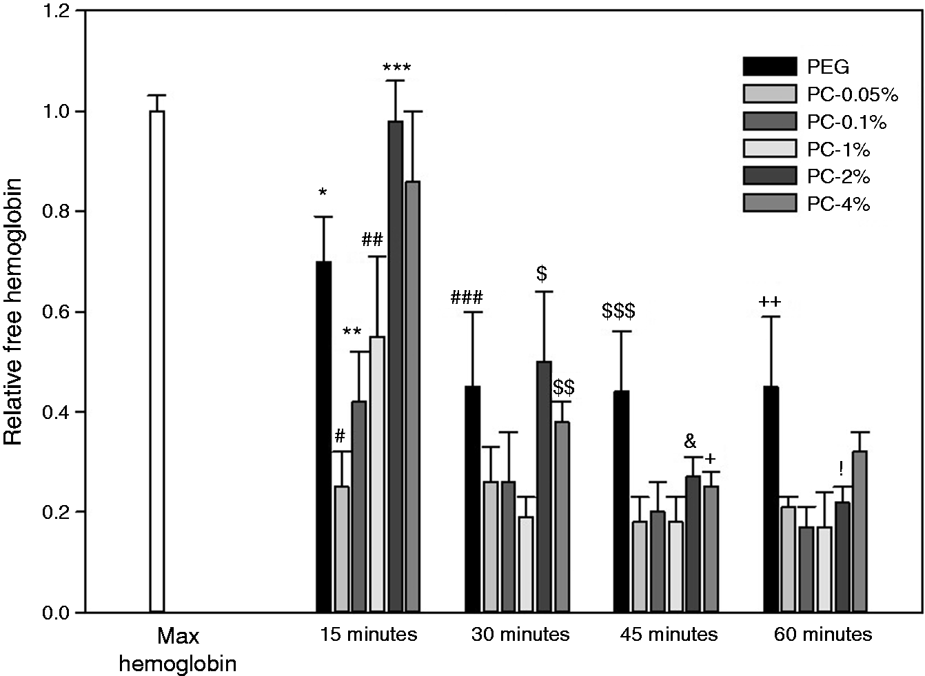
Free hemoglobin test. Whole human blood was put in direct contact with the polymeric networks. Blood was incubated with the samples for 15, 30, 45 and 60 min. The blood was solubilized at each time point and was read at 540 nm. The graphic shows the mean of relative free hemoglobin of all the samples ± SD. *p < 0.05 vs. PC-0.05%, PC-0.1%, PC-1%, PC-2% and PC-4%; #p < 0.05 vs. PC-0.1%, PC-1%, PC-2% and PC-4%; **p < 0.05 vs. PC-1%, PC-2% and PC-4%; ##p < 0.05 vs. PC-2% and PC-4%; ***p < 0.05 vs. PC-4%; ###p < 0.05 vs. PC-0.05%, PC-0.1% and PC-1%; $p < 0.05 vs. PC-0.05%, PC-0.1%, PC-1% and PC-4%; $$p < 0.05 vs. PC-0.05%, PC-0.1% and PC-1%; $$$p < 0.05 vs. PC-0.05%, PC-0.1%, PC-1%, PC-2% and PC-4%; &p < 0.05 vs. PC-0.05%, PC-0.1% and PC-1%; +p < 0.05 vs. PC-0.05% and PC-1%; ++p < 0.05 vs. PC-0.05%, PC-0.1%, PC-1%, PC-2% and PC-4%,! p < 0.05 vs. PC-0.05%, PC-0.1%, PC-1% and PC-2% (two way ANOVA). PC: polyethylene glycol-chitosan.
Discussion
The bands observed in the FT-IR of the PEG, chitosan and PC networks (Figure 2) are similar with the FT-IR reported in literature.15,21–23 The synthesis of a new material was demonstrated with the appearance of bands from both polymers: PEG and chitosan. PEG network showed bands at 1108 cm−1 of C-O-H stretching and at 1724 cm−1 of the carboxylic acid from the acrylation of PEG. PC networks showed the same bands and also peaks at 2844 cm−1, 2919 cm−1 (CH-stretching) and 1656–1596 cm−1 (secondary amide NHCO-), which demonstrate the presence of chitosan in these ones. The bands corresponding to CH-stretching group from chitosan showed more intensity in PC-4%, which proves a higher amount of chitosan in this network.
The importance of SEM lies in its ability to provide high-resolution imaging useful for the evaluation of microstructure morphology and chemical composition characterizations. 24 In this work, SEM was used to analyze the surface of PC networks (Figure 3). The obtained images showed that the higher the amount of chitosan, the higher the crosslinking and, consequently, smaller pores. The presence of pores in the networks was also observed by Gaitán-Tolosa et.al. 21 who synthesized ketoprofen-loaded PC system for wound dressing and they found pores of different sizes. However, the pore size was of 13.7 nm, since they loaded ketoprofen inside the networks, so they decreased the pore size to avoid the diffusion of the drug outside of the PC system. One of the most important parameters that regulate the diffusion of the encapsulated molecule in a hydrogel is the pore size. In the present work, we did not load a drug within the hydrogel network, so we did not decrease the pore size, resulting in pores of 50–100 µm.
This study compared the swelling of PC networks by changing the solution in which they were immersed (PBS at room temperature and PBS at 37°C) (Figure 4). The swelling of all the networks was higher in PBS at 37°C than in PBS at room temperature (25 ± 2°C). This is because the increase in temperature increases the rate of the diffusion of solution to the hydrogels and adsorption. 25 In addition, the swelling time in PBS at 37°C and in PBS at room temperature of the networks was different; PEG network, PC-0.05% and PC-0.1% reached maximum swelling in approximately 24 h. However, PC-1%, PC-2% and PC-4% had the maximum swelling in PBS at both temperatures in 2 h. This behavior can be attributed to the size and number of pores that is determined by the crosslinking between PEG and chitosan. 21 In both solutions, PC-4% exhibited the lowest swelling capability (349.58 ± 47.58% at 37°C; 262.4 ± 52.54% at room temperature). The PEG hydrogel showed the highest percentage of swelling (930.55 ± 324.91% at 37°C; 614.16 ± 57.85% at room temperature). This is because the higher the amount of chitosan the higher the crosslinking with the PEG. The higher the crosslinking, the lower the mobility of the structure interpenetrated of the hydrogels, hence the solvent absorption is lower. In contrast, the highest swelling of all the networks at a higher temperature (37°C) are in accordance with the results obtained by Larrañeta et al. 26 who prepared pH- and thermo-responsive chitosan hydrogels and they investigated the swelling behavior of these ones at different temperatures. They found that raising the temperature increases the swelling of the hydrogels due to an increase of their adsorption capacity and an increase in the rate of the diffusion of solution.
With the aim to test the antibacterial activity of PC networks, a method from the Pharmacopeia of the United States of Mexico (MGA 0100)20 was performed. In this work the antibacterial action of chitosan 27 from PC networks was tested against S. aureus ATCC25922, which is the most common Gram-positive bacteria found in chronic wounds.9,10 The results showed that PC-2% (0.020 ± 0.005) had a higher bacterial inhibition against S. aureus compared with the other networks and the C+ (0.020 ± 0.005; p < 0.05 vs. C+, PEG network, PC-0.05%, PC-0.1%, PC-2% and PC-4%). In contrast, although PC-4% was the network with the highest amount of chitosan added, it was not able to decrease the number of bacteria (0.033 ± 0.001) compared with PC-2% (0.020 ±0.005; p < 0.05 vs. PC-4%). This behavior can be attributed to the loss of chitosan solubility which prevents its interaction with microorganisms. According to Kong et al., 28 there are some factors that can affect the antimicrobial activity of chitosan: intrinsic chitosan factors (molecular weight, solubility, degree of deacetylation, positive charge density and chelating capacity); microorganism (species and stage of development); physical state of chitosan (liquid [colloid] or solid [membrane]); environmental factors (pH, temperature and time). The antibacterial effect of chitosan showed in this work is in accordance with Miguel et al., 29 who produced chitosan-agarose hydrogels varying the amount of chitosan. They evaluated the antibacterial properties of the hydrogels by standard tube dilution method using S. aureus. They observed bactericidal activity of hydrogels containing more than 188 µg/mL of chitosan. Another study by Sautrot-Ba et al. 30 confirmed the antibacterial property of chitosan. They synthesized chitosan-based PEG hydrogels under light irradiation in aqueous medium. They evaluated the antibacterial activity of the hydrogels against S. aureus. The resulting chitosan–PEG hydrogels demonstrated a tremendous inhibition of bacterial growth.
With the objective to test the biocompatibility of the polymeric networks, cell viability assay was performed on HDF for 1, 3 and 7 days. The results of cell viability assays indicate that after 1 day, PC-1% was able to induce a significant increase in cell viability compared to PEG network. Moreover, after 3 days, PC-0.1%, PC-1% and PC-2% increased the cell viability compared with PEG network. PC-0.05% and PC-0.1% increased the cell viability compared with PEG network after 7 days in contact with the cells. The amount of chitosan did not have a significant impact on the cell viability compared to the network without chitosan (PEG network). These results are in accordance with that of Hamilton et al. 31 who correlated fibroblast responses with known chitosan material characteristics, and they demonstrated that chitosan had no effect on HDF proliferation.
In vitro erythrocyte compatibility assay was performed as a requisite to the ISO-10993–4 (2017) for materials intended to be in contact with human blood. The results of this work showed that all the networks induced less than 1% of hemolysis, hence they are highly hemocompatible. The amount of chitosan did not affect the hemocompatibility of the networks. These results are similar to those obtained by Escudero-Castellanos et al. 32 who used PEG, PC and multi-arm PEG networks and reported that all materials tested guarantee a hemolysis percentage below 5%. Another study made by Zhao et al. 33 confirmed the hemocompatibility of photo-crosslinked cell membrane-mimicking chitosan-based hydrogels by measuring their lytic activity against red cells (hemolysis). They found that the hydrogels induced less than 1% of hemolysis, classifying them as non-hemolytic.
Free hemoglobin assay was performed to analyze the hemocompatibility by means of the measurement of blood free hemoglobin which is also related to the clotting process. The higher the free hemoglobin, the lower is the clotting process. According to our results, after 15 min of incubation, PC-2% showed the best hemocompatibility hemoglobin with human blood by having the highest amount of free hemoglobin. In contrast, after 30 min of incubation, there was a similar hemocompatibility between PEG network and PC-2% (ANOVA one way; p > 0.05). However, after 45 and 60 min of incubation, PEG network showed a better hemocompatibility compared with the rest of the networks (ANOVA two way; p < 0.05). The decrease of free hemoglobin and clotting process with the increase of chitosan are in accordance with some comparative studies, for example, Sanandam et al. 34 found that chitosan takes two-fold less time to clot blood than that taken by the natural blood clotting process.
Conclusions
PC networks with antibacterial properties can be prepared under UV-light irradiation in 15 min. The resulting networks had presence of pores of different size (50–100 µm) and good swelling behavior in PBS at 37°C (>315%). The addition of chitosan to PEG promoted the antibacterial properties of the networks. PC-2% showed the highest inhibition of S. aureus after 24 h of incubation. PC-2% also had better in vitro hemocompatibility properties than the other PC networks. However, all the materials showed less than 5% of hemolysis in contact with human erythrocytes.
In conclusion, PC networks proposed here can represent a good alternative as a wound dressing in the treatment of chronic wounds, due to their antibacterial properties and good hemocompatibility.
As a perspective and future direction, the PC networks developed in this work can be used not only as wound closure dressings but also as intelligent controlled-release systems by adding a broad-spectrum antibiotic, for a more efficient treatment of the infection.
Footnotes
Acknowledgements
The authors thank Professor Lidia Sandoval Flores for her participation in performing the method (MGA 0100; Pharmacopeia of the United States of Mexico) used to test the antibacterial activity of the hydrogels. The authors also thank the Laboratory of Microbiology (UAEMéx) for providing the material for developing the antibacterial assay.
Declaration of conflicting interests
The author(s) declared no potential conflicts of interest with respect to the research, authorship, and/or publication of this article.
Funding
The author(s) disclosed receipt of the following financial support for the research, authorship, and/or publication of this article: The authors acknowledge the financial support from UAEMéx (project: 4993/2020/CIB) and CONACyT. This work was partially supported by the Natural Science and Engineering Research Council of Canada Discovery Program (DM).