
Abstract
Rare earth-doped nanoparticles have been widely used in disease diagnosis, drug delivery, tumor therapy, and bioimaging. Among various bioimaging methods, the fluorescence imaging technology based on the rare earth-doped nanoparticles can visually display the cell activity and lesion evolution in living animals, which is a powerful tool in biological technology and has being widely applied in medical and biological fields. Especially in the band of near infrared (700–1700 nm), the emissions show the characteristics of deep penetration due to low absorption, low photon scattering, and low autofluorescence interference. Furthermore, the rare earth-doped nanoparticles can be endowed with the water solubility, biocompatibility, drug-loading ability, and the targeting ability for different tumors by surface functionalization. This confirms its potential in the cancer diagnosis and treatment. In this review, we summarized the recent progress in the application of rare earth-doped nanoparticles in the field of bioimaging and tumor treatment. The luminescent mechanism, properties, and structure design were also discussed.
Introduction
Bioimaging is becoming more and more important in cancer diagnosis, intraoperative guidance, and postoperative evaluation.1–5 It interacts with biological tissues or organs by light, ultrasound, X-ray, and electromagnetic fields, and then the feedback is transmitted to the detector, which is then fed into the computer, thus generating the morphology and structure information about biological tissues, organs, and tumor. New imaging technologies, such as magnetic resonance imaging (MRI), computed tomography (CT), photon avalanche (PA), positron emission tomography (PET), and single-photon emission computed tomography (SPECT), are now available in the clinic, and these models, combined with a variety of cleverly designed imaging contrast agents, can be used for disease diagnosis and therapy monitoring.6–8 But the low resolution and dangerous ionizing radiation of tomography, as well as the high time consumption of CT, MRI, PET, and SPECT imaging, have limited their real-time visualization application in disease therapy. Compared with other imaging modes, fluorescent imaging has attracted more and more attention due to its features of low cost, rapid feedback, high sensitivity, and no radiation.9,10 In the process of fluorescence imaging, the sample is stimulated by excitation light, and the emitted light is collected by the detector. The whole imaging process takes only a few milliseconds while the resolution can reach tens of nanometers. 11 In tumor diagnostics and treatment, fluorescence imaging is widely used to monitor the dynamic interaction between drug molecules and tumor cells, especially near infrared (NIR) fluorescence imaging, which has negligible tissue scattering, absorption, and self-fluorescence, and can monitor the real-time dynamic process in biological tissues. 12 Since the absorption peak of DNA is located at 260 nm and the absorption peak of hemoglobin is less than 600 nm, when the excitation or emission light is located at more than 700 nm, the penetration depth of tissue formation and signal-to-noise ratio will be greatly improved. 13 Therefore, the penetration depth of NIR light in biological tissues is much stronger than that of visible light.
In 1999, NIR imaging was first applied for tumors in vivo, 14 and the imaging penetration depth was 1–2 cm. So far, several kinds of NIR fluorescent imaging contrast agents have been synthesized including: (1) organic dyes and small-molecule-conjugated probes: 15 the preparation and synthesis of some organic dye contrast agents are relatively mature and commercial. Most of them have been approved by Food and Drug Administration for clinical application, such as methylene blue (MB) 16 and indocyanine green (ICG). 17 In addition, some of them are under the stage of preclinical animal experiments, such as cyanine dyes 18 and aggregation-induced emission dots. 19 However, the main limitation is their low quantum yield. (2) Quantum dots (QDs): they possess the advantages including high stability, high quantum yield, and brightly fluorescent emission. A variety of NIR-emission QDs has been widely applied for the bioimaging, such as Ag2S, 20 Ag2Se, 21 CdS, 22 and PbS. 23 Although the Ag2S overcomes the high toxicity of QDs, it still suffers from the quenching effect caused by biological environment.24,25 (3) Carbon nanotubes (CNTs): CNTs were first used in NIR imaging by Dai’s 26 research group in 2009. They applied it to the deep tissue anatomical imaging of mice by the surface functionalization. 27 Nowadays, CNTs have been able to target the brain vessels, lymphatics, and different tumors in vivo through the modification of different targeting ligands.28–30 But their wide length distribution (hundreds of nanometers) and needle-like structure (which can cause tissue damage and long-term chronic toxicity) hinder their biological application. (4) Rare earth (RE)-doped nanoparticles (NPs).31–33 The RE-doped NPs solved all the above shortcomings.34–36 Because of the protection of 5s and 5p orbits, the influence of environment on the fluorescence of Ln3+ is very weak, which makes its fluorescence spectrum sharp (high intensity, narrow emission peak) and stable. Its full width at half maximum is generally 10–20 nm, while transition metal ions are 100 nm and QDs are 25–40 nm. Since the fluorescence of Ln3+ is attributed to atomic transition, it has high anti-light bleaching property. Moreover, the doping of different Ln3+ also makes the emission band of RE-doped NPs particles more customizable, because their rich fluorescence levels cover the ultraviolet (UV), visible (vis), and NIR regions. Our group prepared the Nd3+-doped lanthanide NPs (KNdLiF) with emission at 1060 nm that demonstrated the high contrast fluorescence imaging for whole body and human colon cancer cells in mice under 808 nm excitation.
For applying the RE-doped NPs to the diagnosis and treatment of tumors, the first work is to improve the water solubility and biocompatibility of NPs, which is the prerequisite for NPs to enter the organism and realize their functions safely. The researchers modified the surface properties of NPs by linking hydrophilic, low toxicity ligands including polyethylene glycol (PEG), polyvinyl pyrrolidone (PVP), polyetherimide (PEI), and so on.37–40 The second is to endow NPs with tumor targeting by surface functionalization. Tumor-targeting NPs imaging is able to provide pivotal information not only on the location of the cancer in the body but also on the biological processes of the tumor. The active functional groups located in the outer layer of NPs could be utilized to covalently connect some targeting ligands, including folic acid (FA), Arg-Gly-Asp peptides (RGD), peptides, aptamers, antibody, and so on.41–46 Finally, biocompatible water-soluble tumor-targeting NPs could be combined with photothermal agents and photosensitizers or used as carriers to load drugs for treatment of tumor cells.
Herein, we summarize the properties, structures, luminescence principles of RE-doped NPs as well as the frontier progress in the field of biological imaging and tumor treatment. The recent research progress on the enhancement of fluorescence intensity of NPs, surface modification, and tumor targeted diagnosis and treatment has also been highlighted.
Fluorescence bioimaging
Fluorescence imaging is mainly composed of four parts: (1) excitation light; (2) contrast agent; (3) emission light detector; (4) signal-fluorescence image conversion equipment (computer, software, etc.). There are also four most important elements that affect the quality of fluorescence bioimaging: (1) autofluorescence of tissue; (2) surface reflection; (3) tissue absorption; (4) tissue scattering. The autofluorescence intensity of biological tissue is inversely proportional to the wavelength. For example, reticulin of staining cells fluoresces at 470–520 nm caused by elastin and collagen. Lipofuscin fluoresces at 450–650 nm. Endogenous chromophores such as flavin fluoresce at 500–600 nm and nicotinamide adenine dinucleotides fluoresce at 460 nm. However, at 1500 nm, the autofluorescence of rodent tissues disappeared, especially in organs enriched with endogenous chromophores, such as liver. Surface reflection is caused by the difference of refractive index between biological tissue and air and is mainly affected by surface curvature and roughness. Tissue absorption is mainly caused by photon absorption of endogenous biomolecules. Most chromophore molecules can transform photons into heat and damage cells. Although the absorption spectra of oxyhemoglobin and deoxyhemoglobin are slightly different in the visible band before 600 nm, they all lead to strong absorption of blood. In addition, for hemoglobin, there are some biological macromolecules (such as reduced nicotinamide adenine dinucleotide, heterocyclic flavin, aromatic amino acid residues, and different forms of melanin) that have strong absorption in the UV–vis bands. Tissue scattering originates from the difference of refractive index of different tissues, which is also inversely proportional to the wavelength. As photons pass through different tissues, the longer the wavelength is, the lower the scattering is.
Properties and luminescence principles
The characteristics of RE-doped fluorescent NPs include tunable emission, negligible photobleaching, high fluorescence stability, and long lifetime, and the luminescence modes include upconversion (UC) and downconversion (DC). The 5s25sp6 electron orbital of RE ions can protect the 4f orbital inside from the interference of external crystal field, thus bringing excellent optical and magnetic properties. Apart from La, Gd, and Lu ions, other Ln ions emit in narrow peaks, which is due to the internal f–f transition. For inorganic Ln-doped NPs, the internal 4f orbital electrons hardly participate in the formation of valence, so the change of local environment will not cause the emission of internal f–f transition. The parity forbidden effect of f–f transition results in the long fluorescence lifetime of Ln ions and effectively avoids the interference of autofluorescence. There are five energy transfer (ET) modes for RE ions (Figure 1): excited state absorption (ESA), two-photon absorption (TPA), PA, energy transition upconversion (ETU), cross relaxation (CR), and Stokes process. According to the different ways of ET, ETU can be divided into three types: successive energy transfer (SET), CR, and cooperative upconversion (CU).
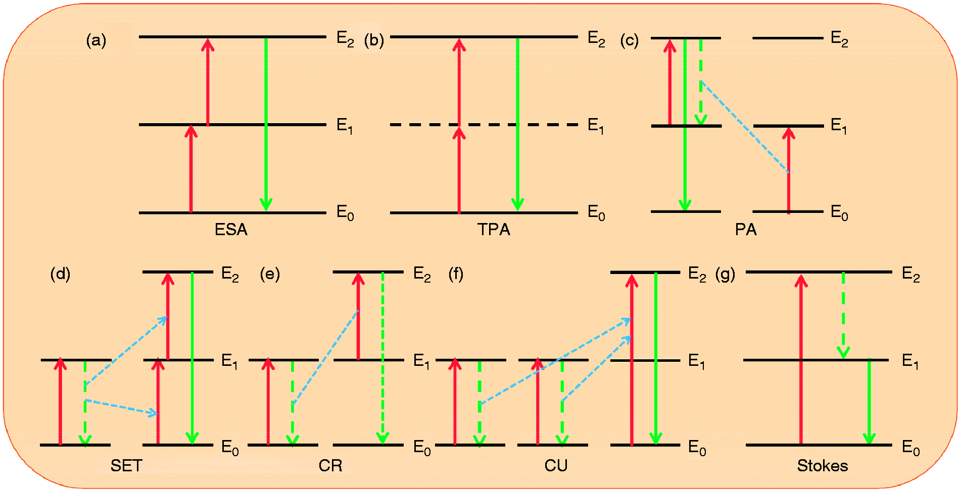
Mechanisms of ESA, TPA, PA, SET, CR, CR, CU, and Stokes process.
Upconversion
UC emission is a process of converting low-energy excitation light into high-energy emission light, which belongs to an anti-Stokes ET process. In general, in order to accomplish this process in RE-doped NPs, two parts are indispensable: activation ion and sensitization ion. UC luminescence is achieved by ET upconversion (ETU) between sensitized ions and activated ions after co-doping. This requires that the sensitized ions have large absorption cross sections in the NIR region. Yb3+ is the most commonly used sensitizer for UC luminescence. The absorption cross section of Yb3+ at 980 nm is much larger than that of other RE ions. At the same time, the energy level difference between the only excited state and ground state of Yb3+ is very close to the adjacent energy-level difference of activator ions, which greatly promotes the realization of energy conversion. In order to enhance the efficiency of energy conversion, the doping concentration of Yb3+ in matrix materials is usually high, which can reach 15–30 mol%. And in order to avoid the fluorescence weakening caused by quenching, the doping concentration of RE ions (activators) is generally not higher than 3 mol%. At present, commonly used UC matrix materials include NaYbF4 34, Yb2O3, 47 NaGdF4, 48 KGdF4, 49 GdF3, 50 BaF2, 51 GdVO4, 52 and LuPO4, 53 And Yb3+, Er3+, Tm3+, and Ho3+54–56 are the most commonly used activators in UC luminescence. They all have ladder-like energy levels. The energy difference between adjacent energy levels is very close, so they can transfer energy with each other for the UC luminescence.
Downconversion
DC process is a Stokes ET process, accompanied by non-radiative and radiative transitions. The energy of emitting light decreases compared with that of exciting light, and the exciting light is usually located in the NIR band. Therefore, compared with UC luminescent NPs, DC luminescent NPs have greater advantages in the field of bioimaging, including higher tissue penetration depth, quantum yield, and signal-to-noise ratio. The doping of RE ions plays a crucial role in the wavelength and intensity of luminescence. For example, Eu3+, Tb3+, Dy3+, Ho3+, Sm3+, and Nd3+ are all widely used due to the long fluorescence life.57–60 Among which Nd3+ is the most ideal, because it has a large absorption cross section at 800 nm. In 2015, Villa and coworkers reported a kind of cubic fluorite structure fluorescent nanoparticle SrF2:Nd3+ with particle size of about 10 nm and quantum yield of about 0.9 ± 0.1% by hydrothermal method. 61 The fluorescence imaging in mice was achieved by using 808 nm (∼500 mW/cm2) excitation and collecting emission at 1340 nm as fluorescence detection signal. Here, the fluorescence signal at 1340 nm also helped avoid fluorescent interference from the mouse feed in vivo imaging (900–1100 nm).
Upconversion and downconversion
The dual-mode RE-doped NPs (UC and DC) have great interest in biological multi-mode imaging. However, according to the reported literatures, there are very few groups researching it. In general, researchers use Yb3+/Tm3+ co-doping to obtain UC luminescence by ET process or DC luminescence by Nd3+ doping. However, it is impossible to obtain UC and DC dual-mode NPs via merely tri-doping. This is because the ET between Tm3+ and Nd3+ results in fluorescence quenching in both UC and DC processes of NPs. To solve the above problems, the cleverly designed core-shell NPs can realize the dual-mode fluorescence of UC and DC. A novel strategy based on core-shell structure NPs β-NaGdF4: Nd3+@NaGdF4:Tm3+, Yb3+ was proposed by Zhou et al., 62 and the dual-mode (UC and DC) fluorescence was achieved by precisely controlling the concentration and distribution of Ln3+ species (Figure 2). The UC fluorescence is realized by the ET between Yb3+ and Tm3+, while the DC fluorescence is attributed to Nd3+. Such core-shell structure not only achieves the ET process among Nd3+, Yb3+, and Tm3+, but also passivates the defects on the surface of the particles through the shell protection layer. In both UC and DC modalities, the NPs have realized the imaging under the chicken breast tissue.
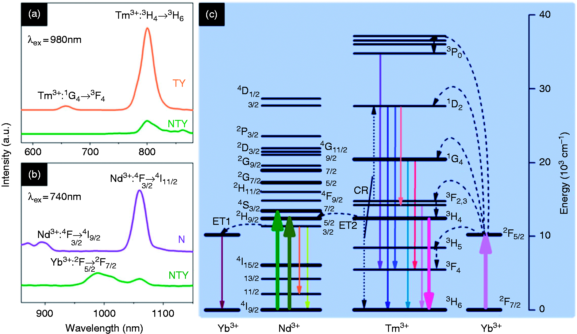
UC emission spectra of NTY and TY under 980 nm excitation (a); DC emission spectra of NTY and N under 740 nm excitation (b); energy level diagrams of Nd3+, Tm3+, and Yb3+ ions (c). Reproduced with permission from Zhou et al. 62 Copyright 2013 American Chemical Society.
In addition, Liu obtained the desired dual-mode fluorescence emission (UC and DC) by controlling the ET process through changing the concentration of Yb3+ in Gd2O3:Yb3+/Er3+. 63 The UC fluorescence is caused by the transition of Er3+ when receiving the energy from Yb3+. The observation of an NIR fluorescence (950–1100 nm) was resulted from the 2 F5/2 → 2 F7/2 transition of Yb3+ in the DC process under the excitation of 380 nm.
Improvement of fluorescence intensity
Design of core-shell structure
Due to the size-dependent fluorescence effect, more and more core-shell structures have been adopted to endow NPs with various functions and improve the fluorescence intensity of NPs. Generally, the shell structure whose lattice constant is similar to the core structure not only increases the size of the fluorescent NPs but also reduces the surface quenching caused by the high-energy vibration bonds of the organic groups in the solvent. At present, the shell structures of NPs can be divided into two sorts: inert shell and active shell.
Inert shell
The inert shell refers to the shell that undoped RE ions, which is classified as homogeneous shell and heterogeneous shell. Being coated with the inert shells could improve the ET efficiency, fluorescence intensity, and quantum yield of NPs by inhibiting the non-radiative transitions caused by surface defects and cross-relaxation between the doped ions. Prasad coated an undoped inert layer NaGaF4 on the nanoparticle NaYF4:Tm3+ to achieve the efficient NIR emission. The growth of the inert shell enhanced the fluorescence intensity of the original NPs by three times. Furthermore, the NaYF4:Tm3+@NaGdF4 is single exponential decay and of long lifetime due to the suppression of surface quenching effects, whereas the NaYF4:Tm3+ NPs show biexponential decay. 64 Since the fluorescence intensity and lifetime of NPs are largely affected by their sizes and surface-to-volume ratios, Damasco et al. 65 explored a method of growing a thin NaYF4 shell on the core NaYbF4:Gd3+ 30%/Tm3+ 0.5% NPs, which makes the fluorescence intensity of this NPs increase by 350 times. There are also some other types of shell materials, and the oxide shell was selected by Liu to coat on the Gd2(WO4)3:Yb3+/Ho3+ NPs via one-pot co-precipitation method. 66 This inert shell could inhibit effectively surface defects and protect the internal luminescent core from surrounding environment, thus significantly improving the fluorescent intensity of NPs (Figure 3). And the effect of the thickness of SiO2 shell on the fluorescent intensity of the NPs has been systematically studied.
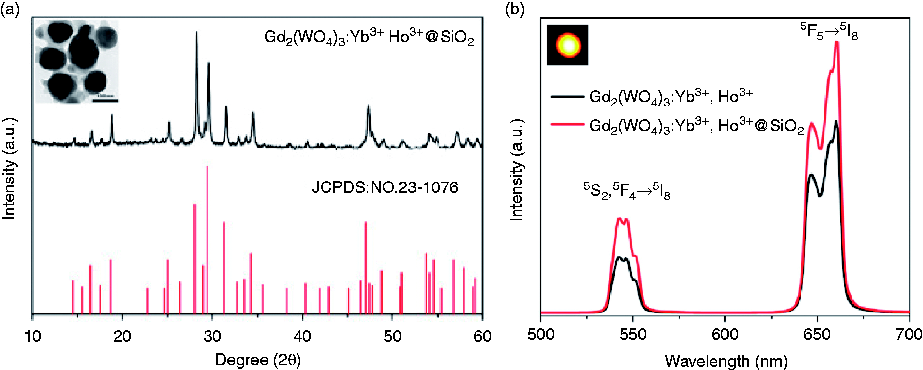
The XRD pattern of Gd2(WO4)3: Yb3+/Ho3+@SiO2 UCNPs. The standard data for Gd2(WO4)3 (JCPDS card No. 23–1076) is shown as reference. And the inset is the TEM image of sample (a). The emission spectra of Gd2(WO4)3: Yb3+/Ho3+ and Gd2(WO4)3: Yb3+/Ho3+@SiO2 UCNPs under 980 nm irradiation (500 μg/mL, 0.5 W/cm– 2 ). Inset is the digital photo of sample upon 980 nm irradiation (b). Reproduced with permission from Liu et al. 66 Copyright 2019 Elsevier.
Active shell
Active shell is a shell doped with active ions. In general, the RE ions with high absorption cross section are doped in the shell, which can transfer excitation energy to the luminescence center in the core, thus greatly improving the fluorescent intensity of NPs. In addition, the active shell can also adjust the excitation wavelength of NPs to the desired band through ET process, thus expanding the application of NPs. By coating an active shell NaGdF4:Yb3+ on the NaYbF4:Tm3+ NPs via a pyrolysis method, Wang increased the fluorescent intensity of the NPs at 800 nm by 7.2 times. 67 Changing the core material, the NaErF4:Tm@NaGdF4:Yb constructed by Xu showed a higher fluorescence enhancement. 68 The active shell NaGdF4:Yb coating on the NaErF4:Tm core enables the efficient ET from NIR photon to upconverting band and suppress the fluorescence quenching caused by surface defects and associated organic ligands simultaneously (Figure 4). This core-active shell NPs exhibit higher (∼20 times) fluorescence intensity than the core-inert shell NPs NaErF4:Tm@NaGdF4, and the quantum yield was up to 3.71%.
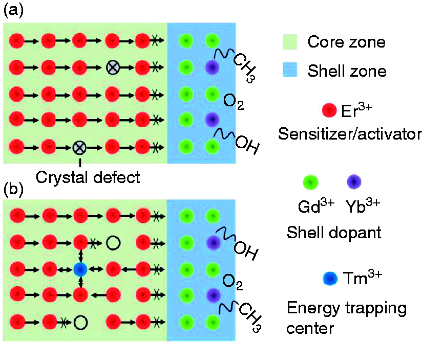
Typical strategy of preventing energy migration to surface defects and eliminating luminescence quenching in Er3+-based nanoparticles through active shell coating (a). Proposed energy transfer mechanism of using Tm3+-mediated trapping center for enhancing upconversion in Er3+-based nanoparticles (b). Reproduced with permission from Xu et al. 68 Copyright 2018 Royal society of chemistry.
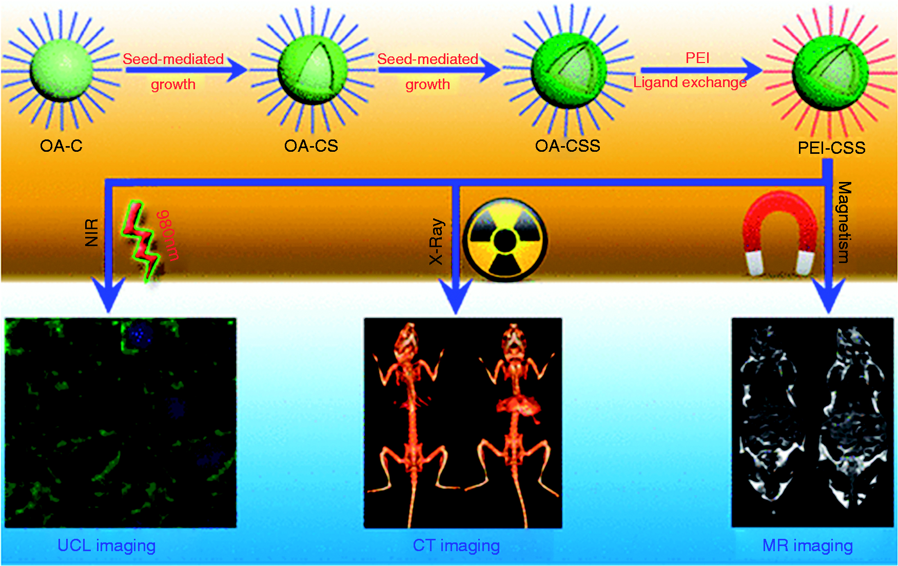
In addition to the double-layer core-shell structure, the multi-layer core-shell structure has also been designed to improve the fluorescence intensity. 69 Li reported a new type of NIR emission NaYbF4:Tm@NaYF4:Yb@NaNdF4:Yb hierarchical-structured NPs that emit at 696 and 980 nm when excited at 808 nm (Figure 5). 70 The ET between Yb3+ and Nd3+ in the shell layer not only changes the excitation light from 980 to 808 nm, but also avoids the thermal damage to the biological tissue. The presence of intermediate shell blocks the CR of Tm3+ and Nd3+, and the sensitizer-rich core facilitates the efficient ET to Tm3+, thus increasing the fluorescence intensity significantly.
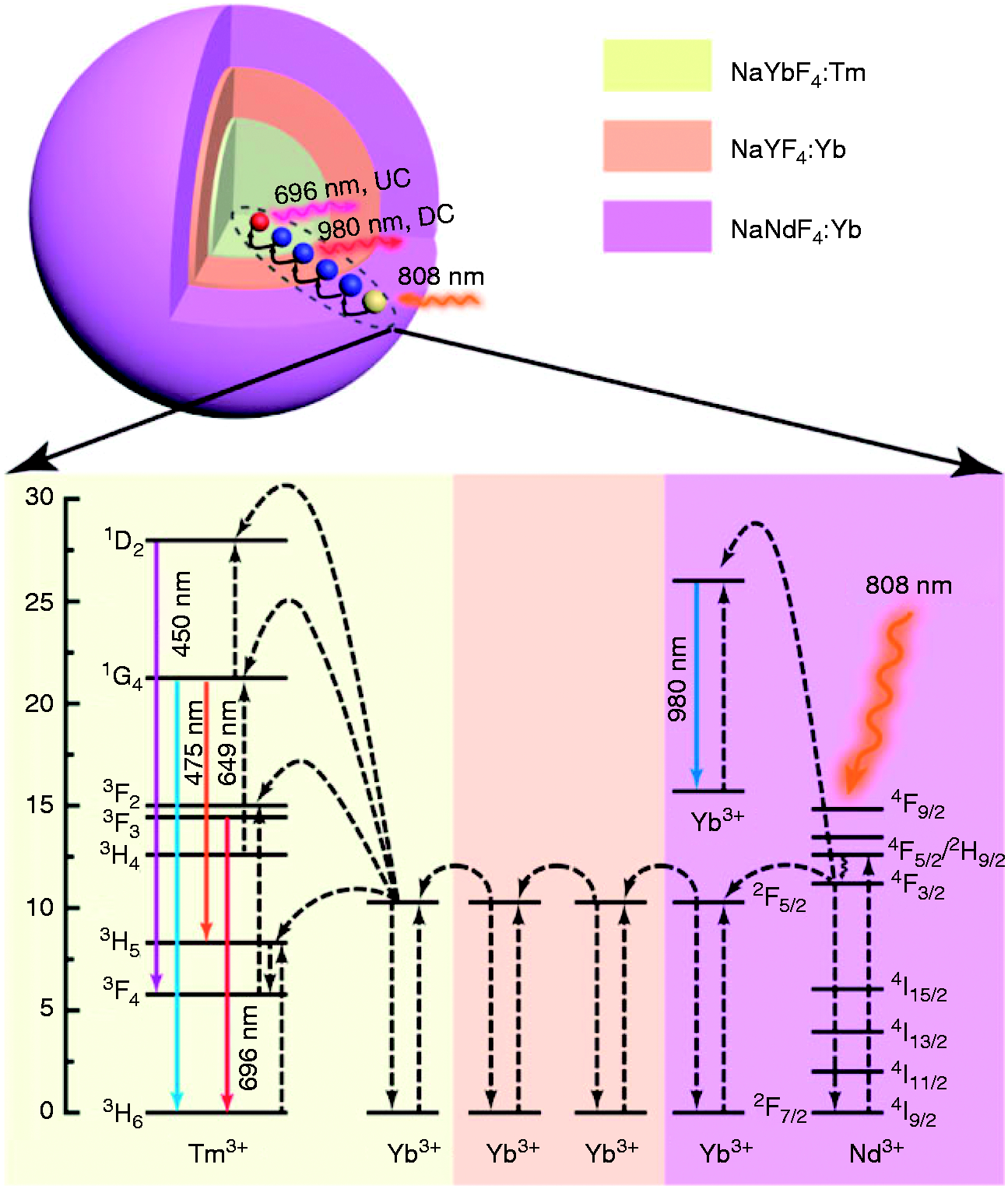
Schematic design (top) and energy-level diagram (bottom) of NaYbF4:Tm@NaYF4:Yb@NaNdF4:Yb(CSS) nanoparticle for dual modes of NIR emission under 808 nm excitation. Reproduced with permission from Li et al. 70 Copyright 2016 American Chemical Society.
Introduction of foreign elements
The application of RE-doped NPs in the biological field is limited by the low fluorescence efficiency. A strategy of doping foreign elements could improve the fluorescence efficiency.71–77 According to the selection rule

Improve the water solubility and biocompatibility
When Ln3+-doped fluorescent NPs are applied in the field of bioimaging, the first problem to be solved is to improve their water solubility and biocompatibility. This is the guarantee that the materials can circulate to the target area through the biological vein and perform fluorescence imaging. But it often has many limitations in the practical application. (1) Organic solvents are often used as dispersants in the preparation process of Ln3+-doped fluorescent NPs, so the hydrophobic organic groups are bonded on the surface of NPs, which makes the prepared NPs insoluble in aqueous solution and hard to be compatible with biological tissues. (2) Generally, the prepared Ln3+-doped fluorescent NPs are very small in size and have high surface energy, so it is easy to agglomerate, which could result in the decrease of water solubility. In order to increase the water solubility and biocompatibility of NPs and reduce the interaction between NPs and biological environment, the surface functionalization is always used to change the surface properties of NPs. Since 2008, Zhang and coworkers 81 modified Ln3+-doped fluorescent NPs NaYF4:Yb3+ and Er3+ with PEI and applied them for the first time in vivo imaging. Scientists now use various modifiers and modification methods to functionalize or coat NPs to improve their water solubility and biocompatibility, such as SiO2, PEG, PEI, PVP, PAA, and so on.82–87
SiO2
SiO2-coated NPs are one of the most important platforms in the field of nanotechnology and biomedicine. SiO2 is one of the ideal materials for surface modification of Ln3+-doped fluorescent NPs because of its high stability, high biocompatibility, water solubility, corrosion resistance, and high optical transparency, and it can avoid the potential toxicity of precursors to cells. Viswanathan 88 creatively integrated co-precipitation, polymerization, and sol–gel technology to fabricate a SiO2-coated fluorescent nanoparticle. The coated SiO2 layer greatly improves the water solubility of NPs, making them promising bioimaging contrast agents. SiO2 also plays an important role in reducing the cytotoxicity of NPs. And it also helps to avoid the photoquenching effect of the surrounding environment on the luminescence center, thus improving the fluorescence intensity and stability of NPs. In 2019, Ansari et al. 89 proposed the exploration on constructing a mesoporous multi-layered silica-coated fluorescent Y2O3:Eu NPs by sol–gel methods. The mesoporous SiO2 layer played a key role in perfecting the solubility, biocompatibility, and non-toxicity of the NPs. The experimental results suggested that the SiO2-coated fluorescent NPs are biocompatible with human cells. By a typical Stober method, a thin layer of SiO2 was coated on the RE-doped nanoparticle NaYF4:Yb and Tm by Shi et al. 90 The NPs-SiO2 exhibited an excellent spherical morphology with good water solubility and biocompatibility, which demonstrated the great potential for applying in bioimging. 90
Polyethylene glycol
PEG consists of two distinct groups: one is hydrophilic and polar and the other one is lipophilic and non-polar. When the RE-doped NPs are coated with this amphiphilic polymer, the non-polar oleophilic group of PEG links to the surface of the NPs by the coordination bonds, while the polar hydrophilic group is dissolved in water phase, which improves the water solubility of the RE doped NPs. PEG coating can also reduce the surface tension of NPs, change their surface properties, and make NPs in a more stable state. Because the long chain structure of PEG can form steric hindrance on the surface of NPs, which generates the strong repulsion between NPs in water, thus preventing the agglomeration and increasing the dispersion of NPs. That is to say, lanthanide-doped fluorescent NPs with good water solubility, dispersion, and biocompatibility can be obtained by coating PEG on the surface of NPs, which makes it possible for the application for bioimaging. The lanthanide oxysulfide NPs Ln2O2S:Ln’ 3 + NPs (Ln = Gd, Tb, and Eu; Ln’ = Tb and Eu, 1 and 5%) was fabricated by Lin et al. 91 via a convenient thermal decomposition method. The surface modification of the Gd2O2S:Eu3+ (5%) NPs with mPEG allowed their effective cellular and in vivo animal luminescence imaging with low bio-toxicity. These novel imaging materials with high water solubility and biocompatibility would be potentially useful for luminescence bioimaging applications. 91 Currently, RE-doped NPs have been widely used in bioimaging. Because they are usually prepared in oleic acid solvents, there will be oleic acid ligands on the surface of NPs, which will seriously affect their water solubility and biocompatibility. This problem was well settled by Wong et al. 49 In his study, the oleate-capped ultra-small multifunctional KGdF4:Tm3+, Yb3+ NPs with NIR to NIR emission were further grafted with PEG to endow them with water solubility, which is necessary for future bioimaging applications. For different matrix materials, PEG can also play an important role in material modification. The melanin particles were selected by Hong et al. as matrix materials. 92 After the modification of PEG, the stability was greatly improved. On this basis, Gd3+ could be loaded stably in different PH for the multimodal imaging in living organisms. In a similar way, Deng et al. constructed a kind of deep-tissue bioimaging PEGylated NaLuF4:Yb/Er NPs. 56 Coating with PEG provides this novel nanoparticle with high biocompatibility that would bring more potential for the bio-applications in clinic.
Polyetherimide
PEI is a hydrophilic polymer with good thermal stability. It can be adsorbed on the surface of NPs to control the growth of NPs. At the same time, the amino group in PEI can improve the water solubility and biocompatibility and prolong the half-life of NPs in vein.93–97 Zhang et al. 96 demonstrated the synthesis of the RE-doped NPs NaYF4:Yb,Er (Ex = 980 nm) coated by the PEI for NIR bioimaging for the first time. The PEI–NPs showed visible fluorescence no matter in the human ovarian carcinoma cells or in the body of rats. 55 In 2018, the PEI-modified NPs NaYF4:Yb3+, Tm3+ prepared by Li’s group98 has been applied in sensitive and selective detection of dopamine in biological fluids. A novel sandwich-like core-shell nanoparticle Sr2LuF7:Yb,Er@Sr2GdF7@SrF2(SLF@SGF@SF) (∼8 nm) was fabricated in Chen’s group. In order to meet the requirements of bioimaging, PEI was modified on the surface of SLF@SGF@SF to endow it with great water solubility and biocompatibility. At the same time, this process will hardly affect the size and shape of SLF@SGF@SFs. The results of in vivo experiments demonstrated the potential of SLF@SGF@SFs-PEI as a contrast agent for multimode bioimaging (Figure 6). 99
Schematic illustration of the preparation of PEI-capped SLF@SGF@SF and its bioapplication. Reproduced with permission from Chen et al. 99 Copyright 2017 American Chemical Society.
Others
Bogdan designed a water-soluble NPs NaYF4:Er3+, Yb3+ by an ingenious method. 100 This method obtains the water-soluble NPs by removing the oleic acid coated on the surface through a simple acid treatment process. And the further modification of the NPs with heparin and fibroblast growth factor offers the potential application in bioimaging and detection in biological assays. In addition, Han chose hairpin DNA as the modifier to construct a kind of bifunctional water-soluble NPs based on NaYF4@SiO2-Au with controllable drug delivery and biofluorescence imaging properties (Figure 7). 101 The hairpin DNA molecules tethered on the surface of particle largely improved the dispersibility of the NPs in physiological environment.
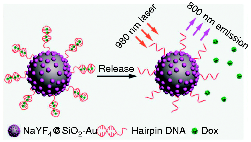
Design of hairpin DNA‐functionalized NaYF4@SiO2–Au nanoparticles for photothermal drug release. Reproduced with permission from Han et al. 101 Copyright 2017 Wiley.
Multimode bioimaging
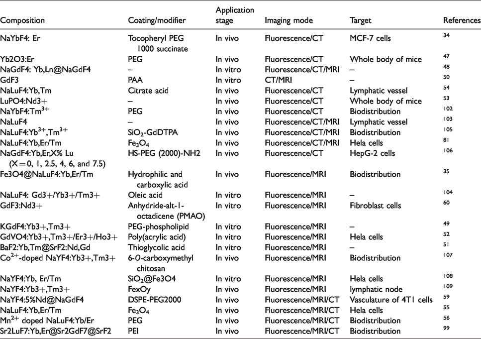
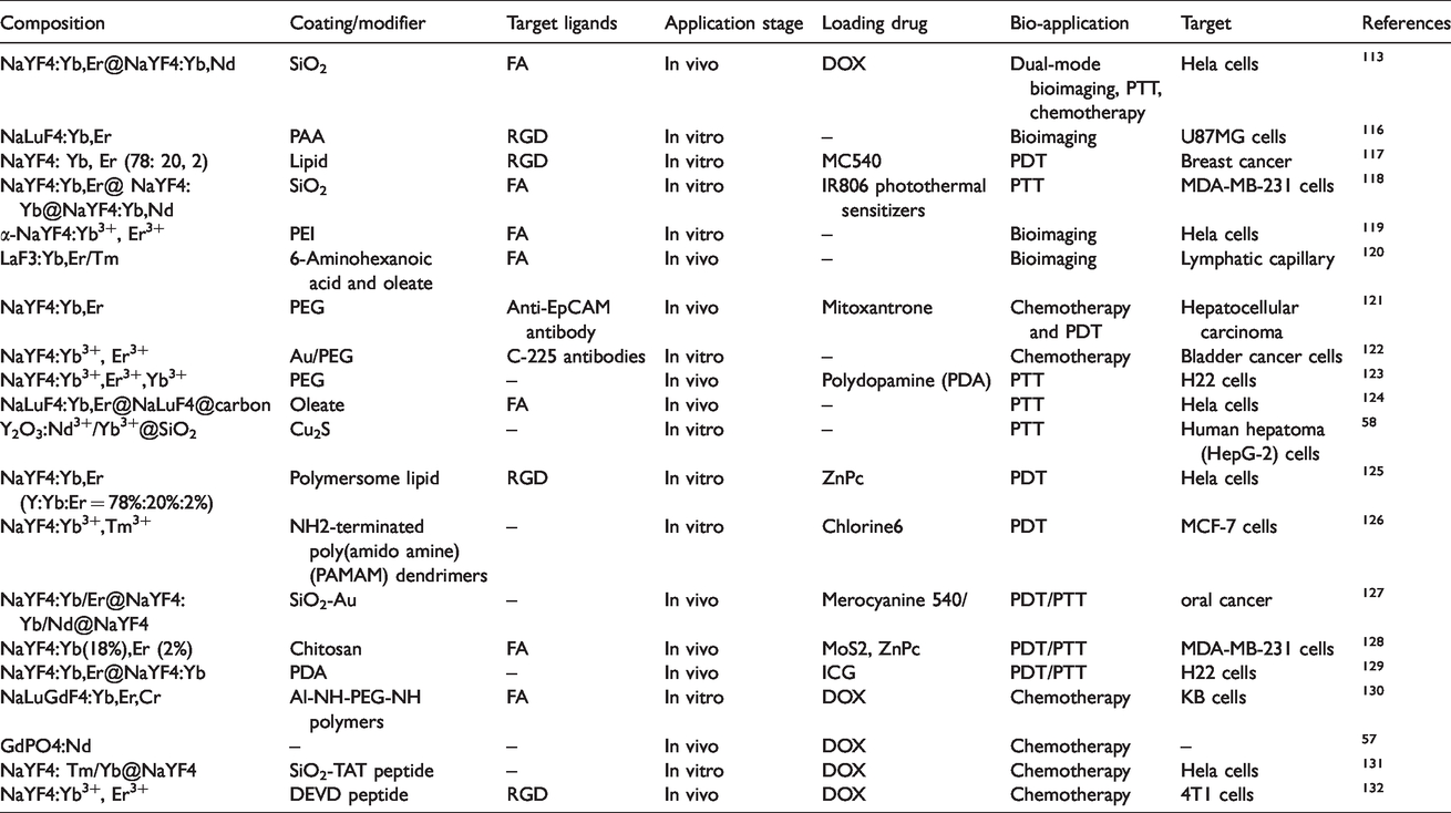
Fluorescence imaging has been widely studied as a convenient, rapid, and high penetration depth biological imaging method. However, a single fluorescent imaging model can no longer meet the current needs of high resolution imaging diagnosis. In order to obtain more complete and high-resolution structural information of biological tissue, various imaging methods are used to collect as many signals as possible from the target to improve the final imaging quality; for example, fluorescence imaging, MRI, CT, and so on.102–104 Table 1 summarizes the features and bioimaging applications of the commonly reported RE-doped NPs.
Summary of RE-doped NPs for multimode bioimaging.
Fluorescence and CT imaging
High-resolution CT imaging requires high X-ray attenuation coefficient. The attenuation coefficient of X-ray depends on atomic coefficient and electron density. Lanthanides have the great potential to block light beams for CT imaging due to the high atomic number. There is a huge gap between biological tissues and RE elements in X-ray absorption coefficient, so that the CT imaging can be achieved with high resolution in vivo. After combined with sensitive fluorescence imaging, it can provide more accurate information in medical diagnosis. Some dual-modality imaging lanthanide NPs have been synthesized and applied in vivo, including NaYbF4, 34 Yb2O3, 47 NaGdF4, 48 GdF3, 50 NaLuF4, 54 and LuPO4. 53 Among these RE elements, Yb plays an important role in imaging. It can not only be used as the sensitizer to enhance the NIR luminescence of NPs but also provide large X-ray absorption for CT imaging. 102 Due to the high atomic number of Lu, a certain concentration of NaLuF4 NPs solution can reach the Hu value which is five times higher than that of the iopromide (a commercial CT contrast agent) of same concentration. 103 Li and coworkers103,105 have realized the high-resolution dual-modality imaging for lymphangioma and Hela tumor with NaLuF4 NPs (Figure 8). They also enhanced the CT imaging signal by 138% via intratumoral injection of core-shell NPs Fe3O4@NaLuF4:Yb,Er/Tm (Figure 9). 81
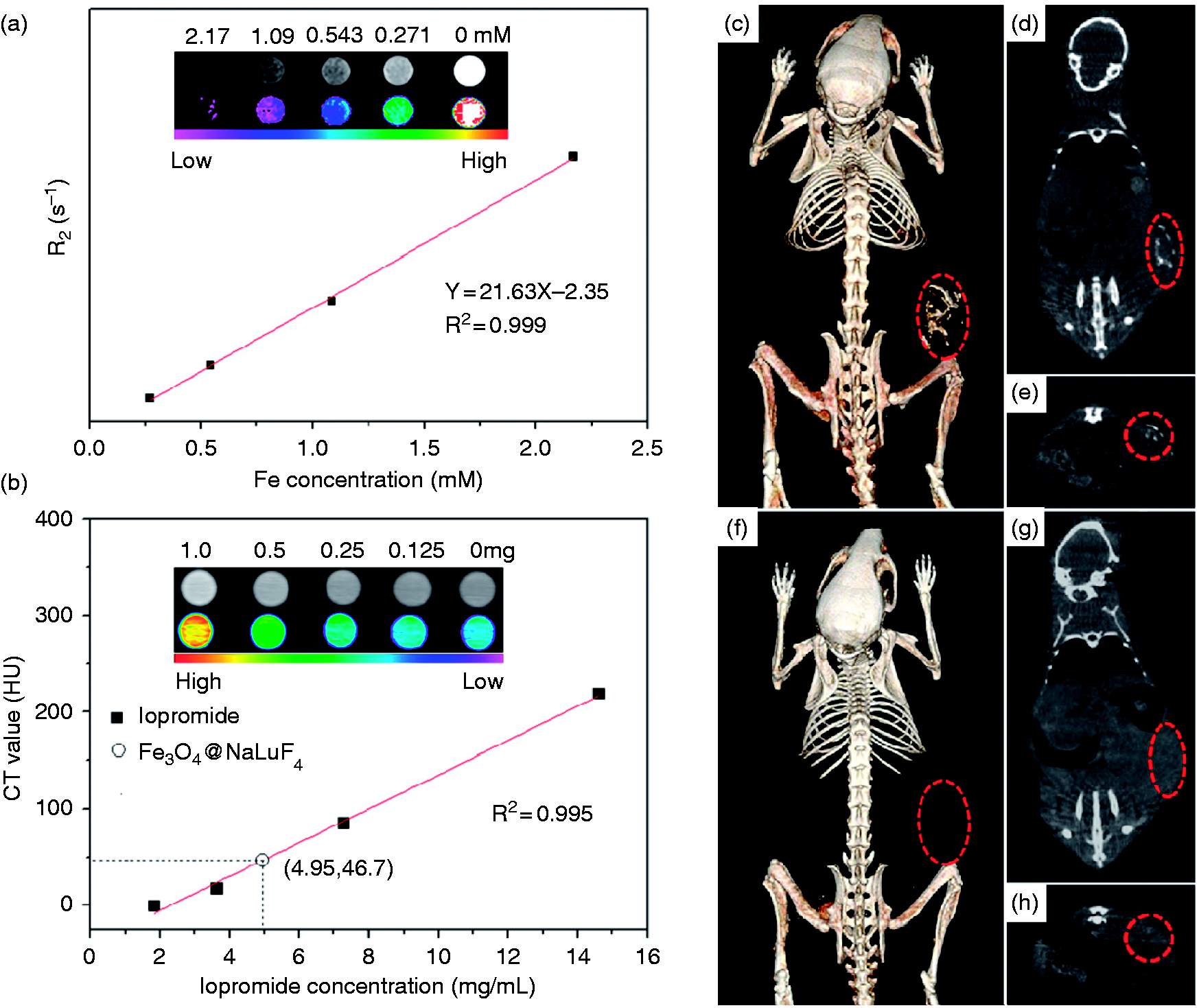
Relaxation rate R2 (1/T2) versus various molar concentrations of MUCNP (2.17, 1.09, 0.543, and 0.271 mM of Fe), inset: the T2-weighted and color-mapped MR images of MUCNP. The CT value (HU) as a function of iopromide concentration (14.6, 7.3, 3.65, and 1.83 mg/mL) (a). The measurement shows that 1 mg/mL of the MUCNP gives an equivalent X-ray absorption as 4.95 mg/mL iopromide. Inset: phantom and colormapped CT images of MUCNP with different concentration of 1.0, 0.5, 0.25, and 0.125 mg/mL (b). In vivo CT volume-rendered (d, f) and maximum intensity projection of corona (d, g), transversal (e, h) images of the tumor-bearing mouse before and 30 min after intratumoral injection. The position of tumor was marked by red circles. Reproduced with permission from Zhu et al. 81 Copyright 2012 Elsevier.
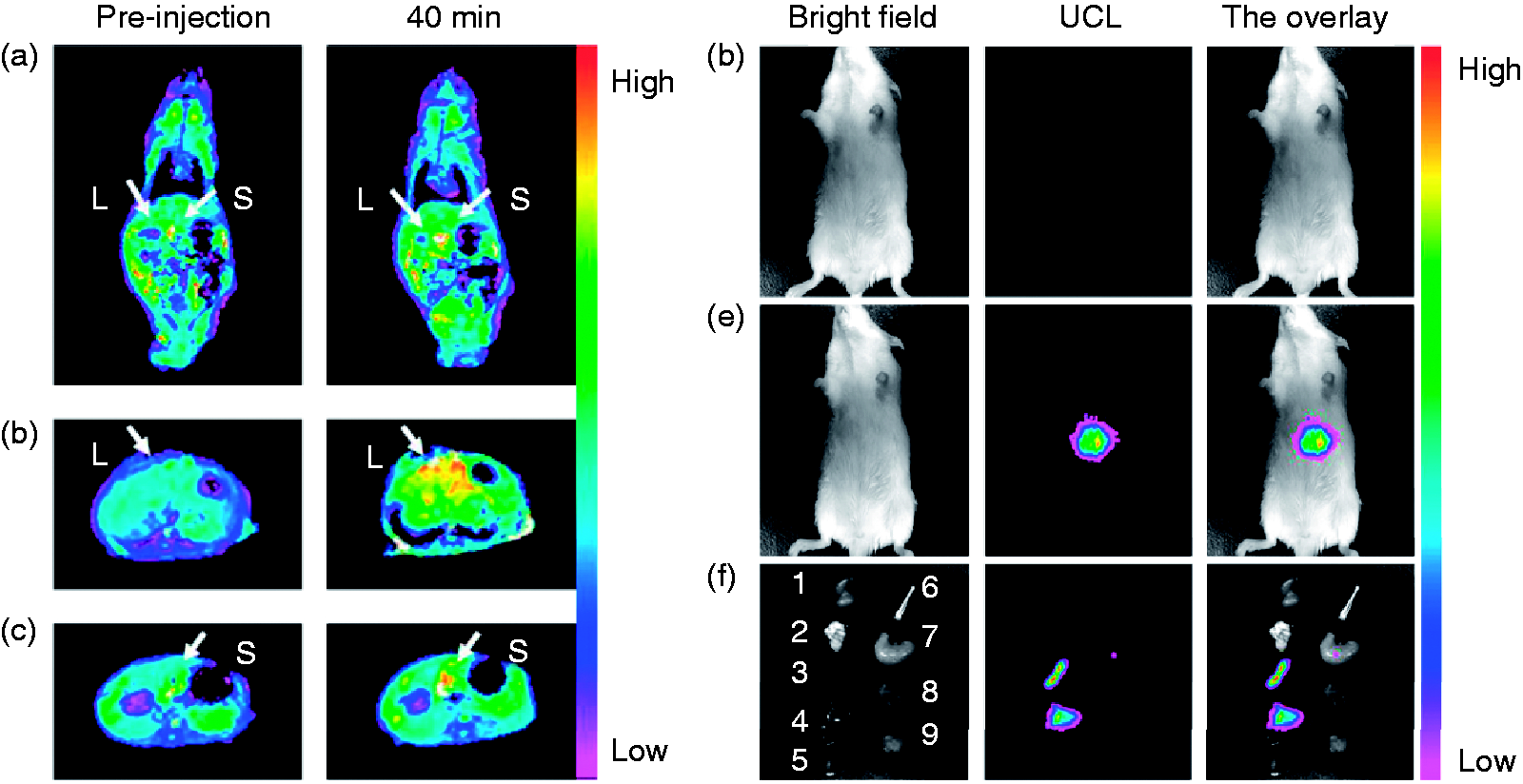
Color-mapped coronal images of the whole body (a) and transversal cross-sectional images (b, c) of the liver (L) and spleen (S) of mice at pre-injection and at 40 min after intravenous injection of AA-NPs at 1.5 mg/kg. In vivo upconversion luminescence imaging of mouse after intravenous injection with and without AA–NPs (d,e). All images were acquired under the same instrumental conditions (power density ≈ 150 mW/cm2 on the surface of mouse). Representative UCL images of dissected organs of a mouse sacrificed 40 min after intravenous injection with AA–NPs (f): (1) kidney; (2) lung; (3) spleen; (4) liver; (5) heart; (6) bone; (7) stomach; (8) intestines; (9) muscle. All images were acquired under the same instrumental conditions (power density ≈ 100 mW/cm2 on the surface of mouse). Reproduced with permission from Zhou et al. 35 Copyright 2010 Elsevier.
Yb can be served as a good CT imaging agent because of its high X-ray absorption. In view of this, a multi-mode imaging nanoparticle NaYbF4: Er was fabricated in Tian’s group for CT/fluorescence dual-mode bioimaging. 34 The observation of strong fluorescence emission results from the transition process of Er3+. In addition, tocopheryl PEG 1000 succinate was used as surface modifier to functionalize the NPs for the first time. It endows the NPs with P-glycoprotein inhibition capability and excellent water-solubility. After loading the chemotherapeutic drug DOX, the as-prepared nanoparticle could serve as a biocompatible dual-mode imaging-guided tumor treatment agent. By a solvothermal method with high-level modulation of both the phase and morphology, Liu constructed a new type of uniform and ultra-small NaGdF4:Yb,Er,X% Lu (X = 0, 1, 2.5, 4, 6, and 7.5) NPs. 106 Its fluorescence emission originated from the Er3+ could be significantly enhanced by doping Lu3+ due to the change of the Yb-Er interionic distance. At the same time, Lu3+ endowed the NPs with high X-ray attenuation coefficient for CT imaging. After being grafted the HS-PEG(2000)–NH2 ligand, this NPs show higher biocompatibility and lower toxicity, which could be applied in the bioimaging and tumor diagnosis.
Fluorescence and MRI imaging
In order to overcome the low resolution and low penetration depth of fluorescence imaging, MRI has been widely used to make up for the deficiency of fluorescence imaging. MRI and fluorescence imaging are two complementary imaging modalities, and they can combine to produce higher resolution, higher penetration depth, and higher sensitivity imaging. Gd is widely used as MRI/NIR dual-modality imaging agent due to its high r1 relaxation and paramagnetism. An NIR/MRI dual-modality, low-toxic, and water-soluble NaGdF4:Tm3+/Er3+/Yb3+ nanoparticle with emission at 800 and 650 nm (under CW excitation at 980 nm) was developed by Zhou and coworkers via hydrothermal and Lemieux-Von Rudloff reagent oxidation methods. 35 The large amount of Gd3+ near the surface provides high r1 relaxation (5.60/s1 (mm)−1) and paramagnetism for MRI. And the fluorescence emission at the NIR band is attributed to the transitions 3H4 → 3H6 of Tm3+. So far, the NaGdF4:Tm3+/Er3+/Yb3+ NPs have been widely applied in mice to achieve MRI and NIR dual-modality imaging (Figure 9). Furthermore, Gd could also be doped into other host materials including NaLuF4: Gd3+/Yb3+/Tm3+, 104 GdF3:Nd3+, 60 KGdF4:Yb3+,Tm3+, 49 GdVO4:Yb3+, Tm3+/Er3+/Ho3+, 52 and BaF2:Yb,Tm@SrF2:Nd,Gd. 51 The host material GdVO4 is selected by Kang for the preparation of multifunctional composites as bioprobe. 52 They filled the modifier PAA into the lanthanide-doped nanoparticle GdVO4:Ln3+ (Ln = Yb/Er, Yb/Ho, and Yb/Tm) by photoinduced polymerization to achieve the pH-dependent delivery in vivo. The fluorescence emission could be tuned by changing the co-doped RE ions. At the same time, the nanocomposites could act as the MRI agent due to the doped Gd3+. The imaging results of cell uptake process demonstrated the potential of this nanoparticle for dual-mode bioimaging. High r2 relaxation ions or compounds can also be used as MRI imaging agent in bioimaging, such as Fe x O y and Co2+.107,108 Xia and coworkers coated NaYF4:Yb3+, Tm3+ NPs with 5-nm thick Fe x O y shell, which provided a T2-enhanced MR imaging with a saturation magnetization of 12 emu/g. 109 Such NPs enable NIR/MRI dual-mode imaging of lymphatic node.
Fluorescence, CT, and MRI imaging
Different imaging modes have different specialties in sensitivity, resolution, penetration depth, and cost, but due to the limitation of the equipment and light source of each imaging modality, there will be some defects in a certain aspect. This will lead to its failure to provide effective and accurate information of biological structure and physiological process to meet the requirements of clinical diagnosis. Therefore, the bioimaging based on RE-doped NPs is developing from single modality to multi-modality. The NPs doped with multiple RE ions (Gd, Yb/Lu/Gd, and Er/Tm/Ho)36,81,109 show excellent bioimaging performance, especially in the fluorescence emission, MR, and X-ray attenuation. These characteristics fueled the NPs’ wide application in the bioimaging as multi-mode imaging contrast agents.110–112 In this research area, Ren’s research group has done an excellent work, in which a small, homogeneous, strong NIR emission core-shell DC NaYF4:5%Nd@NaGdF4 nanoparticle with emission at 1060 nm (under excitation of 808 nm) was synthesized by small nano-cluster medium method. 59 The NPs were successfully applied to demonstrate the variation of tumor vasculature through the NIR II fluorescence imaging in a breast tumor model, and they were also served as contrast agent for MR imaging and X-ray computed tomography imaging to provide complementary anatomic structure of tumor tissue (Figure 10). These results confirmed that the NPs could be served as a new tri-modality imaging nanoplatform for real-time biological diagnosis.
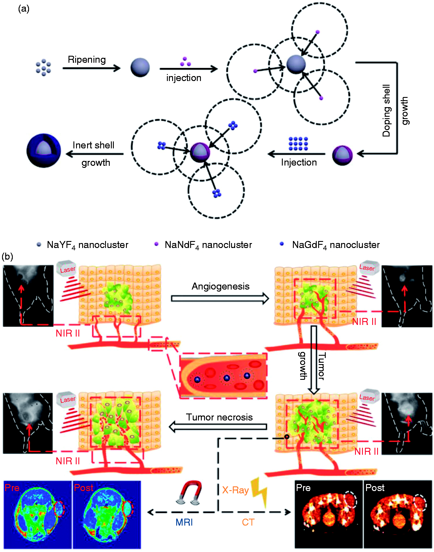
Schematic illustration of the synthesis of NaYF4:5%Nd@NaGdF4 core-shell NCs (a) and their application in NIR II, MRI, and CT imaging of tumor from tumorigenesis, growth, and necrosis (b). Reproduced with permission from Ren et al. 59 Copyright 2018 Elsevier.
A novel step-wise synthetic method was developed by Zhu for fabricating a multifunctional bioimaging agent Fe3O4@NaLuF4: Yb, Er/Tm (MUCNP). 55 The Lu3+ provides high X-ray absorption, and so it can be used for high-resolution CT imaging. The superparamagnetism of Fe3O4 with saturation magnetization of 15 emu/g and T2-enhanced MR effect (r2 = 21.63/s/mM at 0.5 T) provides the MUNCP with high tissue-penetration for MRI. And the NaLuF4: Yb, Er/Tm core can achieve fluorescence imaging under the NIR excitation. In vivo experiments demonstrate that the MUCNP can be successfully applied in multimode imaging. In 2018, Xu et al. 68 reported a multimode imaging nanoparticle based on NaLuF4:Yb/Er doped with PEGylated Mn2+ (PEG–UCNPs). The PEG–UCNPs can not only generate the bright fluorescence signal for fluorescence bioimaging but also be used as T1-weighted MR imaging agent due to its high longitudinal relaxation. The Lu3+ provides the CT imaging modality due to its high X-ray absorption coefficient, which makes the PEG–UCNPs integrate three imaging modalities for the deep-tissue bioimaging. 56
Tumor therapy
Nowadays, RE-doped NPs have developed from the single function NPs of cancer diagnosis to the multi-functional NPs of diagnosis and therapy. 113 The emission of the lanthanide-doped NPs is usually located at a long wavelength, which can convert the absorbed photons into heat energy or reactive oxygen species (ROS). Researchers have used these properties of lanthanide-doped NPs to inhibit and kill tumor cells by means of photodynamic therapy (PDT), photothermal effect, and loading drug molecules. 114 To diagnose and treat various tumor cells, the most important step is to modify NPs to endow them with the ability of tumor targeting. The method is to attach the ligands with targeting ability to vary tumor to the surface of NPs by physical adsorption or chemical bonding, which will not suppress the fluorescence or other signals of NPs while giving them the tumor targeting. At present, FA, RGD, and various antibodies are widely used as target ligands. 115 Table 2 summarizes the targeting ability and therapeutic application of the commonly reported RE-doped NPs.
Summary of RE-doped NPs for tumor treatment.
Tumor targeting of NPs
Tumor-targeting NPs are able to provide pivotal information not only on the location of the cancer in vivo but also on the biological processes of the tumor. The state of the tumor reveals abundant information, including protein kinase activity, hypoxia, angiogenesis, and apoptosis. This could provide clinicians with great information on assessing the diagnosis, therapeutic, and prognosis of cancer patients.133,134 In order to obtain the tumor targeting, the surface of NPs is functionalized, and the active functional groups located in the outer layer of NPs can be utilized to covalently connect some targeted ligands, including FA, RGD, peptides, aptamers, antibody, and so on.135,136
RGD
Receptor is widely used for cancer imaging because the over expression of specific receptors is closely related to cancer progression. Receptor-targeted imaging offers enhanced specificity to tumors of interest and quantitative information on the receptor. Therefore, a substantial amount of effort has been made to develop a receptor-targeted technique. As a tumor-targeting penetration peptide, RGD first accumulates in tumors by initially targeting avb3 integrin receptors, which are specifically expressed in the tumor vasculature and tumor cells. Then it binds to neuropilin-1 (NRP-1), consequently triggering tissue penetration.137–142 Physically binding with iRGD or chemically conjugated to iRGD can not only improve tumor penetration of different NPs but also enhance the tumor-targeting ability through NRP-1-dependent manner.132,143–146 Nowadays, RGD has been widely used as the surface modifier of lanthanide NPs to provide tumor targeting. Yang’s group prepared a sub-20 nm polyhedral cubic NIR nanoparticle NaLuF4:Yb,Er NP (Em = 510–560, 630–670 nm, Ex = 980 nm) coated with PAA by solvothermal method. 116 By the ligand exchange with PAA, the NPs covalently linked to RGD. Based on the high affinity between RGD and integrins αvβ3, the RGD–NPs can be used as a tumor-targeting imaging agents for U87MG tumor cells. Furthermore, RGD can be linked with different photosensitizers and endow the imaging agents with the tumor-targeting therapy at the same time. 147 Wang et al. synthesized a new type of RGD-coated nanoparticle (Em = 570 nm, Ex = 980 nm) consisting of NaYF4: Yb, Er core and targeted RGD polymer shell. 117 The RGD-coated rare-earth NPs provides the advantages of protecting the NPs from catching by reticuloendothelial system and targeting to the breast cancer cells. Besides, it can also be conjugated to photosensitizer to achieve tumor inhibition by photodynamics therapy.
Folate
The folate receptor (FR) is a kind of cell membrane glycoprotein and has been widely used in the medical field as a tumor marker.148,149 It was reported that FR can bind to FA, and its conjugate has high affinity. 150 Normally, FR is found in a low level in normal cells or tissues, but it can be highly expressed in malignant cells, including breast, lung, kidney, brain, and so on.151,152 Therefore, the FR is considered to be a significant therapeutic target, as it could provide an effective method for tumor-targeting therapy by constructing FA-modified NPs for the selective delivery to FR-overexpressing tumor cells. In addition, FA is also an important water-soluble vitamin for organism. 153 To date, some studies have demonstrated that folate can be physically or chemically attached to the NPs for the tumor-targeted imaging and treatment.154–158 A versatile nanoparticle NaYF4:Yb,Er@ NaYF4:Yb@NaYF4:Yb,Nd for bioimaging and tumor treatment with NIR excitation (793 nm) and NIR emission (980 nm) was reported by Lin. 118 In order to obtain tumor targeting, mesoporous silica was coated on the surface of the NPs. Then, the positively charged PAH was absorbed to the NPs by electrostatic interaction. Finally, NP@mSiO2–PAH was further conjugated with FA–PEG–NHS by amide bond formation. In vitro studies confirmed that the NPs could be uptaken by the MDA-MB-231 cells for NIR fluorescence imaging and tumor-targeting therapy. Yang designed a novel NIR α-NaYF4:Yb3+, Er3+ (∼130 nm) nanoparticle with porous shell and hollow internal structure, which can be used for bioimaging and loading drugs by a surface etching and hydrothermal ion-exchange method. 119 The free amino groups on the surface can conjugate to FA through amide reaction to endow the NPs with tumor-targeting ability. The in vitro experiment demonstrated a high uptake amount of NaYF4-PEI-FA-FITC in Hela cells, so that the NPs achieved a high-contrast in vitro cell imaging with negligible background. Using the same doped RE ions but different host materials, a new one-step synthesis method to prepare water-soluble high-quality LaF3:Yb,Er/Tm NPs by a hydrothermal reaction assisted with binary cooperative ligands was established in Cao’s group. 120 The 6-aminohexanoic acid and oleate used in this experiment can not only control the growth and crystallization of NPs, but also be further conjugated with FA for tumor targeting imaging of KB cells.
Antibody
Antibody, a biologically active ligand that could be used to overcome the nonspecific targeting and increase their selectivity for tumor cells by the binding process of antigen and antibody. It is a protective glycoprotein produced by the stimulation of an antigen and it belongs to immunoglobulin. 159 So far, many antibodies have been used as targeting ligands of RE-doped NPs to improve the targeting ability for different tumors, so as to achieve the purpose of diagnosis and treatment.160–162 Epithelial cell adhesion molecule (EpCAM) is a general biomarker of tumor stem cells, which is mainly applied to reflect the proliferation and apoptosis of hepatocellular carcinoma. So, Han and coworkers selected the anti-EpCAM antibody to achieve the tumor targeting. 121 In this study, an RE-doped nanoparticle mitoxantrone (MX)-NaYF4:Yb,Er was fabricated for multiplex imaging-guided treatment. The MX as an antitumor drug and a photosensitizer used here provides the PDT and chemotherapy. The surface of NPs was grafted by anti-EpCAM antibody, which endows the NPs with tumor targeting for the hepatocellular carcinoma. In vivo and in vitro tests demonstrate the potential of this MX–NPs-anti-EpCAM for the future clinical application. In order to improve the efficiency of targeted binding by antibody, Cho constructed an imaging-guided therapeutic UCNPs with shell of Au nanorod by PEGylation process. 122 The C-225 antibody was further attached to the NaYF4 NPs-Au to target epidermal growth factor receptor (EGFR) overexpressed bladder cancer cells. Within 2 h of incubation, it can target binding to EGFR-positive cancer cells rather than EGFR-negative cells. On this basis, it can also achieve tumor treatment by optoporation-assisted chemotherapy. Urothelial carcinoma cells is a kind of tumor with high expression of Glypican-1. Considering this, Glypican-1 monoclonal antibody MIL-38 was used by Polikarpov to target urothelial carcinoma cells. 163 In this method, silica is first coated on the NaYF4:Yb,Er NPs, and then the silica-specific solid-binding peptide is attached to the silica shell to combine anti-Glypican-1 antibody MIL-38, which could well mediate the targeted binding of NaYF4:Yb,Er NPs to Glypican-1 positive urothelial carcinoma cells.
Treatment
Photothermal therapy
Photothermal therapy (PTT) is an effective treatment for tumor, which uses photothermal materials to convert exciting light into heat to achieve thermal ablation of cancer cells. PTT has attracted extensive attention in recent years due to its non-invasive, simple process, and less damage to normal tissues. However, the existing photothermal materials often lack tumor targeting and imaging guidance properties such as MoS2, 164 CuSx, 165 Bi2S3, 166 Au NPs, 167 and so on. Therefore, it is of great significance to develop new photothermal materials with great tumor-targeting property for PTT. The RE-doped NPs can provide the fluorescence guidance for tumor treatment due to their excellent fluorescence properties. 168 A new type of multifunctional core-shell NPs doped with Nd3+ for imaging-guided PTT was constructed by Ding. 123 It has PDA core and UCNPs shell NaYF4:Yb3+, Er3+, and Yb3+. The PDA can be served as a photothermal medium to provide high photothermal conversion efficiency and significant anti-cancer effect on H22 cells. At the same time, it can also endow the NPs with excellent biocompatibility. It is known that if the thermal effects of the photothermal medium produce excessive local temperature, it can not only inhibit the growth of cancer cells but also damage normal surrounding tissues. In order to minimize the damage of photothermal effect on normal tissue, Zhu monitored the real-time local temperature during treatment process based on the temperature-feedback NPs NaLuF4:Yb,Er@NaLuF4@carbon (csUCNP@C). 124 The principle is that when stimulated by 730 nm laser, the carbon shell, as the photothermal medium, can produce thermal effect to kill the csUCNP@C-labeled tumor cells and transmit the heat to the core RE-doped NPs simultaneously. By analyzing the luminescent spectrum, we can obtain the local temperature change so as to control the photothermal treatment process to avoid thermal erosion to the cells or tissues not labeled by csUCNP@C. In 2018, a core-shell photothermal agent Y2O3:Nd3+/Yb3+@SiO2@Cu2S (YRSC) was constructed by Zhang’s group. 58 The core Y2O3:Nd3+/Yb3+ can not only generate bright fluorescence but also transfer the energy of 808 nm excitation to the shell Cu2S to generate thermal effect for PTT. To confirm the heating effect of YRSC NPs, the in vitro tests of thermal imaging and ablation of YRSC to Escherichia coli and HepG-2 cells were carried out. The results suggested the potential of YRSC for applying on PTT and NIR imaging.
Photodynamic therapy
PDT is a potential therapeutic method for many kinds of tumors cells.169–171 In PDT, the interaction of light, oxygen, and the photosensitizer could generates ROSs which can cause oxidative damage to tumor cells and tissues.172,173 Compared with conventional tumor therapies (chemotherapy, radiation, and surgery), the advantages of PDT are its minimally invasive therapy nature and fewer side effects.174,175 Furthermore, the photosensitizers could also act as fluorescent probes for tumor-specific fluorescence imaging.176–178 Photosensitizers are considered to play a decisive role in PDT. An ideal photosensitizer should be non-toxic without irradiation. It can specifically accumulate in tumor tissues and has intense absorptions in the phototherapeutic wavelength window (650–900 nm) for better tissue penetration depth as well as high ROSs generation efficiency.179–181 In order to achieve excellent photodynamic treatment and target imaging of tumor cells, many PDT agents including semiconductor QDs, 182 fullerene C60, 183 metal NPs, 184 and TiO2 185 have been combined with lanthanide-doped NPs for constructing the image-guided tumor-targeting therapeutic agents. 186 A new type of “nanodumbbell” NIR NaYF4@lipid@polymersome nanoparticle with RE NPs core and hydrophilic polymersome lipid shell was designed in Wang’s group. Such polymersome can load photosensitizer ZnPc in large quantity and protect the internal structure from non-specific absorption and corrosion in the process of transmission. The NaYF4 core can convert NIR light with high penetration depth into visible light to ZnPc for PDT (Figure 11). The results of ROS production test and PDT test in Hela cells suggested that this nanodumbbell could provide a new NIR diagnosis and treatment nanoplatform. 125
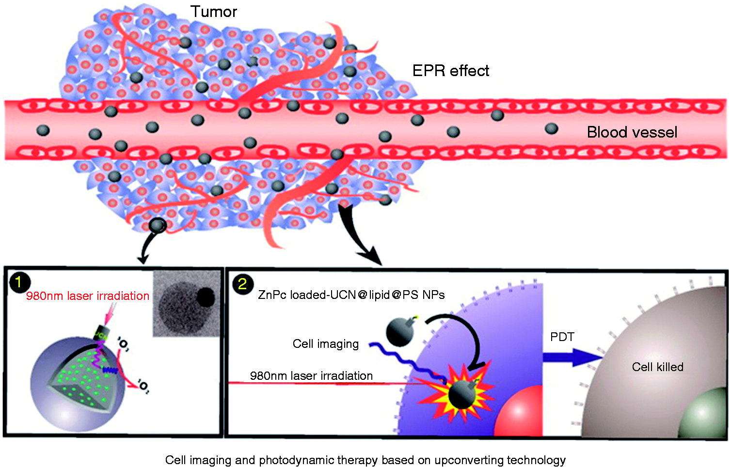
A schematic illustration shows applications of the triple-layer nanoparticles as NIR remote controlled-release nanocarriers for cell imaging and photodynamic therapy. Reproduced with permission from Hou et al. 125 Copyright 2017 Elsevier.
In 2017, Wang et al. linked a water-soluble NIR NPs NaYF4:Yb3+, Tm3+ to NH2-terminated PAMAM dendrimers (G4) and chlorine6 (Ce6) by a layer-by-layer absorption method. 126 The singlet oxygen is generated by Ce6, and the accumulation of singlet oxygen in human breast cancer cells can be regulated by changing the excitation time. When excited by 980 nm laser, the only less than 40% of cells survived under a low Ce6 concentration of 2.5 × 10⁻⁷ M. Therefore, the NPs have great potential as the NIR-responsive PDT agents for tumor treatment.
Dual treatment modality
It has become a mainstream research direction to treat tumor by light because of its high penetration, high sensitivity, and low damage. At present, PTT and PDT are two popular models of tumor treatment, which can induce apoptosis by generating heat effect and ROS. By combining photosensitizer and photothermal agents in one system, PTT/PDT dual therapy modality could supply a more effective therapeutic outcome than either therapeutic modality alone. In 2018, an NIR RE-doped nanoparticle NaYF4:Yb(18%), Er (2%) combined with mesoporous silica shell-coated gold nanorods (AuNR@mS) was constructed by Liu et al. 106 via electrostatic adsorption. When excited by light, NPs can transfer energy to Au and produce thermal effects for PTT. The photosensitizer Merocyanine 540 (MC540) could be loaded in the mesoporous silica layer to generate ROS for PDT, and this effect is further enhanced by the surface plasma resonance of the Au nanorods. 127 Using different photosensitizers and photothermal agents, Han et al. 128 designed a new nanostructure by covalently linking FA and oleic acid on the NPs and then loaded the NPs with oxalic acid. Such a nano system integrates PTT and PDT. It has the fluorescence imaging guidance and tumor-targeting capabilities, which is a versatile nanoplatform for diagnosis and treatment. Liu et al. 129 integrated the tumor treatment (PTT/PDT) and diagnosis (bioimaging) in an RE-doped nanosystem PDA-shelled NaYF4:Yb,Er@NaYF4:Yb, which is able to load ICG molecules by Π–Π stacking, hydrophobic interaction, and electrostatic adsorption. The ICG in this nanosystem could produce both ROS and photothermal effect under the excitation at 808 nm. In vivo and in vitro experiments show its significant therapeutic effect on the H22 tumor, revealing the great potential of application as an imaging-guided dual-mode tumor treatment agent. 129
Drug delivery
Drug-loaded NPs are a combination of nanomaterials and modern medicine. Because the volume is smaller than cells, NPs can be absorbed by tissues and cells, so that the drug-loaded NPs can be transported to the lesion area for therapy. The drug-loaded NPs are mainly composed of carrier materials, therapeutic drugs, and targeted ligands. 187 The carrier materials must have high loading capacity in order to load enough drugs for cancer therapy. For example, mesoporous materials and polymeric micelles are often selected as drug-loaded materials because of their high porosity, which can load a large number of drugs. Among these carriers, the nanoplatform based on RE NPs not only has the ability of loading drugs, but also can track the drugs by bioimaging. It could provide vital information for clinical diagnosis. Furthermore, the drug release can be controlled efficiently because the drug loading system could respond to the endogenous or exogenous stimulus including the pH, light, and so on. 188
pH-responsive drug-loaded NPs
The pH-responsive drug-loaded NPs can greatly improve the drug utilization. Its significant advantage is that it can be delivered without leakage, absorption, or corrosion in physiological environment. The microenvironment of tumor is usually acidic, because the pH of endosomes and lysosome is between 4 and 6. So, at present, many pH-responsive drug-loaded NPs for tumor treatment have been widely studied. In 2019, Tawfik et al. 130 designed a stable and biocompatible NIR NaLuGdF4:Yb,Er,Cr NPs (Ex = 980 nm) modified with Al-NH-PEG-NH-FA polymers and explored it for the successful encapsulation and release of the anticancer drugs doxorubicin (DOX) (Figure 12). The encapsulation efficiency can reach a maximum of 83% in the PBS, and the drug-loading capacity was determined to be 18.3% due to the large cavities of the materials and amphiphilic polymer shell. The loaded DOX can be released efficiently in a highly controlled and selective pH-responsive way by FR-mediated endocytosis. It improved the capability of DOX-loaded NPs to induce apoptosis and abrogation of KB cells. Furthermore, they quantify the uptake and the accumulation of DOX inside KB cancer cells by flow cytometry. The results indicate the efficient cellular uptake of this NPs.
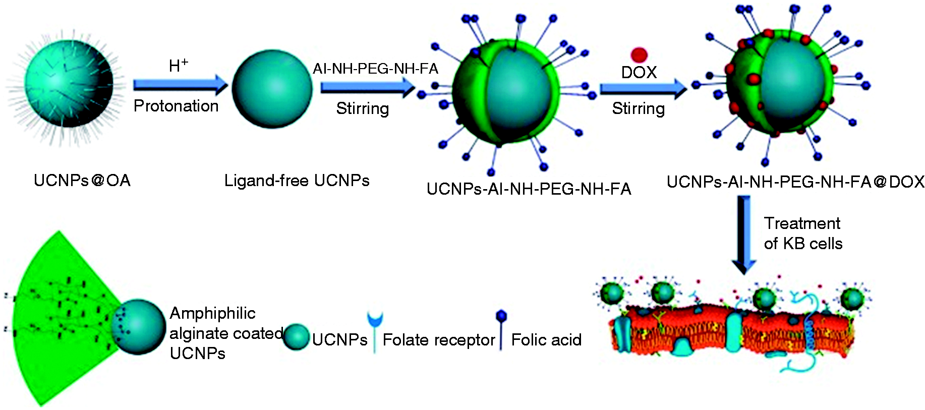
Schematic illustration showing the preparation of UCNPs capped with the Al-NH-PEG-NH-FA polymer and subsequent bio-applications. Reproduced with permission from Tawfik et al. 130 Copyright 2018 Elsevier.
In 2018, Yang et al. 57 and coworkers have made innovative use of GdPO4:Nd NPs as matrix materials. By self-sacrificing template method in which cetyltrimethylamine bromide is served as structure-directing agent, they prepared the egg yolk-shell structure GdPO4:Nd, which can be used as drug carrier to respond to the changes of pH. The NPs can be further linked to anticancer drug DOX by electrostatic interaction. The yolk-shell structure of GdPO4 can provide a large space for loading DOX (Figure 13). In the acidic environment of tumors, the surface of NPs will become neutral due to the protonation of hydroxyl groups, which will weaken the electrostatic interaction between NPs and DOX, thus leading to the controlled release of DOX in the tumor cells.
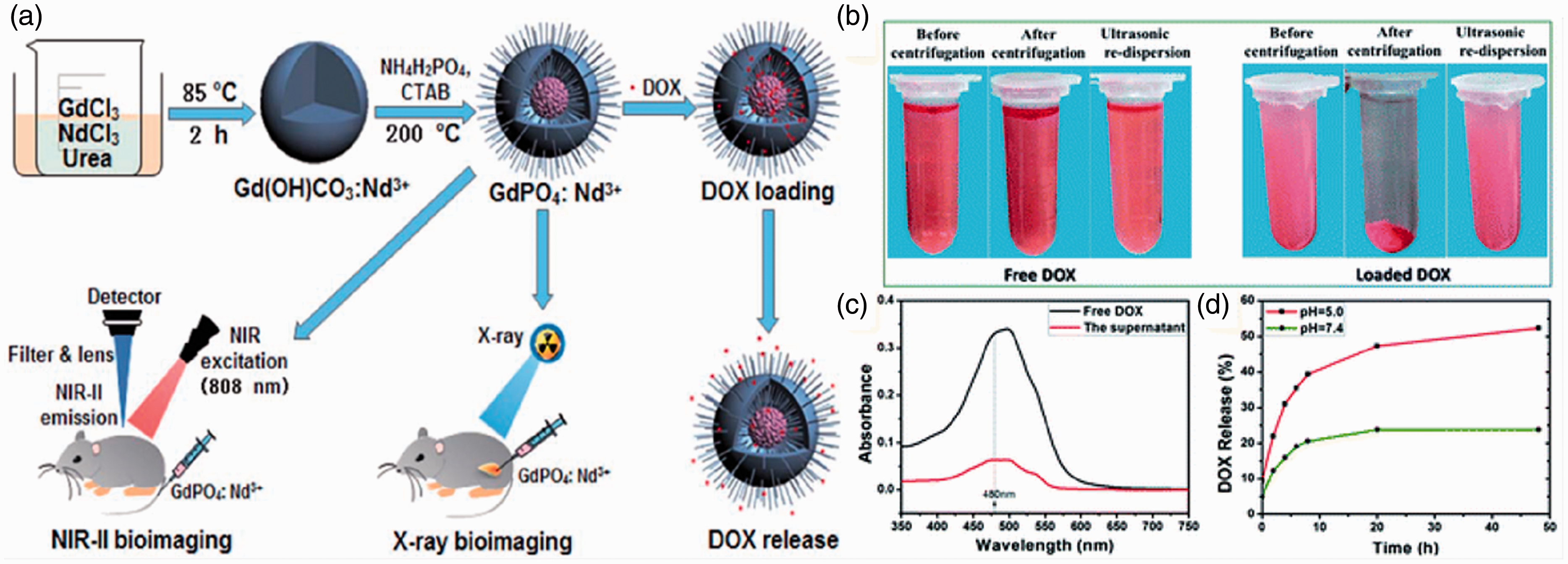
Schematic illustration of designing the theranostic GdPO4:Nd3+ nanoprobes for in vivo NIR-II/X-ray bioimaging, and pH-responsive drug delivery (a). pH-responsive DOX-loading and releasing properties: digital photographs of free DOX and DOX-loaded samples after centrifugation and ultrasonication (b); UV–vis spectra of free DOX (black) and the supernatant transparent liquid of centrifuged DOX-loaded solution (red (c)); pH-responsive DOX release from the DOX-loaded samples over time in PBS at pH values of 5.0 (red) and 7.4 (green) (d). Reproduced with permission from Yang et al. 57 Copyright 2018 Royal society of chemistry.
Light-responsive drug-loaded nanoparticles
Another way to prevent premature release of drugs before delivery to the target areas or tumor cells is to construct photoresponsive drug-loaded NPs. Using NIR light stimulation can provide highly controlled drug release, and it can avoid the damage to biological tissues and interference of scattering, absorption, and autofluorescence from biological tissues. Liu and coworkers coated a shell of mesoporous silica on the NaYF4:Tm/Yb@NaYF4 NPs to obtain a pore network for loading anticancer drugs DOX. 131 Then, the NPs were modified with azobenzene molecules to provide the ability of light-responsive drug release. Under the excitation light of 980 nm, the amount of the released anticancer drug DOX can be well controlled by adjusting the intensity or time of NIR light irradiation while being delivered to the Hela cells for treatment. Furthermore, PDT can also be triggered while the drug is released by the NIR excitation. A versatile tumor-targeting light-responsive drug-loaded nanosystem was designed by Zhao’s group. 132 It consists of the NaYF4:Yb3+, Er3+ NPs, caspase-3-responsive DOX prodrug, photosensitizer, and tumor-targeting ligand cRGD-PEG-DSPE. Upon cellular uptake and NIR irradiation, the caspase-3-responsive drug release can be induced, and the visible light emission of UCNP could excite the photosensitizer to produce ROS for PDT (Figure 14), thus accomplishing the NIR-triggered PDT and chemotherapy to the tumor cells.
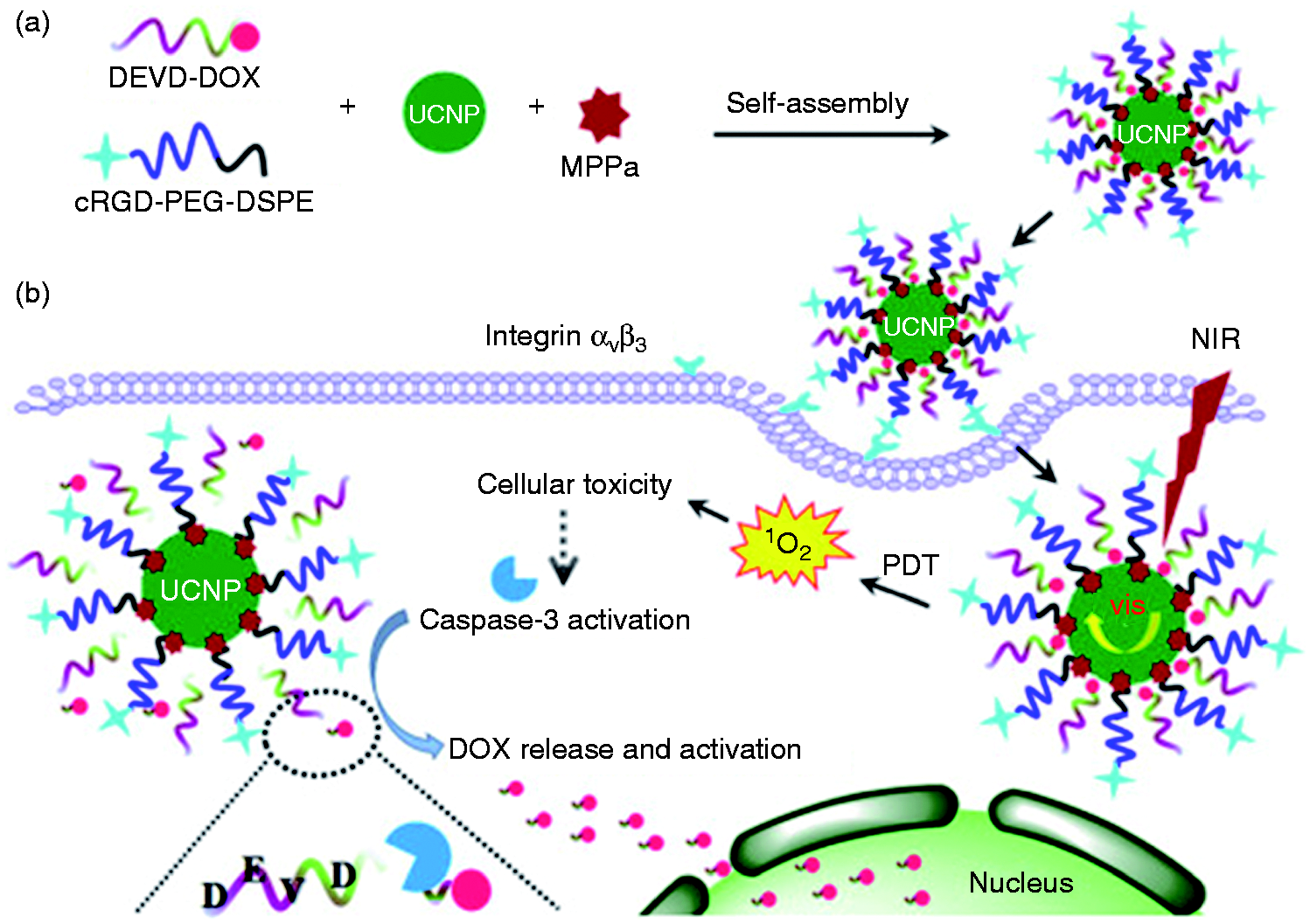
Schematic illustration of NIR-triggered high-efficient photodynamic and chemo-cascade therapy: (a) the aqueous self-assembly and fabrication of caspase-3 responsive functionalized upconversion nanoparticles tethered with anticancer doxorubicin (DOX) (CFUNs, MPPa/UCNP-DEVD-DOX/cRGD); (b) proposed mechanism of NIR-triggered photodynamic therapy (PDT) and the concurrent activation of caspase-3, which further mediated the intracellular DOX release and drug activation for cascade chemotherapy based on CFUNs. Reproduced with permission from Zhao et al. 132 Copyright 2017 Elsevier.
Conclusion and outlook
This review summarized the properties, structures, principles, and methods of improving the fluorescence intensity of RE-doped NPs as well as the progresses in the field of biological imaging and tumor treatment. Fluorescence imaging technology is a powerful tool in biological imaging which can visually display the cell activity and lesion evolution in living animals, and it has been widely applied to medical and biological fields. NIR (700–1700 nm) light has the characteristics of deep penetration due to low absorption, low photon scattering, and low autofluorescence interference. The RE-doped NPs can not only adjust the emission light vary from ultraviolet to NIR by doping different RE ions, but also can be endowed with water solubility, biocompatibility, and tumor targeting for different tumors by surface functionalization. In addition, RE-doped NPs can also be used in the field of drug loading by bonding with anticancer drugs.
Nowadays, more and more RE-doped NPs have been applied in the fields of biological imaging and tumor therapy. However, due to the absorption, scattering, and autofluorescence of biological tissue and biological microenvironment, the main goal is still to improve the signal-to-noise ratio of imaging by enhancing the signal intensity of NPs. Although many methods have been proposed to improve the fluorescence intensity of NPs, such as designing core-shell structure, sensitizing luminescence, introducing exogenous ions, and so on, this is far from enough, because the signal intensity for clinical application is far higher than the current research level. At present, there is a new perspective of improving the signal intensity of biological imaging, which is to explore the intrinsic properties of NPs or endow them with new properties based on various imaging agents, so as to develop new imaging modes. The high quality imaging of the target tissue can be achieved by the cooperation of multi modes. This way can not only improve the spatial resolution of imaging but also improve the depth of imaging and ultimately improve the SNR of biological imaging. The multimodality imaging has become a major research trend.
Many efforts have been made to make RE-doped NPs available for clinical applications. Various modification methods have been used to improve the water solubility and biocompatibility of RE-doped NPs. Although RE-doped NPs have been widely used in small animals, they are rarely used in humans. No matter how much the particle size is and how surface properties of the NPs are, they will more or less leave in the organism such as liver, kidney, and spleen. We cannot estimate whether it will cause a long-term toxicity in the human body, so there is still lot of work to do in the clinical field. One encouraging thing about RE-doped NPs is that they can already target different tumors by binding different ligands according to different needs. This allows us to tailor a variety of diagnostic and therapeutic nanoplatform for different tumors, which is very significant.
Footnotes
Declaration of conflicting interests
The author(s) declared no potential conflicts of interest with respect to the research, authorship, and/or publication of this article.
Funding
The author(s) disclosed receipt of the following financial support for the research, authorship, and/or publication of this article: National Natural Science Foundation of China (No.619350061 and No.61475189), Natural Science Basic Research Plan in Shaanxi Province of China (No.2014JQ8345 and 2019JM-113) and Open Research Fund of Key Laboratory of Spectral Imaging Technology from Chinese Academy of Science (CAS).