
Abstract
A multifunctional targeted nanoplatform combining photothermal therapy and chemotherapy has emerged as a promising strategy for comprehensive therapies of breast cancer. In this study, we constructed human epidermal growth factor receptor 2 (Her2)-targeted gold nanoshelled poly(lactic-co-glycolic acid) hybrid nanocapsules encapsulating perfluorooctyl bromide, superparamagnetic iron oxide nanoparticles, and doxorubicin (Her2-GPDH nanocapsules) as theranostic agent for bimodal ultrasound/magnetic resonance imaging and synergistic photothermal-chemotherapy of Her2-postive breast cancer cells. Her2–GPDH nanocomposites possessed well-defined spherical morphology, and the average diameter was about 296 nm with good dispersion. Targeting assays demonstrated that Her2–GPDH nanocapsules exhibited higher targeting binding to Her2-positive SKBR3 cells than Her2-negative MDA-MB-231cells. The encapsulation efficiency and the loading content of doxorubicin in Her2–GPDH nanocapsules were 39 ± 1.45% and 3.8 ± 0.52%, respectively, and the agent exhibited pH-responsive and near-infrared light-triggered stepwise release behavior of doxorubicin. In vitro, the agent had potential to serve as feasible candidate for ultrasound imaging and T2-weighted magnetic resonance imaging with a relatively high relaxivity. Cell experiments confirmed that the agent had significant photothermal cytotoxicity on SKBR3 cells, and the combined photothermal–chemotherapy could significantly enhance the anti-tumor effect. In summary, the present Her2–GPDH nanocapsules, a novel multifunctional nanoplatform, will offer a new way for early bimodal molecular-level diagnosis and synergistic treatment of Her2-positve breast cancer.
Keywords
Introduction
Breast cancer is the most commonly diagnosed cancer in women with high internal heterogeneity.1,2 An increasing understanding of the molecular pathogenesis of breast cancer has promoted the development of targeted therapies, especially for breast cancer overexpressing human epidermal growth factor receptor 2 (Her2). 3 Her2, a member of the transmembrane epidermal growth factor receptor family, is found to be overexpressed in approximately 25–30% of human primary breast cancers, associated with aggressive tumor behavior, higher remote metastasis, and poor clinical outcomes.4,5 Moreover, it is found that Her2-positive breast cancers are prone to resistance to chemotherapy or hormone therapy.3,6 Therefore, we envisaged an early stage, precise, targeted therapies for Her2-positive breast cancer, while monitoring and evaluating the treatment process in a timely manner.
Chemotherapy is one of the primary adjuvant treatments for breast cancer, but often fails due to incomplete tumor elimination, multi-drug resistance, and systemic side effects caused by non-specific delivery. 7 Recently, noninvasive near-infrared (NIR)-mediated phototherapy (PTT) has gained increasing attention as an effective alternative to conventional therapies by converting optical energy into thermal energy for specific tumor cell ablation with good controllability and low systemic toxicity.8,9 However, due to the scattering and absorption of light, the NIR light-induced PTT has limited effect on deep tumor tissue and the surrounding area of the tumor. 10 So, chemotherapy and PTT alone cannot completely remove tumor cells. Encouragingly, NIR-mediated PTT can not only directly ablate cancer cells but also trigger the release of drugs in the therapeutic agent, which can significantly enhance drug accumulation at the tumor site and reduce side effects, resulting in synergistic therapeutic effects.11,12 PTT combined with chemotherapy has attracted widespread attention as a promising comprehensive treatment strategy for tumors. However, traditional photothermal-chemotherapy is mostly non-targeting and cannot identify breast cancer at the molecular level. In order to achieve specific and targeted breast cancer treatment, the following conditions must be met: (1) early molecular imaging evaluation of breast cancer, (2) specific targeting of breast cancer tissues and cells, and (3) chemotherapy drugs and light-absorbers simultaneously reach the tumor cell, achieving PTT and controllable release of drugs.
Recently, the development of stimulus-responsive nanoparticles (NPs) has elicited significant impacts on the potential applications of cancer theranostics.13–15 Among various PTT agents, gold nanomaterials have attracted tremendous attention as promising photoabsorbers owing to its good biocompatibility and high photothermal conversion efficiency.9,16 Gold nanoshell, one kind of spherical gold nanomaterials, is composed of a spherical dielectric core particle and a thin layer gold shell on the surface. 16 By varying the relative size of the core and shell, the surface plasma resonance absorption of gold nanoshells can be turned to NIR region (690–900 nm) to selectively ablate cancer cells with minimal absorption by human tissues. 17 Poly(lactic-co-glycolic acid) (PLGA) with excellent biocompatibility and biodegradability has been approved by Food and Drug Administration for therapeutic use in humans. 18 PLGA-based nanostructures have been widely developed as carriers for drug delivery and various theranostic agents.19,20 PLGA NPs have the capability to provide sustained drug release and thermal-triggered drug release behavior. 21 Research by Zheng et al. 22 showed that lipid PLGA NPs loaded with doxorubicin (DOX) and indocyanine green could significantly increase the release rate of DOX and prolong the drug retention time in tumor cells under laser irradiation. 22 In addition, Park et al. 23 developed DOX-loaded PLGA-gold half-shell NPs for NIR light-triggered chemotherapy combined with PTT and demonstrated that synergistic treatment on Hela cells was more effective than chemotherapy or PTT alone.
The combination of imaging technology and treatment methods on one nanoplatform to achieve theranostic has become one of the hotspots in nanomedicine recently. 24 Moreover, since a single imaging method cannot provide complete information about disease progression, the combined diagnosis of multimodal imaging is becoming a promising strategy. As we all know, ultrasound (US) and magnetic resonance imaging (MRI) play important roles in the clinical diagnosis and staging of breast cancer. 25 Over the last decade, gold nanoshell PLGA nanocapsules (NCs) have been reported as probes for US and CT molecular imaging.26,27 Besides, liquid perfluorocarbons such as perfluorooctyl bromide (PFOB) have been encapsulated in polymer PLGA NCs to prepare nanoscale ultrasound contrast agents (UCAs) with great stability and echogenicity compared with conventional microbubbles UCAs.28,29 In addition, superparamagnetic iron oxide NPs (SPIOs) have been widely applied as T2-weight MR molecular probes due to their great biocompatibility and excellent magnetic properties.30,31 Numerous reports showed that SPIOs loaded in polymer nanocapsules could significantly enhance its stability and negative contrast enhancement effect.
The goal of our study is to develop a novel multifunctional nanoplatform consisting of Her2-targeted gold-nanoshelled PLGA NCs encapsulating PFOB, SPIOs, and DOX (Her2-GPDH NCs) for US/MR bimodal imaging and NIR light-triggered synergistic photothermal-chemotherapy of Her2-postive breast cancer cells. The nanoplatform was fabricated by encapsulating PFOB, SPIOs, and DOX into PLGA NCs, followed by the formation of gold nanoshells on the surface of PLGA NCs. Then, anti-Her2 antibodies were covalently linked to gold nanoshells surfaces via SH–poly (ethylene glycol) (PEG)–carboxylic acid (COOH) (Figure 1). We investigated the targeting specificity, photothermal effect of the NCs in vitro, and drug release characteristics under pH-responsive combined with NIR-triggered. The study was also designed to verify the feasibility of the nanoplatform for US/MR bimodal imaging and synergistic anti-cancer effect of photothermal-chemotherapy on Her2-positive breast cancer SKBR3 cells.
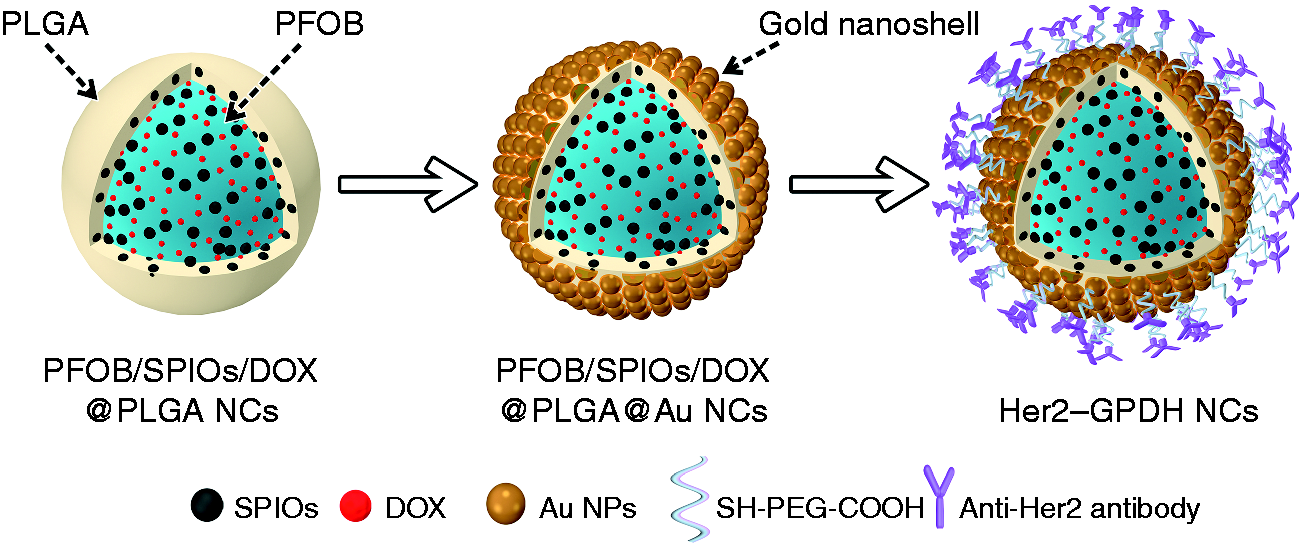
Schematic illustration of the fabrication procedure of Her2-GPDH NCs.
Materials and methods
Materials
PLGA, carboxylic acid terminated, lactide: glycolide 50:50, Mw 24,000–38,000), polyallylamine hydrochloride (PAH, Mw 17,500), and tetrachloroauric (III) acid trihydrate (HAuCl4·3H2O) were supplied by Sigma-Aldrich Trading Co., Ltd. (Shanghai, China). Doxorubicin hydrochloride (DOX·HCl) was purchased from Dalian Meilun Biotechnology Co., Ltd. (Shanghai, China). Oleic acid (OA)-coated SPIOs with a mean diameter of 10 nm were obtained from Shanghai So-Fe Biomedicine Co., Ltd. (Shanghai, China). Polyvinyl alcohol (PVA, 87–89% mole hydrolyzed, low molecular weight), 1-(3-dime-thylaminoprpyl)-3-ethylcarbodiimide hydrochloride (EDC), and N-hydroxysuccinimide (NHS) were supplied from Aladdin Chemistry Co., Ltd. (Shanghai, China). PFOB and carboxyl-terminated (SH–PEG–COOH, Mw 2000) were supplied by J&K Scientific Co., Ltd. and Shanghai Ponsure Biotech, Inc, respectively. Anti-Her2 antibody was purchased from Abcam (Cambridge, MA, USA). All other reagents used were of analytical or chromatographic grade.
Preparation of PFOB/SPIOs/DOX@PLGA NCs
PFOB/SPIOs/DOX@PLGA NCs were prepared according to the methods reported previously.9,26 Briefly, PLGA (100 mg), DOX (10 mg), and excess triethylamine (quadruple the amount of DOX) were dissolved in methylene chloride (5 mL) and stirred for 1 h. Then, OA-coated SPIOs in hexane (0.5 mL and 20 mg/mL) and PFOB (60 µL) were added and thoroughly mixed. Resulted organic phase was added dropwise to precooled aqueous solution of PVA (20 mL, 2%, w/v), followed by emulsification with probe sonication in an ice water bath for 120 s (on/4 s and off/2 s). After evaporation of the organic solvent by magnetic stirring for at least 5 h, PFOB/SPIOs/DOX@PLGA NCs were obtained by centrifugation (24,041 g, 5 min, 15°C, Avanti J-25, Beckman Coulter) and washing three times with deionized (DI) water.
Preparation of PFOB/SPIOs/DOX@PLGA@Au NCs
PFOB/SPIOs/DOX@PLGA NCs were dispersed in PAH solution (20 mL, 1.0 mg/mL in 0.5 mol/L NaCl aqueous solution) for 40 min, then centrifuged (21, 130 g, 10 min, 15°C), and washed twice with DI water. Meanwhile, citrate-stabilized gold NPs with an average diameter of 5 nm were prepared via a redox reaction. 16 Next, PAH-absorbed PFOB/SPIOs/DOX@PLGA NCs were mixed with citrate-stabilized gold NPs suspension (100 mL) for 1 h, and the centrifuge/wash steps were repeated to obtain Au NPs-coated PFOB/SPIOs/DOX@PLGA NCs. Then, the resulted Au NPs-coated PFOB/SPIOs/DOX@PLGA NCs were re-dispersed into HAuCl4 solution (2 mL, 1% w/v) with stirring for 30 min, followed by the addition of hydroxylamine hydrochloride solution (NH2OH·HCl, 0.3 mL, 0.5 mol/L). The mixture was stirred for 15 min to reduce HAuCl4 to form Au nanoshells around the surface of PFOB/SPIO/DOX @ PLGA NCs.
Preparation of Her2–GPDH NCs
SH–PEG–COOH (5 mg) was added to PFOB/SPIOs/DOX@PLGA@Au NCs aqueous solution (1 mL, 2 mg/mL) and stirred at room temperature for 12 h, and then the free SH–PEG–COOH was removed by centrifuge/wash steps. Afterward, the precipitate was re-dispersed in phosphate-buffered saline (PBS), and EDC/NHS coupling activator (10 mg/10 mg) was introduced to activate the carboxylic acid groups on the surface of the pegylated PFOB/SPIO/DOX@PLGA@Au NCs. The mixture was stirred gently at room temperature for 2 h, followed by centrifuge/wash steps to obtain the activated NCs (GPDH NCs). Next, the GPDH NCs suspensions were mixed with anti-Her2 antibodies (10 µL, 0.38 mg/mL) and incubated for 90 min in an isothermal shaker. Finally, the Her2-targeted PFOB/SPIO/DOX@PLGA@Au NCs (Her2–PFOB/SPIO/DOX@PLGA@Au NCs, Her2–GPDH NCs) were obtained by repeated centrifuge/wash steps.
Characterizations
Field emission scanning electron microscopy (FE-SEM, Hitachi S-4800, Tokyo, Japan) was used to observe the morphology and size of Her2–GPDH NCs. The internal structure of Her2–GPDH NCs was evaluated by high resolution transmission electron microscope (TEM, JEM-2100; JEOL, Tokyo, Japan). Energy-dispersive X-ray spectroscopy (EDS) attached to the TEM was used to verify the corresponding elements of Her2–GPDH NCs (without the gold sputtering process). The amount of Au and Fe elements in Her2–GPDH NCs was quantitatively analyzed by inductively coupled plasma atomic emission spectroscopy (ICP–AES, Vistampxicp Varian, USA). The size distribution and zeta potential of Her2–GPDH NCs were characterized by dynamic laser scattering (DLS) instrument (Zetasizer Nano ZS3690; Malvern Instruments, Malvern, UK). The UV–vis absorption spectra of the NCs during the fabrication progress were monitored with a UV–vis spectrophotometer (Beckman Coulter DU 730, USA).
Evaluation of photothermal effect
To evaluate the photothermal effect of Her2–GPDH NCs, different concentrations of Her2–GPDH NCs suspensions (pure water, 0.05, 0.1, 0.15, and 0.2 mg/mL) were dispersed in quartz cuvettes (total volume of 2 mL) and irradiated with an 808 nm NIR laser (1 W/cm2) for 10 min. The temperature changes of the suspensions were monitored using a FLIR A300 thermal camera every 10 s. In order to further assess the photothermal cycle stability of it, Her2–GPDH NCs suspension (0.2 mg/mL) was treated with five cycles of 10-min laser irradiation followed by 10-min cooling, and temperature change was also measured using thermal camera.
DOX loading and release
To investigate the loading content and encapsulation efficiency (EE) of DOX, Her2–GPDH NCs (4 mg/mL) were fully dissolved in DMSO and irradiated under NIR laser (808 nm, 1 W/cm2, 10 min) to quickly release the loaded DOX. Then, the released DOX in the supernatant was collected by centrifugation (21,130 g, 5 min, 15°C), and the content of DOX was determined using a UV–vis spectrophotometer according to the corresponding standard calibration curve. Drug loading content (DLC) and EE were calculated according to the following equations
32


To investigate pH-responsive and thermal effect of Her2–GPDH NCs, the in vitro release behavior of DOX in Her2–GPDH NCs was measured under different pH values (pH 7.4 and pH 5.7) with or without NIR laser irradiation (808 nm, 1 W/cm2, 10 min). To determine the pH-responsive drug release behavior, Her2–GPDH NCs (5 mg) were dispersed in PBS (2 mL, pH = 5.7 and 7.4), sealed in dialysis bags (Mw 5000), and immersed in the same PBS (20 mL) at 37°C under continuous shaking. At regular time intervals of 2, 4, 6, 8, 10, 12, 24, and 48 h, the original PBS (2 mL) was taken out, and fresh PBS (2 mL) was added accordingly. The release behavior of free DOX at pH 7.4 and 5.7 in dialysis bags was also investigated as a control. The amount of DOX released was monitored by a UV–vis spectrophotometer at 490 nm. In addition, NIR-triggered release pattern of DOX from Her2–GPDH NCs was also investigated. Samples in different PBS (2 mL, pH = 5.7 and 7.4) were exposed with or without laser irradiation (808 nm, 1 W/cm2) for 10 min at the specified time points (50, 110, and 170 min) under continuous shaking. The amount of DOX released was measured before and after laser irradiation process. All experiments were performed in triplicate.
Cell culture
Her2-overexpressing human breast cancer SKBR3 cell line (SKBR3 cells) and Her2 low-expressing human breast cancer MDA-MB-231 cell line (MDA-MB-231 cells) were provided by the Institute of Biochemistry and Cell Biology (Shanghai, China). The cells were cultured in Dulbecco’s modified Eagle medium (DMEM, Gibco Life Technologies, Grand Island, NY, USA) containing 20% fetal bovine serum (FBS) and 1% penicillin–streptomycin at 37°C in a humidified incubator containing 5% CO2.
Targeting assays in vitro
SKBR3 and MDA-MB-231 cells were seeded in confocal cell-culture dishes (Φ = 20 mm) at a density of 2 × 10⁴ cells/well for 24 h. They were divided in three groups: non-targeted, targeted, and targeted competition. Two kinds of cells in non-targeted group and targeted group were treated with 100 µL GPDH NCs and Her2–GPDH NCs for 30 min, respectively. The cells in targeted competition group were incubated with 20 µL free anti-Her2 antibodies for 30 min, washed three times with PBS, and then treated with Her2–GPDH NCs. Afterward, the cells in each group were washed again with PBS and fixed with 4% paraformaldehyde for 15 min. Then, nucleus staining agent (DAPI; Beyotime Biotechnology Co., Ltd., Shanghai, China) was added to stain cell nuclei for 10 min. Finally, the samples were qualitative observation under a confocal laser scanning microscopy (CLSM) (Leica TCS SP5 II, Leica Microsystems Ltd., Mannheim, Germany).
NIR light triggered DOX release
The NIR laser irradiation triggered drug release was evaluated in vitro using CLSM. SKBR3 cells were seeded in confocal cell-culture dishes (Φ = 20 mm) for 24 h, and they were divided into two groups: laser and non-laser. After incubation with Her2–GPDH NCs for 1 h, the cells in laser group were irradiated with NIR laser (808 nm, 1 W/cm2, 10 min). All the cells were further incubated for 1 h and then were investigated under CLSM.
In vitro bimodal US/MR imaging and quantitative analysis
In vitro US imaging of Her2–GPDH NCs solution was investigated using Ultrasound System Unit (Mylab 90; Esaote SpA, Genova, Italy) with a LA522 transducer. Her2–GPDH NCs were dispersed in degassed DI water at a concentration of 2 mg/mL in EP tube (2 mL), and ultrapure degassed DI water was filled with the same EP tube served as control. Then, the samples were placed in a tank containing ultrapure degassed water, and real-time ultrasonography was performed in two-dimensional (2D) gray-scale mode and contrast-enhanced ultrasound (CEUS) mode (mechanical index (MI) = 0.01, frequency range: 3–9 MHz). The acquired US images were quantitatively analyzed using QontraXt V3.06 software.
In vitro MR imaging and relaxivity measurements were conducted using a 0.5 T MRI scanner (MiniMR-60, Shanghai Niumag Corporation). Her2–GPDH NCs were diluted in DI water to various Fe concentrations (0, 0.005, 0.01, 0.02, 0.04, and 0.08 mM) and placed in EP tubes (Φ = 1 cm). T2-weighted MR imaging of the samples was carried out using traditional spin-echo sequence, and the parameters were as follows: TR = 2000 ms, TE = 100 ms, slice thickness = 3 mm, and the number of acquisition = 1. The proton transverse relaxation times (T2) of Her2–GPDH NCs aqueous solution with different concentrations were also investigated. Relaxivity (r2) was obtained from the fitting plots of 1/T2 (s−1) versus Fe concentration (mM).
In vitro cytotoxicity assessment
The cell-counting kit-8 assays (CCK-8, Dojindo Molecular Technologies Inc., Japan) were used to evaluate the in vitro cytotoxicity of Her2–GPH NCs (without DOX), DOX, and Her2–GPDH NCs (with DOX) on SKBR3 cells. SKBR3 cells were seeded in 96-well plates at a density of 1 × 10⁴/well and incubated for 24 h and then treated with Her2–GPH NCs at different concentrations (0, 0.05, 0.1, 0.25, 0.5, and 1 mg/mL) or free DOX and Her2–GPDH NCs at the corresponding DOX concentrations (0, 2.5, 5, 10, 15, and 20 µΜ). After incubation at 37°C for 4 h, the cells were washed with PBS and further incubated for 24 h. Then, the culture medium was replaced with CCK-8 solution (100 µL, containing 10% CCK-8) for additional 1 h, and the cell viability was detected by a microplate reader (Thermo scientific Multiskan MK3) at the wavelength of 450 nm. Results are shown as mean ± standard deviation (n = 4).
In vitro synergistic photothermal-chemotherapy
In order to visualize the effect of synergistic photothermal-chemotherapy effect, SKBR3 cells were seeded at a density of 1 × 10⁵ cells/well in confocal cell-culture dishes (Φ = 20 mm) for 24 h. The cells were divided into eight groups, respectively, including DMEM control groups with or without laser irradiation, free DOX groups with or without laser irradiation, Her2–GPH NCs groups with or without laser irradiation, and Her2–GPDH NCs groups with or without laser irradiation. The gold content of the Her2–GPDH NCs group was the same as that of Her2–GPH NCs group, which was about 150 µg/mL, and the DOX content of the Her2–GPDH NCs group was approximately 7.6 µg/mL. After incubation with materials for 4 h, the cells were washed with PBS. Then, the laser irradiation groups were irradiated with an NIR laser (808 nm, 1 W/cm2) for 10 min and cultured for another 4 h. Finally, live/dead cell assay kits (Life Technologies, Grand Island, NY) were added, and the stained cells were investigated under an CLSM.
To quantitatively evaluate the synergistic therapeutic effect of Her2–GPDH NCs on breast cancer cells, SKBR3 cells were plated in 96-well plates at 1 × 10⁴/well and incubated for 24 h. The groupings were consistent with the AM/PI staining assay. SKBR3 cells were treated with culture medium containing Her2–GPH NCs or Her2–GPDH NCs at different concentrations (100, 150, and 200 µg/mL), or free DOX at the corresponding concentrations (3.8, 5.7, and 7.6 µg/mL) for 4 h at 37°C. After incubation, the cells were washed with PBS, and the laser irradiation groups were irradiated with an NIR laser. Then, the cells were further cultured for 4 h, and the cell viabilities were assessed by CCK-8 assay. Results are shown as mean ± standard deviation (n = 4).
Statistical analysis
Quantitative data were expressed as mean ± SD. Statistical differences among multiple groups were analyzed using analysis of variance, and two independent groups were compared using Student’s t-test. Probabilities of P < 0.05 were considered statistically significant. Statistical analyses were performed using SPSS v17.0 (IBM, Armonk, NY, USA).
Results and discussions
Characterizations of Her2–GPDH NCs
Initially, PFOB/SPIOs/DOX@PLGA NCs were fabricated using an adapted oil-in-water emulsion process. SEM images (Figure 2(a)) showed the uniform spherical morphology and smooth surface of PFOB/SPIOs/DOX@PLGA NCs. As illustrated in TEM image (Figure 2(b)), the deep gray spots appeared in the shell and core region of the NCs, indicating the encapsulation of SPIOs in the PLGA matrix. EDS elements mapping (Figure 2(c)) of PFOB/SPIOs/DOX@PLGA NCs clearly revealed the presence of characteristic Fe, F, and Br elements, proving the successful package of SPIOs and PFOB. In addition, compared with PFOB/SPIOs@PLGA NCs, PFOB/SPIOs/DOX@PLGA NCs exhibited a characteristic absorption peak at 490 nm, suggesting that the DOX was successfully encapsulated in the NCs (Figure 2(f)). DLS results confirmed that the average diameter of PFOB/SPIOs/DOX@PLGA NCs was about 260 nm, in agreement with TEM measurement. Au NPs were coated on the surface of PFOB/SPIOs/DOX@PLGA NCs by electrostatic adsorption. Specifically, PFOB/SPIOs/DOX@PLGA NCs were negatively charged with a zeta potential of approximately –18.8 mV, which could be used to absorb positively charged PAH so as to subsequently attach negatively charged citrate-stabilized Au NPs. Afterward, the attached Au NPs with an average size of 5–7 nm were served as seeds to nucleate the growth of an Au nanoshells around PFOB/SPIOs/DOX@PLGA NCs by a seeding procedure. 33
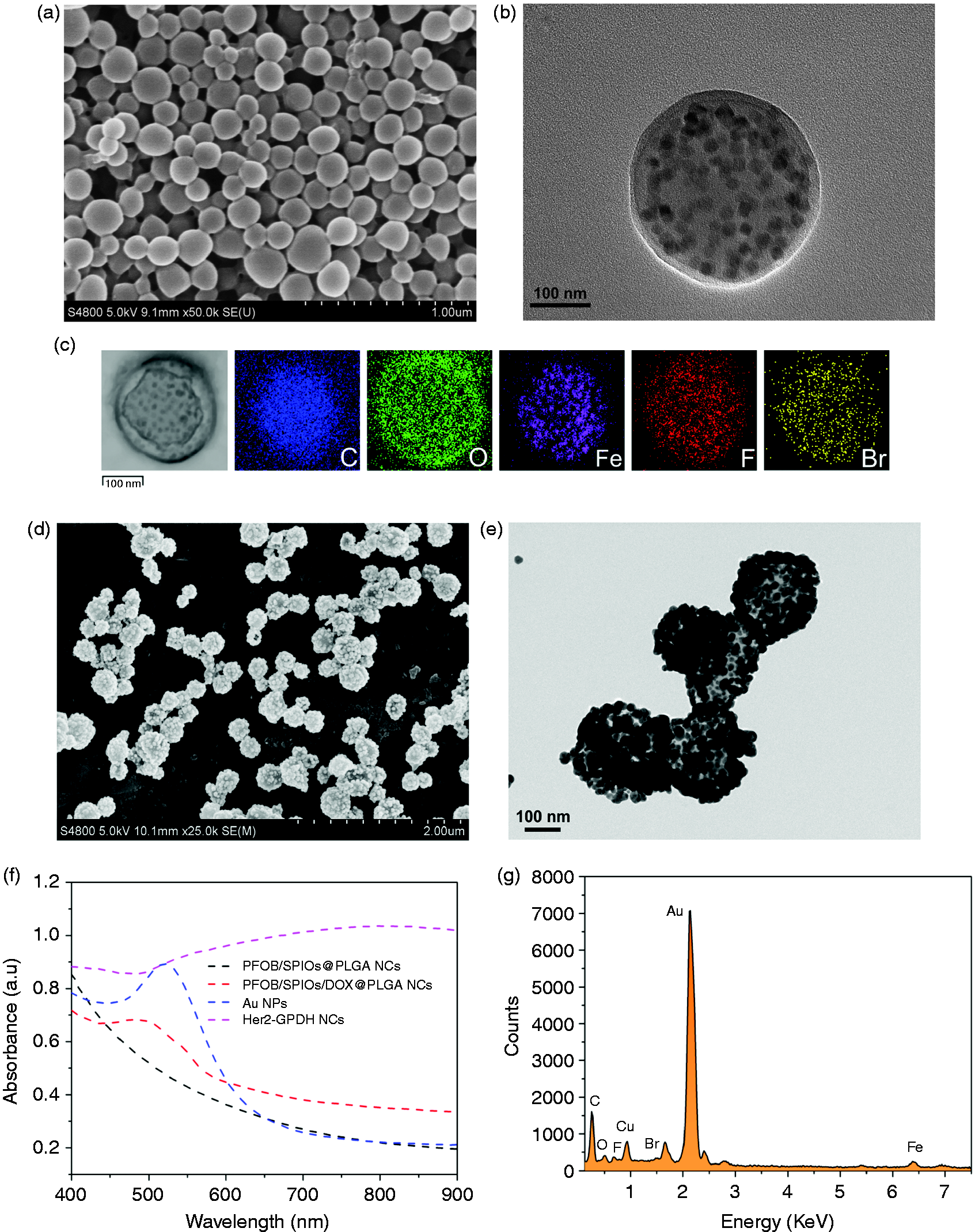
Characterizations of Her2-GPDH NCs: (a) SEM (scale = 1 µm); (b) TEM (scale = 100 nm) images of PFOB/SPIOs/DOX@PLGA NCs; (c) EDS element mapping images of PFOB/SPIOs/DOX@PLGA NCs exhibit the distributions of C, O, Fe, F, and Br elements; (d) SEM (scale = 2 µm) and (e) TEM (scale = 100 nm) images of Her2-GPDH NCs (Her2-PFOB/SPIOs/DOX@PLGA@Au NCs); (f) UV–vis absorption spectra of Her2-GPDH NCs at different stages of synthesis process; (g) EDS elements analysis of Her2-GPDH NCs.
Her2–GPDH NCs were synthesized by linking SH–PEG–COOH to PFOB/SPIOs/DOX@PLGA@Au NCs via Au–S linkage, followed by coupling anti-Her2 antibody with PEG chain via an amide linkage catalyzed by EDC/NHS method. 32 Observed from SEM image (Figure 2(d)), Her2–GPDH NCs maintained their regular spherical morphology but increased the surface roughness due to deposition of aggregated Au NPs. TEM image (Figure 2(e)) clearly illustrated that dense Au NPs with a diameter of tens of nanometers were distributed homogeneously on the surfaces of PLGA NCs to form Au nanoshells. Compared with PFOB/SPIOs/DOX@PLGA NCs, the average size of Her2–GPDH NCs increased to 296 nm with a polydispersity index of 0.124, and zeta potential was about –23.7 mV, which indicated the good solution stability of it. Furthermore, Her2–GPDH NCs exhibited a continuous broad plasmon resonance peak ranging from 600 to 900 nm, which was attributed to the increased size distribution and the aggregation of Au NPs around the surface of PFOB/SPIOs/DOX@PLGA NCs (Figure 2(f)). 34 Moreover, EDS element analysis (Figure 2(g)) showed that Her2–GPDH NCs contained Au, C, O, Fe, F, and Br elements, and ICP–AES revealed that the amount of Au and Fe elements in Her2–GPDH NCs was about 75 and 1.35%, respectively.
Photothermal effect of Her2–GPDH NCs
To evaluate the photothermal conversion effect of our nanoplatform, the temperature variations of Her2–GPDH NCs suspensions were monitored every 10 s with a thermal imaging camera under irradiation of an NIR laser (808 nm, 1 W/cm2, 10 min). As vividly shown in Figure 3(a), after laser irradiation for 10 min, the corresponding photothermal images of Her2–GPDH NCs indicated that the heat increased with the increasing concentration of Her2–GPDH NCs and the heat diffused homogeneous from the center to the surroundings. Observed from the quantitative measurement of Figure 3(b), the temperature of Her2–GPDH NCs suspensions elevated with the increase of irradiation time and concentration. At the highest concentration of 0.2 mg/mL, the temperature increased by about 18.5°C after 10 min of irradiation. In contrast, the temperature increase of the DI water was only 1°C. In order to induce cancer cell to death, the temperature had to rise above 42°C according to previous studies. 16 Her2–GPDH NCs could easily be heated up to 42°C at relatively low concentration in vitro, showing the potential to kill cancer cells by hyperthermia. Furthermore, the photothermal cycling stability of Her2–GPDH NCs was also investigated (Figure 3(c)). After five cycles of on−off irradiation, the temperature increments of Her2–GPDH NCs suspensions did not obviously change, which confirmed the excellent photothermal stability of the nanoplatform.
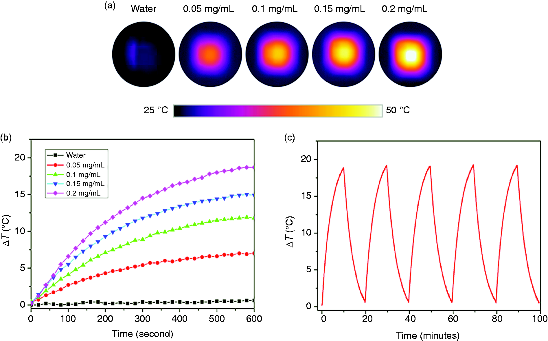
Photothermal conversion effect of Her2-GPDH NCs. Photothermal images (a) and temperature change profiles (b) of water and Her2-GPDH NCs at different concentrations (0.05, 0.1, 0.15, and 0.2 mg/mL) under irradiation of an NIR laser (808 nm, 1 W/cm2, and 10 min). (c) Temperature change profiles of Her2-GPDH NCs (0.2 mg/mL) in five heating-cooling cycles of NIR irradiation (808 nm, 1 W/cm2, and 10 min).
DOX loading and release
DOX was encapsulated into PLGA nanocapsules during the emulsion process. In particular, the addition of excess triethylamine was used to convert DOX·HCl to water-insoluble base (DOX) to favor its encapsulation inside PLGA nanocapsules by improving hydrophobicity. 35 In order to obtain relatively ideal EE and DLC, the weight ratio of PLGA to DOX was selected to be 10:1 according to the previous studies. 32 The DLC and EE of DOX in Her2–GPDH NCs were determined by fluorescent spectroscopy to be 3.8 ± 0.52% and 39 ± 1.45%, respectively. The release behavior of DOX from Her2–GPDH NCs was investigated using a dialysis method in PBS under different pH values (7.4 and 5.7) with or without laser irradiation (808 nm, 1 W/cm2, 10 min). Figure 4(a) displayed the release behavior of free DOX at different pH values (7.4 and 5.7). As a control, free DOX released rapidly from the dialysis bags, and the release rates were very similar at pH 7.4 and 5.7. The cumulative DOX release exceeded 80% within 8 h, which confirmed that the favorable diffusion rate of DOX as low molecular weight molecules across the dialysis membrane was not the limiting factor for DOX release studies. 36 Figure 4(b) showed the cumulative DOX release profile in Her2–GPH NCs under different pH values (7.4 and 5.7). At the physiological condition (pH 7.4), the Her2–GPDH NCs were relatively stable, and only 14% of DOX was released within 48 h. At weak acidic condition (pH 5.7), the DOX release was dramatically increased within the first 5 h (about 15%), and the cumulative release amount was about 25% after 48 h. The enhanced release of DOX in weak acidic conditions indicated the pH-responsive release behavior of Her2–GPDH NCs, which was likely attributed to the degradation of PLGA polymer and accelerated protonation of DOX in the acidic environment.32,37 The pH-responsive release of Her2–GPDH NCs could reduce the side effects of the drug on normal tissues while ensuring the substantial accumulation of drug in the target tumor tissues. To further evaluate the NIR-triggered DOX release, Her2–GPDH NCs in dialysis bags immersing in PBS with different pH values (5.7 and 7.4) were irradiated with NIR laser (808 nm, 1 W/cm2) for 10 min at the specified time points (50, 110, and 170 min). As shown in Figure 4(c), DOX release rate increased significantly under laser irradiation and slowed down after the laser was turned off. At pH 5.7, the cumulative release of DOX in Her2–GPDH NCs increased by 7.5% after the first 10 min irradiation and reached 33% after three cycles of laser on/off, twice higher than that of non-laser irradiation (14%). In addition, at pH 7.4, the cumulative release of DOX reached 24% after three cycles of laser on/off, still below the release at pH 5.7. These results demonstrated the stepwise triggered release of DOX based on NIR laser, which may be due to the degradation of PLGA matrix caused by the autologous photothermal effect of Au nanoshell under NIR laser irradiation. 9 All these data suggested that pH-responsive combined with NIR-triggered had potential to effectively enhance DOX release of Her2–GPDH NCs in the intracellular compartment of acidic cancer cells.
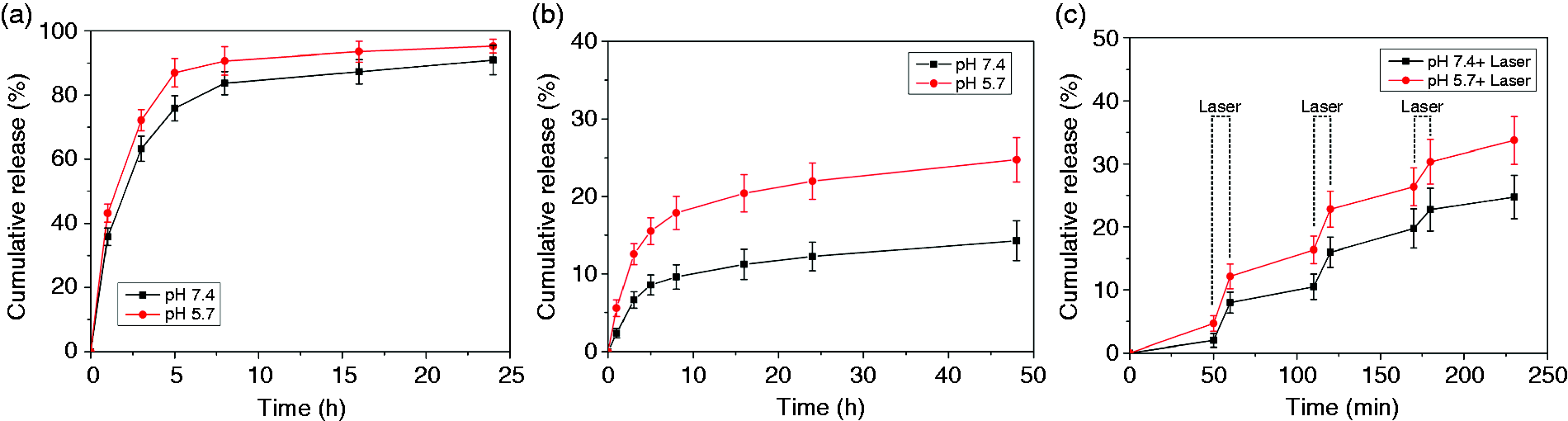
In vitro DOX release profiles. The release profiles of free DOX (a) and DOX from Her2-GPDH NCs (b) at different pH values (7.4 and 5.7). (c) DOX release profiles from Her2-GPDH NCs upon three cycles of NIR laser irradiation (808 nm, 1 W/cm2, and 10 min) under different pH values (7.4 and 5.7).
In vitro specific targeting studies
DOX, excited by the laser of 488 nm, exhibits a red fluorescence and can be used for intracellular tracing. 38 Therefore, we chose DOX to label Her2–GPDH NCs, and the attachment of Her2–GPDH NCs to cells could be clearly determined by immunofluorescence assay. As shown in Figure 5, the aggregated Her2–GPDH NCs can be clearly observed in the bright field. Besides, Her2–GPDH NCs exhibited bright red fluorescence, which demonstrated the successful encapsulation of DOX in Her2–GPH NCs.
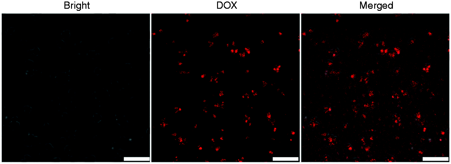
Confocal microscopic images of Her2-GPDH NCs in bright field, DOX fluorescence, and merged channels (scale bar = 5 µm).
CLSM was utilized to evaluate the targeting specificity of Her2–GPDH NCs in vitro. As shown in Figure 6, SKBR3 cells exhibited bright red fluorescence signals on the surface of cell membranes after incubation with targeted Her2–GPDH NCs (a), whereas no fluorescence was observed in SKBR3 cells treated with non-targeted GPDH NCs (b), suggesting that Her2–GPDH NCs could efficiently bind to cells overexpressing Her2. To further clearly observe the combination of Her2–GPDH NCs and cells, we show the images of bright field (a1), DOX channel (a2), DAPI channel (a3), and merged channel (a4). Since Her2 is a transmembrane receptor protein, Her2-targeted NPs specifically binds to cell membranes after a short incubation time (30 min). 39 As seen from Figure 6(a1), Her2–GPDH NCs aggregated into dots and rings on SKBR3 cell membranes. To investigate the targeting mechanism of Her2–GPDH NCs, the competition experiment was further examined by pretreating SKBR3 cells with free Her2 antibody before incubation with Her2–GPDH NCs (c). SKBR3 cells in competition group no longer showed distinctly fluorescence signals, which confirmed receptor-mediated targeting binding of Her2–GPH NCs and could be blocked by excess free anti-Her2 antibodies. 40 In addition, there was little fluorescence in MDA-MB-231 cells incubated with Her2–GPDH NCs (d), which confirmed the targeting specifically of Her2–GPDH NCs to Her2-positive SKBR3 cells.
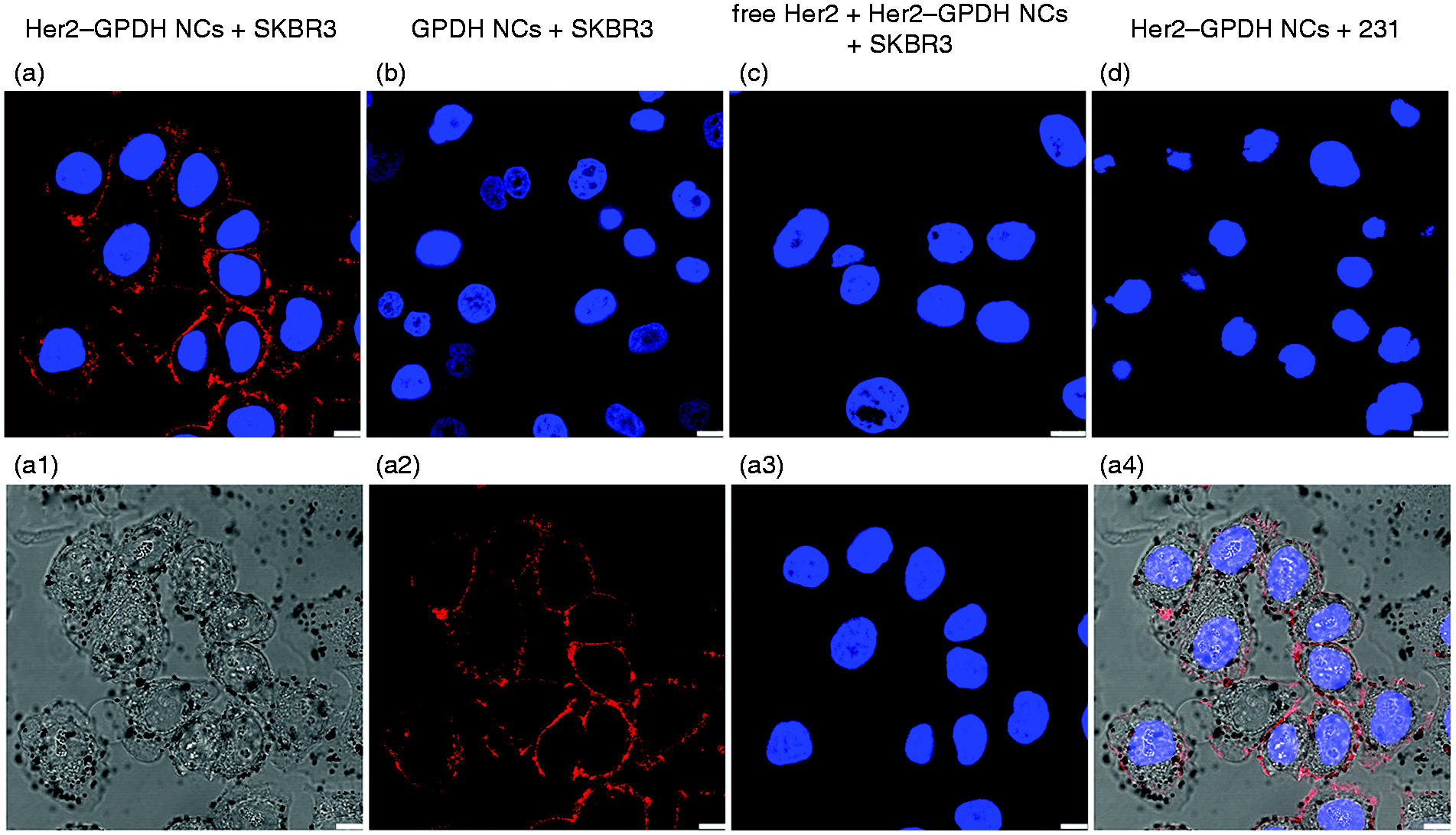
In vitro targeting studies. Confocal microscopic images of SKBR3 cells treated with Her2-GPDH NCs (a) a1–a4 and GPDH NCs (b), free Her2 antibody pre-treated SKBR3 cells incubated with Her2-GPDH NCs (c), and MDA-MB-231 cells treated with Her2-GPDH NCs (d) (scale bar =10 µm): (a1) bright field; (a2) DOX channel; (a3) DAPI channel; (a4) merged channel.
NIR light triggered DOX release
DOX is an effective and widely used anticancer drug, which binds to the nucleic acids and induces DNA damage. 41 We utilized CLSM to observe the NIR light-triggered release of DOX in Her2–GPDH NCs and determined the effective delivery of DOX into intracellular compartments. As illustrated in Figure 7, in non-laser irradiation group, DOX fluorescence was mainly distributed on the cell membrane after SKBR3 cells were incubated with Her2–GPDH NCs for 1 h. Under NIR laser irradiation (808 nm, 1 W/cm2) for 10 min, DOX fluorescence intensity increased significantly and mainly distributed in the cytoplasm and nucleus. These results indicated that NIR laser-induced photothermal effect could promote the release of DOX in Her2–GPDH NCs, and Her2–GPDH NCs could serve as an effective carrier for DOX delivery.
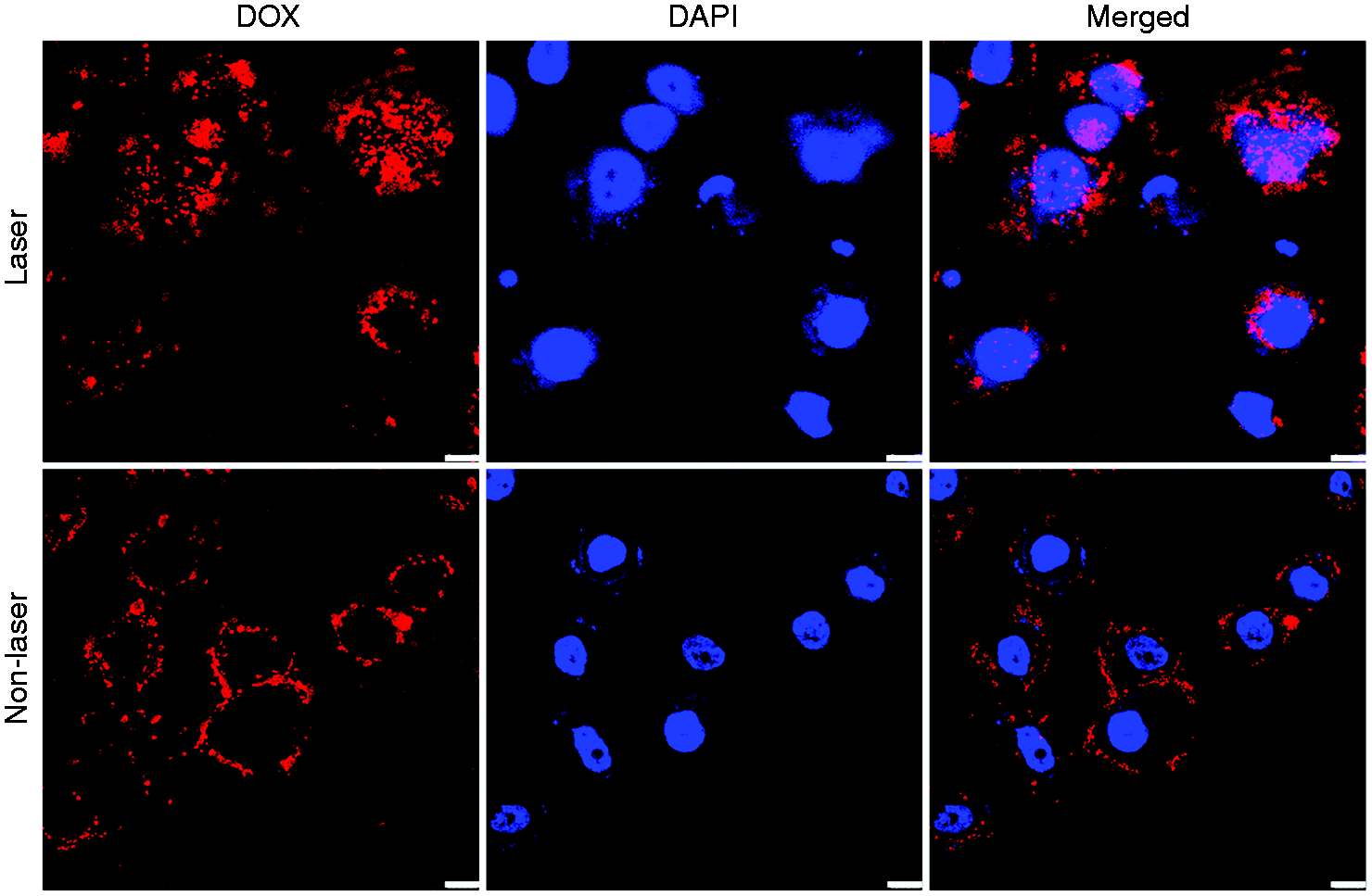
Confocal microscopic images of DOX cellular uptake in SKBR3 cells after incubation with Her2-GPDH NCs for 1 h with or without NIR laser irradiation (808 nm, 1 W/cm2, and 10 min) (scale bar =10 µm).
In vitro US/MR bimodal imaging
The US imaging capability of Her2–GPDH NCs was investigated in vitro under 2D gray-scale and CEUS modes. As shown in Figure 8(a), Her2–GPDH NCs solution (2 mg/mL) exhibited uniform dotted echo in both modes, whereas degassed DI water displayed as anecho. It was worth noting that the US signals in CEUS mode were more obvious and better than 2D gray-scale, which suggested that Her2–GPDH NCs was consistent with the existing US contrast agents and was more suitable for nonlinear CEUS imaging. Moreover, quantitative analysis was performed via time intensity curves (TICs). The mean signal intensity of Her2–GPDH NCs (2 mg/mL) solution was 82 ± 1.45 dB, compared with degassed DI water of 1 ± 1.31 dB. In addition, we also accessed the US imaging of Her2–GPDH NCs with time elapsing. Observed from Figure 8(b), Her2–GPH NCs exhibited satisfactory CEUS signal within 3 min. Although the signal intensity gradually decreased over time, the CEUS signal lasted more than 6 min, which could satisfy the requirement of clinical ultrasonography. These results suggested that Her2–GPDH NCs had the potential to serve as novel US contrast agent with satisfactory CEUS imaging effect in vitro.
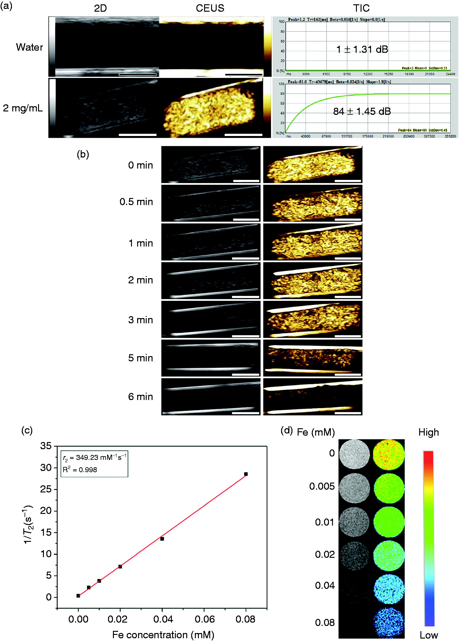
In vitro US/MR bimodal imaging: (a) 2D gray-scale US, CEUS images, and TICs of Her2-GPDH NCs (2 mg/mL) and degassed DI water (scale bar = 0.5 cm); (b) in vitro US images of Her2-GPDH NCs (2 mg/mL) at various time points (scale bar = 0.5 cm); (c) linear fitting between T2 relaxation rates (1/T2) and Fe concentrations of Her2-GPDH NCs at a 0.5 T magnetic field; (d) T2-weighted MR images of Her2-GPDH NCs with increasing Fe concentrations at 0.5 T, 37°C.
Owing to the capability to shorten the transverse relaxation times, SPIOs have been widely investigated as T2 contrast agents. Furthermore, numerous studies have focused on the aggregation of SPIOs in reservoirs to increase the transverse-relaxivity (r2), which is mainly due to the enhanced magnetic interactions between the assembled nanocrystals.42,43 We evaluated the T2-weighted imaging effect of Her2–GPDH NCs with different Fe concentrations under a 0.5 T magnetic field. The transverse relaxation rates (1/T2) of Her2–GPDH NCs aqueous solution as a function of iron concentration are shown in Figure 8(c). According to the fitting curve, the T2 relaxivity (r2) of Her2–GPDH NCs aqueous solution was calculated to be 349.23 mM−1 s−1, 2.3 times higher than that of the commercially available MRI contrast agent such as Feridex (152 mM−1s−1) at the same magnetic field. 44 Furthermore, as shown in Figure 8(d), with the increase of iron concentration, the T2-weighted images of Her2–GPDH NCs aqueous solution exhibited increasing negative contrast enhancement, which confirmed that Her2–GPDH NCs could be served as T2-weighted MRI contrast agent.
In vitro cytotoxicity assessment
We used CCK-8 assay to investigate the cytotoxicity of Her2–GPH NCs, DOX, and Her2–GPDH NCs on breast cancer SKBR3 cells. As shown in Figure 9(a), compared with controls, the cell viability of SKBR3 cells remained more than 85% after incubation with Her2–GPH NCs (without DOX), even at the highest concentration (1 mg/mL) for 24 h, which illustrated the low cytotoxicity and good biocompatibility of Her2–GPH NCs. On the contrary, both free DOX and Her2–GPDH NCs exhibited cytotoxicity against SKBR3 cells in a concentration-dependent pattern (Figure 9(b) and (c)). With the increase of DOX concentration, the cell viability of Her2–GPDH NCs treated SKBR3 cells decreased moderately, which was still higher than that of free DOX group. In detail, at the highest DOX concentration (20 µM) over 24 h, the viability decreased to approximately 23% for free DOX-treated SKBR3 cells and 58% for Her2–GPDH NCs. These results indicated that the gold nanoshells on the surface of Her2–GPDH NCs could delay the release of DOX, thereby having the potential to reducing the toxic and side effects of DOX.

Cell viabilities of breast cancer SKBR3 cells after incubation with Her2-GPH NCs (a) of different concentrations (0, 0.05, 0.1, 0.25, 0.5, and 1 mg/mL), free DOX, and Her2-GPDH NCs at the corresponding DOX concentrations (0, 2.5, 5, 10, 15, and 20 µM) for 12 h (b) or 24 h (c) (data expressed as mean ± SD, n = 4).
In vitro synergistic photothermo-chemotherapy
Initially, live/dead cell assays were performed to visually evaluate the synergistic effect of photothermo-chemotherapy of Her2–GPDH NCs on SKBR3 cells using calcein AM and propidium iodide (PI) staining. Calcein AM can penetrate the cell membrane to produce green fluorescent in live cells, while PI binds to the DNA of dead cells to emit red fluorescence. 45 As illustrated in Figure 10(a), SKBR3 cells emitted bright green fluorescence in the entire region upon NIR laser irradiation, indicating that laser irradiation had little cytotoxic. Besides, there was a small proportion of dead cells in DOX group with or without laser irradiation, which was mainly attributed to the chemical toxicity of DOX. However, there were no obvious dead cells when treated only with Her2–GPH NCs, suggesting that Her2–GPH NCs itself were biocompatible. On the contrary, there were a large number of dead cells when Her2–GPH NCs were combined with laser irradiation, which indicated that the agent could effectively transform light to thermal energy to kill cancer cells. In contrast, we found that almost all cells died after treatment with Her2–GPDH NCs in the presence of NIR laser, which suggested that Her2–GPDH NCs had a synergistic effect of chemotherapy and photothermal therapy.
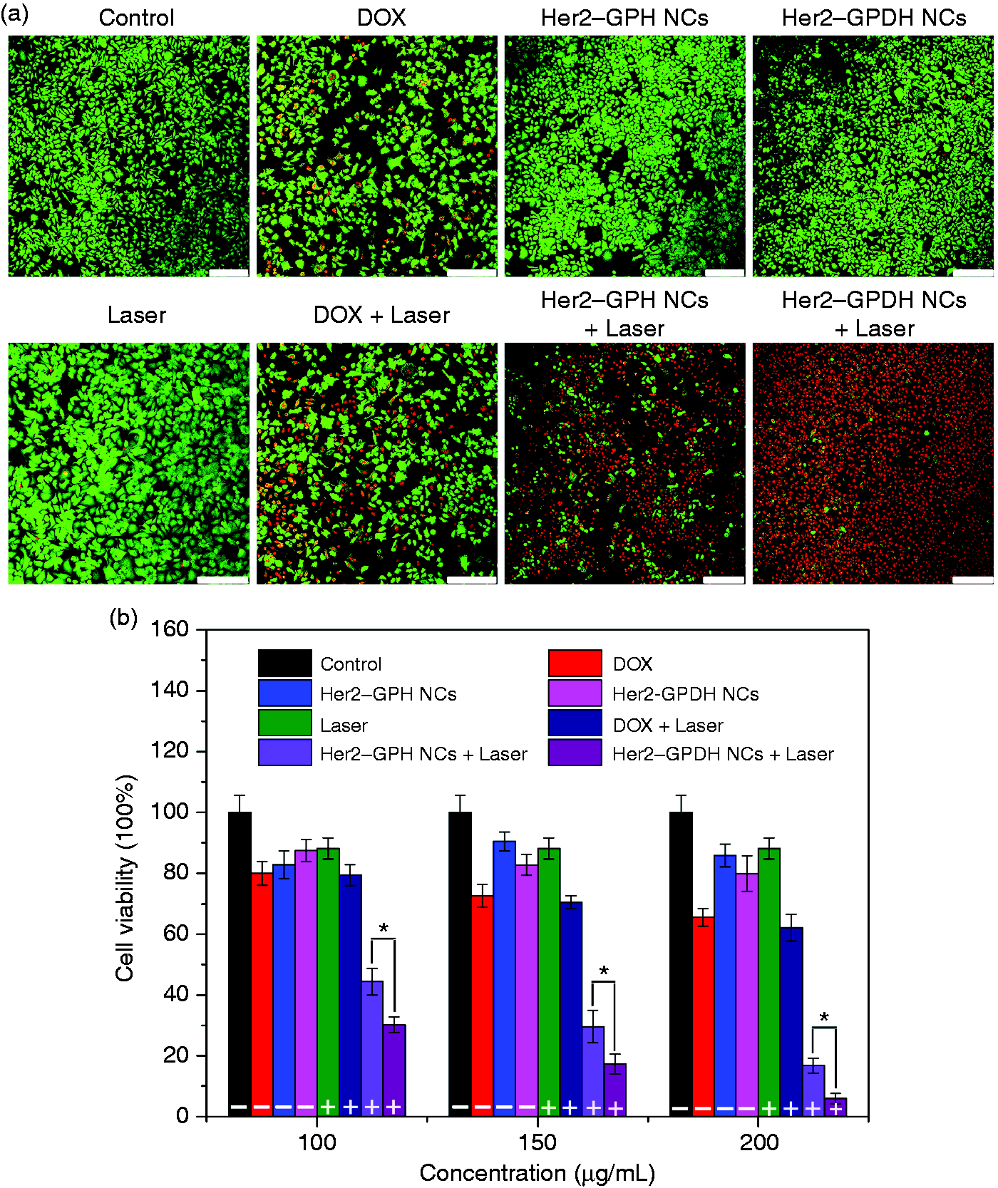
In vitro synergistic photothermo-chemotherapy of breast cancer cell: (a) fluorescence microscopy images of calcein AM/PI-stained SKBR3 cells (green represents living cells, red represents dead cells) incubated with Her2-GPH NCs, DOX, and Her2-GPDH NCs, respectively, without and with NIR laser Irradiation (808 nm, 1 W/cm2, and 10 min). SKBR3 cells in normal culture medium served as control (scale = 250 µm); (b) CCK-8 cytotoxicity assay of SKBR3 cells incubated with DMEM, Her2-GPH NCs, DOX solution, or Her2-GPDH NCs NCs with or without NIR laser irradiation (808 nm, 1 W/cm2, and 10 min) (data expressed as mean ± SD, *P < 0.05, n = 4). The non-laser irradiation group was marked with –, and the laser irradiation group was marked with +.
CCK-8 assay was used to further quantitatively determine the photothermal therapy and chemotherapy of Her2–GPDH NCs. SKBR3 cells were treated with different concentrations of Her2–GPH NCs, DOX, or Her2–GPDH NCs (with DOX), with or without NIR laser irradiation (808 nm, 1 W/cm2, and 10 min). The DOX content of Her2–GPDH NCs group corresponded to the free DOX group, and the Her2–GPH NCs group had an equivalent gold dosage with the Her2–GPDH NCs group. As displayed in Figure 10(b), the cell viabilities of SKBR3 cells remained more than 85% after treatment with NIR laser or Her2–GPH NCs in different concentrations. The cell viability of DOX group decreased with the increase of DOX concentration. However, due to the existence of the gold nanoshell PLGA structure, Her2–GPDH NCs had little effect on cell viability. Upon laser irradiation, the viabilities of SKBR3 incubated with Her2–GPH NCs or Her2–GPDH NCs decreased significantly with increasing concentration of NCs, proving that the agent can induce cancer cell death by photothermal effect. It was worth noting that SKBR3 cells treated with Her2–GPDH NCs in combination with laser irradiation exhibited the lowest cell viability compared with chemotherapy or PTT alone. In detail, under 808 nm laser irradiation, the cell viability decreased prominently to 7.0% for Her2–GPDH NCs (200 µg/mL), significantly lower than that of the Her2–GPH NCs (18%) and DOX (67%) (P < 0.01). The increased cytotoxicity could be attributed to the combination of an enhanced DOX toxicity triggered by NIR irradiation combined with pH-responsive and the intracellular protein denaturation induced by photothermal effect. 42 These results further confirmed that Her2–GPDH NCs was a promising candidate to kill cancer cells by the synergistic effect of PTT and chemotherapy.
Conclusions
In summary, we have successfully developed Her2–GPDH NCs as a novel multifunctional nanoplatform for bimodal US/MR imaging and synergistic photothermal-chemotherapy of Her2-postive breast cancer cells. Her2 functionalization enabled Her2–GPDH NCs to specifically target Her2-positive breast cancer SKBR3 cells for targeted imaging and drug delivery. Besides, the construction of gold nanoshelled-PLGA spherical structure and the co-encapsulation of PFOB and SPIOs provided Her2–GPDH NCs with excellent contrast enhancement in vitro US and T2-weighted MR imaging. In addition, the resulting NCs could serve as effective NIR photoabsorber for PTT and NIR light-triggered stepwise release of DOX due to the formation of gold nanoshells. Furthermore, we found that the combined PTT and chemotherapy had the most significantly anti-tumor cell effect than either treatment alone. The presented Her2–GPDH NCs are promising tools to facilitate early molecular diagnosis and synergistic treatment of breast cancer and still need to be validated in vivo.
Footnotes
Declaration of conflicting interests
The author(s) declared no potential conflicts of interest with respect to the research, authorship, and/or publication of this article.
Funding
The author(s) disclosed receipt of the following financial support for the research, authorship, and/or publication of this article: This study was supported by the National Natural Science Foundation of China (Nos. 81571678, 81801697, 81102014, and 81901747).