
Abstract
Aim
to study non-specific immune response characteristics (serum cytokine profile) in rats after subcutaneous implantation of the decellularized esophagus matrix.
Methods
Data were obtained in Wistar rats. The rats underwent subcutaneous implantation of decellularized esophagus (DE) and native allogeneic esophagus (NE). Explantation of sampling were carried out on the 7th, 14th and 21st day of the experiment. Explanted NEs and DEs were processed for histologic examination. The content of IL1α, IL2, IL4, IL17А, TNFα, IFNγ and GM-CSF in serum samples were tested by ELISA.
Results
In rat serum with DEs on the 7th day of the experiment it was significant increase in IL1α level in comparison with control group, IL2, TNFα, IL4 levels did not differ from the control group levels that indicates the stabilization of inflammation. The content of IL17A, IFNγ and GM-CSF significantly decreased compared to control. On the 14th day, IL17A concentration analysis showed a sharp decrease in comparison with the the 7th experimental day. We found decrease in IL1α level vs control group and decrease in IFNγ level vs 7th day. On the 21st experimental day was shown a significant decrease in the IL17A, IFNγ and IL1α content in DE rats.
Conclusions
It was found dynamic change in studied rat cytokine concentrations that correspond to favourable clinical picture in DE group in comparison with an active inflammatory reaction in NE group. IL1α, IL4, IL17A and IFNγ concentrations reflect positive dynamics of the wound healing process and the absence of local inflammation and rejection of decellularized matrices.
Keywords
Introduction
Damaged esophagus repair is an important problem of modern medicine, including regenerative medicine, since esophagus injuries disrupt the functional integrity of one of the main body systems. Moreover, chest examination for other related injuries (of heart, blood vessels or lungs) may require extensive debridement, which will lead to an even greater defect in the esophagus wall. 1 Small damage detected at an early stage can be repaired, however, defects with significant tissue loss or neoplasms cannot be treated, which arises further instability of the condition of patients and a significant mortality percentage. 2
It is known that to overcome the problem of regeneration in the tissue engineering field, an alternative method of treatment was proposed – implantation of tissues and organs subjected to the decellularization procedure. This method is a removal of cell components to obtain extracellular matrix (ECM) without inflammatory response – a scaffold for cells that promotes their migration, proliferation and differentiation. 3 The method of decellularization followed by recellularization is actively used in experimental studies to produce scaffolds from various tissues and organs (e.g., skin, blood vessels, heart valves and nerves), including an esophagus.4–7
For assessment the tissue response to implantation and the dynamics of scaffold biodegradation a method of subcutaneous implantation is generally used. The method is used to evaluate the systemic reaction and the severity of the inflammatory response to the decellularized matrix.
It is suggested that macrophages are the main cells that determine the biocompatibility of implantable materials. 8 Macrophages take part not only in blood coagulation, fibrinolysis and complement system activation, but also actively produce mediators that can cause proliferation of various cells and the synthesis of molecules involved in inflammation and wound healing. After activation, macrophages synthesize IL1, which has a significant effect on both the inflammatory and immune responses. IL1 is an important mediator in the inflammatory process due to its regulatory effect on fibroblast growth and protein synthesis. By stimulating the activity of fibroblasts, IL1 induces collagen production and causes proliferation of endothelial and smooth muscle cells. Except for IL1, proliferation, tumor necrosis factor (TNF), fibronectin, macrophage growth factor and a number of other factors9,10 affect chemotaxis and collagen production in fibroblasts. For example, IL8 affects the vascular endothelium and polymorphonuclear leukocytes (PNL). All this makes macrophages the main cells that bonds the inflammatory and reparative phases of the regeneration process.
Therefore, it is necessary to take into account the entire spectrum of cytokines – interleukins, interferons, TNF, colony stimulating factors, growth factors and chemokines for assessment the implantation phases. In vivo, there can be no cases of isolated activation of the synthesis of one type of molecules, since the cytokine regulatory network includes various stimulating and inhibitory effects of the cytokines and their receptors within the same biological response. 11
Thus, the aim of our study is to determine the serum cytokine profile in rats after subcutaneous implantation of the decellularized matrix, which will allow us to study the characteristics of the organism systemic reaction to the implant.
Materials and methods
Object of study
Experimental data were obtained in male Wistar rats (a body weight was 230 ± 50 g). All methods and procedures, as well as the use of animals and tissue specimens derived from animals, were carried out in accordance with the general ethical principles of animal experiments in the “European Convention for the Protection of Vertebrate Animals used for Experimental and Other Scientific Purposes” (Strasbourg, 1986) and were approved by the local ethics committee of the Kuban State Medical University, Krasnodar, Russia. The rats were kept under standard laboratory conditions (12 h dark/light cycle) and provided with food and water at libitum. Animals divided into four groups: 1) healthy intact rats - control group (intact, n = 10); 2) sham-operated rats - control group (sham, n = 15); 3) rats with subcutaneous implantation of native allogeneic esophagus fragments - comparison group (NE, n = 15); 4) rats with subcutaneous implantation of decellularized esophagus fragments – study group (DE, n = 15) (Figure 1).
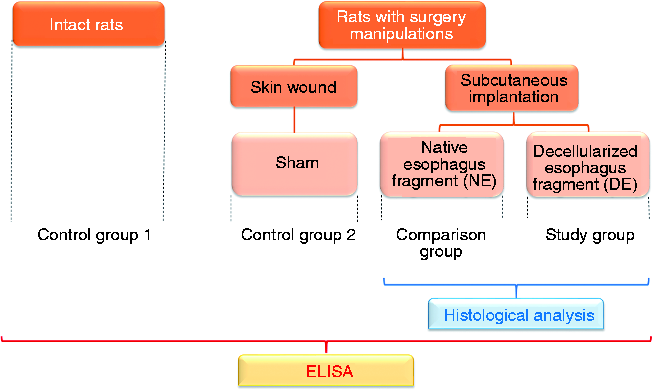
Experimental research design.
NE explantation
Animals were euthanized by intraperitoneal administration of a lethal dose of barbiturates (150 mg/kg). The organocomplex was isolated as a single unit with subsequent esophagus dissection. The esophagus was cut off as close as possible to the pharynx (proximal part) and the esophagus-gastric junction (distal part) to isolate the esophagus part of the greatest extent. The proximal and distal parts of the esophagus were cannulated for fixation in a bioreactor (Harvard Apparatus, Massachusetts, USA).
Esophagus decellularization
Rat esophagus was decellularized by the detergent-enzymatic method using sodium deoxycholate and DNase-I. 4 First, the esophagus was washed with phosphate buffer solution with the addition of 1% penicillin-streptomycin solution. Then perfusion cycles with deionized water (1 h), 4% sodium deoxycholate with the addition of 800 μL EDTA (3 h), phosphate buffer (10 min), phosphate buffer with the addition of 2000 IU/mg of porcine pancreatic DNase-I (1 h) were consequently followed. After that the esophagus was washed with phosphate buffer solution with the addition of 1% penicillin-streptomycin solution (24 h). All solutions were sterile and at room temperature. The feed rate of the solutions was 6 mL/min.
DNA quantification
A standard set of reagents (DneasyBlood and Tissue Kit, Qiagen, Sweden) was used to determine the amount of residual DNA in the decellularized and native esophagus.
NEs and DEs (up to 25 mg) were soaked in 30% ethanol for 10 s. The obtained material was transferred under sterile conditions into Eppendorf tubes, and then the study was carried out according to the manufacturer’s instructions. After sample preparation, the DNA content was measured on a NanoDrop ND-1000 spectrophotometer (Thermo Fisher Scientific Inc., USA).
Subcutaneous implantation
During the operation, the rats were anesthetized by Xilazin 2% (Alfasan International B.V., The Netherlands) - 10 ED, administered subcutaneously and Zoletil (Virbac Sante Animale, France) - 8 ED, administered intramuscularly. The defect application place was carefully prepared for the operation – the rats were shaved, the skin was thoroughly treated with alcohol and Miramistin (LLC Infamed, Russia) to avoid inflammation and infection of the wound. NE and DE fragments (5x5 mm) were placed in the subcutaneous pocket at rat withers and were fixed with two sutures. Skin defect in sham group was applied to the shoulder blades. Skin wounds in all groups were stitched up with a continuous suture.
Implant explantation
Animals were removed from the experiment by euthanasia with chloroform; the explantation of DEs and NEs was performed on the 7th, 14th, 21st day of the experiment. All rats remained healthy, with no overt signs of inflammation over the experimental period. At proper time points, recipient rats were euthanized and the implants with surrounding connective tissue were removed. The size of the explanted NEs and DEs was measured using a ruler and then processed for routine H&E histologic examination.
Histological examination
Histological evaluation of explanted esophagus samples was made using a TP1020 histoprocessor (Leica, Germany). For morphological analysis explanted NEs and DEs were fixed in 10% neutral buffered formalin, dehydrated, followed by incubation with paraffin using a histoprocessor according to standard methods. Paraffin filling was performed on a modular unit EG1150H (Leica, Germany). For a general histological evaluation of the samples using a rotational microtome RM2235 (Leica, Germany), paraffin sections with a thickness of 5 μm were obtained, followed by dewaxing, hydration, hematoxylin and eosin staining (Histolab, Sweden). After sealing, samples were examined by light microscopy at a different magnification to inspect the presence of cells (stained by hematoxylin to a bluish-purple colour) and collagen fibres (stained by eosin to a pink colour). Histological analysis was carried out on samples from six independent decellularization procedures, three slides for each sample, and three sections in each slide.
Cytokine measurement
Peripheral blood sampling was carried out on the 7th, 14th and 21st day of the experiment. Blood serum samples were stored at a temperature of -20°C. IL1α, IL2, IL4, IL17А, TNFα, IFNγ and GM-CSF were determined by ELISA in rat serum with ELISA kits (Platinum ELISA, eBioscience, Austria) according to the manufacturer’s instructions. The method employs the quantitative sandwich enzyme immunoassay technique. Results were evaluated by optical density (OD) on a Multiskan Ascent microplate reader (Labsystems, Finland) at 450 nm. Serum cytokines were calculated from a standard curve.
Statistical analysis
Statistical analysis of the obtained data was performed using the Graph Pad Prism version 6.04 program, the results were presented as the median with the upper and lower quartiles (Me [Q1; Q3]), Mann-Whitney test and Wilcoxon test were used to compare groups. Differences were considered significant at p ≤ 0.05.
Results
The immunogenicity of the decellularized scaffold directly depends on the amount of residual DNA, since nucleic acids are recognized by immune cells of the host – therefore, a quantitative DNA analysis was carried out in the esophagus samples. Quantitative analysis showed that about 92.0% of the DNA was removed during the esophagus decellularization. The native esophagus contained 1576.37 ± 279.6 ng/mg of DNA, while decellularized esophagus – only 123.85 ±22.61 ng/mg of DNA (Figure 2).
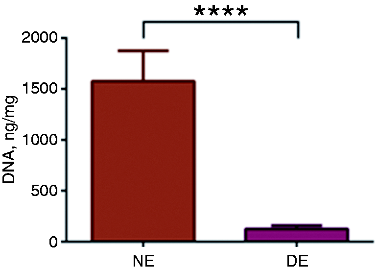
Relative DNA content before and after rat esophagus decellularization. NE – native esophagus, DE – decellularized esophagus (*p < 0.0001).
A severe inflammatory reaction on the 7th day after subcutaneous NEs implantation led to the skin defect formation (Figure 3(a)). Subsequently, a marked connective tissue capsule (a thickness of up to 160–170 μm) was formed around it (Figure 3(b)). On the 14th day, there was a slight decrease in the severity of the implanted native esophagus edema, pronounced mononuclear cell infiltration was detected. The implant was delimited from surrounding tissues by a capsule containing full-blood vessels. On the 21st day, pronounced lymphocytic macrophage infiltration of the sample and surrounding tissues in the implantation zone and a significant decrease in the size of the native esophagus delimited by the connective tissue capsule were revealed.

Subcutaneous implantation of fragments of the native (a and b) and decellularized (c and d) esophagus in rats, the 7th day. (a and c) Skin defect formation and (b and d) connective tissue capsule.
Histological evaluation of NEs revealed that, in the presence of obvious edema, implant infiltration with neutrophils, lymphocytes and macrophages occurred, which was associated with high immunogenicity of NEs and indicative of acute rejection by the host (Figure 4).
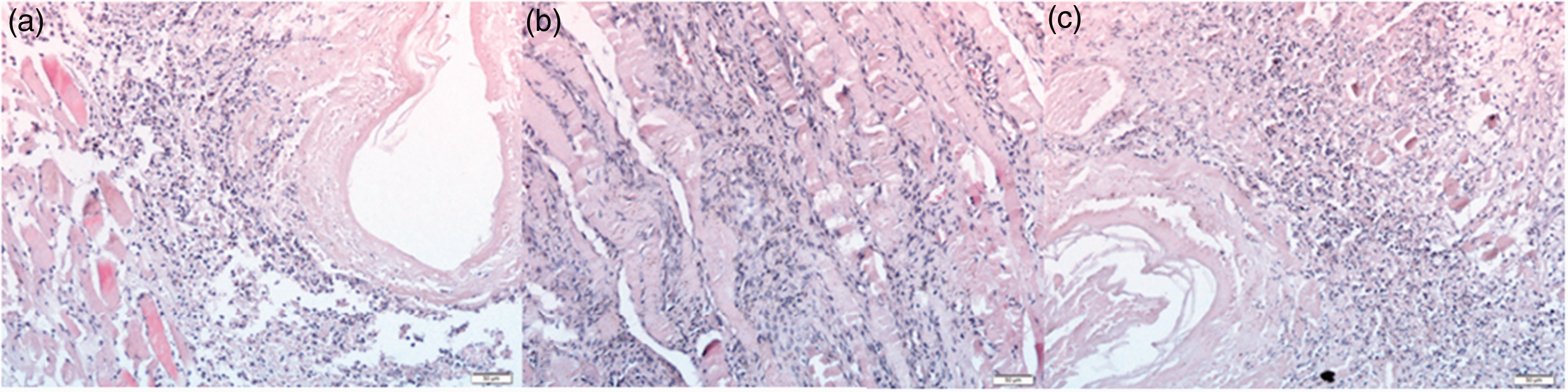
Subcutaneous implantation of the native rat esophagus fragments. (a) The 7th day, (b) the 14th day, and (c) the 21st day; H&E staining; x200.
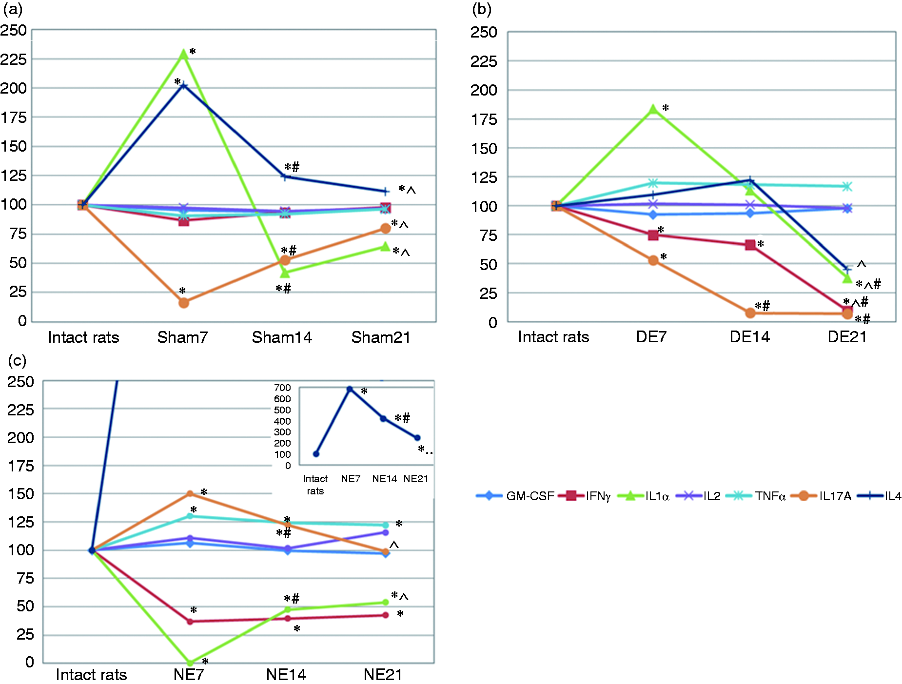
The observed histomorphological picture after the NE implantation was accompanied by a significant increase in cytokines such as IL17A, TNFα and IL4 (p < 0.05), at the same time GM-CSF and IL2 levels were slightly increased (p > 0.05) compare to intact rats. Despite the long-term inflammatory reaction, the concentrations of pro-inflammatory IFNγ and IL1α were lower than the values of the intact control group (p < 0.05) (Table 1). Severe antigenic effect of allogeneic fragments is confirmed by significant differences in cytokine levels compared with sham group: a higher concentration of IL17A, TNFα and IL4, IFNγ; inadequately low concentration of IL1α. On the 14th and 21st day of the experiment, we observed that the level of IL1α stabilized and did not differ significantly from both control groups. It is worth noting the gradual decrease in IL4 levels by the 21st day of the experiment in NE group and in sham operated rats in relation to intact rats.
The cytokine content (pg/mL) in rat serum after subcutaneous implantation of decellularized matrix fragments (Me [Q1; Q3]), DE – decellularized esophagus; NE – native esophagus.
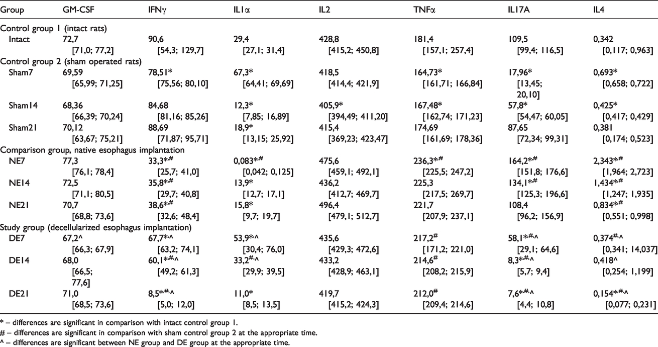
* – differences are significant in comparison with intact control group 1.
# – differences are significant in comparison with sham control group 2 at the appropriate time.
^ – differences are significant between NE group and DE group at the appropriate time.
In contrast, none of the DEs prompted any significant immune response, just inflammation signs. In fact, there were no noticeable differences in visual examination in rats with DE fragments at any given time (Figure 3(c) and (d)). Tissue edema, the formation of a large number of vessels in the implantation zone, a weak tissue reaction and the formation of mature granulation tissue (up to 40–50 μm in diameter) with proliferating fibroblasts and a large number of vessels (up to 18 μm in diameter) were observed around DEs after subcutaneous implantation on the 7th day (Figure 5(a)). Basophilic stained cells of an elongated (fibroblast-like) form were found and lymphomacrophagal infiltration was noted in the implants. On the 14th day, the implant volume was reduced, probably due to a decrease in tissue edema and the distance between the collagen fibers of the decellularized matrix. There was a decrease in the severity of inflammatory cell infiltration and implant germination by newly formed thin-walled vessels (up to 14 μm in diameter, Figure 5(b)). Histology revealed that host cells migrated into all DEs after the first week of implantation. Over the 3-week follow-up there was no evidence of DE rejection by the recipient rat (Figure 5).
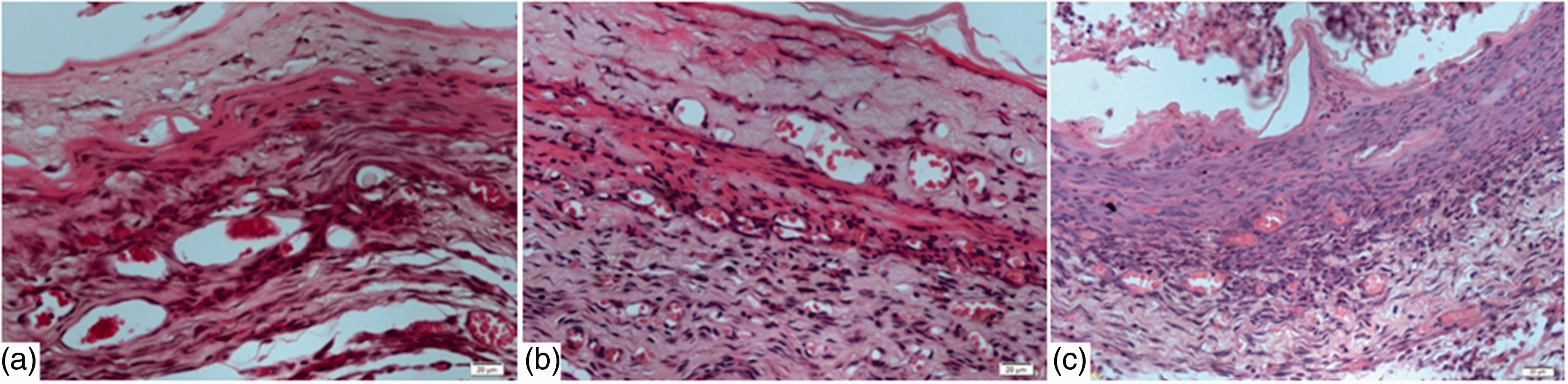
Subcutaneous implantation of rat decellularized esophagus fragments. (a) The 7th day, (b) the 14th day, and (c) the 21st day; H&E staining; x200.
On the 21st day, a further decrease in the implant size and the severity of inflammatory cell infiltration around it were recorded with a predominance of basophilic stained fibroblast-like cells. A large number of vessels with a diameter of up to 10 μm appears in the implantation zone.
In the first week of the experiment in the serum of the rats with DE implantation it was noted that IL1α content was significantly higher in comparison with IL1α values of the intact control group (p < 0.05), comparison group (p < 0.05), but there are no difference from sham operated group; IL2, TNFα, IL4 levels did not differ from the intact control (p > 0.05) levels of the same cytokines, while IL4 concentration significantly exceeded that value in comparison group (p < 0.05) and in sham control group (p < 0.05), which indicates a less intense inflammatory reaction in rats with implantation of decellularized scaffolds. It should be noted that the IL17A, IFNγ content was lower in comparison with intact control values (p < 0.05) (Table 1, Figure 6(a)). Comparative analysis with NE group showed that the IL17A level was lower (p < 0.05), and IFNγ level was higher (p < 0.05) in rats with DE fragments.
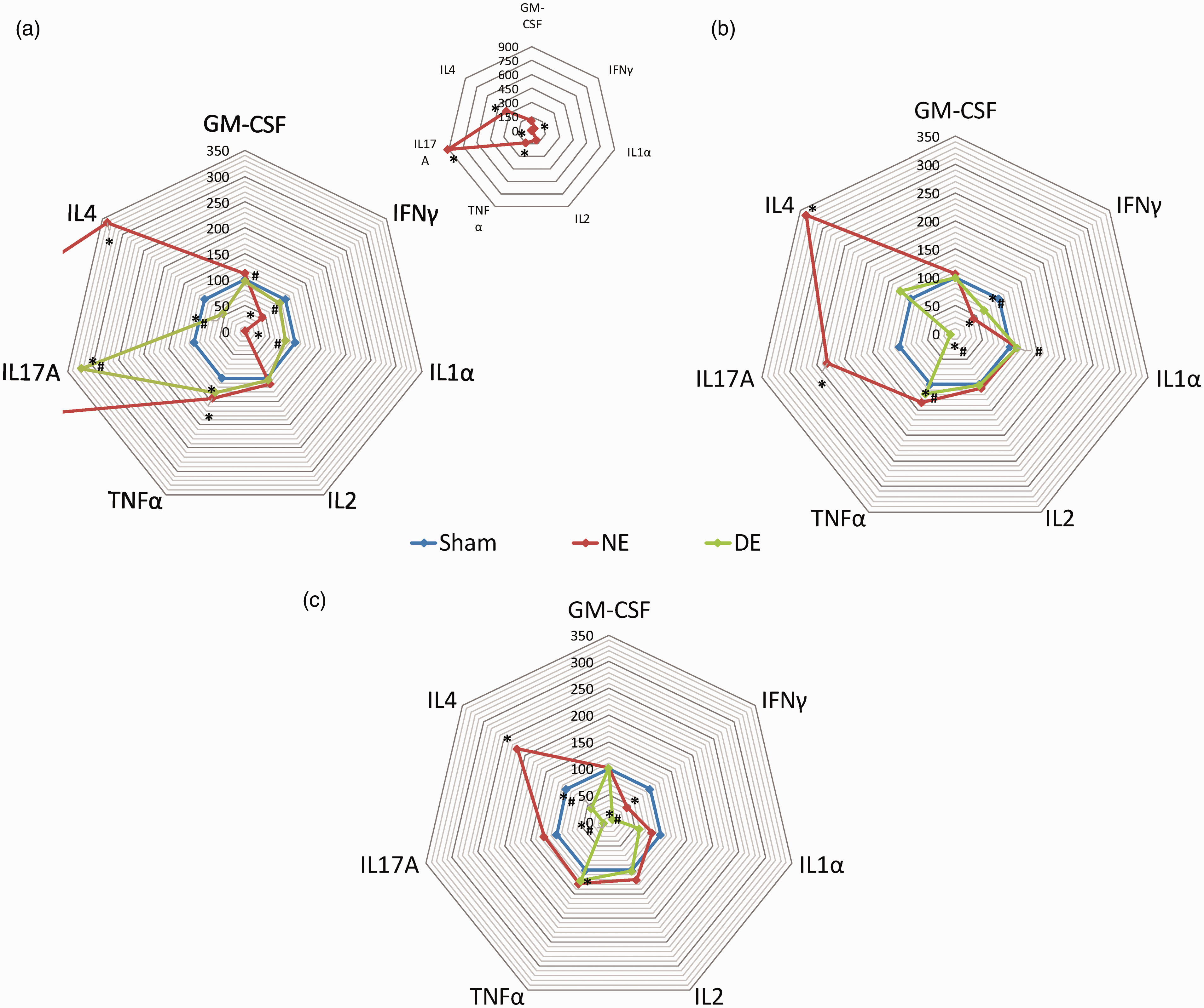
Cytokine profiles at different stages of DE subcutaneous implantation experiment. (a) The 7th day (the data on the NE is presented in the insertion figure), (b) the 14th day, (c) the 21st day. Sham – a sham control group; DE – decellularized esophagus; NE – native esophagus. The data on the NE (day 7) is presented in the insertion figure. The figure shows data on relative values, in which the sham control group has a relative value of 100%.
On the 14th day, IL17A concentration analysis showed a sharp decrease in comparison with the value on the 7th day of the experiment, the sham control corresponding value and intact control data (7 times, 6.9 times and 13 times, respectively). In addition, a decrease in IL1α values to intact control group (p > 0.05) but not reached shame group and a decrease in IFNγ concentration in comparison with the 7th day (p < 0.05) were found (Table 1, Figure 6(b)). No dynamics were observed with respect to IL2, TNFα, GM-CSF, IL4 levels. Higher IFNγ and IL1α levels and lower IL17A and IL4 values in comparison with NE group were also demonstrated, which can be explained as processes of neoangiogenesis in implants occurring in that time interval.
On the 21st day of the experiment, the dynamics of a decrease in IL17A, IFNγ and IL1α content in this group was revealed in comparison with both control group values (Table 1, Figure 6(c)). Besides a decrease in these cytokines, we observed their increased levels in comparison group.
Thus, on the 21st day of the experiment, it is showed mainly a decrease in the concentration of pro-inflammatory cytokines, which reflects the positive dynamics of the wound healing process and the absence of local inflammation and rejection processes in the serum of the rats with subcutaneous DE fragments implantation.
Discussion
The formation of a connective tissue capsule around the implanted material is due to the reaction to a foreign body. It is a healing process that is very long because of the physical presence of the implant. 13 At the implantation zone, macrophages accumulate to neutralize the foreign body; they, however, cannot quickly destroy the implant material, so a macrophage shaft is formatted that delimits the implant from surrounding tissues. If there is no hyperreactivity, fibroblasts form granulomas and surround the implant with connective tissue from inside the macrophages. Granulation is characterized by the presence of a large number of blood vessels, which develop because of both the growth of existing vessels and new ones due to the migration of endothelial and pericyte progenitor cells. 13
Macrophages underwent an activation process upon adhesion to the surface of the biomaterial and the formation of a connective tissue capsule. This process characterizes morphological and cytoplasmic changes leading to the interaction of the complement system and cells and the release of intracellular components. At the same time, pro-inflammatory cytokines activate the connective tissue metabolism and stimulate the proliferation of fibroblasts and epithelial cells, which is important for the healing process. If pro-inflammatory cytokines are secreted and enter the blood in excess, systemic effects are realized.
Immune system humoral factors of specific and nonspecific immunity and local actions of immune cells close to the antigenic information source play a significant role in the development of the inflammatory response. These cells locally produce cytokines, and they distinguish and kill other cells of the surrounding tissues. 14
Cytokines affect almost all cells and play a key role in the progression of inflammation by modulating granulocytes, macrophages, fibroblasts, endothelial cells, epithelial cells, T cells and B cells. 15 Their role in the progression of inflammatory trauma at both the local and systemic levels is almost impossible to estimate. In severe inflammatory conditions, cytokines can enter the bloodstream, causing acute reactions from various organs. The most important role in the inflammatory process belongs to pro-inflammatory cytokines. Therefore, our main interest was in the evaluation of pro-inflammatory cytokines (such as IL1α, IL2, IFNγ, IL17A, TNFα), which enhance the proliferation of T and B cells, antibody synthesis, the production of adhesion molecules and the synthesis of acute phase proteins. In addition, they participate in the processes of specific and nonspecific immunity.
IL1α is one of the most important cytokines for the protection and regulation of wound healing. Obtained data for IL1α level in the rat serum indicate that IL1α level increases compared to intact animals (Figure 7). Our data are found to be a good agreement with published data, which also show an increase in IL1α concentration in rats with inflammatory wounds. 16 It is also known that IL1α plays a critical binding role in connective tissue metabolism; namely, it stimulates the proliferation of fibroblasts and the production of prostaglandin, and also increases the activity of growth factors and the synthesis of cytokines. IL1α also stimulates collagen production and the synthesis of certain enzymes in connective tissue cells. However, wound healing can lead to hypertrophic or keloid scars with the appearance of granular tissue. 17 Our experiments showed that, a decrease in the level of IL1α is observed in dynamics in animals with subcutaneous implantation. This may be perspective for reducing the amount of scar tissue after transplantation of TECs based on decellularized scaffolds. We can assume a negative regulation of the IL1α content by the anti-inflammatory cytokine IL4 regarding subcutaneous implantation of the native esophagus.
IFNγ and TNFα are known because of its exhibit immunomodulatory effects; they are considered inducers of cellular immunity, which are functionally associated with other important pro-inflammatory cytokines. At the same time, we showed that under the conditions of the gradual formation of granulation tissue, IFNγ amount significantly decreases on the 21st day of the experiment, causing a decrease in inflammation and low immunogenicity of DEs. TNFα plays a critical role in the development of inflammation. This cytokine stimulates the proliferation of T and B cells, the antibody production, the synthesis of adhesion molecules and the activation of acute phase proteins. Our study indicated that, TNFα concentration increased slightly at all time points under conditions of subcutaneous implantation, which can be interpreted as the absence of the development of an active adaptive immune response to the matrix. The increased production of this cytokine is mediated by trauma during NE and DE implantation and, probably, indicates the protective and adaptive role of a moderate increase in its production.
In addition, the aim of our work was to evaluate the level of anti-inflammatory interleukin IL4. This cytokine which is produced by Th2 and Th3 cells and is an antagonist of pro-inflammatory cytokines, inhibits the proliferation of T cells and is involved in immune responses to various antigens. They also reduce the levels of IL1α, TNFα, nitric oxide and prostaglandins, which can lead to inflammatory manifestations. The experiment showed that there is a decrease in IL4 in the rat serum in comparison with intact control group and the 7th day, which can be considered as a prognostic factor for successful tissue repair. At the same time, the inflammatory process induces the production of anti-inflammatory cytokines by immunocompetent cells, and the balance between the effects of pro- and anti-inflammatory cytokines affects its outcome. 13 So, on the 7th day, an increase in the IL4 concentration in the rat serum with subcutaneous NE implantation in 6 times was detected compared to both the concentrations of this cytokine in intact control and in DE group. Its increase correlates with an increase in TNFα, which indicates its compensatory role and regulation of TNFα production, which appears as a result of surgical trauma and implant implantation.
Conclusion
The study of the post-implantation systemic immune response to acellular scaffolds to create tissue-engineering structures of the esophagus is crucial for the development of optimal tissue engineering methods, both in gastroenterology and in other fields. As a result of the experiment, it was found that the change in the concentrations of the studied cytokines corresponds to a favourable clinical picture in the group of animals that underwent implantation of the acellular matrices and an active inflammatory reaction in the group with NE implantation. A decrease in the IL1α, IL4, IL17A and IFNγ concentration on the 21st day after DE implantation reflects the positive dynamics of the wound healing process and the absence of local processes of inflammation and rejection of the decellularized matrices.
Footnotes
Acknowledgements
Thanks to our colleagues from the Vivarium of Kuban State Medical University, Krasnodar, Russia, and all those who have participated in the data recording and in the review of this article.
Author Contributions
Karina Melkonyan, Ilya Bykov, Andrey Redko and Sergey Alekseenko: conception and design of the study, analysis and interpretation of data, drafting and critically revising the manuscript, and providing final approval of the version to be published. Ramazan Nakokhov: work with experimental animals (surgical manipulations) and conduction the histological research. Tatyana Rusinova and Yana Yutskevich: conduction ELISA study, analysis and interpretation of data, drafting and critically revising the manuscript.
Declaration of conflicting interests
The author(s) declared no potential conflicts of interest with respect to the research, authorship, and/or publication of this article.
Funding
The author(s) disclosed receipt of the following financial support for the research, authorship, and/or publication of this article: The work was part of a complex research (№АААА-А16-116042550089–5 from 25.04.2016) founded by Kuban State Medical University.