
Abstract
Introduction
Synthetic bone grafts are often used to achieve a well-consolidated fusion mass in spinal fusion procedures. These bone grafts function as scaffolds, and ideally support cell function and facilitate protein binding.
Objective
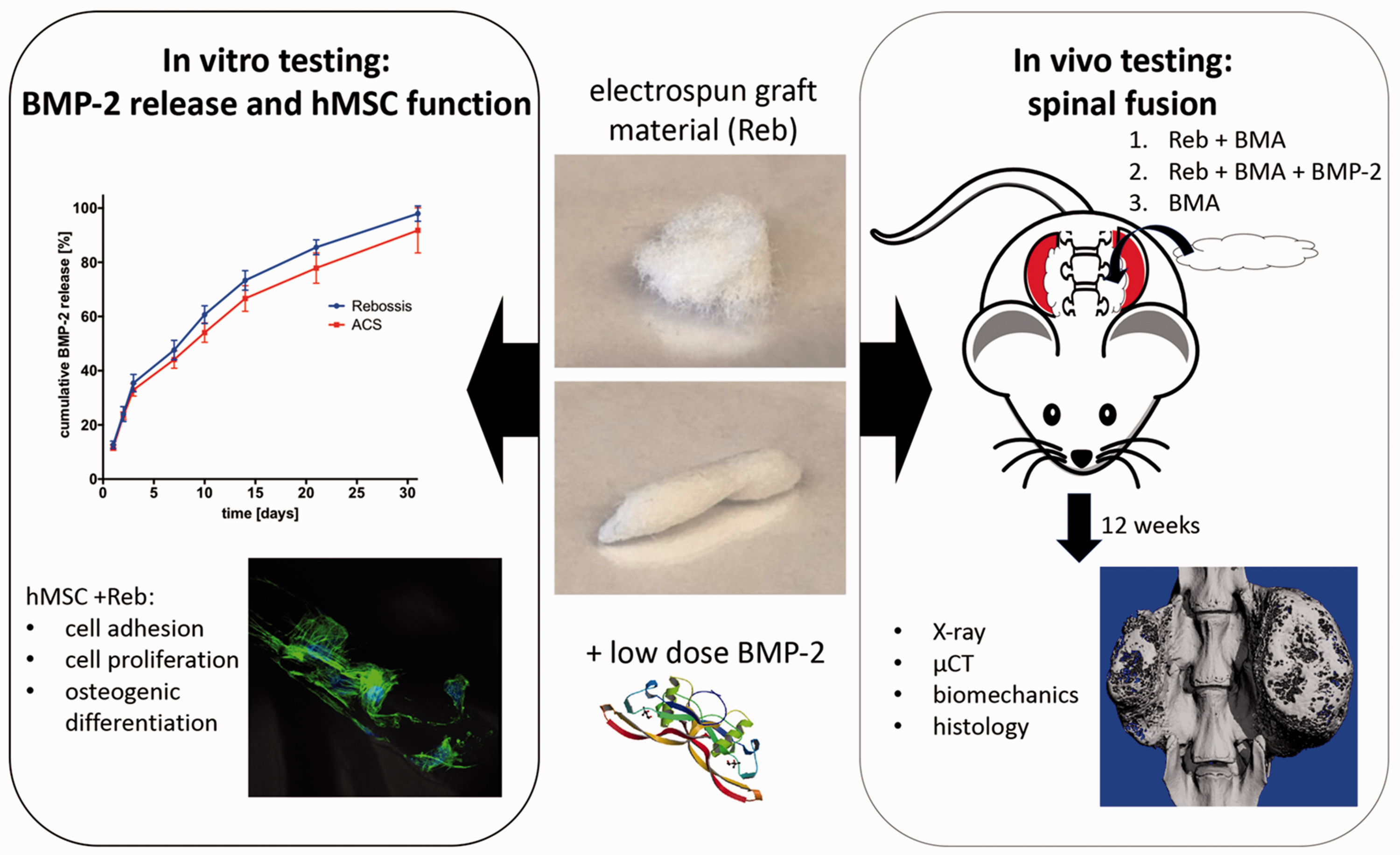
The aim was to characterize an electrospun, synthetic bone void filler (Reb) for its bone morphogenetic protein (BMP)-2 release properties and support of human mesenchymal stem cell (hMSC) function in vitro, and its efficacy in promoting BMP-2-/bone marrow aspirate-(BMA)-mediated posterolateral spinal fusion (PLF) in vivo.
Methods
BMP-2 release kinetics from Reb versus standard absorbable collagen sponge (ACS) was determined. hMSC adhesion and proliferation on Reb was tested using cell counting, fluorescence microscopy and MTS. Cell osteogenic differentiation was quantified via cellular alkaline phosphatase (ALP) activity. For in vivo analysis, 18 Lewis rats were treated during PLF surgery with the following groups: (I) Reb + BMA, (II) Reb + BMA + BMP-2 and (III) BMA. A safe, minimally effective dose of BMP-2 was used. Fusion consolidation was followed for 3 months using radiography and micro-CT. After sacrifice, fusion rate and biomechanical stiffness was determined using manual palpation, biomechanical tests and histology.
Results
In vitro, BMP-2 release kinetics were similar between Reb versus ACS. MSC proliferation and differentiation were increased in the presence of Reb. At 3 months post-surgery, fusion rates were 29% (group I), 100% (group II), and 0% (group III). Biomechanical stiffness was higher in group II versus I. Micro-CT showed an increased bone volume and connectivity density in group II. Trabecular thickness was increased in group I versus II. H&E staining showed newly formed bone in group II only.
Conclusions
Reb possesses a high protein binding affinity and promotes hMSC function. Combination with BMA and minimal dose BMP-2 allowed for 100% bone fusion in vivo. This data suggests that a minimally effective dose of BMP-2 can be used when combined with Reb.
Keywords
Introduction
In order to achieve rapid and successful bone fusion, surgeons have the option to utilize different biologic adjuvants containing the following three components: osteogenic cells, appropriate osteoinductive biological signals (e.g. human recombinant bone morphogenetic protein-2, BMP-2), and osteoconductive biocompatible scaffolds. 1 Autologous bone graft - viable bone tissue harvested from the patient’s own body - is considered the gold standard to support bone fusion. However, the morbidity of the harvesting process causing pain and prolonged duration of surgery has led to a decrease in the frequency of its use. 1 Thus, synthetic bone grafts, such as polymeric synthetic scaffolds, have been developed as an alternative to autograft harvesting. 2 However, synthetic bone grafts are inherently only an osteoconductive scaffold and require supplementation with osteoinductive and osteogenic components. 3
Osteoinductive and osteogenic components such as BMP-2, mesenchymal stem cells (MSCs), and bone marrow aspirate (BMA) are currently being used in pre-clinical and human clinical trials.4,5 MSCs and BMA have the advantage of being relatively safe to use and fairly easy to obtain from iliac crest or adjacent vertebral body. However, the available clinical evidence is insufficient to support the use of MSCs or BMA combined with synthetic or allograft materials as a substitute to autologous bone graft.6,7 BMP-2 is a powerful osteoinductive protein. The Food and Drug Administration (FDA) approved the use of BMP-2 in combination with absorbable collagen sponges (ACS) in 2002 for spinal fusion. 8 The clinical effect of BMP-2 on spine fusion was demonstrated through two large, prospective randomized multicenter trials, which evaluated the use of BMP-2 in anterior interbody lumbar fusion. In these studies, BMP-2 combined with ACS resulted in a higher fusion rate than iliac crest autograft alone. 9
The optimal release characteristics for BMP-2 for spinal fusion is not known at this time. Although BMP-2 release from ACS has been shown to occur within 30 days in vitro,10,11 there is clinical evidence of efficacy for improving spinal fusion outcomes months after. 9 Thus, the release of BMP-2 from ACS seems to improve processes in the initial healing cascade, leading to improved bone consolidation. One disadvantage in the use of BMP-2 includes its high cost. Furthermore, high doses of this protein may have dose-dependent side effect, such as BMP-2 associated inflammation.12,13 Therefore, there is an ongoing interest in the identification and optimization of osteoconductive scaffolds to properly release BMP-2 in order to induce surrounding bone formation.
Several types of scaffolds have been introduced and evaluated to better control growth factor release, including collagen, calcium phosphate ceramics, synthetic and biopolymers.14,15 A limitation of calcium phosphate ceramics is that high amounts of BMP-2 are required, and biopolymers (e.g. collagen) are associated with batch-to-batch variations. 15
Due to the ongoing challenge in bone tissue engineering to fabricate a bone graft that optimally binds bone growth stimulating cells and growth factors, electrospun materials have gained attention in recent years. 16 The electrospinning technology has some remarkable features, such as ease of handling, sufficient porosity with tunable pore size and shape for optimized penetration and diffusion of cells, growth factors and nutrients.17–19 The inherently high surface to volume ratio of electrospun scaffolds can enhance cell attachment, drug loading, and mass transfer properties. 20 Wrapping of BMP-2 releasing hydrogels with a hydrophobic PDLLA electrospun nanofiber membrane was shown to adjust the direction of the BMP-2 release behavior and to be beneficial for guided spinal fusion. 18
For use in posterolateral fusion (PLF) procedures, an electrospun, synthetic bone void filler (ReBOSSIS, Orthorebirth, USA, referred to as Reb throughout the manuscript) received FDA premarket approval (510(k), K172573). According the company’s user information, Reb is a highly porous, biomimetic, and biodegradable scaffold, consisting of (by weight) 40% beta-tricalcium phosphate (β-TCP), 30% siloxane-containing vaterite (a form of calcium carbonate, CaCO3), and 30% poly(L-lactide-co-glycolide). The nanoparticle component of silicon (siloxane) is known to strengthen the adhesion of stem cells on poly-L-lactide. 21 With the consistency, formability and hydrophilic characteristics of cotton, Reb may demonstrate beneficial peptide and cell binding, and release characteristics compared to ACS.
In this study we hypothesize that Reb supports BMP-2 binding and MSC function, allowing for the use of a low dose of BMP-2 to facilitate robust spinal fusion in a BMP-2-/BMA-mediated posterolateral spinal fusion model in rat. Reb was characterized for its BMP-2 release properties and support of hMSC in vitro, and for its efficacy in promoting BMP-2-/BMA-mediated PLF in vivo. For in vivo research, genetically identical Lewis rats were employed, which allow for bone marrow transplantation. 22
Materials and methods
Study design
To determine the biological characteristics of Reb in vitro, BMP-2 release kinetics from Reb compared to ACS, as well as adhesion, proliferation and osteogenic differentiation of human MSC’s seeded on Reb was analyzed.
For the in vivo analysis, Reb was implanted into fusion beds of female Lewis rats (LEW/Crl, ranging from 220–300 g in weight, Charles River Laboratories, USA) undergoing PLF. Rats were randomly assigned to the following groups to test the potential of Reb in the presence BMA with and without a minimally effective dose of BMP-2: (I) Reb + BMA, (II) Reb + BMA + BMP-2, and (III) BMA only. The dosage for BMP-2 for Lewis rat was determined based on a prior study by our team in Lewis rats undergoing PLF, showing that the used BMP-2 dose in combination with ACS resulted in a sub-optimal healing rate (50% for ACS + BMP-2 and 89% for ACS + BMP-2 + BMA, planning and surgical procedure for that study were done by two of our co-authors that repeated the surgical procedure using the same rat strain for our study). 22 In total, 36 Lewis rats (18 BMA donor rats + 18 rats undergoing PLF) were included in the study. In experimental group II, 2.3 µg BMP-2 per side was used for a 300 g weighted rat. After surgery, rats were radiographically followed for fusion consolidation for 3 months. After sacrifice at 3 months, manual palpation, µCT, histological analysis and biomechanical testing were performed (Graphical abstract).
In vitro studies
BMP-2 release kinetics
The kinetics of BMP-2 release from equal volumes of Reb (86 mg, 400 mm3) vs. ACS (Helistat®, Integra Life Science, USA) (10 × 10 × 4 mm, 400 mm3) were determined. Bone grafts were loaded with 2.3 µg BMP-2 (Medtronic, USA), diluted in 100 µl BMP-2 buffer each. Each condition was performed in triplicates. Each loaded graft was incubated for 15 min at room temperature before it was placed into a 2 ml tube containing additional 400 µl of BMP buffer (Medtronic, USA). Tubes were incubated at 37 °C with continuous agitation. After a period of 1, 2, 3, 7, 10, 14, 21 and 31 days, the supernatant was collected, and fresh BMP buffer (Medtronic, USA) added. The amount of BMP-2 in the supernatant obtained at each time point was determined with the human BMP-2 Quantikine ELISA kit (R&D systems, USA).
Cell seeding on bone graft and determination of cell adhesion
MSCs were isolated from human bone marrow of three healthy donors obtained from Lonza, USA. Density gradient centrifugation using Ficoll was applied for cell isolation. Cells were expanded using DMEM, supplemented with 10% fetal calf serum. For cell seeding, 20,000 cells were dripped on Reb bone graft, as described by Seebach et al. 23 After short incubation at 37°C, the medium containing the non-adhering cells was removed and the bone graft carefully rinsed with PBS. Next, the grafts were transferred to another well containing medium. The remaining cells in the supernatant and the bottom of the initial seeding were counted and the percentage of adherent cells calculated as follows: [(initial cell number – remaining cell number)/initial cell number) ×100%]. For visualization of cell adherence, hMSC-seeded bone grafts were incubated for a period of 3 days in a CO2 incubator at 37°C. After the indicated time, a DAPI/β-actin (Life Technologies, USA) staining was conducted. Cell staining was visualized in terms of fluorescence microscopy.
hMSC proliferation and differentiation
The adhesion and proliferation rate of cells seeded on Reb was measured by using MTS (Promega, USA). Assays were conducted according to the manufacturer’s instructions. Sample analysis was performed on days 0, 3 and 7. For osteogenic differentiation, cells were seeded on 24-well tissue culture plates in the presence and absence of Reb, separated by a 0.8 µm transwell insert. For induction of osteogenic differentiation, culture medium containing 1% (v/v) ITS + 1, 100 nM dexamethasone, 50 μg/mL ascorbate-2-phosphate, and 100 ng/mL BMP-2 was used. 24 Total cellular alkaline phosphatase (ALP) activity was quantified during osteogenic differentiation using a colorimetric alkaline phosphatase assay (Abcam, USA). 25 The assay was performed on days 1 and 14 after MSC seeding. ALP values (day 14–day 1) were analyzed and normalized to cell metabolic activity, which was evaluated via MTS test.
In vivo studies
Posterolateral intertransverse process fusion surgery
Surgery was performed in accordance to protocol #IACUC007419, approved by Cedars-Sinai Medical Center’ Institutional Animal Care and Use Committee prior to study start. Dissection was performed to expose the transverse processes of L4 and L5 of Lewis rats. The transverse processes were decorticated with a high-speed burr until punctate bleeding. Prepared implants were placed bilaterally in the paraspinal muscle bed, between and touching the transverse processes of L4 and L5 (Figure 1). The surgeon was blinded to the experimental treatment groups during operation. No internal fixation was used.

Images of the surgical procedure of spinal fusion using Reb. (a) midline posterior longitudinal incision over lumbar spine. (b) Localization of the transverse processes of the spinal segments L4 and L5 (forceps tips), which were subsequently decorticated with a high-speed burr until punctate bleeding. (c) Bilateral implantation of prepared Reb into the paraspinal muscle bed, between and touching the transverse processes of L4 and L5. (d) Final result of Reb implantation prior to suturing (cranial is top and caudal is bottom in each image).
After all surgical procedures, muscles and skin were closed with non-absorbable sutures. After anesthesia and the morning after surgery, rats were medicated with subcutaneous injections of buprenorphine (0.05 mg/kg) to control pain. To prevent infection, 0.1 mg/ml enrofloxacin was given in drinking water to each rat and rats were housed in singles for a duration of 2 weeks. Rats were sacrificed at 12 weeks post-surgery.
Implant preparation and bone marrow isolation
Simultaneously at the start of surgery on a separate sterile field, implants were prepared by adding BMP-2 and/or BMA to 86 mg of Reb graft (for a 300 g rat, adjusted according to rat weight) in an 2 ml vial under careful agitation until the liquid was fully absorbed. Implants were used within 15 min.
To prepare 50 µl of BMA, a donor rat was euthanized, and its femurs were harvested. BMA was obtained by centrifuging the femurs at 400 g for 3 min after resecting the proximal and distal aspects of each femur. 50 µl BMA were combined with either 100 µl PBS (group I) or with 2.3 µg BMP-2/100 µl BMP buffer prior to loading on graft (for a 300 g rat, adjusted according to rat weight, group II). In the BMA only group, 50 µl BMA was carefully injected into the implantation site (group III; see also online Supplemental Table 1).
High resolution radiographs
Radiographical fusion were determined in vivo via high-resolution radiographs (LX-60, Faxitron X-Ray, LLC Lincolnshire, IL) obtained in the prone position at 8 and 12 weeks postoperatively under inhalation anesthesia.
Manual palpation
Fusion quality and quantity was assessed via manual palpation testing of spine segments by two independent observers that were blinded to the experimental groups. 22 Any motion detected between the L4 and L5 segment and adjacent segments, including the transverse process or vertebral bodies, was considered to indicate a fusion failure. Each side was tested separately; the observation of no motion on both the right and left sides indicated a successful fusion.
Biomechanical testing
Fused specimens were potted and loaded in flexion with a four-point bending apparatus (MTS 642.001 A-02 MTS Corp., Eden Prairie, MN) on a servo-hydraulic actuator (MTSMTS Bionix 370.02, MTS, Eden Prairie, MN), as previously described.26,27 Briefly, specimens were loaded at a rate of 3 mm/min, while force was recorded and the first linear region of the resultant moment-deflection curve was used to determine stiffness. The researcher performing biomechanical testing was blinded to the experimental groups.
Micro-computed tomography
Ex vivo µCT (vivaCT 40, Scanco USA Inc., Wayne, PA) was performed on explanted spines to determine the structural properties of the fusion mass. Histomorphometric 3D evaluation was performed on a cylindrical volume of interest (VOI) including the upper and lower vertebral bodies around the fusion bed at the transverse processes of L4-5. A constrained 3D Gaussian filter (σ = 0.8, support = 1) was used to partly suppress VOI noise. The trabecular bone tissue was segmented from marrow and soft tissue by using a global thresholding procedure. The researcher was blinded to the experimental groups. Direct 3-dimensional morphometry was used to determine (A) volume of mineralized bone tissue (BV; ΔBV = BV (week 12) – BV (pre-surgery), (B) bone volume/total volume (BV/TV), (C) trabecular thickness, (D) trabecular spacing, (E) bone mineral density (BMD) and (F) connectivity density.
Histological preparation
For analysis of fusion groups, spines were fixed with 10% formalin and decalcified with Surgipath Decalcifier II (Leica, USA) for a duration of 7 days. For assessment of new bone formation, the specimens were sliced (5 µm of thickness) in the sagittal plane near the transverse processes at levels L4-5.
Statistics
All statistical analyses were performed using Prism 7; p < 0.05 was considered to be statistically significant. The outcome measurements were 1) BMP-2 protein amounts, 2) MSC function and 3) µCT measures. Separately for each dependent measure, analysis of variance (ANOVA), mixed model, or T-Test were performed using mean values with grouping of implant group; for multiple comparisons, appropriate post hoc tests were used. In figures, data are displayed as mean of biological replicates ± SEM.
Results
In vitro studies
BMP-2 kinetics are similar between reb and ACS
Release characteristics of BMP-2 from Reb vs. ACS were investigated. Both materials demonstrated a similar liquid absorption capacity of about 60% of their volume (no data shown). BMP-2 release curves between Reb and ACS were comparable, with an initial BMP-2 release of 10–12% on day 1 (corresponding to 230–270 ng out of 2.3 µg added to the materials). Complete BMP-2 release was reached after 30 days. No significant differences in BMP-2 release between these two carrier material groups were detected (Figure 2).
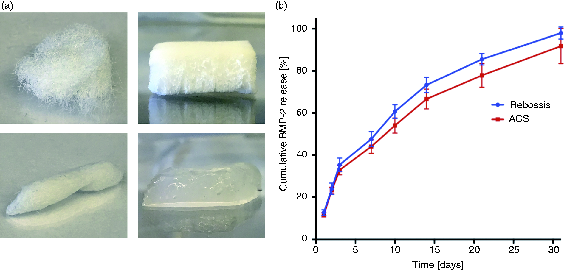
Cumulative release of BMP-2 from Reb is not different from ACS in vitro. (a) Pictures of Reb (left) and ACS (right) in dry (top image) and in wet (bottom image) condition. (b) Shown is the percentage release over a time period of 31 days. Number of samples per group: n = 3.
Reb supports hMSC adhesion, proliferation and differentiation
Ninety percent of hMSCs adhered to the Reb graft after 3 days of cell culture on the graft (mean: 90 ± 1.3%, p < 0.05) (Figure 3(a)). Human MSC proliferation increased between days 1, 3 and 7 (meanOD490 day 1: 0 ± 0, day 3: 0.18 ± 0.01, day 7: 0.43 ± 0.05; day 1 vs. 3: p < 0.001; day 3 vs. 7: p < 0.0001; day 1 vs. 7: p < 0.0001) (Figure 3(b)). ALP activity was increased in the presence of Reb between days 1 and 7 (meanday1: 5.2 ± 3.2, meanday7 2.3 + /–3.0; p < 0.05) (Figure 3(c)).
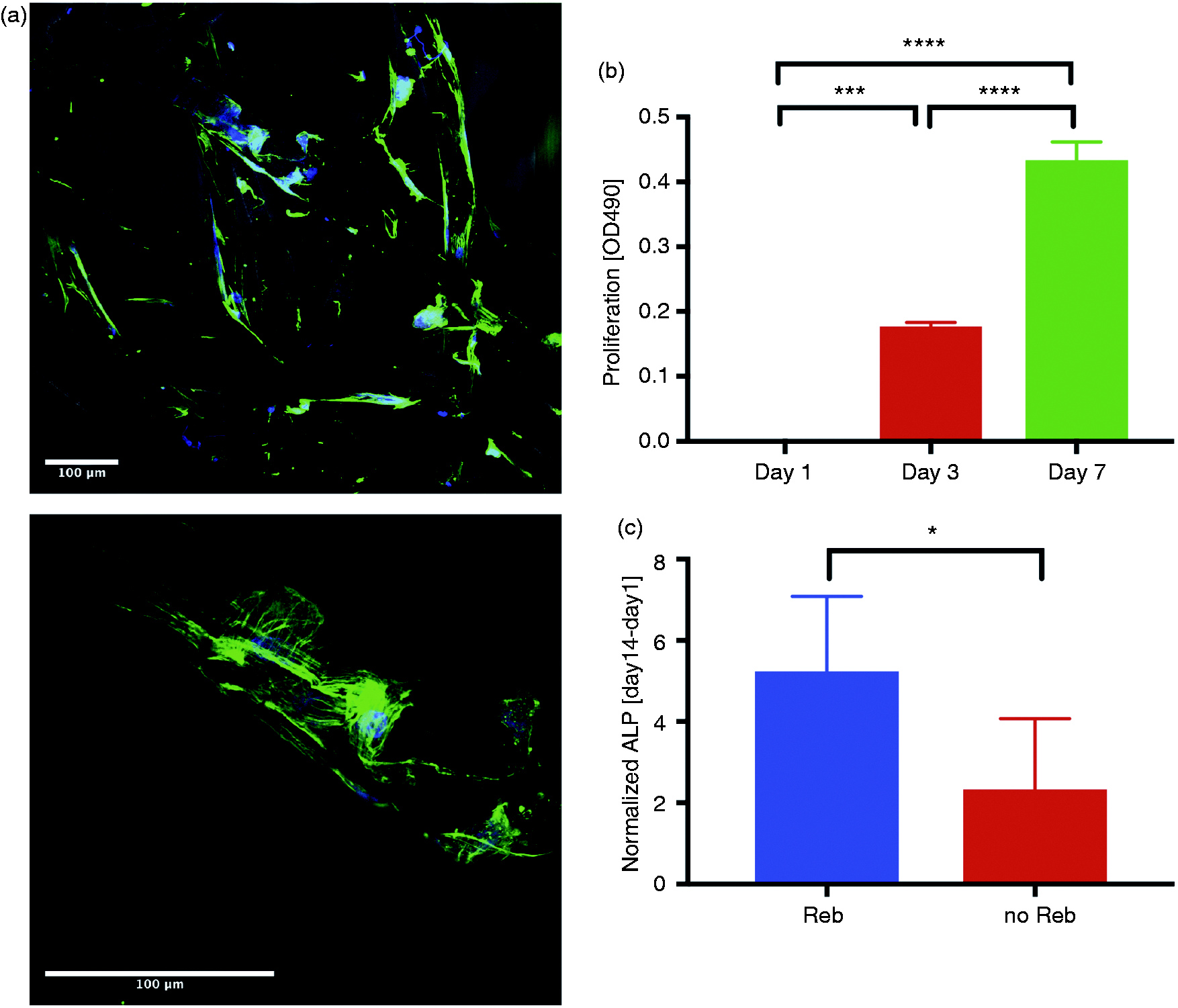
Reb supports hMSC adhesion and proliferation and stimuates osteogenic differentiation. (a) DAPI/β-actin immunofluorescent stain of of hMSCs adhering to Reb on day 3 after seeding. (b) hMSC adhesion and proliferation on Reb over time is shown, determined via MTS assay. (c) Quantification of ALP activity of hMSCs in the presence and absence of Reb, investigated via transwell assay. Per condition, the increase of ALP activity between days 1 and 14 was measured. Results were normalized to cell metabolic activity. Cells from 3 patients were included per group.*p < 0.05, ***p < 0.001, ****p < 0.0001.
In vivo studies
Implantation of BMA- and low dose BMP-2-loaded reb promotes spinal fusion, demonstrated by radiography and biomechanical stiffness testing
At week 12, explanted spines showed a 100% fusion rate in the graft + BMA + BMP-2 group (7 out of 7 spines), a 29% fusion rate in the graft + BMA group (2 out of 7 spines), and no fusion in the BMA only group (0 out of 4 spines), which was detected via manual palpation (Figure 4(a)). Biomechanical stiffness of the fused spine samples was 286.90 ± 72.17 Nmm/mm in the graft + BMA + BMP-2 group and 198.25 ± 7.70 Nmm/mm in the graft + BMA group (Figure 4(b)). X-ray evaluation of the spines at weeks 8 and 12 confirmed the signs of new bone formation in the graft + BMA + BMP-2 group (online Supplemental Figure 1).
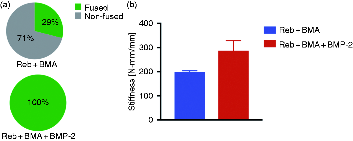
Reb+BMA+low dose BMP-2 result in 100% spinal fusion. (a) Manual palpation (n = 18) and (b) Biomechanical stiffness results from fused samples are shown (n ≥ 2).
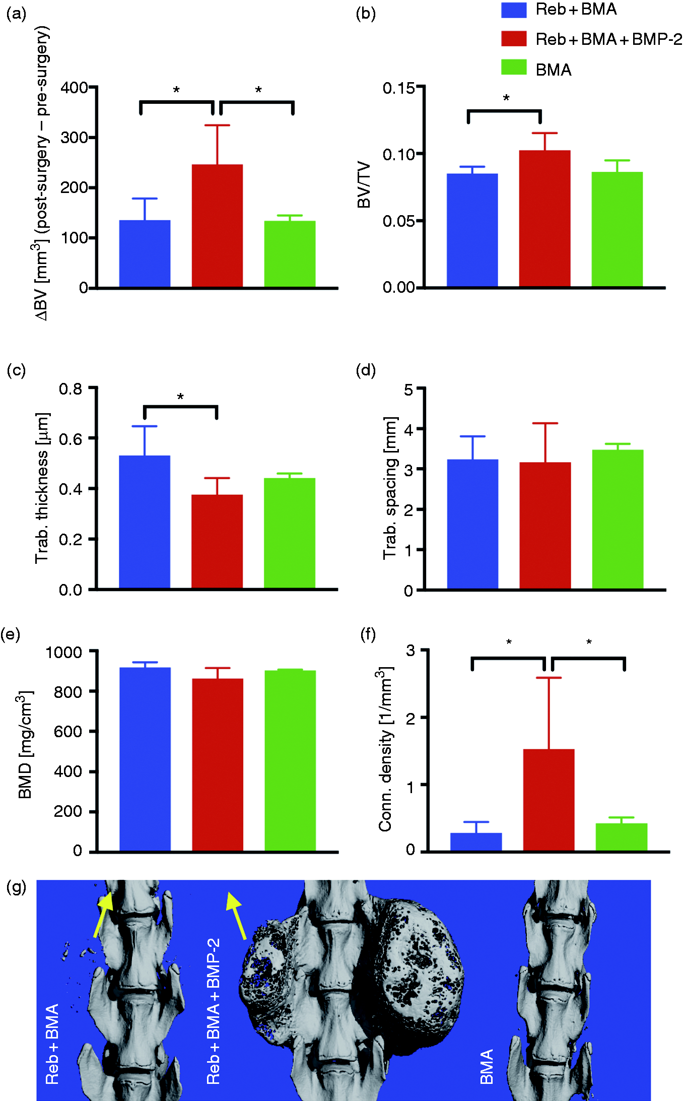
Bone volume, bone volume/total volume and connectivity density are increased in the Reb+BMA+BMP-2 group and trabecular thickness is increased in the Reb+BMA group. (a–f) µCT parameters of the implantation sites at the transverse processes of L4-5 of the three experimental groups are shown. (a) Delta Bone Volume (BV post-surgery-BV pre-surgery), (b) bone volume/total volume, (c) trabecular thickness, (d) trabecular Spacing, (e) bone mineral density, and (f) connectivity density. Number of samples per group: n ≥ 4. G. Representative pictures of 3 D reconstructed images of all groups. Yellow arrows indicate paraspinal bone formation at the implant site in the Reb+BMP+BMA group. *p < 0.05.
Bone quality and quantity are increased after implantation of BMA- and low dose BMP-2-loaded reb
Micro-CT analysis of the spine explants at week 12 showed an increased bone volume in the graft + BMA + BMP-2 group (group II) vs. graft + BMA (group I) vs. BMA only groups (group III) (group I: 135.6 ± 43.0, group II: 216.0 ± 88.1, group III: 133.9 ± 10.9, p < 0.05, Figure 5(a) and (g)). Bone volume/total volume was increased in group II vs. group I (group I: 0.085 ± 0.0005, group II:0.0098 ± 0.144, p < 0.05, Figure 5(b)). Trabecular thickness was increased in group I vs. group II (group I: 0.53 ± 0.42, group II: 0.42 ± 0.11, p < 0.05, Figure 5(c)). Connectivity density was increased in group II vs. group I (group I: 0.28 ± 0.16, group II: 1.26 ± 1.10, group III: 0.42 ± 0.09, p < 0.05, Figure 5(f)). No differences in trabecular spacing and bone mineral density were observed between the experimental groups (Figure 5(d) and (e)).
New bone formation detected within implants containing BMA- and low dose BMP-2
Histological evaluation performed on spine explants at week 12 post-surgery demonstrated new bone formation within the graft in the graft + BMA + BMP-2 group (Figure 6(a) and (c)). In the graft + BMA group, no evidence of new bone formation was detected (Figure 6(b) and (d)). In both experimental groups, Reb was not fully degraded after 12 weeks. No new bone formation was detected in the implant-free BMA group (no images shown).
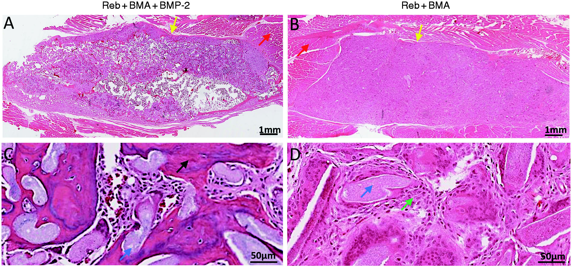
H&E staining of the implant region detects new bone formation in the Reb+BMA+BMP-2 group. Sagittal cuts through the implant region of Reb+BMA and Reb+BMA+BMP-2 are shown. (a and b). Whole implant areas are shown. In the Reb+BMA group, bone formation did not occur. In Reb+BMA+BMP-2, new bone formation was detected within the implant area. (c and d) Magnification of tissue structural properties within implants. Arrows: red: muscle tissue, yellow: host-implant interphase, green: mononuclear cells, blue: Reb implant, black: bone.
Discussion
As alternative to autogenous bone graft, synthetic osteoconductive bone grafts with and without osteoinductive components are being developed for musculoskeletal applications. To support bone healing and minimize risks for the patient, the release time and concentration of osteoinductive components needs to be well-controlled. This is especially important with regards to BMP-2, a highly potent growth factor, which may have serious adverse effects with high-dose or off-label use. Due to these reasons, there is an ongoing interest in the identification and optimization of osteoconductive scaffolds to properly release osteoinductive factors into the surrounding tissue, as well as support osteogenic cell proliferation and differentiation.
In this this study we demonstrated that an electrospun, synthetic bone void filler (Reb) has similar BMP-2 binding and release characteristics to the well characterized collagen sponge (ACS). Furthermore, Reb promotes human MSC attachment, differentiation and proliferation in vitro. In vivo, a minimally effective dose of BMP-2 in combination with Reb and BMA was shown to be sufficient to achieve a 100% spinal fusion in a rat model.
Our study demonstrated similar in vitro BMP-2 release profiles from Reb compared to standard ACS. Both Reb and ACS showed a sustained release rate of BMP-2 for a duration of 30 days, a time period that includes the different biological stages of bone healing in vivo, including inflammation, angiogenesis and osteogenic differentiation. 28 The maximum loading efficiency of BMP-2 was not evaluated in this study, but it has been demonstrated that 129 ug can be loaded onto similar size ACS without showing a strong initial burst effect, 11 indicating that much higher doses can be efficiently bound to the material than the 2.3 µg used in the present study.
Although our study investigated the BMP-2 release behaviors in vitro only, successful new bone formation and slow implant degeneration was demonstrated in the Reb + BMP-2 + BMA group via micro-CT, biomechanical and histological analyses. In contrast to Reb, other carriers such as hyaluronic acid hydrogel have been shown to degenerate quickly, resulting in poor bone healing.29,30 For example, binding of BMP-2 to a poly(phosphazene) hydrogel demonstrated limited BMP-2 release and support for bone generation due to its complete degradation in 21 days. 30
Herein, an increased BMP-2 release rate from Reb and ACS was detected within the first 3 days in vitro. These results are in line with the literature, showing a burst release of BMP-2 from ACS in the first 3 days. 10 While the optimal release profile of BMP‐2 for safe and effective clinical application is unknown, a recent study demonstrated increased subcutaneous bone formation after implantation of a phosphorylated hydrogel showing a BMP‐2 burst release compared to other materials without this effect. 31 In contrast, strong burst effects resulting in low release rates or lack of release at later time points have been shown to negatively affect BMP’s bone inducing potential. 32
In order to investigate cell function within Reb, hMSC adhesion, proliferation and differentiation was analyzed. In this study, approximately 90% of hMSCs adhered to Reb after 3 days of cell culture. Compared to various other synthetic bone graft materials showing an MSC adhesion rate of 20–80% after 2 days, 33 the detected adhesion rate is high. This may be explained by two special properties of Reb: structurally, Reb has a micro three-dimensional structure using the electrospun technology, which provide as much contact area as possible for adhesion for stem cell in a restricted space. Chemically, the nanoparticle component of silicon in Reb is known to strengthen the adhesion of stem cells on PLGA, which is the main ingredient of Reb. 21
Moreover, we demonstrated an increased hMSC proliferation and osteogenic differentiation in the presence of Reb between days 1–7. Similar to our study, Yoshimoto et al. demonstrated MSC adhesion and osteogenic differentiation when cultured on an electrospun poly(ε-caprolactone) (PCL) scaffold. 34 In contrast, analysis of MSC binding on different bone graft materials performed by Schultheiss et al. demonstrated no cell proliferation on most materials tested. 33
The effect of Reb on spinal fusion was investigated using a posterolateral spine fusion procedure in a rat model, which is well established in our lab. 22 Three experimental implant groups were included in this study: Reb + BMA, Reb + BMA + BMP-2 and BMA only. To evaluate the impact of Reb on BMP-2-mediated fusion in comparison to standard ACS as carrier, BMP-2 was added in a low dose of 2.3 µg/side/300 g rat to each implant. This dose has been shown to be suboptimal for stimulation of spinal fusion in this rat model: a prior study performed by our team using the same animal model detected an 89% fusion rate when applying 1.68 µg/side/220 g rat (equals 2.3 µg/side/300 g rat) of BMP-2 to ACS in the presence of BMA. 22 In the present study, implantation of Reb + BMP-2 + BMA resulted in 100% fusion as demonstrated by radiography and manual palpation. This shows that BMP-2 dosing can be reduced in rats when used in combination with Reb and BMA. Furthermore, our study detected a 29% fusion in the Reb + BMA group compared to 0% fusion in ACS + BMA group that we presented in our prior study. 22 No fusion was found in rats undergoing the same surgery with BMA application only-control. Since we did not detect any differences in the BMP-2 release kinetics in vitro, we hypothesize that an improved binding and release of cells or other growth factors, support of cell proliferation and differentiation as well as the slow degradation of the material may contribute to the observed effect.
Biomechanical analysis of the fused lumbar spines treated with Reb + BMA + BMP-2 had an average stiffness of 286.90 ± 72.17 Nmm/mm, which was comparable to stiffness values recorded for rat lumbar fusion treated with an autograft. 26 The resultant lumbar fusions of specimens treated with Reb + BMA alone were 44% less stiff, indicating a reduction in quality of the formed fusion mass.
Micro-CT analysis to verify the bone quantity and quality showed an increased bone volume, bone volume/total volume and connectivity density in the Reb + BMP-2 group compared to Reb + BMA and BMA only. These results suggest that new bone is formed and tissue remodeling is more robust in the Reb + BMP-2 + BMA group compared to controls.
A reduction of BMP-2 dosing for fusion could have different clinical benefits. Considering several major and minor adverse effects, such as as ectopic bone formation in the neural canal, dysphagia after anterior cervical fusion, prevertebral swelling, seroma/hematoma formation, radiculitis, osteolysis etc., potential complications may be decreased by lowering BMP-2 doses.13,35 Furthermore, BMP-2 may be applied at multiple sites, e.g. for multi-level fusion surgery and deformity correction surgery for degenerative kyphosis, scoliosis and kyphoscoliosis. In future research, the combinatory release of growth factors and other biologic agents from Reb should be considered, as previously evaluated using ACS and other scaffolds with similar applications and desired outcomes in addition to single application of BMP-2.11,36,37
Conclusion
Our results demonstrate that Reb possesses a similar BMP-2 protein binding affinity and release profile as ACS. Furthermore, Reb promotes hMSC binding, proliferation, and osteogenic differentiation. Our in vivo data in a rat PLF model show that low dose BMP-2 is sufficient to induce spinal fusion in 100% of animals when combined with Reb + BMA. Therefore, we suggest that the structural properties of this synthetic graft support cell responses to the delivered BMP-2, which may allow for a reduction in BMP-2 dosing.
A reduction of the minimally effective dose through combination with new carriers may reduce the occurrence of potential side effects and save healthcare utilization costs. However, systematic dosing studies are needed to appropriately delineate BMP-2 dosing-carrier response curves to optimize release for a biological response in targeted bone formation. Furthermore, immune responses to the Reb bone graft and the duration to complete biodegradation of Reb beyond the 12-week timepoint should be addressed in future studies.
Supplemental Material
sj-pdf-1-jba-10.1177_0885328220937999 - Supplemental material for Electrospun, synthetic bone void filler promotes human MSC function and BMP-2 mediated spinal fusion
Supplemental material, sj-pdf-1-jba-10.1177_0885328220937999 for Electrospun, synthetic bone void filler promotes human MSC function and BMP-2 mediated spinal fusion by Juliane D Glaeser, Khosrowdad Salehi, Linda EA Kanim, Derek G Ju, Jae Hyuk Yang, Phillip H Behrens, Samuel A Eberlein, Melodie F Metzger, Yasaman Arabi, Tina Stefanovic, Dmitriy Sheyn and Hyun W Bae in Journal of Biomaterials Applications
Footnotes
Declaration of conflicting interests
The author(s) declared no potential conflicts of interest with respect to the research, authorship, and/or publication of this article.
Funding
The author(s) disclosed receipt of the following financial support for the research, authorship, and/or publication of this article: This work was supported by Orthorebirth USA, the Foundation for Spinal Restoration, and NIH/NIAMS [grant no. K01AR071512].
Supplemental material
Supplemental material for this article is available online.
References
Supplementary Material
Please find the following supplemental material available below.
For Open Access articles published under a Creative Commons License, all supplemental material carries the same license as the article it is associated with.
For non-Open Access articles published, all supplemental material carries a non-exclusive license, and permission requests for re-use of supplemental material or any part of supplemental material shall be sent directly to the copyright owner as specified in the copyright notice associated with the article.