
Abstract
Objective
Since the systemic drugs have been used to reduce the hyperplasic response in the tunica intima, the periadventitial local drug applications to the vascular wall have gained more popularity. In this study, we investigated the effect of bovine serum albumin-glutaraldehyde and polyethylene glycol polymer on neointimal hyperplasia in rabbit carotid artery anastomosis to explore the effects of these two different agents.
Methods
21 New Zealand male rabbits were randomly divided into three groups. The carotid artery transection and anastomosis was performed onthe control group. The bovine serum albumin-glutaraldehyde and the polyethylene glycol polymer were applied locally on the other two groups seperatley after transection and anastomosis of the carotid arteries. At the end of 28-day follow-up, the histological and the immunohistochemical results related to neointimal hyperplasia were compared.
Results
The glue residues were detected in the BSA-glutaraldehyde group, but in the PEG polymer group there was no glue residue. The intima thickness and the intima/media thickness ratio in the control group was significantly higher (p<0.05) than the other groups. These values did not differ significantly between the BSA-glutaraldehyde group and the PEG polymer group (p>0.05). The lumen diameter and the area in the control group were significantly higher (p < 0.05) than the BSA-glutaraldehyde group. These values between the control group and the PEG polymer group did not differ significantly (p>0.05). aSMA-positive staining score in the Control group was found to be significantly lower (p < 0.05) than the BSA-glutaraldehyde and PEG polymer group and the VEGF-positive staining score in the control group was found to be significantly higher (p < 0.05) than the BSA-glutaraldehyde and the PEG polymer group.
Conclusions
Although the both agents have positive results on neointimal hyperplasia, it would be favorable to use polyethylene glycol polymer, since it does not seem to affect the lumen area and the lumen diameter of the vessel.
Introduction
It has been shown that the main cause of the luminal narrowing in the arterial damage models (in animals and in humans) is the smooth muscle cell proliferation and the connective tissue accumulation in the intima. 1 This condition, also called as neointimal hyperplasia, is a characteristic feature of healing process of arterial injury and it is also a normal adaptive feature of arteries against hemodynamic stress. 2 For this reason, many studies have been conducted to explore how to minimize the neointimal hyperplasic response after vascular surgical interventions. In these studies, systemic drugs have been used to reduce the hyperplasic response in the tunica intima, but the periadventitial local drug applications to the vascular wall have gained more popularity recently. 3
Cardiovascular surgical interventions have developed rapidly over the past 50 years. 4 Effective treatment of bleeding by hemostasis especially in cardiovascular surgery has been an importantl step. Due to the increasing number and variety of the operations it is critical to obtain the best possible results after the operation. This can be achieved by ensuring better visualization of the surgical field by proper hemostasis, this also improves operation times (cardiopulmonary bypass time, total circulatory arrest time etc.), transfusion requirement and treatment costs and hospital stay. A wide variety of agents have been developed in the last 15 years to achieve proper hemostasis in surgical operations.5,6 These agents vary greatly by their composition, the mechanisms of action, the ease of application, adhesion to wet or dry tissue, immunogenic properties and costs. An ideal hemostatic agent should act quickly with minimal side effect, maintain bleeding control, have tissue compatibility and be biodegradable. 7
BioGlue® (CryoLife Inc., Kennesaw, GA, USA), a natural polymer based hemostatic agent was developed for this purpose. It is a transparent liquid solution consisting of 45% bovine serum albumin (BSA) and 10% glutaraldehyde mixture. Glutaraldehyde molecules first covalently bind BSA molecules and then they bind to tissue proteins in the application area. In this way, it creates a flexible mechanical seal on both tissue and synthetic materials. Upon contact with the tissue, polymerization begins within 20–30 seconds and it is completed within 2 minutes. In addition to standard surgical tecniques, it is used as a tissue adhesive and an hemostatic agent. 8
CoSeal® (Baxter Healthcare Corp., Deerfield, IL, USA), a synthetic polymer-based hemostatic agent, consists of 2 polyethylene glycol (PEG) polymer, a dilute hydrogen chloride solution and a dilute sodium phosphate/sodium carbonate solution. During application, these components form a hydrogel that covalently bonds to tissue or to any synthetic graft. It polymerizes in 60 seconds and it is absorbed within 30 days after application. In various clinical and preclinical studies, PEG polymer has been shown to provide a fast and strong seal while maintaining flexibility and elasticity. It also has no adverse effect on wound healing. PEG polymer is a fully synthetic sealant that does not contain human/animal protein or glutaraldehyde and it does not induce observable tissue response. It has been shown to have minimal toxicity and completely absorbed within four weeks.9,10
Although, there is available data regarding the comparison of tissue reactions of BSA-glutaraldehyde and PEG polymer, 11 which are frequently used as hemostatic agents after vascular reconstruction, there is no comparative data on the effect of neointimal hyperplasia and endothelial proliferation. We believe that the safe application of these two agents in vascular reconstruction should be evaluated and the effects of other hemostatic agents on neointimal hyperplasia and endothelial proliferation should be examined as well.
In line with all these data and purposes, we investigated the effect of BSA-glutaraldehyde and PEG polymery used as a hemostatic agent on neointimal hyperplasia and endothelial proliferation in rabbit carotid artery anastomosis.
Methods
Animal care and welfare
This experimental randomized research was started after obtaining permission from DokuzEylül University Faculty of Medicine Experimental Animals Ethics Committee and it was carried out in the Experimental Animals Laboratory in the same facility.
21 New Zealand randomly selected male rabbits, weighing 2–3 kg, were used in this animal experiment model due to the well-documented response of this rabbit carotid artery to vascular reconstruction, they also have the tendency to develop atherosclerosis and neointimalhyperplasia.This race has comparable size of carotid arteries to human’s. They are also suitablefor cage preservation in short and long-term experiments. During the study period, all animals were cared in the same place (at a temperature of 20 ± 2 °C, in a room with proper ventilation system, following 12-hour night and 12-hour day conditions) and the animals were given ad libitum access to pelleted food and drinking water.
Wound care was performed with hypochlorous acid (Crystalin®; Natural Health Products, Izmir, Turkey) every day for the first 7 days in the post-operative period and then 3 times a week for the following week until a proper healing achieved. The wounds in all subjects healed properly. No subjet needed debridement and additional intervention. The subjects were closely monitored for infection/abscess development, general weakness, convulsion, coma, paresis/paralysis, ataxia, incontinence and they were routinly checked by the staff veterinarian. Appropriate disinfection/sterilization measures were taken during the operation. Prophylactic antibiotherapy was applied. Possible complications were prevented accompanied by appropriate surgical technique and close post-operative follow-up.There was no experimental animal loss during our study.
Study protocol
The control group was defined to obtain comparable results in our experimental study. The anastomoses were performed without using any hemostatic agent in the control group. The vascular anastomoses were performed on all animals by a single surgeon using the same technique (simple interrupted suture and end to end anastomosis) and using 8/0 polypropylene sutures (Propilen®; Doğsan, Trabzon, Turkey). The arterial flow was confirmed by duplex ultrasonography (iU22®; Philips Healthcare, Bothell, WA, USA) after each anastomosis. The distal segment of the carotid artery was clamped and transected adjacent to the clamp. In this way the patency of the anastomosis was confirmed by the proximal pulsatile blood flow and the histological samples were collected at the end of the procedure. In our study, the effect of hemostatic agents applied locally on the anastomosis site was investigated regarding the neointimal hyperplasia and the presence of coagulopathy. The other conditions that may cause neointimal hyperplasia were excluded. A careful use of both hemostatic agents is recommended due to thromboembolic complications and the intravenous administration is strongly discouraged. For this reason, the use of hemostatic agents during the experiment was limited to periadventitial area and the applied volume was 0,5 cc on the each subject.
Three different study groups were planned for the procedure filling up each day of the experiment schedule.
Surgical procedure
The venous catheters (24 G; Beybi, Istanbul, Turkey) were inserted through the rabbits’ dorsal marginal ear vein preoperatively.The general anesthesia was achieved with 50 mg/kg intramuscular ketamine hydrochloride (Ketalar®; Pfizer, Istanbul, Turkey) and 5 mg/kg intramuscular xylazine (Alfamin®; Atafen, Izmir, Turkey). Cefazolin (Sefazol®; Gensenta, Istanbul, Turkey), 50 mg/kg, was used preoperatively for prophylactic antibiotherapy. In order to provide better exposure during the surgery, the neck midline of the subjects were shaved and the disinfection was provided with povidone iodine (Figure 1(a)). The same researcher using the same technique performed all anastomoses in this study. The right main carotid artery was used for anastomosis in all animals. A vertical midline neck incision was performed on all subjects and the right main carotid artery was explored (Figure 1(b) and (c)). Following, the main carotid artery was dissected and intravenous heparinization (Vasparin®; VEM Ilac, Tekirdag, Turkey) at a dose of 100 IU/kg was performed. The main carotid artery was clamped on the proximal and on the distal segments by bulldog clamps and then transected. End-to-end anastomosis was performed using simple interrupted suturing technique (Figure 1(d)) with six pcs 8/0 polypropylene suture (Propilen®; Doğsan, Trabzon, Turkey) to avoid iatrogenic stenosissince the average carotid artery diameter in New Zealand male rabbits, weighing 2–3 kg, vary between 1,5-2mm. The arterial flow was evaluated by duplex ultrasonography (iU22®; Philips Healthcare, Bothell, WA, USA) after completion of each anastomosis. No hemostatic agent was applied locally on the right main carotid artery anastomosis site of the Control group. 0.5 cc BSA-glutaraldehyde was applied locally on the right main carotid artery anastomosis site of BSA-glutaraldehyde group to completely covering the anastomosis line (Figure 1(e)) and 0.5 cc PEG polymer was applied locally on the right main carotid artery anastomosis site of PEG polymer group to completely covering the anastomosis line (Figure 1(f)). The vertical neck incisions of the subjects were closed regarding the anatomical plans following proper hemostasis.
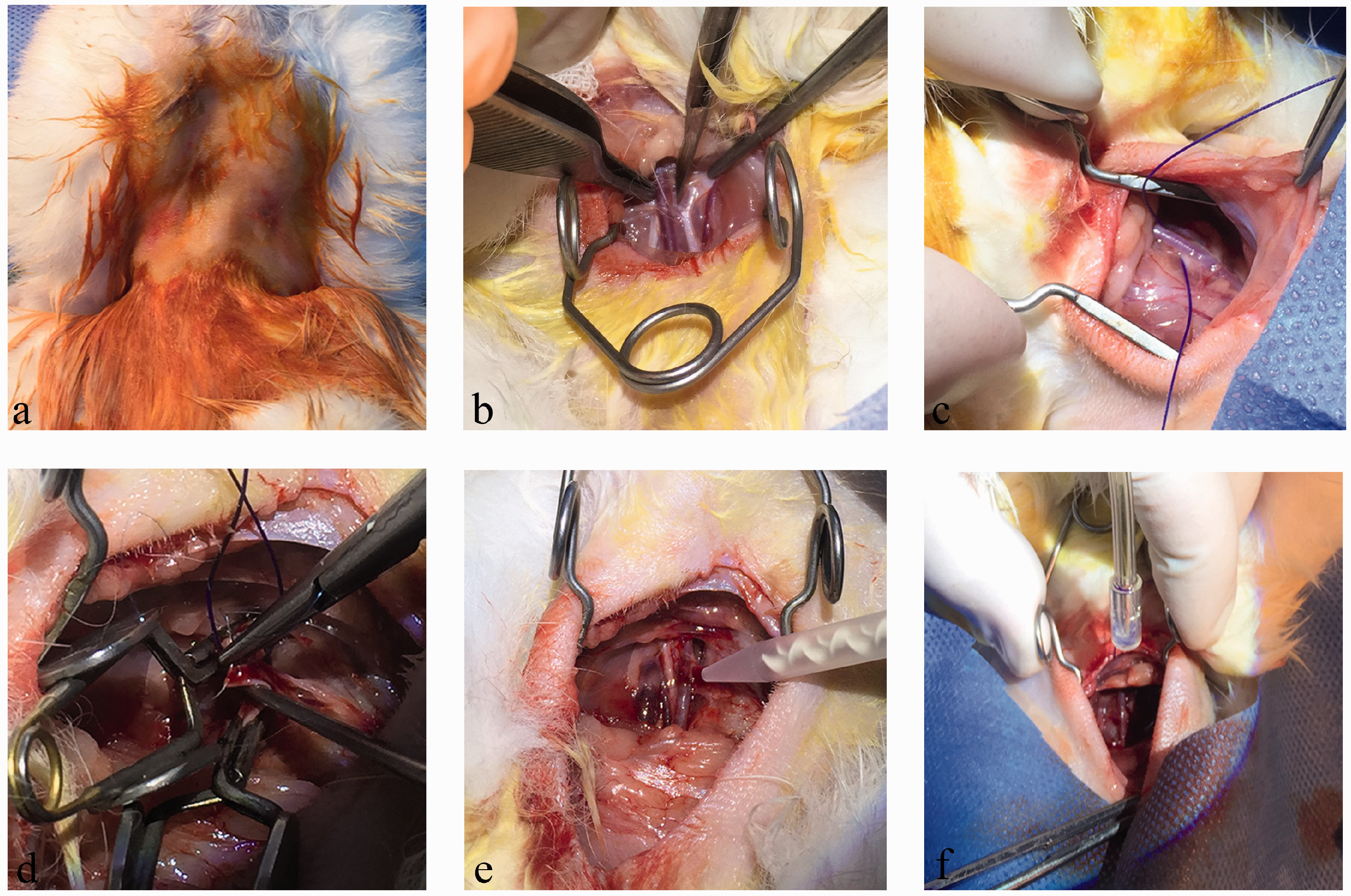
The surgical process of the study. (a) Disinfection was provided with povidone iodine. (b,c) Right main carotid artery was explored. (d) End-to-end anastomosis was performed. (e) 0.5 cc BSA-glutaraldehyde was applied locally to BSA-glutaraldehyde group. (f) 0.5 cc PEG polymer was applied locally to PEG polymer group.
The right main carotid artery segments, which were used in all groups, were removed and fixed for histological examination, and then sent to the histology laboratory at the end of 28 days. After collecting the tissue samples, the subjects were sacrified with high dose sodium thiopental (Pental®; İbrahim EtemUlugayIlac, Istanbul, Turkey).
Hematoxylin-eosin staining
The carotid artery samples were embedded in paraffin. Hematoxylin-Eosin was chosen as a routine histological stain. The morphometric analysis was performed in three-hematoxylin eosin stained cross-sections of each carotid artery by using computeraided planimetry. The values related to lumen, tunica intima and tunica media were calculated and compared among the groups. In this way, cellular increase, decrease and thickening and thinning occurred in the layers were observed. The scar tissue and inflammation marker which are macrophage, mast cell and fibroblasts were observed. For detailed description see the online supplementary data.
Masson's trichrome staining
Masson's trichrome staining was used specifically to selectively identify muscle, collagen fibers and fibrin. The connective tissue formed due to neointimal hyperplasia in the tunica media was evaluated with this method. We interpreted fibrosis by looking the increase in the amount of collagen fibers and fibrin. For a detailed description see the online supplementary data.
Immunohistochemical staining
Immunohistochemical analysis was performed in three representative cross-sections of each carotid artery and the average values were calculated for each subject. Samples were taken from the anastomosis area of each group so that the effect of different biomoomaterials on the anastomosis area was observed. A distance of 10 µm was left between samples. Since the hyperplasia areas in the samples showing lower intimal hyperplasia ended at the forth and at the fifth samples, so the samples were limited to 3. A-smooth muscle actin (aSMA) and vascular endothelial growth factor (VEGF) stainings were performed and were evaluated by a semiquantitative scoring system by two analysts blinded to the groups. For a detailed description see the online supplementary data.
Statistical analysis
Average, standard deviation, median lowest, highest, frequency and ratio values were used in the descriptive statistics of the data. Distribution of variables was measured by Kolmogorov Simirnov test. In the analysis of quantitative independent data, Kruskal-Wallis and Mann-Whitney test were used. SPSS 26.0 program was used in the analysis.
Results
Macroscopic findings
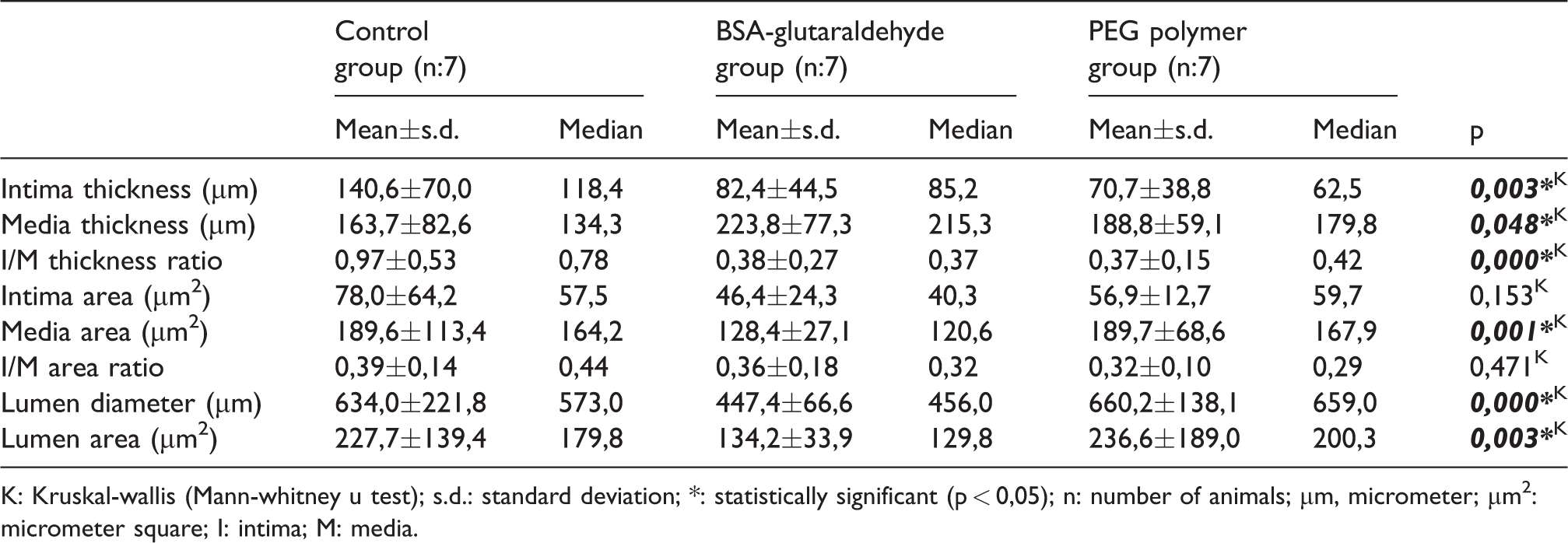
The surgical field was evaluated macroscopically before removing carotid artery samples on the 28th postoperative day. While there was no difference regarding the macroscopic findings of the control and PEG polymer groups (Figure 2(a) and (c)), there were glue residues in the BSA-glutaraldehyde group (Figure 2(b)).
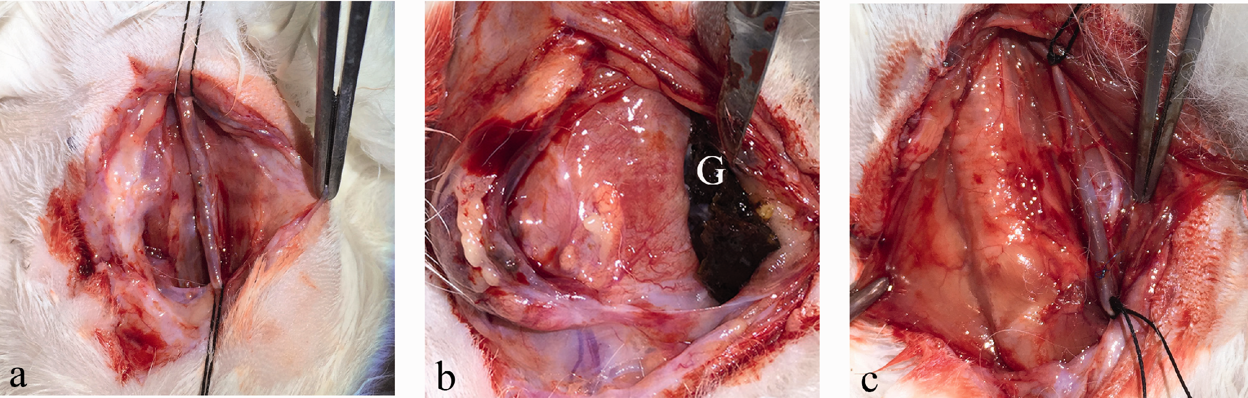
Macroscopic examination of groups. (a) No hemostatic agent was used in the Control group. (b) Glue residues was observed macroscopically in the BSA-glutaraldehyde group. (G) Glue residues of BSAglutaraldehyde (c) Any glue residue was not observed macroscopically in the PEG polymer groups.
Histomorphological findings
The intima thickness in the Control group was significantly higher (p < 0.05) than the BSA-glutaraldehyde group. The media thickness in the Control group was significantly lower (p < 0.05) than the BSA-glutaraldehyde group. The I/M thickness ratio in the Control group was significantly higher (p < 0.05) than the BSA-glutaraldehyde group. The intima area between the Control group and the BSA-glutaraldehyde group did not differ significantly (p >0.05). The media area in the Control group was significantly higher (p < 0.05) than the BSA-glutaraldehyde group. The I/M area ratio between the Control group and the BSA-glutaraldehyde group did not differ significantly (p >0.05). The lumen diameter in the Control group was significantly higher (p < 0.05) than the BSA-glutaraldehyde group. The lumen area in the Control group was significantly higher (p < 0.05) than the BSA-glutaraldehyde group (Table 1).
Statistical analysis of the histomorphological findings in Control group and BSA-glutaraldehyde group.
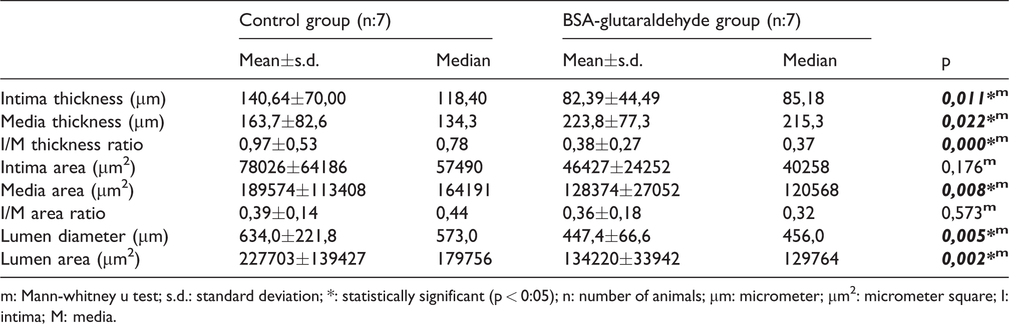
m: Mann-whitney u test; s.d.: standard deviation; *: statistically significant (p < 0:05); n: number of animals; µm: micrometer; µm2: micrometer square; I: intima; M: media.
The intima thickness in the Control group was significantly higher (p < 0.05) than the PEG polymer group. The media thickness between the Control group and the PEG polymer group did not differ significantly (p >0.05). The I/M thickness ratio in the control group was significantly higher (p < 0.05) than the PEG polymer group. The intima area between the Control group and the PEG polymer did not differ significantly (p >0.05). The media area between the Control group and the PEG polymer group did not differ significantly (p >0.05). The I/M area ratio between the Control group and PEG polymer group did not differ significantly (p >0.05). The lumen diameter between the Control group and the PEG polymer group did not differ significantly (p >0.05). The lumen area between the Control group and the PEG polymer group did not differ significantly (p >0.05) (Table 2).
Statistical analysis of the histomorphological findings in Control group and PEG polymer group.
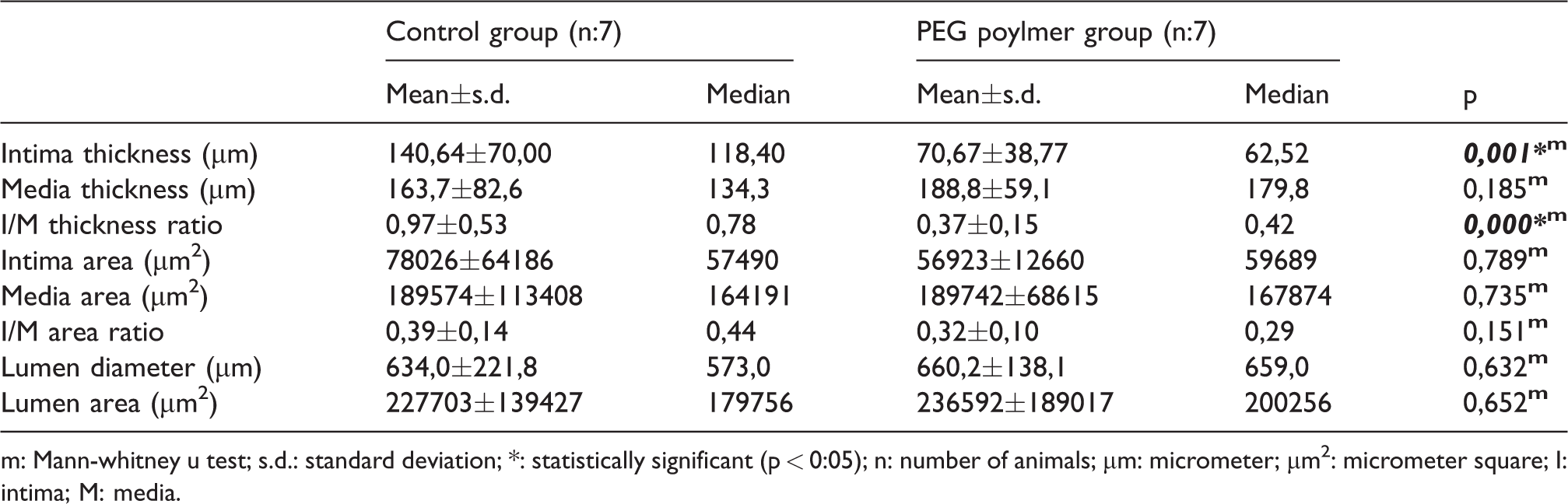
m: Mann-whitney u test; s.d.: standard deviation; *: statistically significant (p < 0:05); n: number of animals; µm: micrometer; µm2: micrometer square; I: intima; M: media.
The intima thickness did not differ significantly between the BSA-glutaraldehyde group and the PEG polymer group (p >0.05). The media thickness between the BSA-glutaraldehyde group and the PEG polymer group did not differ significantly (p >0.05).The I/M thickness ratio between the BSA-glutaraldehyde and the PEG polymer group did not differ significantly (p >0.05). The intima area in the whole group did not differ significantly (p >0.05). The media area in the PEG polymer group was significantly higher (p < 0.05) than the BSA-glutaraldehyde group.The I/M area ratio in the whole group did not differ significantly (p >0.05). The lumen diameter in the PEG polymer group was significantly higher (p < 0.05) than the BSA-glutaraldehyde group. The lumen area in the PEG polymer group was significantly higher (p < 0.05) than the BSA-glutaraldehyde group (Table 3).
Statistical analysis of the histomorphological findings in Control group, BSA-glutaraldehyde group and PEG polymer group.
K: Kruskal-wallis (Mann-whitney u test); s.d.: standard deviation; *: statistically significant (p < 0,05); n: number of animals; µm, micrometer; µm2: micrometer square; I: intima; M: media.
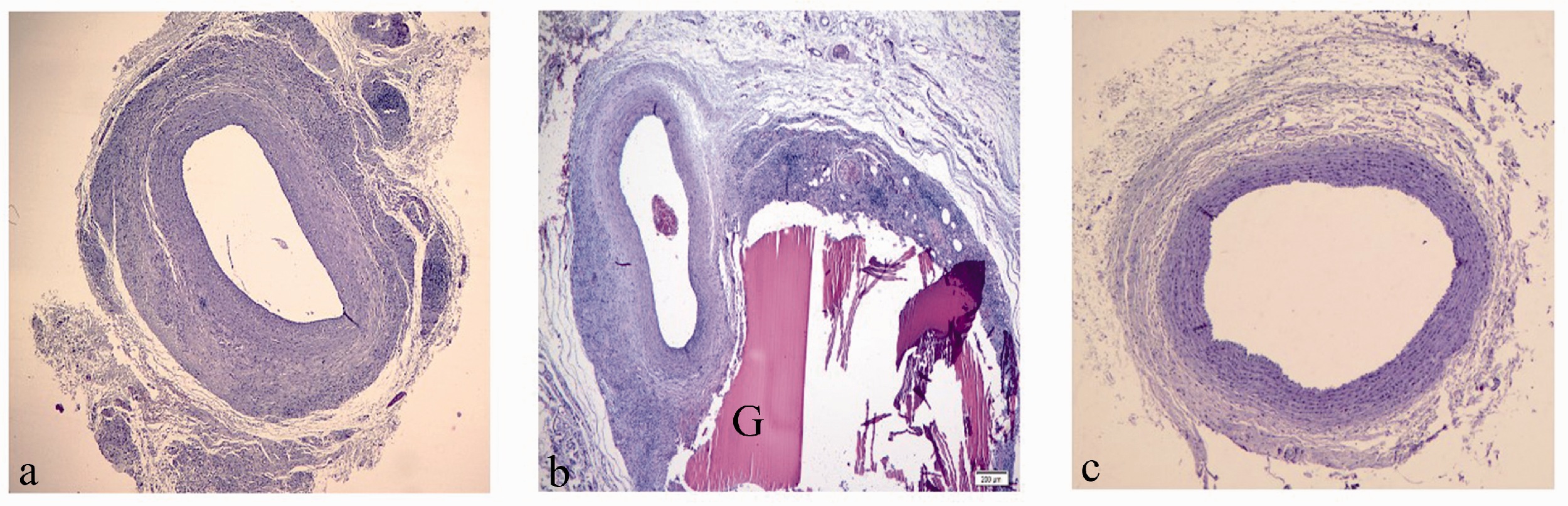
The glue residues in the BSA-glutaraldehyde group have been detected in hematoxylin eosin staining (Figure 3(b)). However, no glue residue was found in the PEG polymer group (Figure 3(c)). In addition, the differences between the groups in terms of the development of neointimal hyperplasia are clearly seen in hematoxylin eosin staining (Figure 4).
Comparison of glue residues in hematoxylin eosin staining. (a) Control group (No hemostatic agent was used.) (4x). (b) BSA-glutaraldehyde group (BSA-glutaraldehyde was used as a hemostatic agent.) (4x). (c) PEG polymer group (PEG polymer was used as a hemostatic agent.) (4x). (G) Glue residues of BSAglutaraldehyde.
Histomorphological differences in the development of neointimal hyperplasia between groups. (a) Control group (40x). (b) BSA-glutaraldehyde group (40x). (c) PEG polymer group (40x). (Arrowheads) internal elastic lamina.
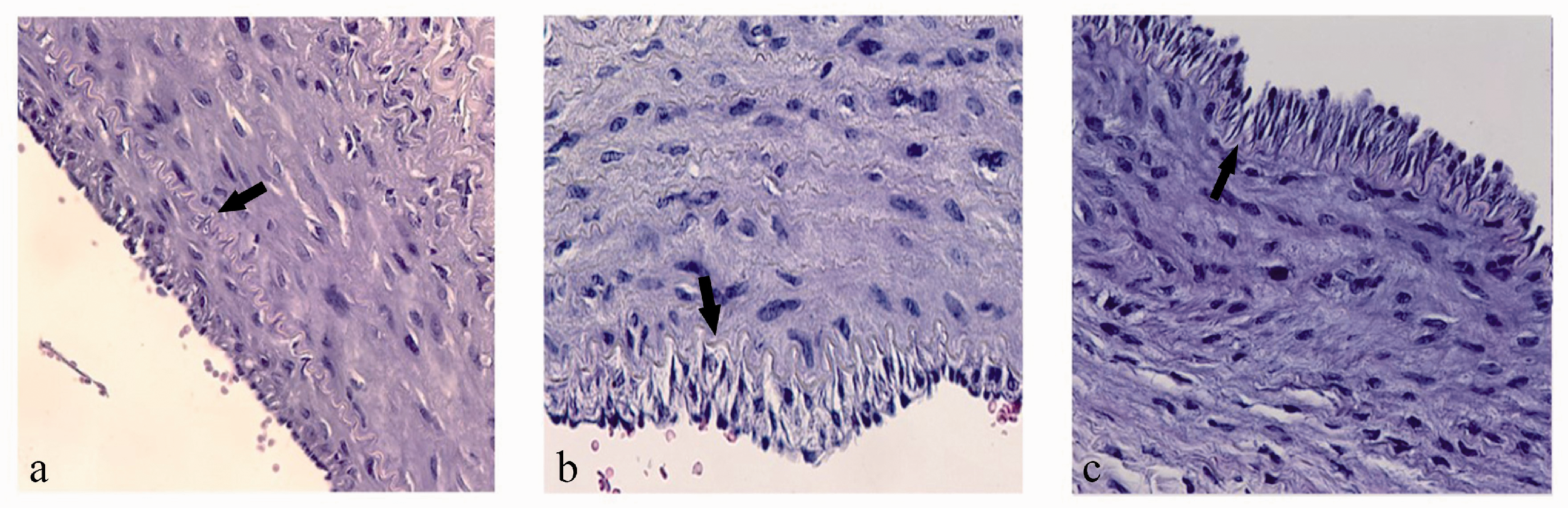
In preparations evaluated by Masson's trichrome staining, dense blue staining was detected in the tunica media of the Control group with increased connective tissue formation, while there was a small amount of connective tissue formation and minimal blue staining in the tunica media of the BSA-glutaraldehyde group. The blue staining was not observed in the tunica media of the PEG polymer group, and a limited blue staining was observed in the tunica adventitia (Figure 5).
Differences between groups in terms of connective tissue formation in Masson's Trichrome staining. (a) Control group (Dense blue staining was observed on whole layers.) (10x). (b) BSA-glutaraldehyde group (Blue staining was observed in tunica media and tunica interna.) (40x). (c) PEG polymer group (Although blue staining was observed in tunica adventitia, no blue staining was observed in tunica media/interna.) (40x). *Also, blue colored areas are marked with star symbols on whole sections for the description of staining.
Immunohistochemical findings
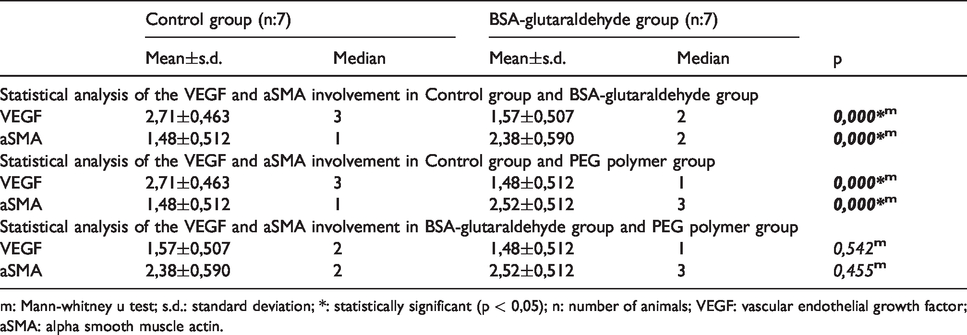
Scoring the amount of VEGF-positive staining in the Control group was found to be significantly higher (p < 0.05) than the BSA-glutaraldehyde and PEG polymer group. There was no significant difference (p >0.05) between the BSA-glutaraldehyde and the PEG polymer group (Table 4) (Figure 6).
Statistical analysis of the VEGF and aSMA involvement in groups.
m: Mann-whitney u test; s.d.: standard deviation; *: statistically significant (p < 0,05); n: number of animals; VEGF: vascular endothelial growth factor; aSMA: alpha smooth muscle actin.
Results of VEGF-positive staining (as seen in brown color) on vessel wall in all groups. (a) Control group (Mean±s.d.: 2,71 ± 0,463) (40x). (b) BSA-glutaraldehyde group (Mean±s.d.: 1,57 ± 0,507) (40x). (c) PEG polymer group (Mean±s.d.: 1,48 ± 0,512) (40x).
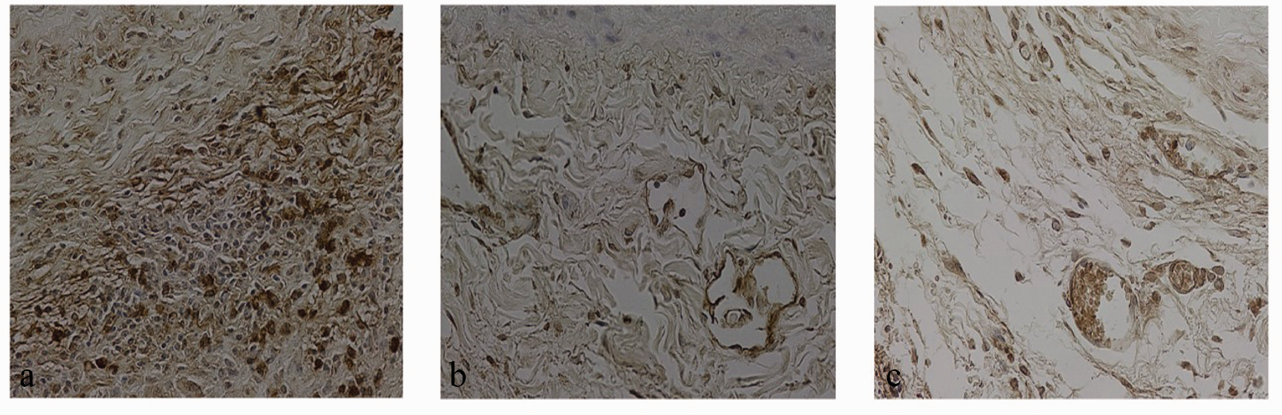
Scoring the amount of aSMA-positive staining in the Control group was found to be significantly lower (p < 0.05) than the BSA-glutaraldehyde and the PEG polymer group. There was no significant difference (p >0.05) between BSA-glutaraldehyde and PEG polymer group (Table 4) (Figure 7).
Results of aSMA-positive staining (as seen in brown color) on vessel wall in all groups. (a) Control group (Mean±s.d.: 1,48 ± 0,512) (20x). (b) BSA-glutaraldehyde group (Mean±s.d.: 2,38 ± 0,590) (20x). (c) PEG polymer group (Mean±s.d.: 2,52 ± 0,512) (40x).
Discussion
Although smaller animals such as mice and rats are used in animal experimental models of vascular anastomosis, rabbit carotid artery is more commonly used by researchers in neointimal hyperplasia models due to its similar structure to human coronary artery.12,13 End-to-end and simple interrupted anastomosis which causes less neointimal hyperplasia14,15 and it achieves full lumen clearance with this anastomosis technique,this technique also supports the use of rabbit carotid artery in neointimal hyperplasia studies. Due to this well-known relationship between anastomosis technique and neointimal hyperplasia, we used the end-to-end and simple interrupted anastomosis technique to decrease neointimal hyperplasia risk. We also standardized the operation team. The adventitial damage causes neovascularization in tunica adventitia. It is also known that the adventitial vasa vasorum damage serves as an entry point for inflammatory cells and. This neovascularization process enables the reshaping of vascular structure and it facilitates the development of neointimal hyperplasia.16,17 We obtained comparative data by defining the control group, which excludes the effects of adventitial damage due to hemostatic agents.
The balloon catheterization and the vascular anastomosis are the two most commonly used methods to create vascular damage in experimental studies on intimal hyperplasia. The balloon catheterization causes intima damage, and vascular anastomosis causes entire vessel wall damage.18–20 The pressure, which causes intimal damage, cannot be applied equally to all subjects since the balloon is inflated manually in the balloon catheter model. Also the vascular segment where the balloon is inflated can not be determined clearly. This may complicate the undamaged vascular segment during histological examination. 21 The advantage of the anastomosis model that we used in our experimental study is that the intimal damage is higher since the entire vessel wall will be included. For this reason, we did not prefer balloon catheter methoddue to standardization problems. In a study by Sullivan et al., 22 they showed that estrogen may reduces neointimal hyperplasia. So, we preferred to use male rabbits to increase neointimal hyperplasia in our study.
In a study, by More et al., 19 they showed that reendothelization begin in 3 days after endothelial damage in rabbits and the neointimal hyperplasia reaches maximum levels in 1 month due to extracellular matrix accumulation and it decreases within 3 months. In our study, we sacrificed the rabbits on 28th days and it is ensured that the neointimal hyperplasia was on the highest level.
As seen in the literature, the intima thickness, the media thickness, the I/M thickness ratio, the intima area, the media area, the I/M area ratio, the lumen diameter and the lumen area are the comparable variables for histological evaluation of neointimal hyperplasia.23–27 We also used these histological measurements in our study. We found that intimal hyperplasia was significantly lower in the BSA-glutaraldehyde and in the PEG polymer group compared to the Control group (p < 0,05). In addition, we immunohistochemically evaluated VEGF28,29 and aSMA involvement30–32 associated with neointimal hyperplasia.
Angiogenesis, the growth of new blood vessels from existing vessels, is an important precursor of the tissue repair process. Restoration of blood flow to damaged tissues provides the oxygen and nutrients needed to support the growth and function of regenerative cells. VEGF is one of the strongest proangiogenic and vascular permeability growth factors. The amount of VEGF found in damaged tissue can significantly affect healing. VEGF stimulates microvascular endothelial cells to proliferate and to migrate. It also changes gene expression.VEGF makes microvascular endothelial cells over-permeable, and this causes to secretion of plasma proteins into the extravascular space through the endothelial cells. It leads to fibrinogen clotting and accumulation of fibrin gel. The fibrin accumulation in the extravascular space acts as a temporary matrix which supports the growth of new blood vessels and other mesenchymal cells that produce mature and vascularized stroma. Increased VEGF in tissues and VEGF associated angiogenesis are important factors for the formation of unbalanced atherosclerotic lesions and neointimal hyperplasia. 33 This is why the amount of VEGF in the samples of the groups with different applications was determined. The total amount of VEGF in the tissue was used to observe the healing differences in equal time periods.
In a study by Li et al. 28 They showed that the increase in the adventitial VEGF signal was associated with the development of neointimal hyperplasia and they showed that VEGF was less retained in healthy healing tissues. In our study, VEGF involvement in the BSA-glutaraldehyde and in the PEG polymer group, where neointimal hyperplasia detected, were lower compared to the Control group (p < 0,05).The BSA- glutaraldehyde and the PEG polymer groups did not show significant difference in this regard. Following these data, our study resulted in similar findings to Li et al. 28 in terms of the relationship between the VEGF involvement and the neointimal hyperplasia. In another study by Zhou et al., 29 it is showed that Tongxinluo reduces VEGF by inhibiting adventitial angiogenesis and it reduces neointimal hyperplasia.This relationship between decreased adventitial VEGF involvement and decreased neointimal hyperplasia showed similar results with VEGF involvement of the BSA-glutaraldehyde and the PEG polymer group in our study. aSMA is a marker which is non-proliferative and contractile phenotype of the adult smooth muscle cells. After vascular reconstruction, expression of aSMA and other structural proteins reduce. In this way, smooth muscle cells acquire a proliferative phenotype. 30 Yan et al. 32 used the combination of Astragalus–Angelica to reduce intimal hyperplasia in their study. They found that aSMA showed high staining in the control group, but the intensity of the staining decreased in the experimental groups. It was observed thataSMA staining in the recovery groups were higher than in the experimental groups. As a result of their study, lower levels of aSMA involvement was found to be associated with the increase neointimal hyperplasia. Similarly, in our study, aSMA involvement was higher in the BSA-glutaraldehyde and PEG polymer group where the neointimal hyperplasia was less prominent (p < 0,05).
The critical healing phase after vascular anastomosis is the first 2 weeks after surgery. Rapid and complete hemostasis is of great importance in vascular surgery. Today, local application of hemostatic agents is used in vascular surgery effectively. These agents close the gaps between the sutures.Thus, safely and effectively simplify surgical procedure, shortens vascular surgery time and they effectively provide hemostasis. 34 Although there are extensive studies regarding tissue reactions against these agents, inflammation and hemostatic efficacy of PEG polymer and BSA-glutaraldehyde. They are frequently in use as hemostatic agents in vascular reconstruction; there is no data about the effect of these agents on neointimal hyperplasia.11,35,36 Therefore, we investigated the effects of these commercially available synthetic and biological based hemostatic agents to investigate neointimal hyperplasia.
In an experimental study, Wippermann et al. 37 compared histological effects of cyanoacrylate, BSA-glutaraldehyde and gelatin-resorcinol, which are used as hemostatic agents on distal anastomosis of swine coronary artery. At the end of the 3-month period, it was found that BSA-glutaraldehyde causes severe inflammatory response and widespread fibroblastic proliferation compared to the other hemostatic agents. Gelatin-resorcinol showed minimal tissue reaction. For this reason, they suggested applying BSA-glutaraldehyde on to the anastomosis line (max 2 cc). In addition, a similar proportion of adhesive residues were observed in all hemostatic agent groups. In our study, glue residues were observed in the BSA-glutaraldehyde group in the preparations examined using H&E staining (Figure 3). The lumen area and the lumen diameter in the BSA-glutaraldehyde group were significantly lower compared tothe Control group and to the PEG polymer group (p: 0.003 p: 0,000).We believe that the glue residues of BSA-glutaraldehyde may cause mechanical pressure on the vessel wall from the outside. The degradation time of BSA-glutaraldehyde is stated to be between 3–6 months. 8 Considering that the neointimal hyperplasia has reached its maximum level after 4 weeks, we may conclude that BSA-glutaraldehyde narrows the vascular lumen due to its long degradation time, despite decreasing neointimal hyperplasia. In animal studies on goats by Gundry et al., 38 it was found that BSA-glutaraldehyde produced a minimal inflammatory response at the end of the 3-month period. Vokrri et al. 39 reported that a low-grade inflammatory response in the anastomosis region compared tothe control group in a rabbit abdominal aorta modelwhere BSA-glutaraldehydewas used.In another study by Hewitt et al., 40 BSA-glutaraldehyde was used as a hemostatic agent in thoracic aortic repair in sheeps.It was showed thatit caused minimal inflammation after third month of anastomosis. Although there was no scaling of inflammation in our study during 28-day period, the connective tissue increase in the BSA-glutaraldehyde group was lower than the Control group in Masson's trichrome staining although it shows increased inflammation (damaged vascular wall, tissue fibrosis etc.).41,42 In addition, VEGF involvement associated with inflammation in the immunohistochemical examinationwas lower in the BSA-glutaraldehyde group compared to the Control group (p < 0,05).43–47 These results indicate that less inflammatory response occurred in the BSA-glutaraldehyde group. Cytotoxic effects of BSA-glutaraldehyde causing cell necrosis were detected in a study examining the toxic effects of BSA-glutaraldehyde on human embryonic fibroblast and mouse myoblast by Fürst et al.. 48 In our study, cell necrosis was not observed in the BSA-glutaraldehyde group, but inflammation was observed. Sutureless anastomosis using BSA-glutaraldehyde in the rabbit carotid artery and sutured anastomosis were compared by Schiller et al. 49 and they found no significant difference in terms of intra-luminal thrombosis. But, they showed that there was more inflammatory cell in the sutureless anastomosis group. Also early calcifications were observed in both anastomosis groups. Another experimental study which investigated the BSA-glutaraldehyde by scaling the inflammatory response concluded that use of BSA-glutaraldehyde in human coronary artery bypass surgery was not currently safe and may have disadvantageous in other vascular surgeries in the long term period. 50 As a result of our study,the lumen area and the lumen diameter in the BSA-glutaraldehyde group were significantly lower compared to the Control group. Therefore, we may conclude that BSA-glutaraldehyde should not be used as a hemostatic agent on vascular anastomosis such as distal or proximal coronary anastomosis. There is a need for more studies about these agents. However, the lumen area and the lumen diameter in the PEG polymer group increased compared tothe Control group, it is not statistically significant after 28 days (p: >0,05). In addition, theintima thickness and the I/M thickness ratio in PEG polymer group were statistically lowercompared tothe Control group (p: 0.001 p: 0,000).In the light of these results, we may conlude that PEG polymer may have favorable effects on anastomosis compared to BSA-glutaraldehyde and, it may be used more safely in case of necessity.
In a study investigating the effectiveness of PEG polymer on human saphenous vein under ex vivo cardiopulmonary bypass by Stooker et al.. 51 It is predicted that PEG polymer can be used in coronary artery bypass operations due to its biocompatibility, biodegradability, elasticity and transparency. They also concluded that smooth muscle cell damage was less prominent compared to the control group. It is possible that PEG poylmer may be combined with drugs which will prevent cell proliferation for periadventitial local applications, since it is used as a drug carrier pharmacologically. There are other studies stated that the PEG polymer may reduce neointimal hyperplasia with combined drugs, but we need more long-term studies about this topic. 51 In our experimental study, we showed that neointimal hyperplasia was significantly lower in the PEG polymer group compared to the Control group (p: 0.001 p: 0,000). In addition, aSMA involvement in PEG polymer group was found to be higher compared tothe Control group (p < 0,05). VEGF involvement in PEG polymer group was found to be lower compared tothe Control group (p < 0,05). Masson's trichrome staining showed that connective tissue increase in the tunica media was observationally lower compared tothe Control group.Our result shows that there is lower tissue damage in the PEG polymer group compared to the control and to the BSA-glutaraldehyde group. Long-term studies are also needed for its safe usage as a hemostatic agent in vascular anastomosis.
In an another study evaluating the tissue reactions to BSA-glutaraldehyde and PEG polymer used for hemostasis in abdominal aortic anastomosis of New Zealand type rabbits, by Slezak et al. 11 was found that the BSA-glutaraldehyde group has more inflammation and less biodegradation than the PEG polymer group. Granulomatous reaction was observed in both groups in this study. However, eosinophilic infiltration was significantly higher in the BSA-glutaraldehyde group. But,no data was obtained in this study for neointimal hyperplasia. In our study, despite the lack of data about eozonophilic infiltration, we found more connective tissue increase in the BSA-glutaraldehyde group using Masson’s trichrome staining compared to the PEG polymer group. But, aSMA and VEGF involvement of these two hemostatic agents were not showed statistically significant difference.
Conclusion
It is known that the most important condition affecting the long-term success of vascular surgery is restenosis caused by neointimal hyperplasia.We showed the effects of hemostatic agents used locally to control bleeding in vascular anastomosis, on neointimal hyperplasia. This experimental animal study showed that biological based hemostatic agent, BSA-glutaraldehyde, and synthetic based hemostatic agent, PEG polymer, may inhibit the development of neointimal hyperplasia. But, BSA-glutaraldehyde may cause a decrease in the lumen area and lumen diameter due to mechanical compression. The PEG polymer has no negative effect on the lumen area and on the lumen diameter according to our results. We think that these two locally applied hemostatic agents may reduce the development of neointimal hyperplasia due to their effects on VEGF and aSMA signals. Because of this role of tunica adventitia, we think that periadventitial local drug administration may have positive or negative effects on neointimal hyperplasia and restenosis. Therefore, the effects of the agents, which are applied periadventially for vascular reconsruction, must be investigated in terms of cytotoxicity, neointimal hyperplasia and restenosis.
As a result of this study, PEG polymer has favorable results on neointimal hyperplasia with associated markers such as lumen area, lumen diameter, intima thickness, I/M thickness ratio, connective tissue formation in the tunica media, VEGF and aSMA signals in the vessel wall. PEG polymer does not have a negative effect on the lumen area and on the lumen diameter. These results, that we detected in PEG polymer group, might have not be seen in the BSA-glutaraldehyde group due to mechanical compression effect of the glue residues in BSA-glutaraldehyde group. Therefore, the PEG polymer may be used safely in vascular anastomosis. These comparative data will assist the clinical use of these hemostatic agents, which have commercial forms, and they are already in use on surgical operations. There is also need for larger experimental and long-term studies.
Supplemental Material
sj-pdf-1-jba-10.1177_0885328220964913 - Supplemental material for The effect of bovine serum albumin-glutaraldehyde and polyethylene glycol polymer on neointimal hyperplasia in rabbit carotid artery anastomosis
Supplemental material, sj-pdf-1-jba-10.1177_0885328220964913 for The effect of bovine serum albumin-glutaraldehyde and polyethylene glycol polymer on neointimal hyperplasia in rabbit carotid artery anastomosis by Erturk Karaagac, Yuksel Besir, Meltem Kurus, Orhan Gokalp, Sahin Iscan, Yasar Gokkurt, Cagri Kandemir, Fatih Esad Topal, Erdi Keselik, Bortecin Eygi and Ali Gurbuz in Journal of Biomaterials Applications
Footnotes
Ethics approval
All applicable international, national, and/or institutional guidelines for the care and use of animals were followed. The study was approved by Dokuz Eylül University Faculty of Medicine Experimental Animals Ethics Committee. This article does not contain any studies with human participants performed by any of the authors.
Availability of data and material (data transparency)
The authors declare that they accept all data transparency.
Code availability
Antibody: Thermo Fisher Scientific Cat# MA5-13182, RRID:AB_10981661; Sigma-Aldrich Cat# A2547, RRID:AB_476701
Software: ImageJ V1.8.0_112, RRID:SCR_003070
Authors' contributions
All authors contributed to the study conception and design. Material preparation, data collection and analysis were performed by Erturk Karaagac, Yuksel Besir, Sahin Iscan, Meltem Kurus, Erdi Keselik. The first draft of the manuscript was written by Erturk Karaagac and all authors commented on previous versions of the manuscript. All authors read and approved the final manuscript.
Declaration of conflicting interests
The author(s) declared no potential conflicts of interest with respect to the research, authorship, and/or publication of this article.
Funding
The author(s) disclosed receipt of the following financial support for the research, authorship, and/or publication of this article: This study was funded by İzmir Katip Çelebi University Scientific Research Projects Unit with the project number 2018-ÖNAP-TIPF-0005.
Supplemental material
Supplemental material for this article is available online.
References
Supplementary Material
Please find the following supplemental material available below.
For Open Access articles published under a Creative Commons License, all supplemental material carries the same license as the article it is associated with.
For non-Open Access articles published, all supplemental material carries a non-exclusive license, and permission requests for re-use of supplemental material or any part of supplemental material shall be sent directly to the copyright owner as specified in the copyright notice associated with the article.