
Abstract
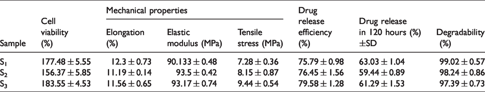
In this study, gelatin/hyaluronic acid (HA) scaffolds containing different amounts of atorvastatin-loaded nanostructured lipid carriers (NLCs) coated entirely with polycaprolactone (PCL) film were fabricated for skin regeneration. 12 atorvastatin-loaded NLCs formulations were synthesized, and particle size, zeta potential, drug entrapment efficiency (EE), and drug release of the formulations were determined. The optimum freeze-dried atorvastatin-loaded NLCs were added in 3 different weight percentages to the gelatin and HA membranous scaffolds. Thereafter, the membranes were coated entirely by a thin layer of the PCL. They were characterized, and then mechanical properties, in vitro degradation and in vitro drug release were assessed. Moreover, human dermal fibroblasts (HDF) were cultured on the prepared nanocomposite scaffolds in order to investigate the cytotoxicity by the MTT assay after the first day, third day, and fifth day. Results revealed that the most favorable atorvastatin-loaded NLCs had 99.54 nm average particle size, −24.30 mV zeta potential, 97.98% EE, and 75.24% drug release within 237 hrs. Mechanical tests indicated that all the three scaffolds had approximately a 90 MPa elastic modulus which was more than two-fold of tensile modulus of normal human skin. The in vitro degradation test demonstrated that the membranes were degraded up to 98% after 5 days, and the scaffolds drug release efficiency (DRE) was in a range of 75–79% during those 5 days. The MTT assay results confirmed the cytocompatibility of the scaffolds. The scaffold containing 54.1 wt% NCLs was the optimum sample (S3). Scanning Electron Microscopy (SEM) images of the latter one showed the uniform distribution of the NLCs with an average size of 150 nm, and the images of cultured HDF illustrated the good cell attachment. In conclusion, suitable physicochemical and biological properties of the novel gelatin/HA/PCL nanocomposite scaffold containing 54.1 wt% atorvastatin-loaded NLCs (S3) can be a good candidate for skin regeneration.
Keywords
Introduction
Skin is the greatest organ of the body which injures as a consequence of burn, trauma, radiation, etc. and needs therapies such as medications as well as interventions.1–3 Although some remedies like autograft, allograft, and xenograft have been utilized to repair skin wounds, they suffer from restrictions including donor sites, high risk of disease transmission, and immune rejection. 4 , 5
Tissue-engineered replacements such as integra, alloderm, 6 epicel, and apligraft are currently employed for the wound healing, but their use is restricted due to their high cost and low mechanical properties. 7 Tensile elastic modulus of the human leg skin was measured in the range of 6–22 MPa. 8 Therefore, in this study an attempt was made to fabricate a cost-effective membrane with suitable mechanical properties and easy manipulation in practice. To fabricate the tissue substitutes, several techniques have been used with some advantages and disadvantages. 9 , 10 In this design, a nanocomposite core made up of gelatin/HA/atorvastatin-loaded NLCs was coated with the PCL film through a simple spin coating process, and then several holes with the diameter of 200 μm were made by inserting microneedles in the PCL coating. Therefore, the medium can penetrate and reach to the nanocomposite core in order to release the drug and other nanocomposite components for suitable wound healing.
The mechanical characteristics of the tissue-engineered skin are evenly matched to human skin, but they do not fully correspond to native skin properties yet. The greatest contrast has been found in vitro and in initial stages of grafting. In fact, the engineered skin turns out to be more fragile than the native skin. This insufficiency in mechanical properties leads to a difficulty in the surgical procedure, reducing the elasticity and resistance after grafting. Degradation should also be noticed and appropriately adjusted in order to maintain a suitable structural integrity for an enough period of time so that the freshly developed skin can be formed by means of its supporting function. 11
Atorvastatin, a second-generation synthetic statin, has been evaluated with a declining blood cholesterol level in order to prevent cardiovascular disorders (i.e., heart attack, ischemia, etc.) and healing efficiency on various skin wounds like other statins.12–14 Topical use of statins could be a sensible substitute for oral administration in order to enhance their influences on the skin and to diminish the side effects. For instance, it was reported that rats with a diabetic foot ulcer gained advantageous effects of topical statins on wound repair.13–15 In an in vivo study, 20 male Sprague Dawley rats were divided into two groups: the atorvastatin and control group. Each rat obtained a surgery of a skin flap incision, elevation, and resuturing to the original position. Compared with the control group, atorvastatin treatment increased skin flap blood perfusion, vascular density, and necrotic area dependent on VEGF mRNA expression. 16 In another study conducted on animals, it was shown that Emulgel of atorvastatin calcium has excellent wound healing activity. 17 Furthermore, novel statin formulations encapsulated in liposomes were effectively delivered through topical administration, considerably decreasing hypertrophic scarring in a rabbit ear model. 18
Solid lipid nanoparticles (SLNs) are the first generation of lipid-based nanocarriers that are synthesized from lipids, which are solid in the body temperature and stabilized by emulsifiers. NLCs are the second generation of lipid-based nanocarriers fabricated from a mixture of solid and liquid lipids and have unstructured matrix due to the various moieties of the constituents of NLCs. NLCs have a greater drug loading (DL) capacity than SLNs due to an imperfect crystal structure and could prevent the drug expulsion by avoiding lipid crystallization during the manufacture and storage periods. They have a low melting point than SLNs and more space for drug dissolution as well as the loading capacity. 19 It was shown that the use of NLCs has a synergistic effect on the chosen permeation enhancers and promoting more hydrophobic drugs penetration through the skin than the drugs in the solution. Also, the in vitro skin experiments illustrated that NLCs dispersed in the scaffold can strongly affect permeation.20–23 Strohbach et al. 24 reported that in a medical device made of biodegradable polymers the selected materials can alter the atorvastatin function on the cultured cells. Therefore, in this research, the atorvastatin loaded NLCs were dispersed in a biodegradable polymeric blend of gelatin/HA coated with a PCL thin film as a novel biodegradable membrane for the wound healing.
Gelatin is a naturally occurring polypeptide which is non-toxic, biocompatible, biodegradable, hydrophile, and soft. 20 , 21 It is also used as a component of an artificial skin in order to improve epithelialization in the wound healing. 21 The collagen-gelatin-PCL scaffold has been known for tissue engineering and successful in the wound healing. In fact, it induces cell proliferation and retains the characteristics of the shape of the fibroblasts. 4 The PCL is a biodegradable polymer with suitable mechanical properties such as elongation and strength for tissue engineering applications. Meanwhile, it has been used as a component in the preparation of membranes for the wound dressing.23–25
Hyaluronan or HA is a linear and natural polysaccharide that has multiple biological functions such as cell adhesion regulation, cellular mobility, cell proliferation, and cell differentiation and provides mechanical properties for the tissue composition. HA interacts with a number of surface receptor cells and affects cellular processes including morphogenesis, wound healing, inflammation, and metastasis.26–31 Addition of HA to the scaffold composed of chitosan and gelatin increased the fibroblast adhesion and mechanical/biological properties of the scaffold. 32 Furthermore, the biodegradable HA scaffold of multipotent stem cells is a safe strategy in repairing damaged tissues. 33
In this research, an attempt was made to fabricate and assess a novel gelatin/HA/PCL nanocomposite scaffolds containing atorvastatin-loaded NLCs for the skin tissue engineering.
Materials and methods
Materials
Atorvastatin calcium trihydrate (99.3%) was purchased from Amin Pharmaceutical Company (Iran). In addition, methanol (MW = 32.04g/mol), dichloromethane, stearic acid (SA), caprylic acid (CA), oleic acid (OA), di-sorbitol, chloroform, sodium hydroxide, and Tween 80 were purchased from Merck (Germany). Glyceryl monostearate (GMS) was used as a liquid lipid and supplied by MP Biomedicals (the Netherlands). Also, gelatin type B (Bovine skin), PCL (MW = 80,000 g/mol), and HA (rooster comb) were purchased form Sigma Aldrich (US).
Synthesis of NLCs containing atorvastatin
NLCs containing atorvastatin were prepared by the emulsion-solvent evaporation technique. First, the drug was dissolved in a suitable amount of methanol in order to form the inorganic phase. Since methanol does not form emulsion in water, dichloromethane was used as the second solvent with the ratio of dichloromethane to methanol equal to 6:1. The weight ratio of lipids to the drug was 10:1. Table 1 presents the different NLCs formulations with various lipids and their weight percentages. In sum, solid lipid and liquid lipid were mixed and then transferred to water bath and heated at around 2–3°C higher than the melting point of the solid lipids. Then, the solvents and drug were added to the mixture. To prepare the water phase, 1v/v% Tween 80 was added to deionized water as the surfactant and was stirred for 20 mins (800 rpm and 60 °C) in order to dissolve the surfactant well. In the next step, the prepared solution was gradually added to the stirring aqueous phase in 15 mins in order to form O/W nano emulsion (the volume ratio of aqueous phase to organic phase was 10:1). 34 The solution was cooled down to 25 °C and kept stirring (400 rpm) for 3 hrs in order to completely remove the solvents.
Different NLCs formulations with various lipids and their weight percentages.
Pharmaceutical evaluation of the NLCs containing atorvastatin
Measurement of particle size and zeta potential of the NLCs containing atorvastatin
Particle size, polydispersity index (PDI), and zeta potential of NLCs were determined by the dynamic light scattering technique using a Zetasizer instrument (Zetasizer 3600, Malvern Instrument Ltd., Worchestershire, UK) at 25 °C. All measurements were done three times.
Drug loading in NLCs
1 mL of NLCs dispersion was centrifuged at 10,000 rpm for 10 mins. Then, the absorbance of the supernatant solution containing the unloaded drug was read at λmax = 239 nm wavelength. To avoid adsorption interference from other formulations, a blank sample was prepared for each formulation of NLCs, and the absorbance was checked at the same wavelength.
The amount of drug in the solution was calculated by a calibration curve in the deionized water. To get the amount of DL efficiency in the NLCs, the following equation was used:
DL percentage was calculated using equation 2
35
.

Drug release from formulations
Drug release from NLCs was studied in vitro using the dialysis bag method in phosphate-buffered saline (PBS) (pH = 7.4). To this end, 2 mL of NLCs dispersion was poured in a dialysis bag (MWCO12400), and its two ends were closed. The bags were placed in 40 mL of PBS and stirred at 500 rpm. The temperature was adjusted at 37 °C during the drug release test. At different intervals between 15 mins to 237 hrs, the absorptions of the sample solutions were determined at λmax = 239 nm. The concentration of each sample was determined using the concentration-absorption equation obtained from the standard curve in the phosphate buffer saline medium.
36
The test was repeated three times for each formulation. The percentage of DRE was determined by calculating the percentage of drug released from the nanoparticles. DRE was calculated by Equation 3, which shows the ratio of the area under the drug release versus time diagram of a definite time to the time in which the drug is released completely (100%).
37

Atomic force microscopy (AFM) analysis of NLCs containing atorvastatin
An atomic force microscopy (AFM, Dualscop, C-26, Germany) was used to study surface topography and morphology as well as particle size determination of NLCs. Two μl of NLCs were poured onto a graphite substrate and were placed in a vacuum oven in order to dry NLCs. This graphite substrate was viewed from different angles. This test was carried out at the room temperature (25 °C) and in the contact mode.
Fabrication of nanocomposite scaffolds
The solvent casting technique was used to fabricate the nanocomposite scaffolds. 38 To do so, gelatin was first dissolved with di-sorbitol (30%w/w of gelatin) in a minimum amount of deionized water in an ultrasonic bath at 50 °C. The solution was then placed on a magnetic stirrer at 400 rpm at the room temperature (25 °C). pH of gelatin solution was adjusted at 7.4 by NaOH solution (0.1 molar) prior to the addition of HA (20%w/w).
After complete dissolution of HA, optimum NLCs-loaded atorvastatin (GMS55OA45) was gently added to the solution in three weight percentages (Table 2). After solution homogenization on the stirrer, it was transferred to the Teflon mold and vacuum dried in order to obtain nanocomposite membranes. Then, the membranes were coated with a thin layer of PCL by a spin coater. In membrane design of the present research, a novel method was used in order to optimize the degradation rate of the inner nanocomposite film, and several holes with the diameter of 200 μm were made immediately after coating by inserting microneedles in the upper PCL film.
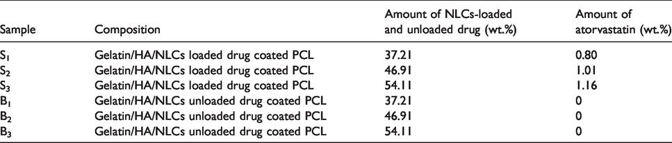
The amount of NLCs-loaded atorvastatin in nanocomposite membranes; the samples (S) and blanks (B) contain NLCs loaded atorvastatin and NLCs unloaded atorvastatin, respectively.
Characterization of the prepared scaffolds
Degradation test
Degradation of the nanocomposite scaffolds was investigated according to the American Society for Testing and Materials (ASTM, F1635-04). The samples (S1, S2, and S3) were prepared with the dimensions of 1 × 1cm2 and immersed in PBS solution (pH 7.4, temperature 37 °C) after 1, 3, and 5 days. After each time period, the samples were removed from PBS solution, rinsed slowly with deionized water, and then placed in an oven at 37 °C for 1 day in order to dry, and their weights were measured (n = 3). Equation 4 was used to calculate the weight loss percentage or the degradation rate (DR) percentage of the samples:

Drug release from the scaffolds
Drug release from the scaffolds was measured by the Franz diffusion cell method, and four Franz cells were used for each sample. Twenty-five mL of PBS with pH = 7.4 was placed in each cell, and it was adjusted on a stirrer with the speed of 110 rpm. The cells were connected to the bain-marie circulator adjusted at 37 °C. The samples with the same dimensions of the cell opening were placed on filter papers which were previously located on the cells.
As mentioned earlier regarding the drug release from NLCs, aliquot of the PBS solution was taken at different intervals between 15 mins to 237 hrs, and subsequently the absorptions of the sample solutions were determined at λmax=239 nm wavelength in order to measure the drug release from the scaffolds. The test was repeated three times for each sample including S1, S2, S3, B1, B2, and B3.
Chemical analysis
A Fourier Transform Infrared Spectroscopy (FTIR, JACSO 6300, Japan) was used for polymer analysis in order to determine the type and amount of compound elements and in order to investigate functional groups in the structure of optimum nanocomposite scaffolds.
Morphology of the scaffolds
A Field-emission Scanning Electron Microscope (FE-SEM, Mira 3-XMU, Czech) was used in order to investigate the morphology and distribution of the NLCs in the nanocomposite scaffolds. Furthermore, SEM (Ziess, Germany) images were prepared from the scaffolds’ surface in order to observe the porosities created in the thin PCL coating on the nanocomposite film.
Mechanical properties in wet condition
The prepared membranes were soaked in PBS (pH 7.4) at 37 °C for 3 hrs After the 3-hr soaking period, the samples were evaluated for their mechanical properties in wet conditions. Mechanical characteristics (i.e., tensile strength, modulus of elasticity, and elongation percentage) of the prepared scaffolds were determined by the tensile test using universal testing machine (Hounsfield, Germany) at 25 °C according to the ASTM F451-86. The samples were sectioned into rectangular strips (dimensions: 20 × 50 mm), and the distance between the gripping points was 30 mm. Mechanical testing was fulfilled on the grips moving at drawing speed of 10 mm/min and a load cell of 100 N. The data on modulus of elasticity, tensile strength, and elongation demonstrated the average results of the three scaffolds. Indeed, mechanical features were assessed at 50% strain. Also, to investigate the effect of the drug on the mechanical properties of the scaffolds, B1 and S1 specimens were tested.
Cytotoxicity evaluation of the scaffolds
The HDF cells (Pasteur Institute, Iran) were used to assess the cytotoxicity of the prepared samples after 1, 3, and 5 days. S1, S2, S3, and B1 samples were cut with the dimensions of 1 × 1cm2. B1 was selected to investigate the effect of drug free specimen on the HDF cells. Then, they were sterilized by being exposed to UV light for 2 hrs, and it was followed by their overnight soaking in 70% ethanol according to standard techniques. 27 To evaluate the cytotoxicity of the prepared samples, the direct contact method was performed. MTT (3–(4, 5-dimethylthiazol-2-yl)-2, 5-diphenyl tetrazolium bromide, Sigma, USA) assay was conducted to measure cell viability and proliferation. For evaluating cytotoxicity through the direct contact, the samples were situated in a 48 multi-well plate and then seeded at a cell density of 5 × 10² cells/cm2. In other words, after reaching confluency, the cells were finally enzymatically extracted from the flasks, counted, and seeded onto the sterile specimens. The polystyrene standard culture plate was utilized as the negative control. Then, 300 μl of cell suspension was used on every sample, and the cells were permitted to stick to the underlying substrate for 2 hrs. Then, the complete culture medium was meticulously added to each well. The MTT was prepared as a 0.5 mg/mL stock solution in the PBS, sterilized by the millipore filtration, and kept in the dark. At the end of the incubation periods (1, 3, and 5 days), the supernatant was removed, and the 500 μl MTT solution was added into each well. The cells were then incubated for 5 hrs at 37 °C. The formazan crystals were formed and dissolved by adding dimethyl sulfoxide (DMSO) (Sigma, USA) per a well under shaking. MTT reacted with tetrazolium ring and produced blue formazan crystals. The solutions were collected and put in 96 well plates. The optical density of the formazan solution was read on an ELISA plate reader (STAT FAX 2100, USA) at 570 nm. 40 The obtained results were normalized in relation to the control sample. The viability percentage of the cells was calculated using Equation 4.27
Absorption of control samples = Ac
Absorption of samples = Asample
Absorption of DMSO solution = Ab
Cell adhesion test
In each time period (1, 2, and 3 days), morphology and attachment of the cells seeded on the prepared nanocomposite scaffolds were investigated by the SEM. For fixation of cells after removal of the medium, the samples loaded with the HDF cells were washed twice in PBS (Bioidea, Iran). The specimens were then exposed to the 500 μL glutaraldehyde solution of 2.5% (diluted 50% glutaraldehyde solution, Sigma, USA) and stored in a refrigerator at 4 °C for 3 hrs until the cells were fixed on the surface of the scaffolds. After the cell fixation, the glutaraldehyde solution was removed, and the samples were again washed with the PBS. Subsequently, the samples were dehydrated in an ethanol solution (30%, 70%, 90%, 96%, and 100%, respectively) for 10 mins. Then, the specimens were dried at the room temperature and got ready for the SEM analysis.
Statistical analysis
The results were statistically analyzed using analysis of variance (ANOVA). The post-hoc multiple comparisons were performed using Tukey’s test. A p value less than 0.05 was considered statistically significant.
Results and discussion
Particle size, PDI, and zeta potential of the NLCs
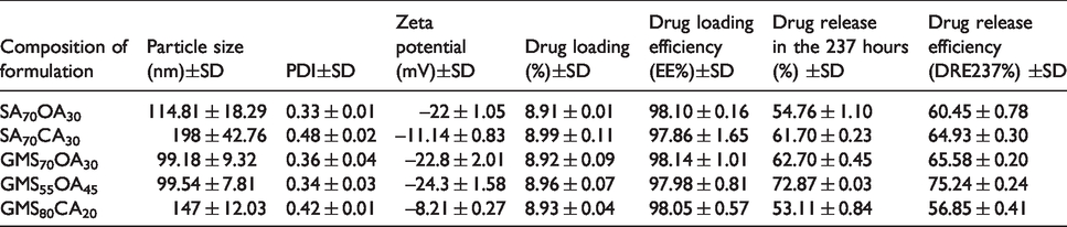
Mean particle size, PDI, and zeta potential of the studied formulations are presented in Table 3. The test variables were the concentrations of not only the solid SA and GMS lipids but also liquid OA and CA lipids. The ratios of solid lipid to liquid lipid were chosen at 80:20, 70:30, and 55:45. As shown in Table 3, the smallest particle sizes are associated with the GMS70OA30 (99.2 ± 9.3 nm) and GMS55OA45 (99.45 ± 7.81 nm) formulations, which include GMS and OA lipids in weight ratios of 70:30 and 55:45, respectively. In contrast, the largest particle size was related to the SA80CA20 (229.05 nm) formulation, which consists of SA and CA lipids with a weight ratio of 80:20. The lowest zeta potential is related to the GMS55CA45 (−6.33 mV) formulation, which consists of 55% w/w GMS and 45% w/w of CA, and the highest zeta potential is related to GMS55OA45 formulation (−24.30 mV) with GMS and OA at a weight ratio of 55:45. Negative values of zeta potential for all formulations were due to the negative charge generated by the lipids at the nanoparticle surface.
The average results of particle size, PDI, and zeta potential of NLCs containing atorvastatin (n = 3).
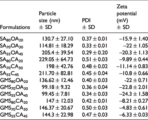
Formulations with small particle size, low PDI, and high zeta potential were selected as optimum formulations. Accordingly, SA70OA30, GMS70OA30, GMS55OA45, and GMS80CA20 were the optimum lipid ratios.
Drug loading and release from NLCs
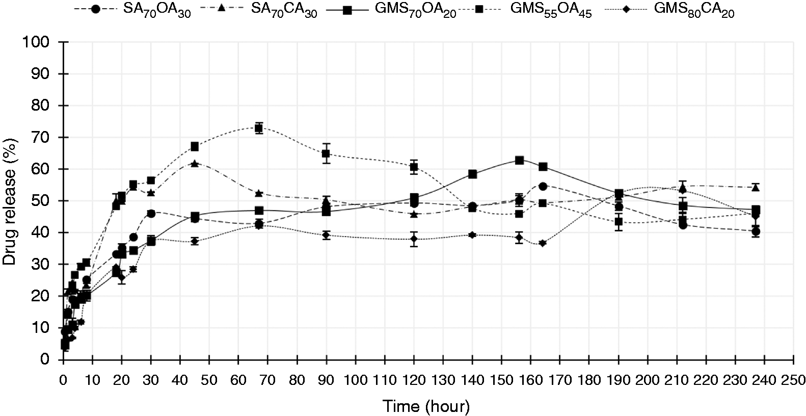
DL percentage, DL efficiency, drug release, and DRE during 237 hrs for the selected formulations are presented in Table 4. The percentage of DL in the formulations was high, and there was no significant difference between different groups. The lowest percentage of DL efficiency was related to SA70CA30 formulation (97.86 ± 1.65%), and the highest percentage of DL efficiency is related to GMS70OA30 formulation (98.14 ± 1.01). Figure 1 and Table 4 show drug release profile and efficiency during 237 hrs in PBS (pH = 7.4), respectively. The lowest and the highest percentage of drug release were related to the GMS80CA20 (56.85 ± 0.41%) and GMS55OA45 (75.24 ± 0.24%) formulations, respectively. The highest and lowest DRE were related to GMS55OA45 and GMS80CA20 formulations, respectively.
Determined parameters related to different NLCs formulations and their efficacy of drug release over a period of 237 hours (n = 3).
Percentage of atorvastatin released from NLCs in PBS (pH = 4.7) (n = 3).
GMS has a much lower hydrophilic-lipophilic balance than SA, which results in greater solubility of atorvastatin in GMS. As a result, the percentage of DL in it was increased. Furthermore, the higher molecular weight of GMS compared to SA led to more DL due to increased empty spaces 41
Likewise, using a higher amount of solid lipid in NLCs formulation caused larger particles to synthesize with a higher viscosity that raised DL efficiency. 42 To find out the effect of liquid lipid type on DL efficiency, OA was used as an alternative to CA. In fact, it may also be related to the higher molecular weight of OA than CA with longer carbon chains which creates more empty space for drug accommodation. In other words, it provided a more heterogeneous mixture with more empty spaces for drug settlement and an increased DL. These findings were in accordance with Rania et al. 43 Meanwhile, using a higher amount of solid lipid in the NLCs formulation enhanced the solid portion of the synthesized particles and its viscosity which reduced the drug release.
Higher amount of OA in NLCs induced faster drug release due to both more amount of liquid lipid in the formulation and little particle size. However, particle size reduction was achieved using more than 30 wt% OA. 44 , 45 The lower the particle size, the more the surface area contact with the release medium, and the higher the drug release. 45 Generally, the rate of drug release from NLCs depends on three factors including the partition coefficient of drug between the lipid phase, release medium, and the diffusion rate of drug across the dialysis membrane.46,47 Figure 1 showed that GMS55OA45 formulation had a higher release rate and release efficiency than the other formulations due to the greater area below its release profile curve. Previous research findings illustrated that formulations containing more liquid lipids have a faster release rate compared to the ones containing lower liquid lipids. 44 , 47
SA in the solid lipid form was used to synthesize SA70CA30 formulation. Its high drug release rate may be attributed to the hydrophilicity of this compound, which tends to favor lipophilic atorvastatin outflow from the NLCs. In fact, hydrophilicity of SA can increase the partition coefficient of the drug between the lipid phase and the release medium. GMS80CA20 had the lowest percentage of release efficiency and release rate, which can be due to having larger particle size than the other formulations. As a result, it reduced the contact surface of the NLCs with the release medium and lower amount of the dissolution rate. Meanwhile, another reason was the lower amount of liquid lipid used in GMS80CA20 formulation.
The optimal formulation selection was based on a combination of five parameters: the smallest particle size, the smallest amount of poly dispersity index, the greatest absolute zeta potential, the highest percentage of DL efficiency, and the highest percentage of DRE. Accordingly, GMS55OA45 was selected as the optimal formulation and was used to prepare the nanocomposite scaffolds.
Morphology of optimum synthesized NLCs
Figure 2(a) (surface image) and Figure 2(b) (three-dimensional image) illustrated the morphology and particle size of the optimum NLCs formulation (GMS55OA45) evaluated by AFM. The majority of the NLCs were relatively spherical with smooth surface, but some of them had an irregular shape with sharp edges. This irregularity of NLCs’ shape could be related to vacuum condition in their preparation. The average particle size of the synthesized NLCs was measured at 100 nm with some aggregates. The two-dimensional images showed a low resolution due to the probe tip contact with the lipids and Tween 80 used in their formulation.
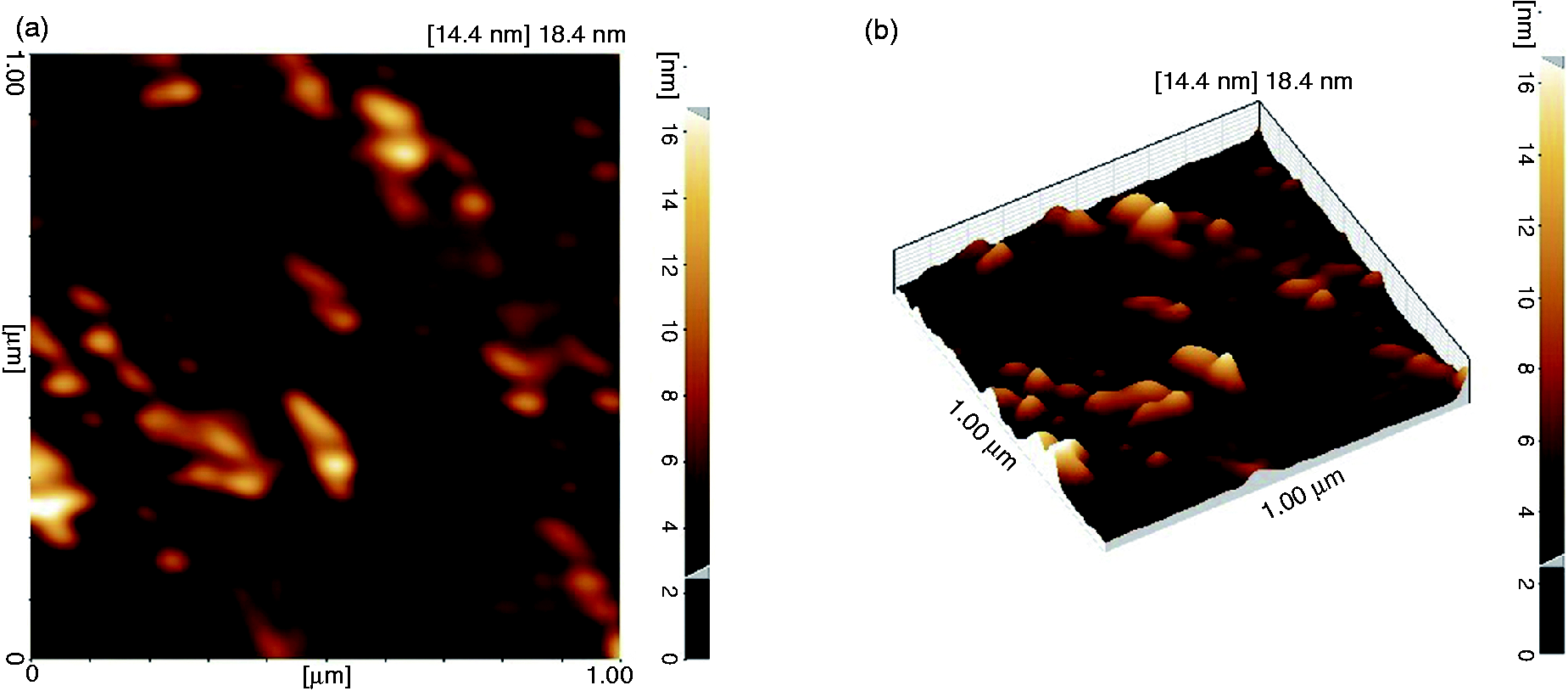
(a) Surface image and (b) three-dimensional image of the of the NLCs (F4:GMS55OA45) containing atorvastatin obtained by AFM.
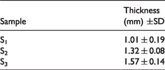
Nanocomposite scaffolds’ thickness
Thickness measurement of the scaffolds by SEM encountered some problems including the separation of the PCL coating and nanocomposite core during the freezing procedure in liquid nitrogen. Therefore, the thickness of the samples was individually determined by measuring the thickness of PCL coating and nanocomposite core. Figure 3 illustrated the PCL coating thickness obtained by SEM, and Table 5 showed the average thickness of the nanocomposite core of the samples which is obtained by a micrometer. The thickness of PCL coating was measured at around 8.98 μm. As can be seen in Figure 3, the thickness of the nanocomposite core was about 1 to 2 mm.
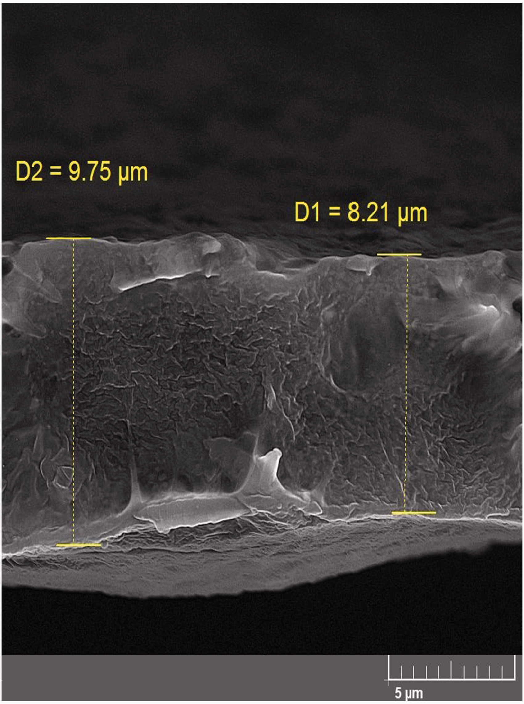
Cross-section image of the PCL coating obtained by SEM.
Thickness of the nanocomposite core of the samples obtained by micrometer (n = 3).
Prepared samples degradability
S1, S2, and S3 nanocomposite scaffolds with PCL coating were soaked in the PBS at 37 °C for 5 days. Then, samples' weight loss was determined at predetermined intervals. Figure 6 illustrated the weight loss percentage of the samples over a 5-day period. The main constituents of the synthesized scaffolds were gelatin and HA, which are highly hydrophilic natural polymers with a considerably high degradation rate in body fluids. In scaffold design of the present research, PCL coating not only improved the mechanical properties but also played a key role in regulating the scaffold degradability. Therefore, the nanocomposite core degradation was controlled by fluids penetration through the coating holes and self-degradation of the PCL coating. However, the main factor in the nanocomposite core degradation was fluids diffusion through the pores, and self-degradation of PCL coating had an inferior role in this phenomenon due to the low degradation of PCL. The spin coating can create relatively uniform and non-porous coating, but in areas far from the samples' center and especially on their edges, non-uniformity and pores could be observed due the lack of PCL coverage. Experiments showed that the optimum degradation rate could be achieved by making some other porosities in the PCL coating which can be observed in Figures 4 and 5.
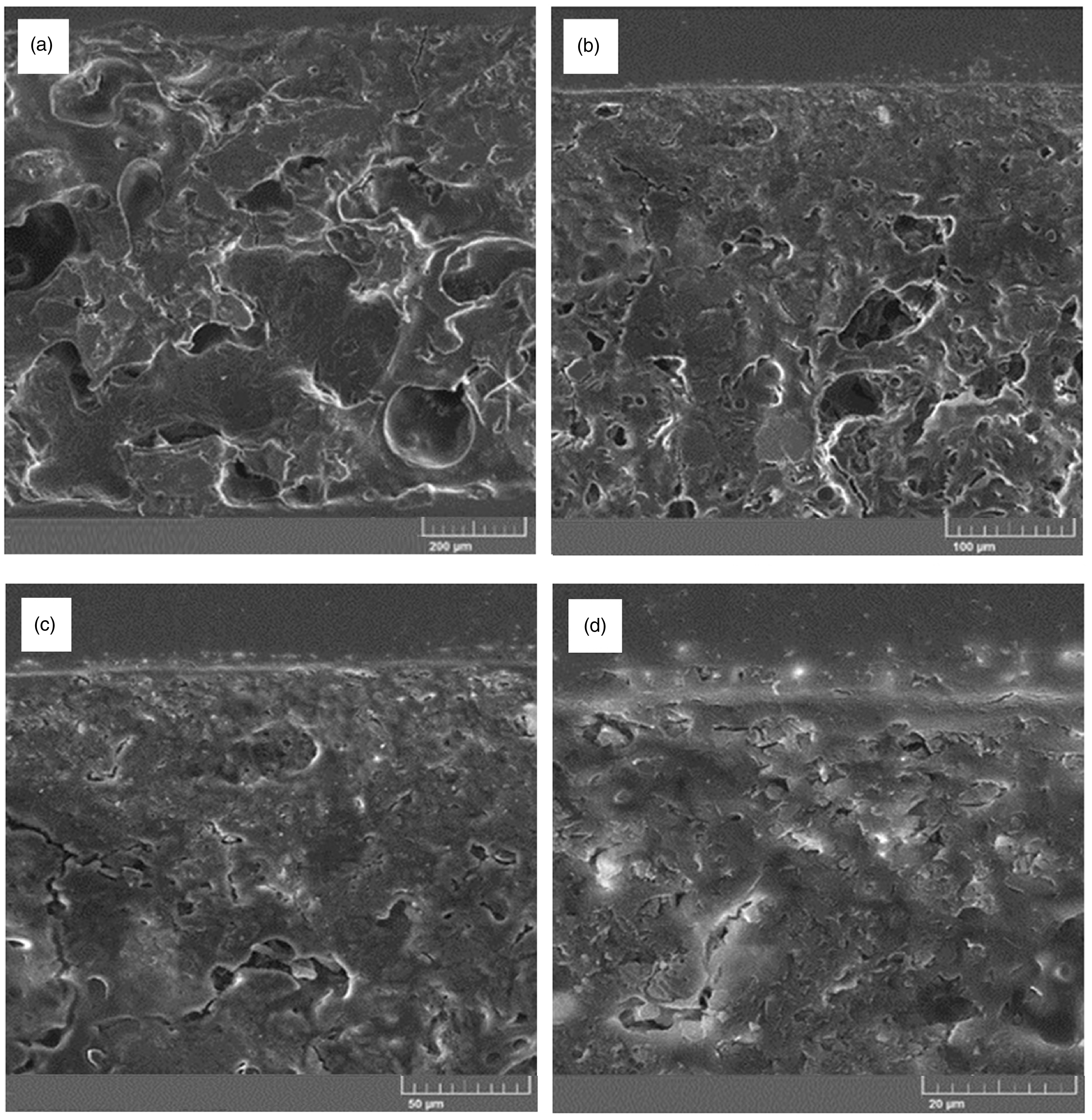
PCL coating morphology obtained by FE-SEM with different magnifications: (a) 200×, (b) 500×, (c) 1000×, and (d) 1000×.
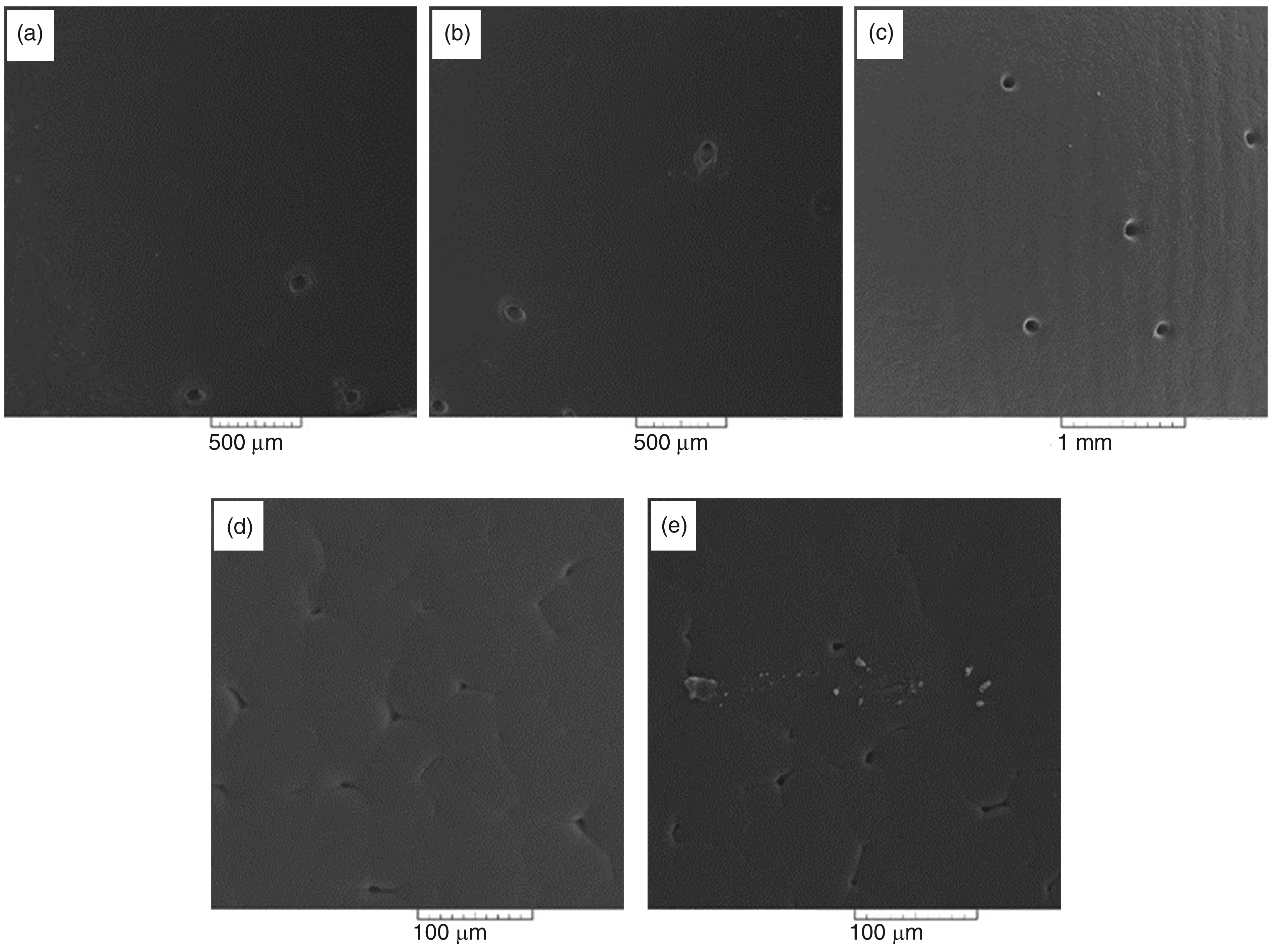
PCL coating morphology obtained by SEM, different magnifications: (a) 45×, (b) 45×, (c) 94×, (d) 400×, and (e) 440×.
Drug release from nanocomposite scaffolds
Figure 7 depicted the drug release percentage versus time profile of the nanocomposite scaffolds (S1, S2, and S3) over a period of 5 days obtained by the cell Franz method. According to Figures 6 and 7, drug release from the scaffolds and their degradation profile displayed a high level of similarity, possibly due to the diffusion process which the samples encountered in the fluids. In a study, 48 drug release from an in-situ injectable formulation was evaluated based on poly (lactide-co-glcolide, PLGA) (50:50) containing doxycycline hyclate. They proposed four steps in this drug release process: 1) a burst drug release, 2) polymer matrix formation, 3) polymer degradation, and 4) drug depletion of the system. 48 Drug release from the prepared samples could be interpreted according to the above research. Preliminary, fast, and sudden drug release was observed due to the early degradation of the scaffolds exposed to the release medium through PCL coating porosities. In fact, the nanocomposite core made up of Gelatin/HA/atorvastatin-loaded NLCs was coated with PCL by means of a simple spin coating process, and then several holes with the diameter of 200 μm were made immediately after coating nanocomposite core by inserting microneedles in the upper PCL film. Therefore, the medium can penetrate and reach to the nanocomposite core in order to release the drug and other nanocomposite components for the suitable wound healing.
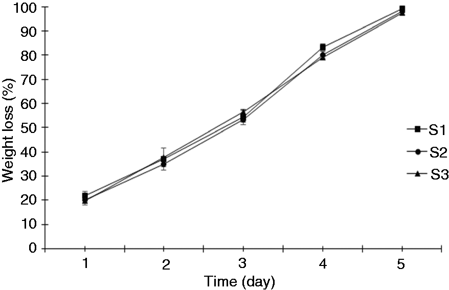
Weight loss percentage of the S1, S2, and S3 nanocomposite scaffolds over a period of 5 days immersion in PBS at 37 °C and pH = 7.4 (n = 3).
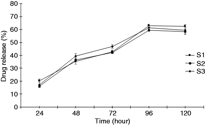
Drug release percentage from the nanocomposite scaffold (S1, S2, and S3) in PBS at 37 °C and pH = 7.4 over a period of 120 hrs (n = 3).
Secondly, slower drug release occurred due to the depletion of free drug in the previous step, and diffusion of atorvastatin through the NLCs controlled the release process of the drug. After three days, the slope of the drug release curve increased because of higher degradation of the samples which resulted in a higher amount of NLCs extracted from the deep layer of the samples. In this step, degradation was more effective than the diffusion process in drug release. Finally, the rate of drug release was declined followed by a drug concentration decrease in the samples.
Table 5 showed the drug release percentage and DRE percentage of each sample over a period of 120 hrs. The drug release percentage of the prepared samples showed no difference statistically. However, a slight difference of the drug release rate between the samples was by the diffusion and degradation processes. According to Table 6, S1 had the highest drug release percentage and the lowest DRE. In other words, drug release was high and rapid for S1. These findings are in accordance with the high degradation rate of S1 in the degradation test, and as a result more drug release is obtained.
Specifications of nanocomposite scaffolds.
Despite the lowest amount of NLCs in S1, the highest degradation rate of this sample induced more medium exposure of the nanoparticles and consequently greater drug release that showed the predominant role of the degradation process over the diffusion process.
Mechanical properties of the scaffolds
Table 6 and Figure 8 represent the wet mechanical properties of the prepared membranes (S1, S2, and S3) including the tensile strength, modulus of elasticity, and elongation percentage at the break. Statistical analysis of the data shown in Table 6 revealed that S3 had the highest tensile strength among the samples (9.44 ± 0.54 MPa) (p < 0.05). Its elastic modulus was measured at 93.17 ± 0.74 MPa. Furthermore, S2 and S3 exhibited the same modulus of elasticity and elongation at the break but more than the data mentioned for S1 (p < 0.05). The ultimate tensile strength and the elastic modulus of the human skin were measured at 21.6 ± 8.4 MPa and 6–22 MPa, respectively. 8 , 49 In fact, differing strain rates can considerably alter the mechanical properties of the skin and skin surrogates. 49 The mechanical test indicated that all the three scaffolds had an approximately 90 MPa elastic modulus which was more than two-fold of tensile modulus of the normal human skin. But, S3 presented the maximum ultimate tensile strength (9.44 ± 0.54 MPa) among the fabricated samples. These mechanical properties could be acceptable for a short period of time that these constructs would be used on the ulcer, and its application in the wound healing needs further investigation in vivo.
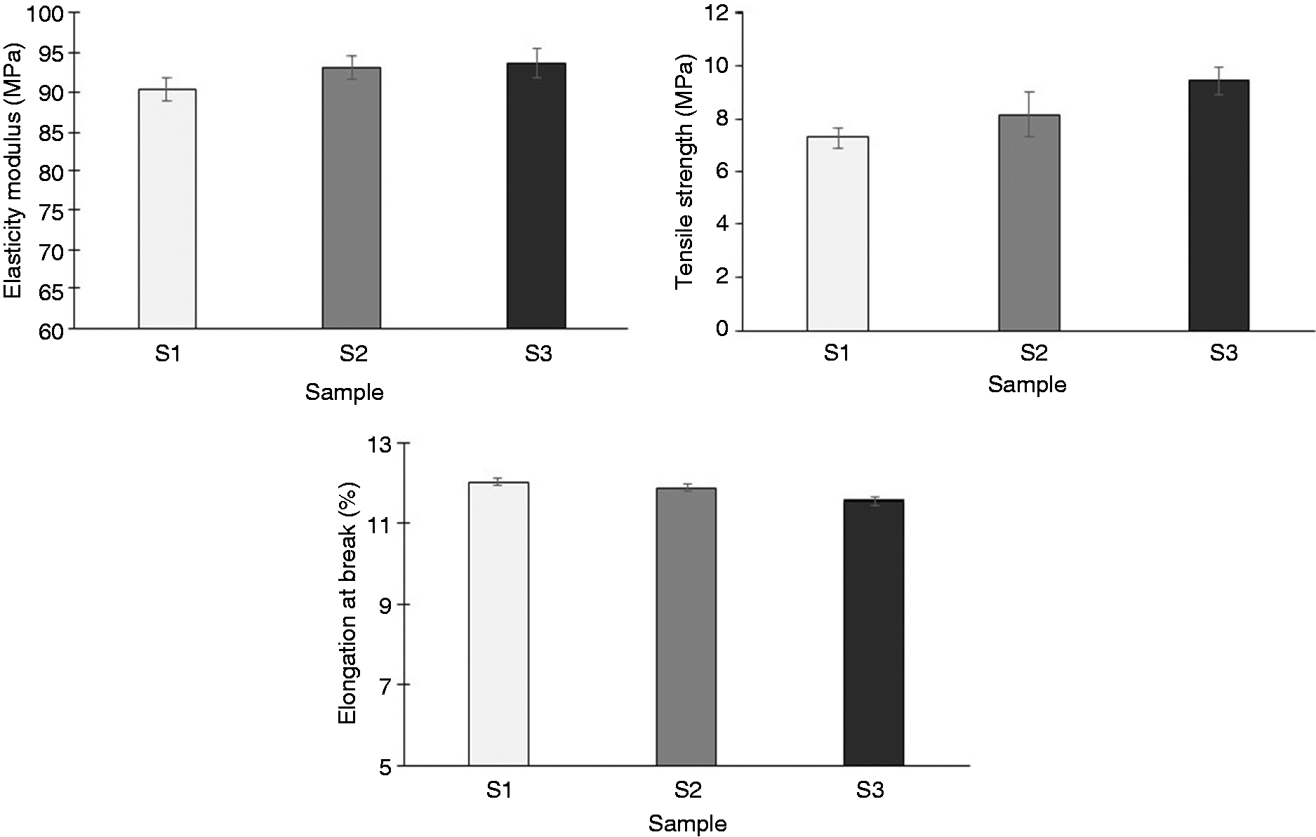
Mechanical properties (tensile strength, elasticity modulus, and elongation at break) of the nanocomposite scaffolds (S1, S2, and S3).
These differences in the mechanical properties may be related to the presence of the NLCs in the nanocomposites due to the scaffolds' composition similarity based on G, HA, and PCL. In fact, they varied with regard to NLCs amount in their structure.
In a research, a drug-loaded hydrogel containing lipid nanoparticles (LNP) was fabricated, and the effect of LNP content on drug release and mechanical properties was investigated. Results indicated that LNP had no effect on the hydrogel mechanical characteristics but influenced the drug release kinetics. 50
It was reported that proteins, polysaccharides, and lipid biopolymers showed certain disadvantages according to their mechanical and physicochemical properties. Therefore, to improve the mentioned properties, plasticizers, nanoparticles, lipids, and antimicrobial compounds were added to them. For example, lipid addition is anticipated to enhance the above films hydrophobicity, to diminish the water vapor permeability, and to influence mechanical as well as physicochemical properties. For example, the incorporation of lipids in such films more than a definite concentration can lead to deteriorate the mechanical properties of the films. Previous studies illustrated that candelilla wax addition in protein films more than 40 wt.% could destroy the mechanical properties. 51 , 52
In the present research, NLCs containing atorvastatin addition to the nanocomposites made the films more hydrophobic and, as a result, less water permeable and, as a result, enhanced mechanical properties. Although more than 40 wt.% of NLCs were used to prepare S2 and S3 nanocomposite scaffolds, mechanical properties were improved in contrast to the above study. Two reasons could describe such a behavior. Firstly, hydrophobicity increased by incorporating more NLCs into the nanocomposite structure. And secondly, the NLCs were uniformly distributed in the polymer matrix. Not only did water permeability decrease, but also crack propagation can be hindered by NLCs in the propagation path of the crack.
To investigate the effect of atorvastatin in the NLCs on the mechanical properties of the samples, the mechanical properties were measured for B1 in comparison to the scaffold containing the drug (S1). Table 6 and Figure 9 showed that S1 had a higher tensile strength and elastic modulus than B1, but B1 depicted more elongation at break than S1. However, there was no significant difference between the mechanical properties of the two samples according to the statistical analysis. In other words, the presence of atorvastatin in the samples has no significant effect on mechanical properties.
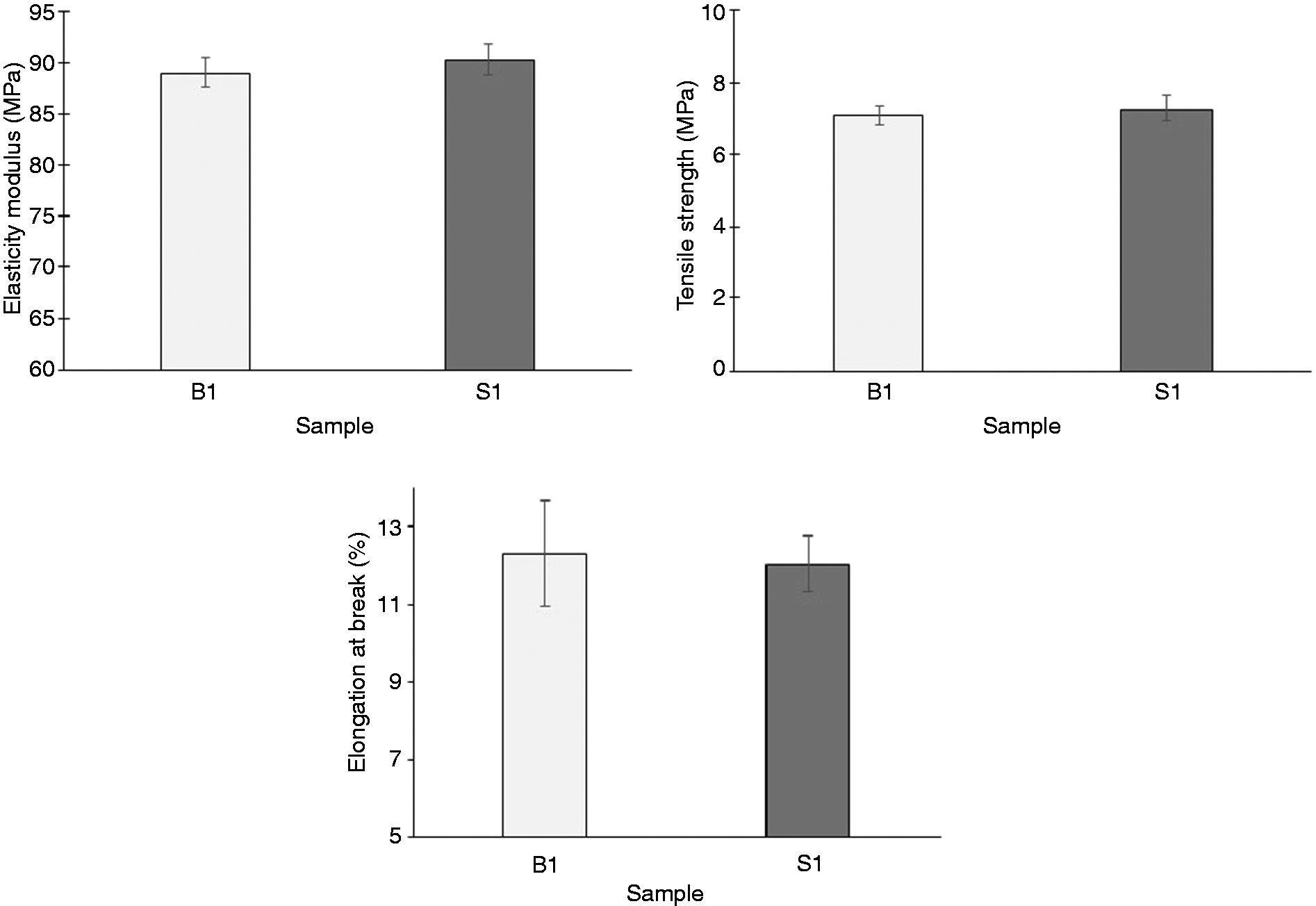
Mechanical properties (tensile strength, elasticity modulus, and elongation at break) of the nanocomposite scaffolds (B1 and S1).
Nanocomposite scaffolds morphology
Figure 10 showed FE-SEM images of the S3 nanocomposite scaffolds. As can be observed, the nanoparticles were not aggregated. The prominent shape of the NLCs was spherical, with some of the amorphous and shapeless particles. NLCs distribution was fairly uniform. The uniform distribution of the NLCs in the primary solvent casting suspension can lead to fabrication of such samples. But nanoparticles accumulation in some parts of the samples could be related to the uneven casting mold. Figure 10 illustrated NLCs' average particle size of about 195 nm. This increase in the particle size could be attributed to the freeze-drying process preparation of the NLCs nanoparticles. In general, factors such as the freezing rate, type, concentration of cooling protective agent, and freeze-drying parameters can affect the amount of particle size rise after the freeze-drying process. 53 , 54

FE-SEM images of S3 nanocomposite scaffold with different magnifications: (a) 2.00 KX, (b) 10.0 KX, (c) 35.0 KX, (d) 35.0 KX, and (e) 35.0 KX.
Chemical composition of the scaffolds
Figure 11 displayed the FTIR-ATR pattern of S3. The initial broadband absorption peak at 3273.57 cm−1 refers to the N-H stretching bond in atorvastatin or the amide A group in gelatin. 55 The peak at 2922.59 cm−1 is related to the C-H stretching bond of methyl group in atorvastatin, gelatin, and PCL substances. A weak peak at a wavelength of 2859.92 cm−1 was observed, which is similar to the preceding peak of the C-H stretching bond from methyl in the PCL, gelatin, and HA materials. The sharp peak at 1724.58 cm−1 is related to the carbonyl group (C = O stretching) in PCL. This peak was displaced to higher wavelengths from the original peak in the pure PCL absorption spectrum, which can be related to PCL interaction with the nanocomposite core of the scaffold. 56 , 57
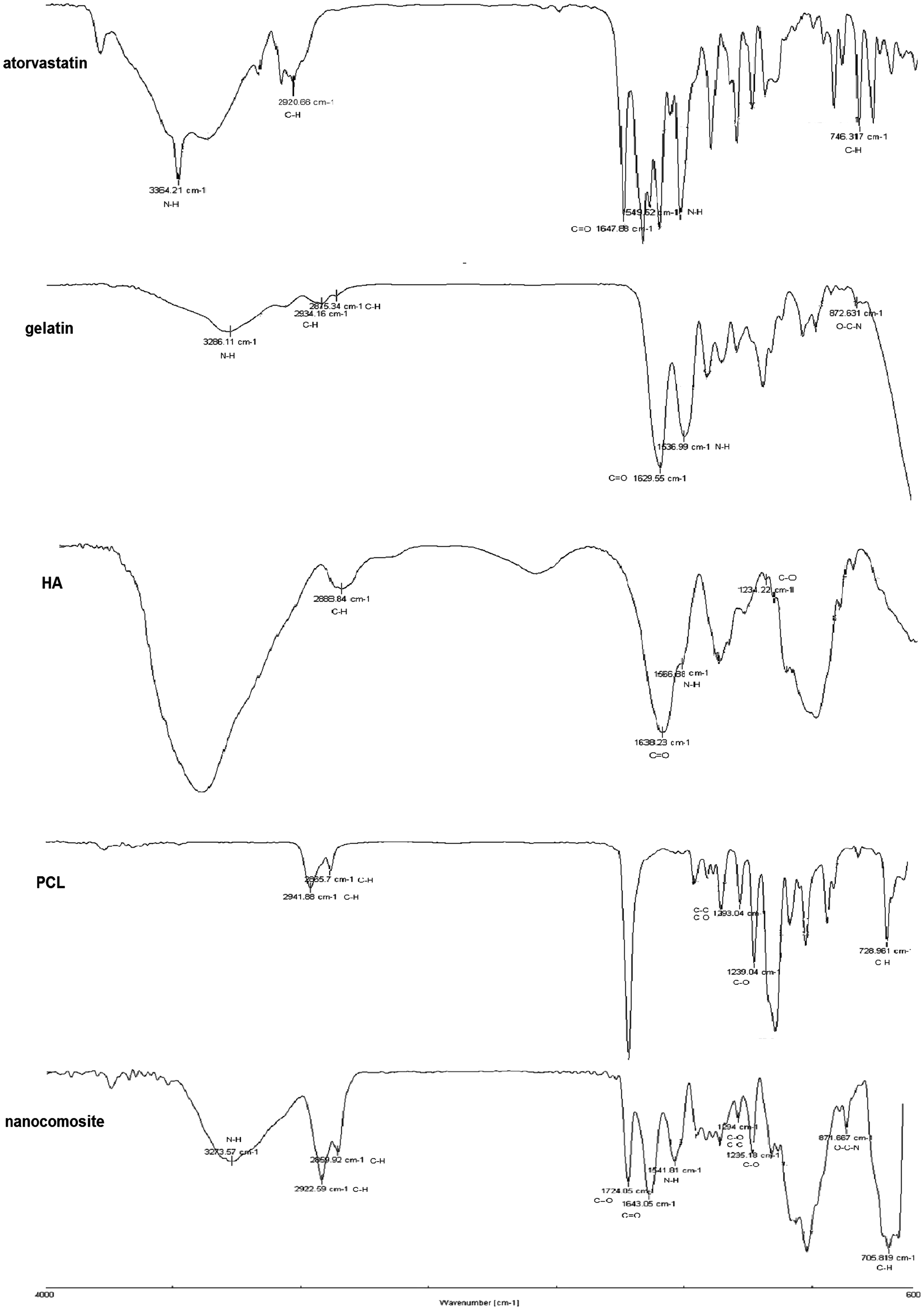
FTIR-ATR spectroscopy analysis of atorvastatin, gelatin, HA, PCL, and nanocomposite (S3).
A sharp peak at the wavelength of 1643.05 cm−1 may refer to gelatin with a wavelength of 1629.55, atorvastatin with the wavelength of 1647.88 cm−1, and HA with the wavelength of 1638.23 cm−1. 58 Due to a double-carbon bond with oxygen, all of these materials in this wavelength have a strong peak, and it can be said that the peak was related to the C = O stretching bond. A relatively weak peak at the 1541.81 cm−1 wavelength related to the N-H bending bond possibly belonged to three substances including gelatin with the wavelength of 1536.199 cm−1, atorvastatin with the wavelength of 1549.52 cm−1, and HA with the wavelength of 1566.88 cm−1. 58 , 59 The small peak at 1294 cm−1 was pertained to C-O and C-C stretching bonds in the PCL crystalline phase. 57 A sharp peak at a wavelength of 1235.18 cm−1 can be attributed to the C-O stretching bond both in HA and PCL. It was also possible that this peak was related to the vibration of the amide III group in the gelatin structure. A small peak at the wavelength of 871.766 cm−1 referred to the O-C-N bending bonds of the amide IV group in gelatin. 27 A sharp peak at 705.819 cm−1 can be attributed to the C-H stretching bond in atorvastatin or PCL. 57
Cytotoxicity assessment of the nanocomposite scaffolds
Figure 12 represents cytotoxicity evaluation of S1, S2, S3, and negative control using the HDF. Results indicated not only cell viability but also enhanced proliferation of the cells on the nanocomposite scaffolds. Statistical analysis of degradability and mechanical tests showed that there were no differences between the specimens, but in the MTT assay, there was a significant difference on the third day of cell culture among the S3 and three other samples (S1, S2, and the control) (p < 0.05). On the fifth day of the culture, both S3 and S1 indicated significant differences with S2 and control (p < 0.05).
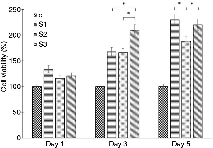
The viability of human dermal fibroblast cells on the S1, S2, and S3 nanocomposite scaffolds in the first, third, and fifth days of culture (p < 0.05).
To evaluate the effect of atorvastatin on the HDF viability and proliferation, the MTT assay on B1 was also performed in order to compare the results of cytotoxicity test with the drug containing S1 specimen. Figure 13 illustrates the results of the MTT assay on S1 and B1 samples. It can be observed that cell viability for the S1 was higher than B1 and negative control after 5 days of cell culture. In fact, on the fifth day, when the drug reached the highest level of release in S1 in accordance with previous drug release test of the scaffolds, its beneficial effect on the HDF viability and proliferation could be confirmed. Likewise, the drug-free B1 sample and the control showed no cytotoxicity. As a consequence, cytocompatibility of the nanocomposite scaffolds' components (G, HA, and PCL) without drug can be affirmed.
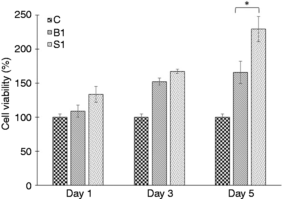
The viability of human dermal fibroblast cells on the S1 and B1 nanocomposite scaffolds in the first, third, and fifth days of culture (p < 0.05).
In a study, 60 the influence of rosuvastatin calcium as a statin drug and scaffolds on proliferation of HDF cells was investigated by the MTT assay. Scaffolds containing drug exhibited a considerable raise in the proliferation of HDFs after 72 hrs of cell culture compared with the scaffolds-without-drug group and the control group (p < 0.05). Therefore, the effect of rosuvastatin calcium on increasing the proliferation of the HDFs was proved. Furthermore, the scaffold without drug displayed more cell growth than the control. This could be attributed to the capability of the cells to proliferate and penetrate through the porous structure of the scaffolds. Also, in vivo study illustrated that medicated scaffolds showed faster wound closure compared to the scaffolds-without-drug group as well as the control group during the first week of the wound healing. Results of the present research were in line with the previous research regarding the effect of rosuvastatin calcium on HDF. 60 S1 containing atorvastatin showed an increased HDF viability and proliferation compared with the scaffold without drug (B1). The exact mechanisms of the atorvastatin effect on the HDF viability and proliferation need further investigations.
Adhesion and morphology of HDF cells on S3 optimal nanocomposite scaffold
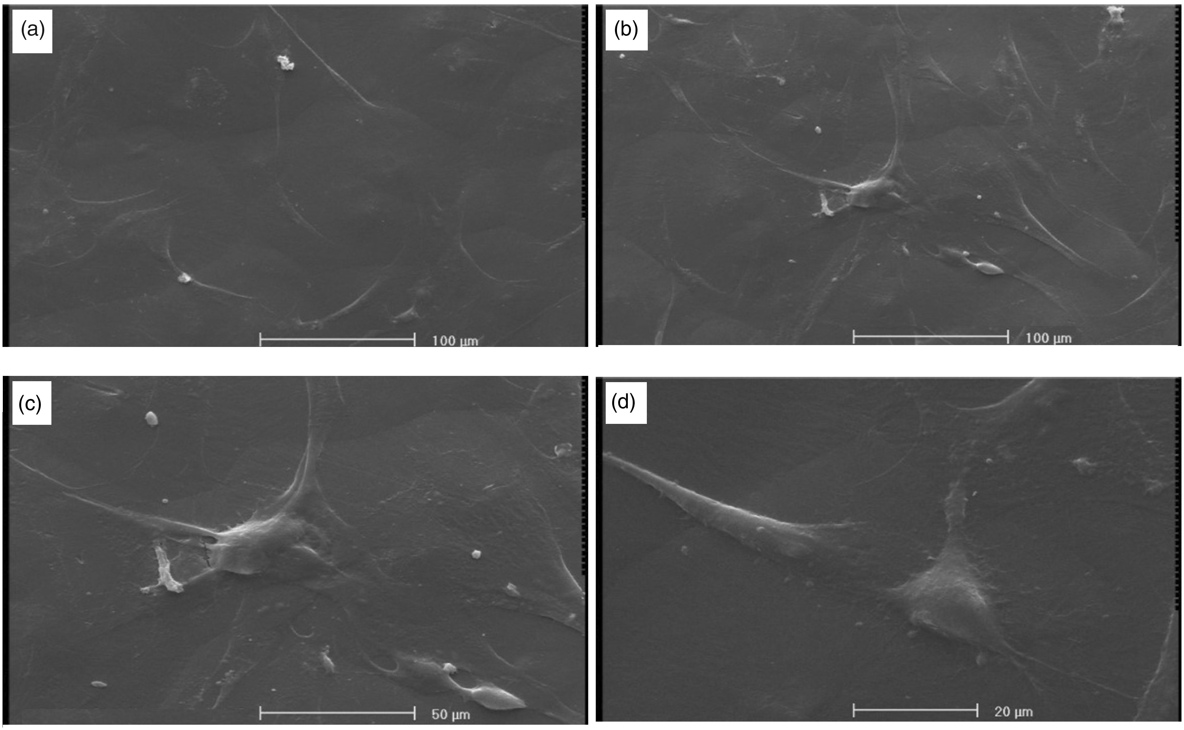
SEM images of HDF cells fixed on the S3 nanocomposite scaffold after 1, 2, and 3 days of the cell culture were presented in Figures 14 to 16, respectively. The cell attachment and proliferation of the cells seeded on the scaffolds have a prominent role on the cells’ survival, function, and extracellular matrix formation. Figure 14 indicated adherence of the HDFs on the scaffold after 24 hrs of incubation. HDFs were distributed on a scaffold surface with a spherical-spindle shape, with some filopodia projections. Figure 15 depicted the morphology of the seeded HDF cells on the S3 sample after two days of the cell culture. Cells with more filopodia connected to each other and formed a mesh interconnected structure on the surface of the scaffold. On the third day of culture, HDFs completely covered the surface of the S3 which indicated that the HDF cells had an excellent adhesion and growth on the surface of S3 scaffold (Figure 16). Also, in a study, PCL fumarate/gelatin-based nanocomposite showed a good cell attachment, and the cells were well spread and display flattened sheet morphologies without any in vitro toxicity. 61
SEM images of HDF cells formed on S3 nanocomposite scaffold after one day of culture with different magnifications: (a) 250×, (b) 250×, (c) 500× and (d) 1000×.
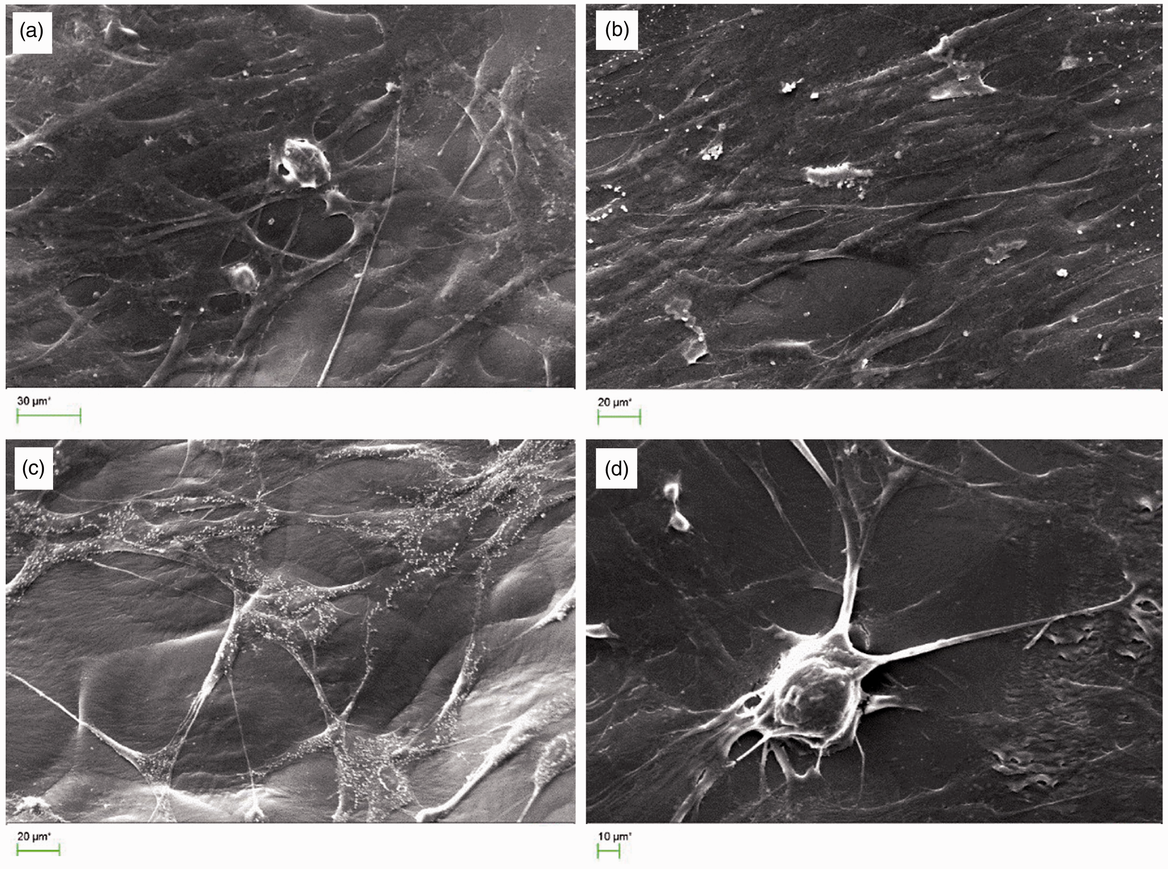
SEM images of HDF cells formed on S3 nanocomposite scaffold after the two day of culture (1000× magnification).

SEM images of HDF cells formed on S3 nanocomposite scaffold after the three days of culture with different magnifications: (a) 1000×, (b) 500×, (c) 1000× and (d) 2000× magnification.
Conclusion
NLCs with suitable properties such as low toxicity, biodegradability, drug preservation, high DL, and controlled drug release can be an appropriate atorvastatin dermal delivery system. NLCs containing 55 wt.% GMS solid lipid and 45 wt.% OA liquid lipid were prepared by the solvent evaporation method, and it showed good physicochemical properties such as small particle size, high zeta potential, proper loading efficiency, and good drug release. G/HA/54.11 wt.% atorvastatin-loaded NLCs nanocomposite scaffold coated with PCL (S3) containing 1.18 wt.% atorvastatin showed the best degradation, drug release, mechanical properties, and HDF cell response among the fabricated samples, and it could be used as an appropriate construct for the skin tissue engineering.
Footnotes
Declaration of conflicting interests
The author(s) declared no potential conflicts of interest with respect to the research, authorship, and/or publication of this article.
Funding
The author(s) disclosed receipt of the following financial support for the research, authorship, and/or publication of this article: The authors wholeheartedly acknowledge the financial support for this work by University of Isfahan and Isfahan University of Medical Sciences, Isfahan, Iran.