
Abstract
Spinal fusion cages are commonly used to treat spinal diseases caused by degenerative changes, deformities, and trauma. At present, most of the main clinical spinal fusion cage products are non-degradable and still cause some undesirable side effects, such as the stress shielding phenomenon, interference with postoperative medical imaging, and obvious foreign body sensation in patients. Degradable spinal fusion cages have promising potential with extensive perspectives. The purpose of this study was to fabricate a degradable spinal fusion cage from both polycaprolactone (PCL) and high-proportion beta-tricalcium phosphate (β-TCP), using the highly personalised, accurate, and rapid fused deposition modelling 3 D printing technology. PCL and β-TCP were mixed in three different ratios (60:40, 55:45, and 50:50). Both in vitro degradation and cell experiments proved that all cages with the different PCL:β-TCP ratios met the mechanical properties of human cancellous bone while maintaining their structural integrity. The biological activity of the cages improved with higher amounts of the β-TCP content. This study also showed that a spinal fusion cage with high β-TCP content and suitable mechanical properties can be manufactured using extruding rods and appropriate models, providing a new solution for the design of degradable spinal fusion cages.
Introduction
In recent years, spinal diseases caused by degenerative changes and deformities of the spine have gradually been afflicting younger individuals more frequently. 1 The clinical surgical method traditionally used for treating patients with spinal diseases is autologous bone graft fusion (also known as autogenous bone grafting). 2 However, an autogenous bone graft often results in a high rate of non-fusion, pain at the transplant site, and misalignment of the transplanted bone. 3 Later, allogeneic bones were used for bone graft fusion. 4 Although fusion cages based on allogeneic bones could effectively reduce the collapse phenomenon after their implantation, they still caused immune rejection and disease transmission in patients. Therefore, titanium alloys were used to make interbody fusion cages. 5 However, despite that titanium alloy-based interbody fusion cages can effectively open the vertebral space and maintain the spine height, their compression modulus is too large and they do not match the natural bone tissue of the human body, resulting in the stress shielding phenomenon. Moreover, these fusion cages still require autologous bone transplantation at the time of surgery to promote spinal fusion, and the inability of X-rays to pass through titanium alloy material can result in their interference with postoperative medical imaging.6–8 In addition to titanium alloys, polyetheretherketone(PEEK) is also used to make intervertebral fusion cages, compared with titanium alloy, the PEEK cages have better X-ray penetration, which is beneficial for postoperative medical imaging assessment, and its mechanical properties can fully open the intervertebral space without causing shielding phenomenon, but due to its low biological activity, its osseointegration effect is not as good as titanium alloy interbody fusion cage, and will cause a series of complications such as: fusion cage collapse or nerve root injury. 9
In recent years, researchers have been focussing on the use of degradable materials for spinal fusion cages. The main degradable materials currently used in interbody fusion cages are polylactic acid(PLA), poly-
The polymer PCL ((C6H10O2)n),
17
which has good biocompatibility and degradation properties, is often used in bone tissue repair together with materials that have good osteoinductive and osteoconductive properties.18–21 Another degradable material with good biocompatibility is β-TCP, the tissue composition of which is similar to the inorganic composition of bone tissue. The pores between connected β-TCP
Therefore, in order to develop degradable spinal fusion cages with an elastic modulus close to that of human cancellous bone, high bioactivity, and good bone conduction and osteoinduction, we used fused deposition modelling 3 D printing technology to fabricate fusion cages with different PCL–β-TCP ratios. A series of in vitro experiments, such as compression and cytotoxicity tests, were carried out to check the mechanical and osteogenic properties of these scaffolds. Furthermore, we have provided a new fusion cage structure with improved mechanical properties by designing a crossbeam and an outer frame structure on the cage. 25
Materials and methods
Design and preparation of the PCL–β-TCP spinal fusion cages
Polymer and inorganic salt composite materials are too brittle to be made into wire materials for fused deposition printing. The general method is to change the wire feeding device to the method of extrusion and powder material silo. However, the preheating time of the powder material bin is too long, and the inorganic salt
The printing step is shown in Figure 1. PCL (
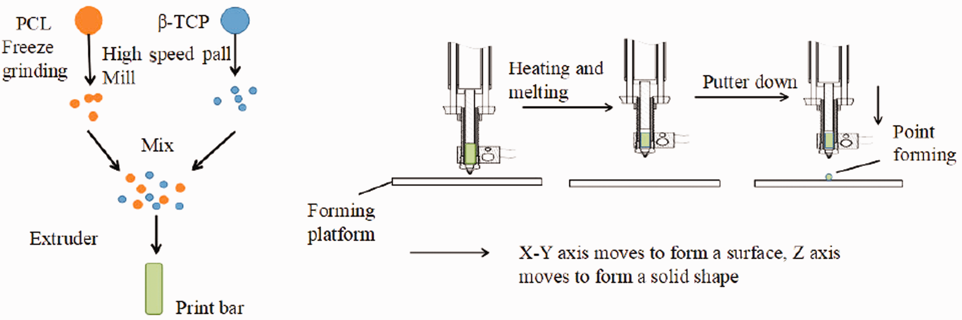
Schematic of the molding process of the PCL−β-TCP spinal fusion cage.
Tests of the mechanical compression and degradation of the spinal fusion cages
The three fusion cage samples with different PCL:β-TCP ratios (designated the 60:40 group, 55:45 group, and 50:50 group, respectively) were tested for their compression performance, using a mechanical testing machine (Instron 5575, USA) with the following settings: compression rate, 1 mm/min; height, 5 mm; and compression percentage, 60%. Before each compression, clear options were first set.
For the degradation experiment, samples of the three fusion cages and of PCL alone (as the control group) were weighed out (m0) and placed individually into 50-mL centrifuge tubes. An equal volume of 10× phosphate-buffered saline (pH0 = 7.42) was then added to each tube. After 3 months, the fusion cages were removed from the centrifuge tube and the degradation solution was collected for measurement of its pH (recorded as pH1). The change in pH can be determined by comparing the pH0 and pH1 values.
After using filter paper to absorb the moisture from the cages, the samples were placed in a vacuum drying box for 1 week. Thereafter, they were removed and weighed (recorded as m1) and their weight loss rate was calculated according to equation (1).

The mechanical compression experiments were performed before and after the degradation of the various cages. Stress–strain curves were obtained for each of the cage groups, with three samples tested per group. The slope of the curve and the highest point of elastic deformation were calculated to obtain the compressive strength and compressive modulus of each cage sample.
The dried samples before and after degradation were cut into smaller block samples, after spraying gold, and placed under a scanning electron microscope to observe the surface morphology of the fusion cage.
Test of the cytocompatibility of the cage extraction solutions with different material proportions
PCL–β-TCP composite stents with different material ratios (mass ratios) were used to prepare stent extracts with different material ratios according to the Biological Evaluation of Medical Devices of China (GB/T16886.12–2005/ISO 10993–12: 2002): The stent extracts were obtained by immersing the stents into alpha minimal essential medium (containing 10% foetal bovine serum) at a concentration of 10 mg/mL. In separate 48-well plates, well-grown MC3T3-E1 cells were first inoculated (after digestion) into each well at a density of 8000 cells/mL and cultured in a cell incubator at 37 °C (under 5% CO2) for 1 day. Then, the original medium was replaced with the stent extracts. In total, five sample groups were set up (3 parallel samples each), with PCL set as one of the groups. Normally growing cells were set as the control group, and the group without the cell culture was the blank group. Cell Counting Kit-8 (CCK-8) was used to measure the cells after 1, 3, and 5 days of culture, and the optical density at 450 nm (OD450) of each group was measured with a microplate reader to calculate the relative growth rate (RGR) according to equation (2).

MC3T3-E1 cells were cultured in the same way as described above for the detection of alkaline phosphatase (ALP), osteocalcin (OC), and collagen I (Col-I) using commercial kits (Wuhan ColorfulGene Biological Technology, Wuhan, China). The cells used for ALP detection were cultured for 7 days, whereas the cells used for OC and Col-I detection were cultured for 1, 3, and 5 days. The differentiation status of the osteoblasts was investigated using a commercial kit according to the manufacturer’s instructions. The groups created for these latter tests differed from those of the CCK-8 test in that the PCL group was not set as one of the sample groups and the blank group was not set for the calculation process.
Observation of the osteoblast morphology on the composite scaffolds
MC3T3-E1 cells were seeded at a density of 280,000 cells/mL onto the various PCL–β-TCP composite scaffolds. After 4 h of cell culture, the remaining cell culture medium on the scaffolds was washed away with physiological saline and the cells were fixed with 5% glutaraldehyde for more than 4 h. The scaffolds with adherent cells were then washed 3 times with physiological saline followed by 2 times with deionised water. After staining with acridine orange (AO) or 4ʹ,6‐diamidino‐2‐phenylindole (DAPI), the distribution and morphology of the cells on the PCL–β-TCP composite scaffolds were observed at 100× magnification under a fluorescence microscope.
Results and discussion
Modelling of the spinal fusion cages
Images of the designed cages by Solidworks 2016 are shown in Figure 2(a). The width of the outer frame and the three beams was 1.1 mm, the total height was 5 mm, the cross-sectional area was 57 mm2, and the internal mesh had a lay-down pattern of 60/90/120° and porosity of 65.1%. Stefan’s research indicated that this fiber network connection can withstand greater shear forces after bone healing. 26 Figure 2(b) shows the cell scaffold model used in the cell adhesion experiment. The diameter of the model was 10 mm, and the mesh porosity was approximately 70.2%. 27
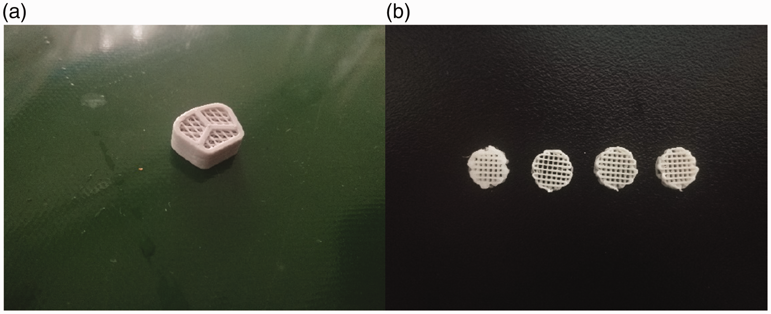
(a) Three-beam spinal fusion cage model; (b) Cell scaffold model used in cell adhesion experiments.
Mechanics of the spinal fusion cages and their mechanical properties after degradation
The degradation of the materials inevitably leads to the degradation of the mechanical properties of the scaffold. 28 , 29 The bone repair material needs not only to meet good biocompatibility but also to maintain certain mechanical properties during the degradation process.30–33
The mechanical properties of the spinal fusion cages were observed to have changed to some extent after degradation (Figure 3). As shown in Figures 4 and 5, with the increase of β-TCP content in composite material group, the compressive modulus of the cage increases, the compressive strength decreases, and the mechanical properties after degradation were all lower than before. Unfortunately, there had no significant differences between the composite material groups (p < 0.05). We also found that the compression modulus of the PCL group samples increased with the degradation of 3 months. This phenomenon was also found in Lin Lu’s research, they explained this phenomenon as the crystalline region in the biopolymer is difficult to be contacted by the surrounding degradation liquid, the degradation starts from the non-crystalline region, and the compression of the PCL The increase in modulus may be due to the increase in crystal area. 34
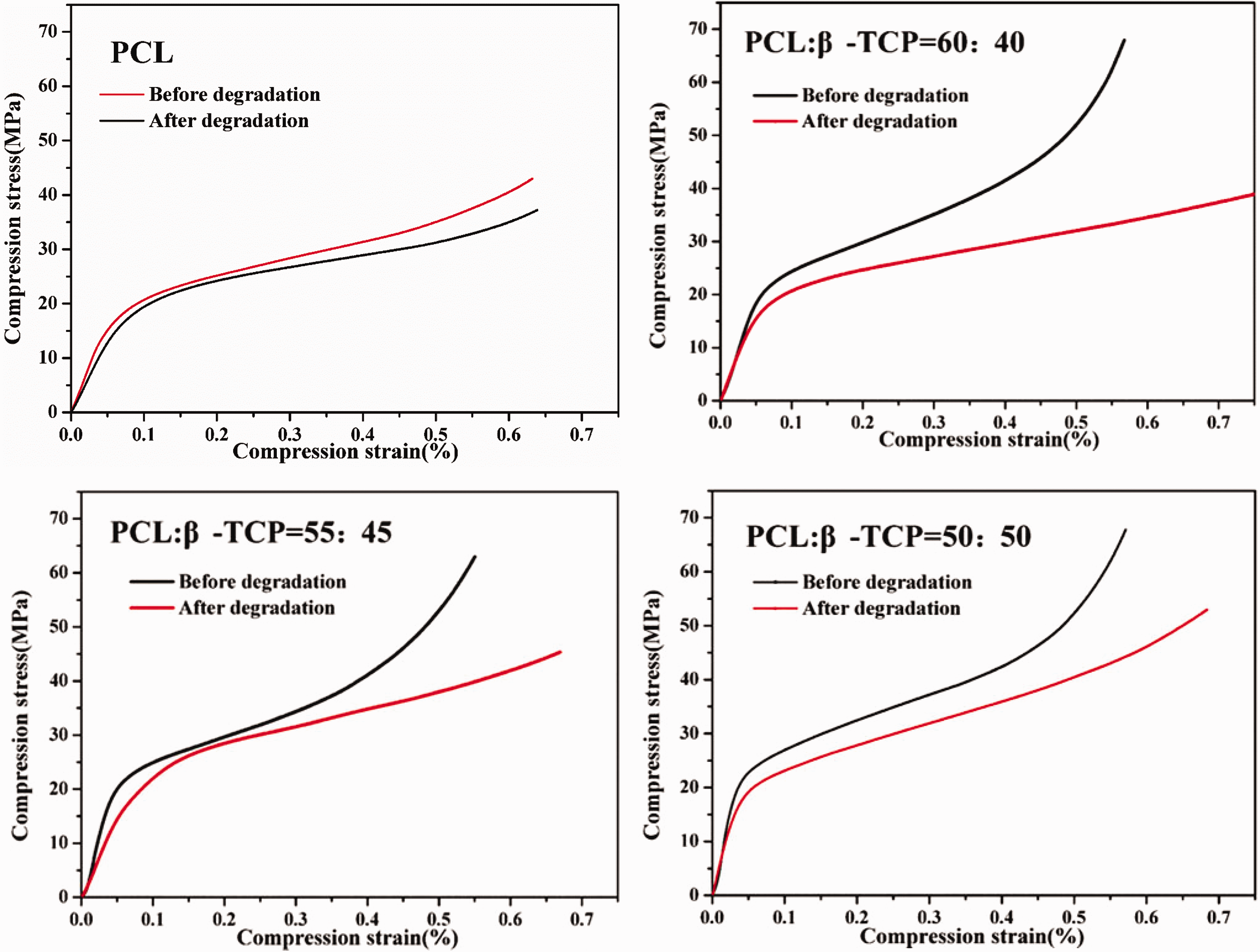
Stress-strain curve before and after degradation.
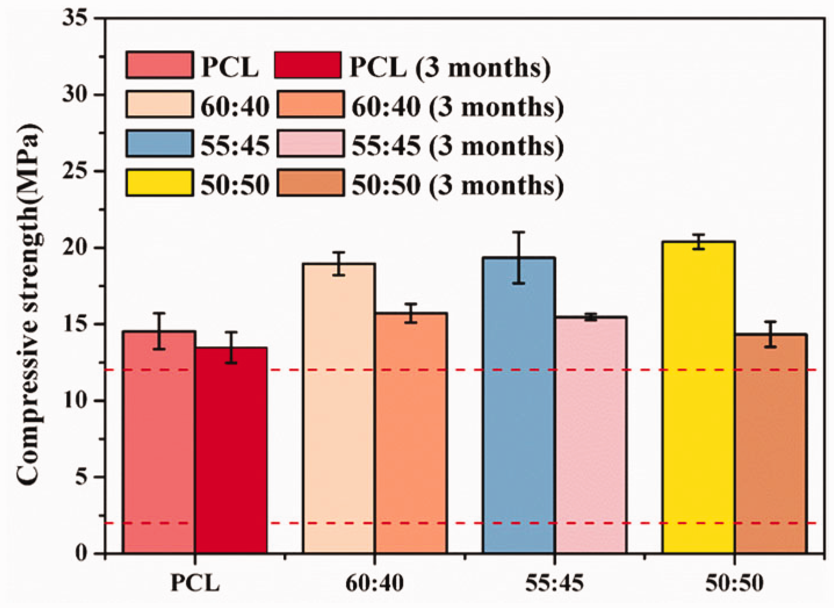
Compressive strength of the cages before and after degradation.
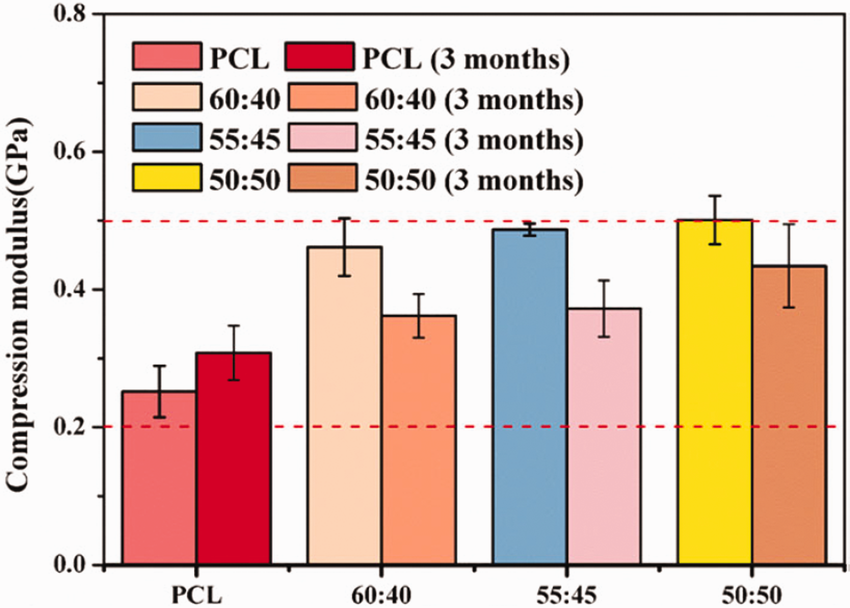
Compression modulus before and after the cages degradation.
The compressive strengths of all the groups met the requirements of the mechanical properties of human cancellous bone (the range of the red dotted line in Figure 4 35 ), indicating that the prepared spinal fusion cages can provide sufficient mechanical support for the human body. Additionally, the compression modulus before and after the degradation of the cages was within the normal range of human cancellous bone (the range of the red dotted line in Figure 5 35 ), indicating that stress shielding will not occur. As far as we know, most of the previous studies used extruded filament 3 D printing technology to complete the printing process of PCL-β-TCP composite materials, but when the β-TCP content exceeds 30%, the filament may be too brittle and fractured, thus terminating printing. 36 The printing method of our extruded bar material has completed the printing of the fusion device with higher β-TCP content, and the mechanical properties of the fusion device can satisfy the requirement for the human cancellous bone.
Changes in other indicators of cage degradation
In the process of PCL degradation, some small-molecule monomers (6-glycolic acid) or oligomers are generated from the breaking of the chemical bonds. 37 These monomers and oligomers are dissolved in the degradation solution, resulting in a change in the solution pH and the loss of quality of the entire fusion vessel, which means it might cause local acidosis so that the side effect happened for treatment.
As indicated in Table 1, with the degradation of the cages, the content of β-TCP increases, which leads to greater quality loss. It might that β-TCP is a water-soluble substance. 38 This means that the incorporation of β-TCP will result in faster degradation of the entire fusion system, and the main body will still be PCL; hence, the mass loss rate of each group fluctuates only by approximately 1%.
Mass loss for 3 months degradation.

Table 2 shows the pH change of the degradation solution after 3 months of degradation of the cages. It was found that the pH values of the three experimental cage groups containing β-TCP were lower than that of the PCL cage group, which was due to the accelerated degradation rate after β-TCP incorporation. 34 , 39 However, as the β-TCP concentration increased in the groups of this article, the pH value changed less. We believe that β-TCP consumes part of H+ during the degradation process. This would reduce any local inflammatory response caused by a reduction in the pH. 40 Compared with the mass loss for 3 months degradation, this result of pH change also proved that it should have the best proportion of β-TCP to balance the mass loss and pH change.
pH change of degradation solution after 3 months of degradation.
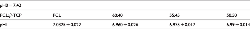
Figure 6 shows the surface morphology of different materials before and after degradation. It can be seen that the pre-designed fiber network structure of each group of fusion devices after degradation is still intact, and the surface of the pure PCL group has not changed much as expected smooth surface. After degradation of composite material samples, some small particles are exposed on the surface of fiber network, which are β-TCP inorganic particles encapsulated by PCL. The degradation of PCL lowers the pH in the system, which accelerates the degradation of β-TCP, so the more β-TCP content, the smaller the size of the exposed granular objects. 41
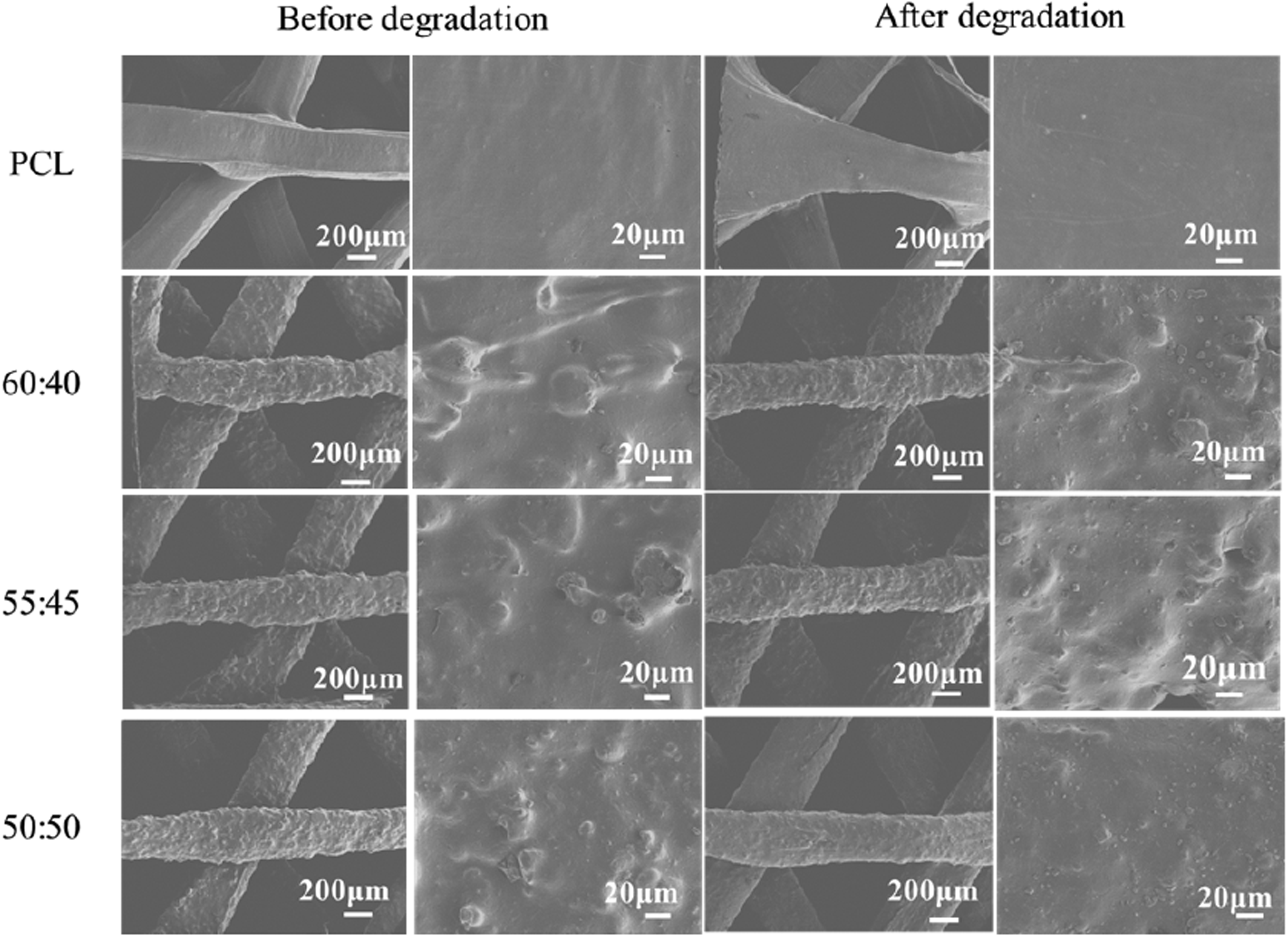
Surface morphology of interbody fusion cages with different materials before and after degradation.
Cell viability test (CCK-8 test)
CCK-8 is one of the methods commonly used for detecting cell viability. Moreover, β-TCP is known to effectively promote osteoblast proliferation. 42 Therefore, we used the CCK-8 method to determine the cytotoxicity of the fusion cages toward MC3T3-E1 cells.
Figure 7 shows the proliferation of MC3T3-E1 cells cultured together with the different stent extracts for 1, 3, and 5 days. It can be seen that the MC3T3-E1 cells grew very fast, and the incorporation of β-TCP accelerated the growth of the cells. The RGR of the 50:50 group exceeded 100% only when the cells were cultured until the fifth day (Table 3). This indicated that the scaffolds had almost no cytotoxicity, had good biocompatibility, and could promote MC3T3-E1 cell proliferation. Yuan and Wang, et al. constructed the PCL-β-TCP (90:10, 80:20) cell scaffold using 3 D printing technology. Through the cultivation of mesenchymal stem cells, it was found that the cell proliferation rate of the 80:20 group was better than that of the PCL group and 90:10 group. 36
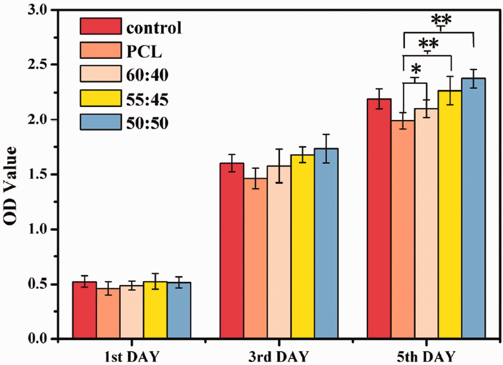
CCK-8 results of PCL−β-TCP with different material ratios.
Cell proliferation rate of scaffold extracts with different material ratios.

Effects of the scaffolds on the alkaline phosphatase activity of MC3T3-E1 cells
ALP can promote the hydrolysis of organic phospholipids, increase the concentration of PO43–, and further promote the deposition of calcium ions on the surface of the material, thereby initiating calcification. Its content can be used to evaluate bone formation ability. 43
Figure 8(a) shows the ALP standard curve (constructed with the ALP standard provided in the kit), for which the correlation coefficient (R2) was 0.989. The linear regression equation for the standard curve was used to calculate the basic phosphate levels of the experimental and control groups. The histogram of the enzyme concentrations recorded (Figure 8(b)) revealed that the ALP contents of the three experimental groups were significantly higher than that of the control group, indicating that the PCL–β-TCP composite scaffold could promote osteoblast differentiation and increase ossification at the same time. In the 50:50 group, the ALP content was as high as 1027.58 ± 72.81 pg/mL, whereas that in the control group was only 502.57 ± 77.42 pg/mL. These results were consistent with the function of β-TCP. This result is consistent with the law derived by Yuan. 36
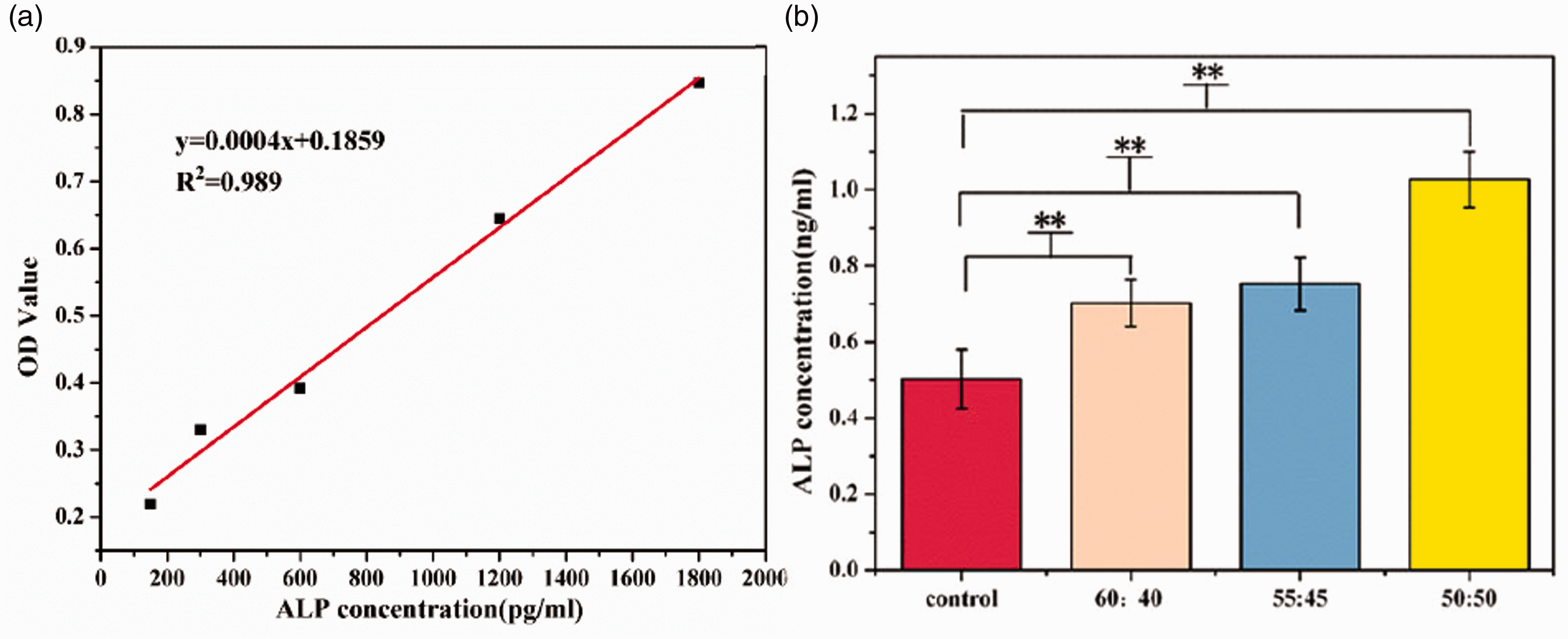
Detection of alkaline phosphatase (ALP) activity in MC3T3-E1 cells: (a) ALP standard curve; (b) ALP content in control and experimental groups.
Effects of the scaffolds on osteocalcin secretion from MC3T3-E1 cells
OC is a non-collagenous protein secreted specifically by osteoblasts. It can maintain the normal rate of bone mineralisation and directly reflects the activity of osteoblasts. 44
Figure 9(a) shows the linear regression equation of the OC standard curve (constructed using the OC standard in the kit), for which R2 was 0.94. Using this equation, the concentrations of OC secreted by the MC3T3-E1 cells on the experimental scaffolds at 1, 3, and 5 days were calculated. As shown in Figure 9(b), with the increase in culture time, the amount of OC secreted by the MC3T3-E1 cells increased gradually. Compared with the cells in the control group, the cells in the experimental group released more OC. This indicated that the PCL–β-TCP composite scaffolds promoted bone metabolism and accelerated the osteogenic process. When the PCL: β-TCP ratio was 50:50, the secretion of OC was as high as 5.23 ± 0.06 ng/mL at 5 days. The experimental results are consistent with the osteogenesis characteristics of β-TCP. 45
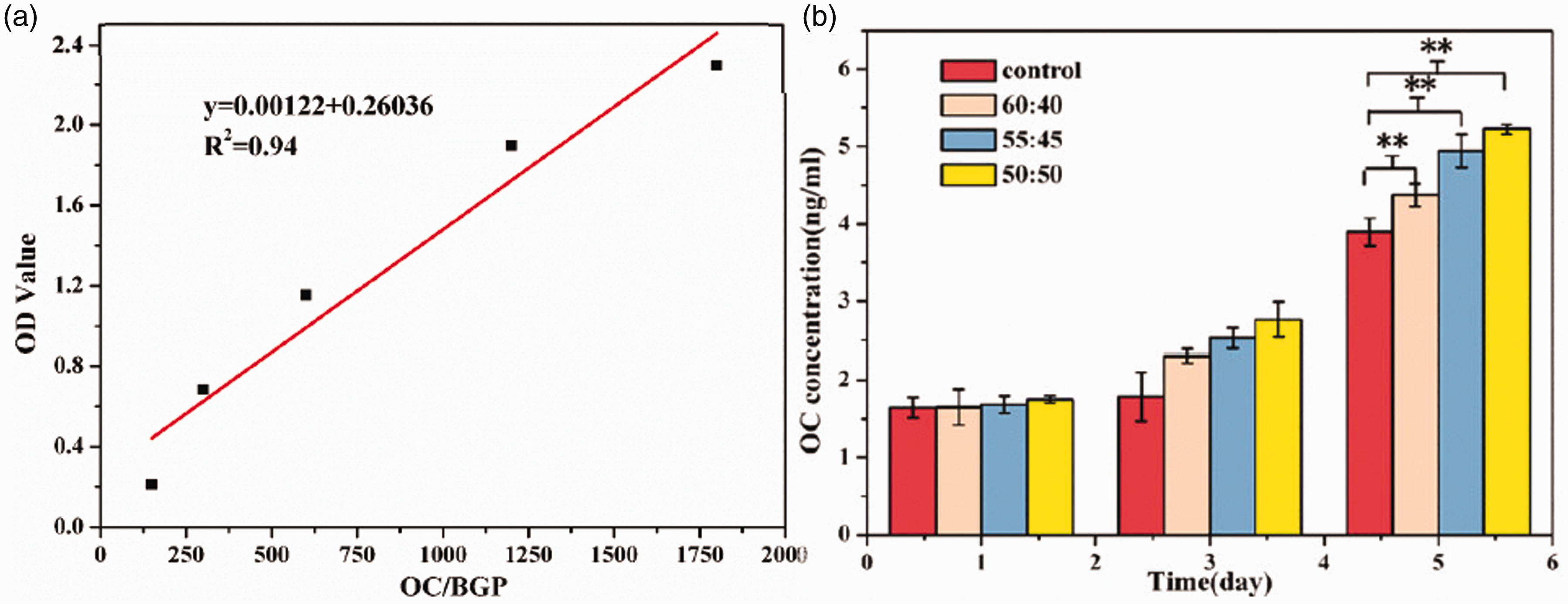
MC3T3-E Cells secretion of osteocalcin (OC): (a) Standard curve of OC standards; (b) OC content in control and experimental groups.
Effects of the scaffolds on collagen I secretion from MC3T3-E1 cells
Col-I is the main component of the bone matrix, and its content expresses the functional status of osteoblasts. 45 Figure 10(a) shows the good linearity of the standard curve that was constructed with the Col-I standard provided in the assay kit. The concentrations of Col-I in the experimental groups and the blank and control groups were calculated using this standard curve. As shown by the histogram (Figure 10(b)), the amounts of Col-I secreted by the cells increased gradually with the increase in culture time, where the amounts secreted by the 55:45 and 50:50 groups were significantly higher than the amount secreted by the control group on the fifth day. These results indicated that the PCL–β-TCP composite scaffold could promote the maturation and differentiation as well as increase the activity of osteoblasts. The amount of Col-I secreted by the cells of the 50:50 group on the fifth day of cell culture was 2.96 ± 0.13 ng/mL. The experimental results are also consistent with β-TCP's osteogenesis-promoting properties. 46
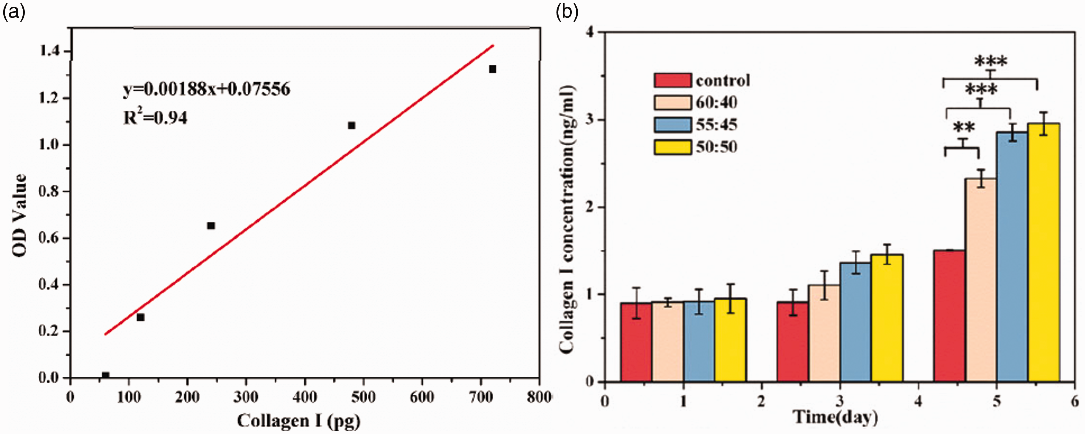
MC3T3-E cells secretion Collagen I: (a) Col-I standard curve; (b) Col-I content in control and experimental groups.
Morphology of MC3T3-E1 cells on the PCL–β-TCP composite scaffolds
The chemical composition of β-TCP is similar to that of bone, which is conducive to the increase and adhesion of osteoblasts and promotion of the osteogenic process. 47 To examine the morphology of the MC3T3-E1 cells on the PCL–β-TCP composite scaffolds, 280,000 cells/mL were seeded onto each group of scaffolds and cultured for 4 h. After staining with AO or DAPI, the distribution of the cells on the scaffolds was observed under a fluorescence microscope at 100× magnification. As shown in Figure 11, the cells on the β-TCP-containing scaffold were fusiform or triangular in shape and more spread on the scaffold than the cells on the PCL scaffold. The statistical results from the analysis of the Image J data are shown in Figure 12. Compared with the number of cells on the PCL scaffold (243.12 ± 31.44 cells/mm2), the numbers of cells adhered to the mixed PCL–β-TCP scaffolds were significantly higher; that is, 298.33 ± 21.81 cells/mm2 on the 60:40 scaffold (P = 0.047), 348.80 ± 21.79 cells/mm2 on the 55:45 scaffold (P = 0.002), and 368.02 ± 27.44 cells/mm2 on the 50:50 scaffold (P = 0.001). These results showed that mixed-material scaffolds can effectively promote cell adhesion and spreading. On the one hand, β-TCP can promote cell adhesion by activating Ras/MAPK signaling pathway. 45 On the other hand, when β-TCP is incorporated into PCL, the surface of the sample is rougher. This rough surface can also promote cell adhesion. 48
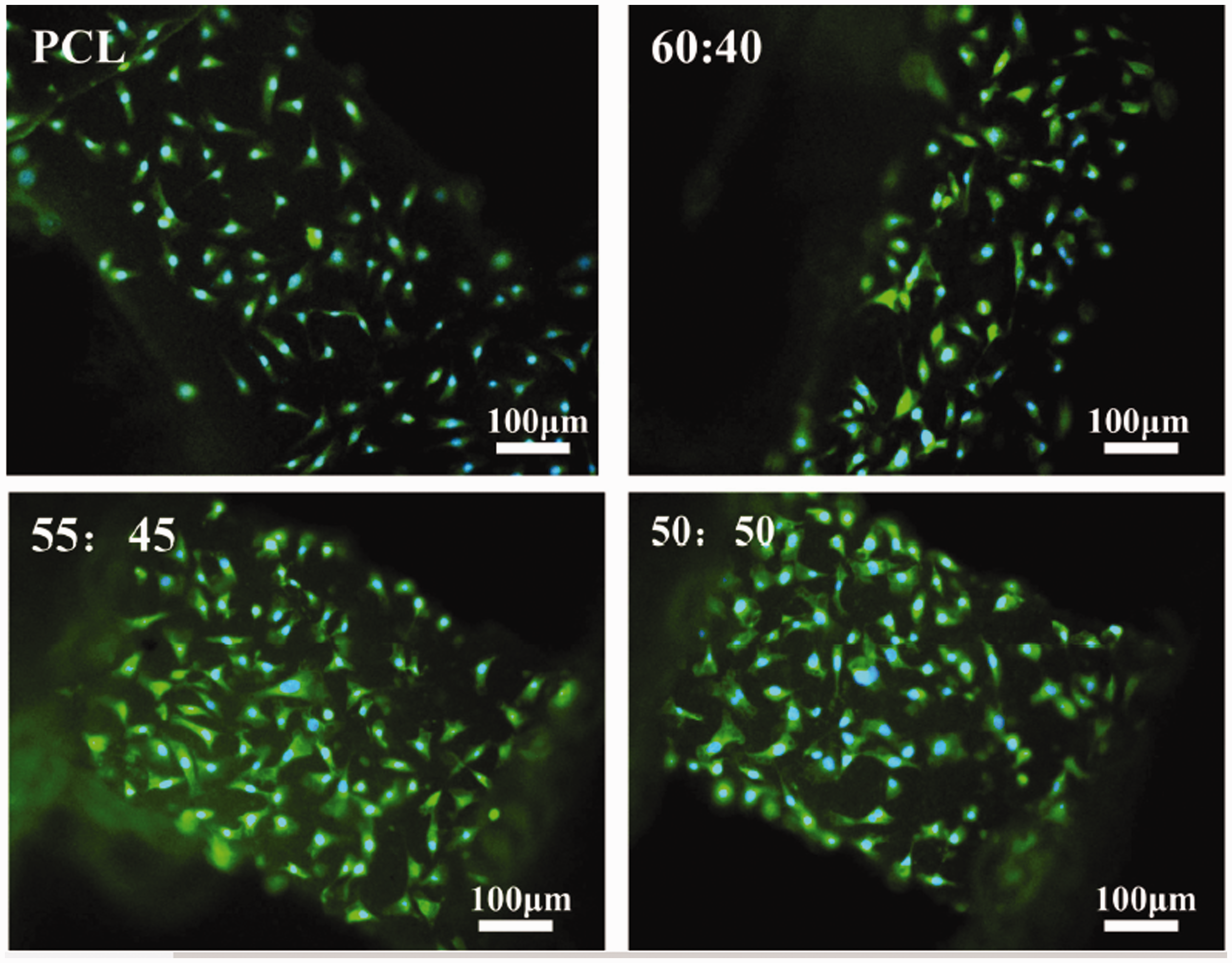
Adhesion of MC3T3-E1 cells 4 h after seeding on scaffolds(AO, green; DAPI,
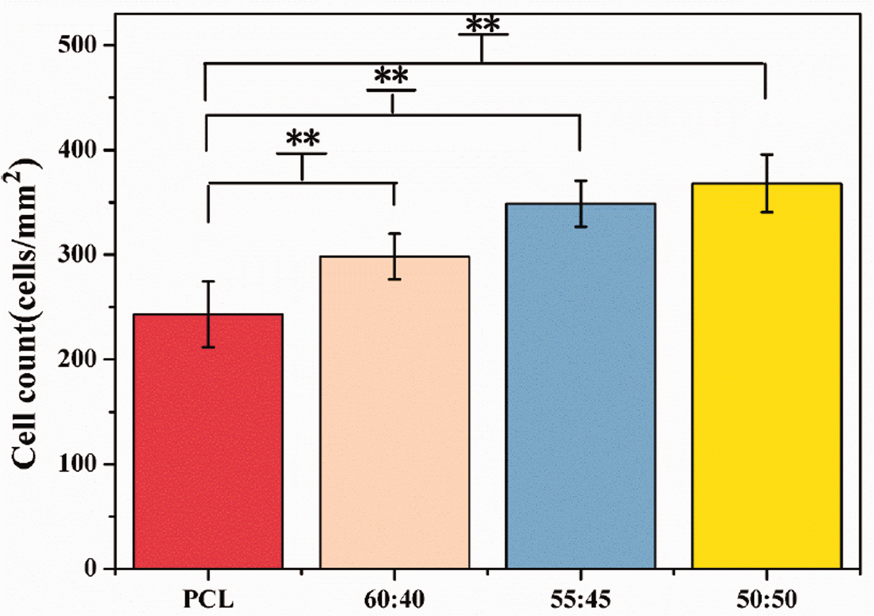
Adhesion density of MC3T3-E1 cells after 4 h of seeding on scaffolds.
Summary
This study solved the problem of how to form filaments from brittle materials in 3 D printing, effectively allowing increases in the content of β-TCP in PCL–β-TCP scaffolds. Compared with the method of transformation in a powder material bin, our rod formation method is more conducive to the extrusion and moulding of materials and provides a new solution for 3 D printing strategies. Our outer frame and three-beam structural designs are expected to further strengthen the stability of the mechanical properties of the fusion cages during degradation. In the future, we will be designing animal and long-term degradation experiments for the further verification of our degradable spinal cage fabrication design.
Footnotes
Declaration of conflicting interests
The author(s) declared no potential conflicts of interest with respect to the research, authorship, and/or publication of this article.
Funding
The author(s) disclosed receipt of the following financial support for the research, authorship, and/or publication of this article: This study was supported by the National Natural Science Foundation of China (81771988), and the Science and technology program of Sichuan Province (20GJHZ0268).