






Abstract
Chemotherapeutic agents and photosensitizers often suffer from poor tumor selectivity, high side toxicity, or low water solubility. To address these problems, various drug delivery systems (DDS) have been explored but most of them are toxic, difficult to synthesize, or of single function. In order to design a highly biocompatible, conveniently prepared, multi-functional drug delivery system, herein, an aptamer of vascular endothelial growth factor (VEGF) and a cytosine (C)-DNA fragment were grafted on the surface of superparamagnetic iron oxide nanoparticles (SPION), and then a chemotherapeutic agent daunomycin (DNM) and a photosensitizer 5, 10, 15, 20-tetra (phenyl-4-N-methyl-4-pyridyl) porphyrin (TMPyP) were self-assembled with the hybridized VEGF-based DNA structure. By loading DNM and TMPyP, the DDS displayed strong chemotherapeutic/phototherapeutic capability against cancer cells via mechanisms such as mitochondrial dysfunction and ROS elevation, which triggered the apoptosis of the tumor cells. The dual delivery of chemotherapeutical agents and photosensitizers with aptamer/C-rich DNA successfully integrated the functions of pH stimuli-responsive drug release and chemotherapeutic/phototherapeutic modalities into one single system and thus could be considered as an ideal drug delivery vehicle with great potential in clinic.
Keywords
Introduction
The poor tumor selectivity, low water solubility, and high side toxicity have limited the clinic application of many chemotherapeutic agents and photosensitizers. Drug delivery systems (DDS) are proved effective in dealing with the above problems and thus have aroused the attentions of many researchers. Among the various DDS reported, nucleic acids-based, artificial structures catch researchers’ eyes since they are highly biocompatible, conveniently prepared, multi-functional, and thus exhibit great potential in DDS.1,2 Especially, aptamers are essentially short RNA or single-stranded DNA oligonucleotides (usually 20–80 nucleotides with 6–30 kDa molecular weights) that can fold into unique three-dimensional conformations. In recent decades, aptamers have played critical roles in DDS because of their unique characteristics such as programmability, flexibility, and low toxicity. 3 For example, AS1411, a well-studied aptamer, can form a dimeric G-quadruplex structure to target high nucleolin-expressing cancer cells and thus has been frequently used as a tether for the drug nanocarrier.4,5 Recently, AS1411 has aroused particular interests since it can effortlessly capture the G4-ligand, such as 5,10,15,20-tetrakis(4-N-methylpyridiniumyl)porphyrin (TMPyP) which has been proved by our group.6,7 TMPyP is a broadly used photodynamic therapy (PDT) reagent which has been identified as an effective G-quadruplex DNA-binding ligand. TMPyP has poor selectivity and is toxic to some normal cell lines, especially normal fibroblasts and epithelium, when exposed to light. Therefore, to increase the accumulation at the target site and to prevent the toxicity to the neighboring tissues, TMPyP has been frequently targeted delivered with drug delivery system (DDS).8-10
However, the landscape of TMPyP delivery has been dominated by AS1411; currently, there are few lights which were shed on other G-rich aptamers, such as vascular endothelial growth factor (VEGF), named V7t1. VEGF is a protein that plays a key role in promoting the vascular permeability and vascularization of tumor cells. V7t1 is a VEGF aptamer with KD values in a low nanomolar range for the most abundant VEGF isoforms VEGF165 and VEGF121.
11
Since V7t1 comprises several G-rich tracts (Figure 1(a)), it has been predicted to fold into a G-quadruplex structure as AS1411 aptamer.12,13 However, currently, V7t1 has not been employed as nanocarrier for its special G-quadruplex structure. (a) The DNA diagram and conformational polymorphism of V7t1-gc34; (b) Schematic illustration of the synthesis and the anti-cancer working principle of the TMPyP&DNM@V7t1-gc34@SPION.
On the other hand, the conformational polymorphism of nucleic acid is also exhibited in the flexible conformational switch of cytosine (C)-quadruplex (known as i-motif) structure upon pH change. 14 It is well known that unlike healthy tissues which usually have a normal physiological pH of ∼7.4, disease regions such as tumors and intracellular endosomal/lysosomal compartments often exhibit abnormally high local acidities. Thus, pH is an attractive environmental stimulus for cancer cells. The pH-controlled conformational switch of C-rich structure makes it well-suited for anticancer drug targeting release via the acidic local environment. Researchers have sought to take advantage of this unique feature to construct a DNA-based carrier for drugs in pH-controlled reversible binding and releasing manner using a C-rich hairpin DNA. 15
Despite enormous research progress in enhancing the anticancer efficiencies of different chemotherapeutic agents, the chemotherapy still has presented disadvantages in terms of irreversibility, high side effects, multidrug-resistance, etc. Combinational therapy of chemotherapy with other modalities is still a burgeoning research area. The conformational polymorphism of nucleic acid gives us the inspiration of co-delivering the agents of different modalities to achieve multi-therapies. As references reported, anthraquinone anticancer family, such as doxorubicin (DOX) and daunomycin (DNM), can intercalate into a DNA strand (preferentially bind to double-stranded 5′-GC-3′ or 5′-CG-3′ sequences) due to its flat aromatic rings and positive charge. 16 Meanwhile, many researchers including our group have previously evidenced that TMPyP, an effective photosensitizer, was conformationally suitable to be loaded by quadruplex DNA structures.17,18 These work evidenced the feasibility of co-delivery for the chemotherapeutic and photodynamic agents by nucleic acid.
Herein, we hybridized G-rich aptamer V7t1 with a C-rich 34-mer DNA fragment (gc34) and then grafted the hybridized DNA structure with polyetherimide-superparamagnetic iron oxide nanoparticles (PEI-SPION) through simple electrostatic strength. Chemotherapeutic drug, DNM and photosensitizer TMPyP were delivered by the gc34 and V7t1 structure, respectively. The novelties of this work are as following: 1) The system was built mainly through the electrostatic strength and self-assembly, avoiding the use of chemical conjugation or encapsulation steps and improving bioavailability and efficacy. 2) VEGF aptamer was focused as a G-quadruplex drug loading structure for the first time and can even keep this conformation after grafting on the SPION. 3) The designed DDS successfully integrated the functions of proton-driven drug release and chemotherapeutic/phototherapeutic dual-therapies in one single system and thus could be considered as an ideal drug delivery vehicle with great potential in clinic.
Experimental
Materials
TMPyP was prepared by the method we previously reported. 19 DNM was purchased from Sigma-Aldrich. Nucleic acid sequence of single-stranded (ss) V7t1-gc34 (5′-CGC CCC TAA CCC TAA CCC TAA CCC TGC GTT TTT TTG TGG GGG TGG ACG GGC CGG GTA GA-3′) was purchased from Sangon Biotech Co. Ltd (Shanghai, China). PEI-SPION was purchased from Nanoeast Biotechnology Co., Ltd (Nanjing, China). 3-(4,5-Dimethylthiazol-2-yl)-2,5-diphenyltetrazolium bromide (MTT), Dulbecco’s modified eagle medium (DMEM), fetal bovine serum (FBS), trypsin-EDTA solution, and penicillinstreptomycin solution were purchased from Gibco Life Technologies. 4′-6-diamidino-2-phenylindole (DAPI), EL-ABTS kit, and BCA kit were obtained from Beyotime Biotech (Shanghai, China). All the other chemicals were of analytical grade. Millipore water (18.2 MΩ) and nuclease-free water were used throughout the experiments.
Instruments
The transmission electron microscope (TEM) analysis was performed with a JEM-2010HR transmission electron microscope (JEOL, Japan). Fluorescence spectroscopy was recorded on a Perkin-Elmer L55 spectrofluorophotometer (PE, USA). MTT assay was performed with a microplate reader (BioTek, USA). Fluorescence microscopy of apoptosis assays was performed with an IX 70 fluorescence microscope (Olympus, Japan). Gel electrophoresis experiment was performed with electrophoresis apparatus trophoresis and gel imaging system (Tanon, China). Flow cytometry analysis was performed by flow cytometer (BD Biosciences, US). Statistical analysis was performed using origin 8.0.
Methods
Annealing of the DNA oligonucleotide
The V7t1-gc34 ssDNA oligonucleotide was dispersed in annealing buffer (200 mM KCl, 4 mM MgCl2, and 28 mM Tris-HCl), annealed at 95 oC for 5 min, and then slowly cooled to room temperature to form the G-quadruplex structure of the V7t1 aptamer as well as the double-stranded gc34 hairpin. The annealed V7t1-gc34 nucleic acid was store at 4°C until needed.
Synthesis of DNA-grafted SPION nanospheres
PEI-SPION was mixed with the annealed V7t1-gc34 in nuclease free water at different weight ratios of 1:15, 1:20, 1:30, and 1:45, respectively. Then, the mixtures were gently shaken for 4 h at room temperature. Unreacted DNA was removed by centrifugal filtration (four times, 10,000 r/min for 10 min). The mixture was run on a 1% agarose gel and visualized with a gel imager, and the mixture with the best DNA grafting efficiency was obtained, namely, V7t1-gc34@SPION.
Stability analysis
V7t1-gc34@SPION (2 μg/mL) was mixed with 10% fetal bovine serum (FBS) and incubated at 37°C. After incubation for 0, 6, 12, 24, 48, and 72 h, the mixture was run on a 1% agarose gel and visualized with a gel imager.
EL-ABTS detection
Low concentration of annealed V7t1-gc34 and V7t1-gc34@SPION were mixed with 2 μM hemin (in 10 μL water) and reacted in the dark for 120 h. After that, 100 μL EL-ABTS (color-substrate solution) was added to the mixtures. After the mixtures reacted at 37°C for 30 min, 50 μL of stop solution from the EL-ABTS kit was added. The absorbance of these solutions was read at 405 nm on a microplate reader.
Loading of drugs with nucleic acid structure
DNM and TMPyP dispersed in millipore water were loaded with V7t1-gc34@SPION under ice bath and room temperature, respectively. Generally, V7t1-gc34@SPION (10 μg/mL, 5 mL) was added to DNM (50 μM, 10 mL). After gently shaken for 10 min in ice bath, the remaining DNM was removed by centrifugal filtration (four times, 10,000 r/min for 10 min) and the DNM@V7t1-gc34@SPION was thus fabricated. Similarly, DNM@V7t1-gc34@SPION (10 μg/mL, 5 mL) was added to TMPyP (50 μM, 10 mL) under room temperature, and then the mixture was shaken for another 10 min. After TMPyP was removed by centrifugal filtration, the TMPyP&DNM@V7t1-gc34@SPION was thus constructed. The prepared drug delivery system was stored at 4°C until needed.
The loading quantity of DNM (Q
DL
) or TMPyP (Q
TL
) was determined by the UV spectrophotometer at 480 nm and 420 nm, respectively. The Q
DL
and Q
TL
were calculated by using the following formulas
pH-controlled drug release
DNM@gc34 system was dissolved in distilled water to get a final concentration of 5 μM for DNM. The drug release was monitored by fluorescence spectroscopy at pH 7.4 and 5.6 at 4°C. The fluorescence emission intensity of DNM@gc34 system at 595 nm on excitation at 480 nm was detected.
For all the cellular experiments, MCF-7 cells (1 × 10⁵ cells/dish) were maintained and cultured in DMEM supplemented with 100 units/mL penicillin-streptomycin and 10% fetal bovine serum.
Biocompatibility research
The biocompatibility of the drug-free V7t1-gc34@SPION against MCF-7 cell lines under dark/light was determined using an MTT assay. For both groups, the cell culture medium was replaced with 100 mL medium containing different concentrations of V7t1-gc34@SPION ranging from 12.5 to 200 μg/mL. For the dark groups, after incubating with the V7t1-gc34@SPION in the dark for 4 h, the viability of cells was determined using the MTT assay at 490 nm. As to the light group, after the adding of V7t1-gc34@SPION, the cells were firstly illuminated with 450 nm light for 15 min and then were incubated in the incubator for another 4 h. The viability was then determined by a microplate reader.
Cell uptake analysis
TMPyP&DNM@V7t1-gc34@SPION mixed with the cell culture medium were prepared at a final concentration of 5 μg/mL and incubated with the MCF-7 cells for 4 h. 300 μL of DPI dye solution were added to each dish at 37°C for 20 min. After the stain was aspirated, the cells were washed three times with PBS. Cell imaging was scanned through the fluorescence of DNM (λEx = 495 nm) by the laser scanning confocal microscope.
Cell apoptosis analysis
Cells without any treatments were used as negative controls. Cells were divided into dark and light groups, both of which were incubated with the TMPyP&DNM@V7t1-gc34@SPION (3 μg/mL) for 6 h. Then, the light group was illuminated by 450 nm light for 15 min, and both the two groups incubated with the drug for another 24 h. After that, the cells were treated with trypsin-EDTA for 2 min and then re-suspended in PBS. Probe loading was performed according to the protocol of the apoptosis kit and tested on a flow cytometer. Data was evaluated using FlowJo 7.6.1 software.
Evaluation of mitochondrial membrane potential
MCF-7 cells (1 × 10⁵ cells/well) were treated with TMPyP&DNM@V7t1-gc34@SPION for 24 h in the incubator. 100 μL JC-1 working solution was then added and incubated for another 20 min at 37°C in dark. After washed twice with PBS, the cells were filtered through a strainer and detected with fluorescent enzyme labeling instrument by a flow cytometer (λex = 488 nm; λem = 530 nm for JC-1 monomers or 590 nm for JC-1 Monomers.
ROS detection
ROS productions of TMPyP&DNM@V7t1-gc34@SPION in dark/light groups were comparatively detected. For the dark group, MCF-7 cells were firstly treated with 5 μg/mL of TMPyP&DNM@V7t1-gc34@SPION for 2 h. For the light group, cells were then irradiated with 450 nm light for 15 min, and then both the two groups incubated with the TMPyP&DNM@V7t1-gc34@SPION for another 24 h. The cells were then washed with PBS 3 times, and probe loading was performed according to the ROS kit. Finally, the cells were observed by fluorescence microscopy.
Statistical analysis
All of the experiments were done in triplicate. The results were expressed as mean ± standard deviation (SD) of the mean value. The statistical significance of the observed differences was analyzed by t-tests. Statistical significance was set at p< 0.05 (*p < 0.05, **p < 0.01).
Results and discussion
The conformational polymorphism of nucleic acid has been fully harnessed in the design of DNA structure. Herein, V7t1, a VEGF aptamer, was hybridized with a C-rich gc34 DNA fragment, and then the DNA structure was grafted on the surface of SPION nanospheres. The DNA diagram and conformational polymorphism of V7t1-gc34 and the synthetic path of TMPyP&DNM@V7t1-gc34@SPION are given in Figure 1. Since SPION has positive charges while the DNA structure is negative, DNA grafted on the surface of SPION through electrostatic strength, avoiding the use of chemical conjugation or encapsulation steps which may affect the bioavailability and efficacy of the system. The V7t1 aptamer domain, which folds into a G-quadruplex structure after annealing, is a good candidate to load the TMPyP with large planar core.6,7 Meanwhile, under neutral or slightly alkaline conditions, DNM can preferentially intercalate into the duplex stem part of the gc34 and will be released under acidic conditions due to the breaking of the hydrogen bonds between the partial GCG-CGC duplex stem. 17
The preparation of the nanospheres
Oligonucleotide grafting on SPION
V7t1-gc34 with negative charges was loaded in the positive PEI-SPION via electrostatic interaction. The combination between V7t1-gc34 and PEI-SPION was detected with different nanoparticle/nucleic acid ratios (N/P ratios by weight) of 1:15, 1:20, 1:30, and 1:45. From the agarose gel electrophoresis assay (Figure 2(a)), there remained a considerable amount of unbounded DNA for N/P ratio of 1:20. In contrast, the unbounded DNA band was almost indiscernible for N/P ratios at 1:30 or 1:45. Thus, 1:30 was selected as the optimum N/P ratio in the preparation work. (a) The gel images of free V7t1-gc34 and the V7t1-gc34 grafted on SPION Nps at different ratios. Lanes 1, 3, 5, 7 for V7t1-gc34, and lanes 2, 4, 6, 8 for V7t1-gc34@SPION. (b) Resistance of the V7t1-gc34@SPION Nps after incubation with 10% FBS for 0 h, 6 h, 12 h, 24 h, 48 h and 72 h. (c) Zeta potentials of the studied Nps in PBS (pH 7.0) at room temperature (25 °C). (d) The TEM of TMPyP&DNM@V7t1-gc34@SPION Nps; (e) The magnetic hysteresis loops of SPION (A), V7t1-gc34@SPION (B) and TMPyP&DNM@V7t1-gc34@SPION (C) Nps; (f) The solid electronic absorption spectra of V7t1-gc34@SPION, TMPyP, DNM and TMPyP&DNM@V7t1-gc34@SPION. (g) The solid electronic absorption spectra of DNM (A), TMPyP (B), V7t1-gc34@SPION (C), and TMPyP&DNM@V7t1-gc34@SPION (D) Nps. (h) Absorptions at 405 nm for hemin, V7t1-gc34 and V7t1-gc34@SPION mixed with hemin analyzed by ABTS assay. (i) The fluorescence of unloaded DNM control, DNM released from DNM@V7t1-gc34 system at pH 7.4 and pH 5.6.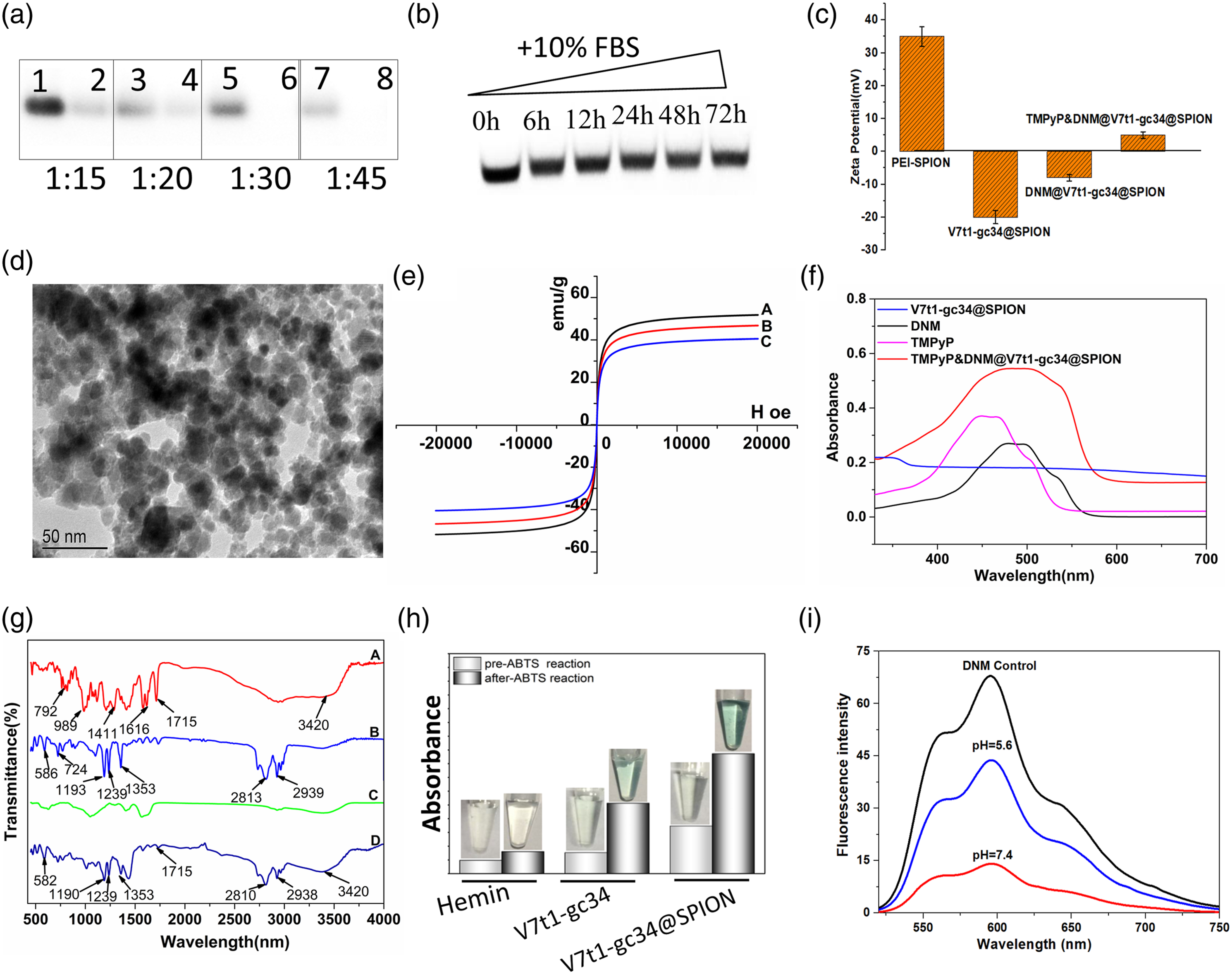
Serum resistance analysis
The studied assembly was incubated with FBS since the serum resistance for medicines is critical when used in living cells and blood circulation. The serum resistance for V7t1-gc34@SPION was analyzed by agarose gel electrophoresis after incubating the assembly with 10% FBS at 37°C for 6 h, 12 h, 24 h, 48 h, and 72 h (Figure 2(b)). The results were given in. Clear and no obvious changed DNA bands were observed in the studied period, indicating that the designed V7t1-gc34@SPION system was not degraded by FBS after incubating for up to 72 h. Thus, we supposed that the designed drug-DNA assembly is relatively safe for the circulation in blood.
Characterization and drug loading
The successful grafting of the oligonucleotide to the PEI-SPION was proved by the zeta potential change (Figure 2(c) and Figure S1). PEI-SPION has a much higher positive ζ potential than V7t1-gc34@SPION (33.9 ± 1.2 mV vs −20.6 ± 1.0 mV), which results from the electrostatic sorption of negative nucleic acid on the cationic surface of the SPION. This negative potential of V7t1-gc34@SPION does a great favor for the loading of DNM and TMPyP, since both them are cationic drugs in the aqueous solution.6,7,16 As expected, after the incubation with DNM and TMPyP, the potential of the system increased to −8.21 ± 1.1 mV and 4.98 ± 0.4 mV, respectively, suggesting the successful loading of the drugs. The loading efficiencies of DNM and TMPyP to V7t1-gc34@SPION had been calculated 73.4% and 42.9%, respectively. The typically high loading efficiency of drug molecules can be attributed to the following two factors: (1) the electrostatic strength between the negative V7t1-gc34@SPION and the cationic drugs, which we can easily find from the above zeta potential changes. (2) The intercalaction between DNM and gc34, and the outside stacking between TMPyP and V7t1, have also done a great favor for the drug loading processes.12,13 These results indicated the high potential of the V7t1-gc34@SPION system as a nanocarrier.
Figure 2(d) gives the TEM image of V7t1-gc34@PEI-SPION, which shows well-dispersed uniform spherical shape, with an average diameter of approx. 50–60 nm. Meanwhile, magnetic responsibility is the key factor for the targeting of the drug delivery nanospheres. The magnetic responsiveness of free cargo PEI-SPION and the synthesized TMPyP&DNM@V7t1-gc34@SPION were detected by the electronic absorption method. Both the free cargo and the drug-loaded system could be well dispersed in the water and rapidly attracted by an external magnet placed close to the vessel (Figure S2), demonstrating that the loading of drugs has negligible effects on the high magnetic responsiveness of the PEI-SPION nanospheres. Moreover, Figure 2(e) shows the magnetic hysteresis loops of SPION, V7t1-gc34@SPION and TMPyP&DNM@V7t1-gc34@SPION Nps, and the saturation magnetizations are 51.6 emu/g, 46.2 emu/g, and 40.7 emu/g, respectively. TMPyP&DNM@V7t1-gc34@SPION has lower saturation magnetization than SPION or V7t1-gc34@SPION, which could be explained by the DNM and TMPyP coating on the nanospheres.
The presence of the two drugs on the nanosphere support was further evidenced by the solid UV-Vis and FTIR spectra shown in Figure 2(f) and (g), respectively. From Figure 2(f), DNM and TMPyP exhibit characteristic absorbance peaks at 490 nm and 450 nm, respectively. Negligible absorption was observed for V7t1-gc34@SPION in this area. In contrast, TMPyP&DNM@V7t1-gc34@SPION gives a significant absorption ranging from 450 nm to 500 nm, indicating the successful loading of the drugs. Meanwhile, in the IR spectrum of free DNM (Figure 2(g)), a pair of bands at 1616 cm−1 and the band at 1725 cm−1 can be attributed to the characteristic C–N and N–H stretching, respectively. The band at 3420 cm−1 can be ascribed to the stretching vibration of -OH group. 20 For the IR spectrum of free TMPyP, the bands near 2939 cm−1 and 2813 cm−1 can be attributed to the stretching vibration of the four -CH3 groups, while the three peaks at 1353, 1239, and 1193 cm−1 are the characteristic vibration of C=N in the pyrrole of porphyrin, and the bands ranging from 700 cm−1 to 1100 cm−1 can be ascribed to the pyrrole ring’s vibration.19,21 Comparing with the IR spectrum of V7t1-gc34@SPION which has few obvious bands, the IR results of TMPyP&DNM@V7t1-gc34@SPION exhibit the characteristic bands of both DNM (bands around 1725 cm−1 and 3420 cm−1) and TMPyP (bands near 2939 cm−1 and 2813 cm−1, 1353 cm−1, 1239 cm−1and 1190 cm−1). The drugs were further convinced to be successfully loaded on the solid support by these spectral results.
The G-quadruplex structure of the aptamer is essential for the assembly of TMPyP with V7t1. In order to investigate the final structure of V7t1 in the long DNA chain grafted on the surface of SPION, we carried out ABTS experiment, which has been widely accepted as an efficient method to detect the G-quadruplex structure of DNA. The complex of hemin/G-quadruplex can catalyze the H2O2− mediated oxidation of ABTS2− to the colored product ABTS−. The chromogenic substrate of peroxidase, which exhibits a color change in the presence of hydrogen peroxide, forms a green soluble product with a strong absorption at 405 nm. From Figure 2(h), it was demonstrated that hemin exhibited negligible color when incubated with ABTS. In contrast, after annealing, the absorptions of both V7t1-gc34 and V7t1-gc34@SPION at 405 nm were significantly increased and green color were obviously observed, confirming the formation of G-quadruplex structure for V7t1 on the surface of SPION. 12
Sensitive pH-dependent DNM release
The pH-dependent releasing of DNM from DNM@gc34 under acid/neutral conditions was monitored in Tris buffer at pH 7.4 (mimicking the pH of blood plasma) and 5.6 (mimicking the pH of cancer cells) by fluorescence spectroscopy (Figure 2(i)). The release at pH 7.4 is relatively slow and sustained, only about 9.9% of original DNM releases after incubation for 5 min. In contrast, the DNM releasing burst at pH 5.6, with the releasing ratio reached 59.6%. The observed pH sensitivity is easy to understand by the fact that under acidic conditions, the C-rich segments of the gc34 part forms an i-motif quadruplex, which breaks down the hydrogen bonds between the partial GCG-CGC duplex stem where DNM intercalated (see Figure 1(a)). This result endows the designed system biosafety in the normal cells while controlled release in the cancer cells.
Anticancer assessment
Assessment of in vitro cytotoxicity and photocytotoxicity
The in vitro cytotoxicity of drug-free V7t1-gc34 cargo, free TMPyP and DNM, TMPyP&DNM@V7t1-gc34 against MCF-7 cells under dark and light (450 nm, 20 mW/cm2) were determined by the MTT assay (Figure 3). No obvious cellular damage was observed for the cells treated with V7t1-gc34 under both dark and light at a concentration at 200 μg/mL, convincing the high biocompatibility of the designed cargo (Figure 3(a)). TMPyP displays no considerable cytotoxicity in the dark but remarkable phototoxicity under illumination with light to MCF-7 cell lines (Figure 3(b)). TMPyP&DNM@V7t1-gc34@SPION and DNM exhibit dose-dependent cell cytotoxicity in the dark. We understand this by the fact that the cytotoxicity of the co-delivery system without light exposure mainly results from the effect of DNM. Under the illumination, TMPyP&DNM@V7t1-gc34@SPION exhibits higher cytotoxicity against the MCF-7 cell lines than the individual DNM and TMPyP (Figure 3(b)), indicating that the system has a synergistic enhancement in the cellular cytotoxicity through the combination of chemotherapeutic/photodynamic therapies.21,22 (a) Cell viability of various concentrations of V7t1-gc34@SPION Nps on MCF-7 cells under dark and 450 nm illumination. (b) Cell viability of various concentrations of TMPyP&DNM@V7t1-gc34@SPION Nps on MCF-7 cells under dark and 450 nm illumination. (c) Cell uptake and subcellular localization of the TMPyP&DNM@V7t1-gc34@SPION Nps after 4 h incubation with MCF-7 cells by confocal laser scanning microscopy. Scale bar: 20 μm.
Cellular internalization
The cellular uptake can affect the biological activity of a drug.23,24 To investigate the mechanism of the therapeutic synergism of the drugs against MCF-7 cells, we monitored the intracellular internalization of TMPyP&DNM@V7t1-gc34@SPION using the signal of DNM after incubating with the cancer cells (Figure 3(c)). DAPI can easily pass through the cell membrane and was employed to stain the MCF-7 cells. In the control group, the cells exhibit bright blue fluorescence of DAPI. In the presence of the TMPyP&DNM@V7t1-gc34@SPION, red fluorescence of DNM was detected around the cellar nucleus, indicating the designed nanospheres can effectively enter the cancer cells and DNM can efficiently accumulate in the nucleus. The cell uptake of the nanospheres could be associated with the cytotoxicity and various cellular and apoptotic pathways involving various mechanisms of action. A detailed apoptotic investigation of TMPyP&DNM@V7t1-gc34@SPION against MCF-7 cells was conducted to explore the possible mechanism of action of this system.
Induction of apoptosis
Apoptosis is a conserved process that regulates the growth of a multicellular organism by eliminating unwanted and abnormal cells through a controlled sequence of molecular events.25,26 In cancer cells, the process of apoptosis is dysregulated so that it could be exploited as an anticancer target. 27 The anticancer ability against MCF-7 cells of TMPyP&DNM@V7t1-gc34@SPION was verified by employing flow cytometry with propidium iodide (PI) and Annexin V-fluorescein isothiocyanate (Annexin-V-FITC) fluorescence staining. Cells with drugs were divided into light/dark groups, and the cells without any treatment were used as a negative control.
As shown in Figure 4(a) and (b), the flow cytometry results showed that the ratio of the survival ratio decreased from 97.8% of the controlled cells to 87.3% for cells treated with TMPyP&DNM@V7t1-gc34@SPION in the dark, which could be mainly contributed to the anticancer capability of DNM. Moreover, as shown in Figure 4(c), under the illumination of 450 nm, the ratio of live cells was further decreased to 69.7%. The apoptotic ratios (including early and late apoptosis) were significantly enhanced, with increasing from 2.89% to 16.2% for early apoptosis and from 9.81% to 14.0% for late apoptosis. The cellular apoptosis induced by bare nanocarrier or free chemotherapeutic agents are given in the supplemental materials as Figure S3. Figure 4(d) summarizes the apoptosis of the bare carrier, free drug and drug-loaded DDS. It is found that, the drug free nanocarrier can negligibly induce the cell apoptosis. Moreover, TMPyP&DNM@V7t1-gc34@SPION can induce the cell apoptosis more significantly than the individual drugs, indicating the chemotherapeutic/phototherapeutic synergistic effects of the designed DDS. Detection of apoptosis in MCF-7 cells stained with annexin V and PI after treatment with 300 μg/mL of TMPyP&DNM@V7t1-gc34@SPION Nps for 24 h by Flow cytometric. Annexin V (λex = 488 nm, λem = 510 ± 20 nm), PI (λex = 488 nm, λem = 610 ± 20 nm). (a) Control, (b) dark, (c) 450 nm illumination, (d) histogram.
Evaluation of changes in mitochondrial membrane potential
Mitochondria play a major role in the induction of apoptosis and are regarded as a new target for anticancer drugs.28,29 The membrane potential of mitochondria is tightly controlled and well-regulated in normal cells. Any damage to the membrane potential can activate apoptotic pathways that ultimately lead to apoptotic death by the release of pro-apoptotic factors. We have further studied the effects of TMPyP&DNM@V7t1-gc34@SPION on the change of mitochondrial membrane potential (MMP) for MCF-7 cells. Flow cytometry was used to detect the intensity of red/green fluorescence after JC-1 staining (Figure 5(a)–(c)), and then the relative ratio of red/green fluorescence was calculated to measure the change of MMP (Figure 5(d)). R1 represents of the red fluorescence region, and R2 represents of the green fluorescence region. In the control group, the ratio of R1/R2 was 5.67, while this value was significantly decreased in the presence of TMPyP&DNM@V7t1-gc34@SPION under both dark (5.57 vs 1.30) and light (5.67 vs 0.57), indicating that the MMP of cancer cells decreased and the drug delivery system had both chemotherapeutic and photodynamic anticancer activity. The results demonstrated that apoptosis induced by TMPyP&DNM@V7t1-gc34@SPION was through a mitochondrial-mediated pathway, although other pathways may also be involved. Effects of TMPyP&DNM@V7t1-gc34@SPION on MMP analyzed by JC-1 staining and flow cytometry. MCF-7 cells were treated with 5 μg/mL of TMPyP&DNM@V7t1-gc34@SPION for 12 h at 37 °C for 24 h. (a) Control, (b) dark, (c) 450 nm illumination, (d) R1/R2 ratios. JC-1 monomers (Green): λex = 488 nm, λem = 510 ± 20; JC-1 aggregates (Red): λex = 488 nm, λem = 580 ± 20 nm.
Measurement of reactive oxygen species generation
The mitochondrion is the primary source of production of reactive oxygen species (ROS), and the unnecessary production of ROS is considered a warning feature leading to mitochondrial damage, genetic instability, and ultimately apoptotic cell death.
30
Reactive oxygen species cause oxidative damage to the cell and play a crucial role in cellular biological function and signaling.
31
Therefore, ROS targeting is accepted as a viable strategy for the treatment of cancer. It is widely accepted that the photosensitizer induce apoptosis through ROS generation.
32
Intracellular ROS levels of TMPyP&DNM@V7t1-gc34@SPION under both bright and dark groups were investigated with a fluorescence microscope, with H2DCFDA using as a probe in microplate analyzer. As shown in Figure 6, the light group has much stronger green fluorescent than that of the dark group, indicating that the light illumination to TMPyP&DNM@V7t1-gc34@SPION significantly elevated the ROS level while the ROS production without light is insignificant. Therefore, it is suggested that ROS-induced apoptosis was activated by the exposure of the designed TMPyP&DNM@V7t1-gc34@SPION under the light and thereby damaging the function of the mitochondria. Generation of intracellular ROS caused by TMPyP&DNM@V7t1-gc34@SPION treatment. MCF-7 cells were treated with 5 μg/mL of TMPyP&DNM@V7t1-gc34@SPION at for 24 h. Then the cells were stained with H2DCFDA for microscopic observation. DCF: λex = 488 nm; λem = 530 ± 20 nm. Scale bar: 50 μm.
Conclusion
In the present contribution, classic photosensitizer TMPyP and chemotherapeutic agent DNM were successfully delivered by a new drug delivery system based on VEGF-aptamer and i-motif DNA structures. For the first time, the G-quadruplex structure of V7t1 was utilized to deliver the photosensitizer TMPyP. The gc34 domain was assembled with the chemical anticancer drug DNM and controlled the drug releasing in cancer cell environment. Due to the pH-dependent conformational change of i-motif, the designed system exhibited smart pH-controlled DNM release in the micro-environment of cancer cells. This system displayed both chemotherapeutical and photodynamic anticancer capability via mechanisms of mitochondrial dysfunction and ROS elevation, which finally induce the cell apoptosis. We suppose this contribution will broaden the application of aptamer-based DDS and facilitate the discovery of new synergistic anticancer drug system.
Footnotes
Declaration of conflicting interests
The author(s) declared no potential conflicts of interest with respect to the research, authorship, and/or publication of this article.
Funding
The author(s) disclosed receipt of the following financial support for the research, authorship, and/or publication of this article: This work was financially supported by the Natural Science Foundation of Guangdong (2016A030313807), Science and technology innovation strategy of Guangdong Province (51361212), and Key Laboratory of New Drug Discovery and Evaluation of ordinary universities of Guangdong province (No. 2017KSYS002).
