






Abstract
Vascularization is one of the most important factors greatly influencing scaffold regeneration. In this study, a precise network of hollow vessels was printed by digital light processing (DLP) with poly(ethylene glycol) diacrylate (PEGDA)/gelatin-methacryloyl (GelMA), and dark pigmentation absorbers were added to ensure printing accuracy. First, the compound bio-inks of the PEGDA-GelMA hydrogel were prepared for direct vascular printing, and a high-precision DLP system was established. Second, the printing effects of three dark absorbers, namely, nigrosin, brilliant black, and brilliant blue, on the x-, y-, and z-axes were studied. By printing models with different densities, it was determined that 0.2% nigrosin, 0.1% brilliant black, and 0.3% brilliant blue had better effects on the x- and y-axes accuracy, and the absorbance of the absorbers played a decisive role in adjusting the accuracy. Additionally, to solve the problem of uneven curing on the upper and lower surfaces caused by the addition of an absorber with high absorbance, a model of the difference in curing width between the upper and lower surfaces of a unit-layer slice based on high-absorbance absorbers was established, and the reference value for the slice thickness was calculated. Third, the biological and mechanical properties of the bio-inks were verified with scanning electron microscopy and Fourier transform infrared, and by tensile, swelling, degradation, and cytotoxicity tests on different concentrations of PEGDA-GelMA hydrogel and absorbers. The results showed that 30% PEGDA-7% GelMA/0.1% brilliant black was the optimal preparation to print a hollow vascular network. The error of the printing tube wall and cavity was between 1% and 3%, which demonstrates the high precision of the method. Human umbilical vein endothelial cells were planted in the lumen, and the survival rate achieved 107% on the seventh day, demonstrating the good biocompatibility of the composite hydrogel.
Keywords
Introduction
Bone tissue engineering (BTE) is a relatively effective approach for treating bone defects.1,2 The rapid realization of functionalized regeneration of the transplanted bone scaffold for BTE may depend on a certain amount of angiogenesis.
3
If the implanted scaffolds rely on the blood vessels of the host to grow inward, the scarcity of oxygen and nutrients required for cell metabolism may lead to the ischemic death of cells away from the blood vessel.
4
Therefore, it is necessary to construct a microchannel in artificial tissues to increase the survival of large-scale multicellular organisms.3,5–7 Taking the skull vascular network as an example, there are a large number of multi-scale venous networks in the diploe (called the diploic vein, Figure 1(a)) between the inner and outer laminas of the natural skull
8
(Figure 1(b)). Hollow blood vessels are particularly relevant for modeling macroscale organ biology,
9
including the cell growth mechanism in the blood vessels of the artificial tissue.10,11 In the vascularization of skull scaffolds, the three-dimensional (3D) vascular network has better nutrient exchange and cell perfusion capabilities.3,9,10 (a) A certain number of multi-scales venous networks in the diploe. (b) An xz-plane scanning image of the skull.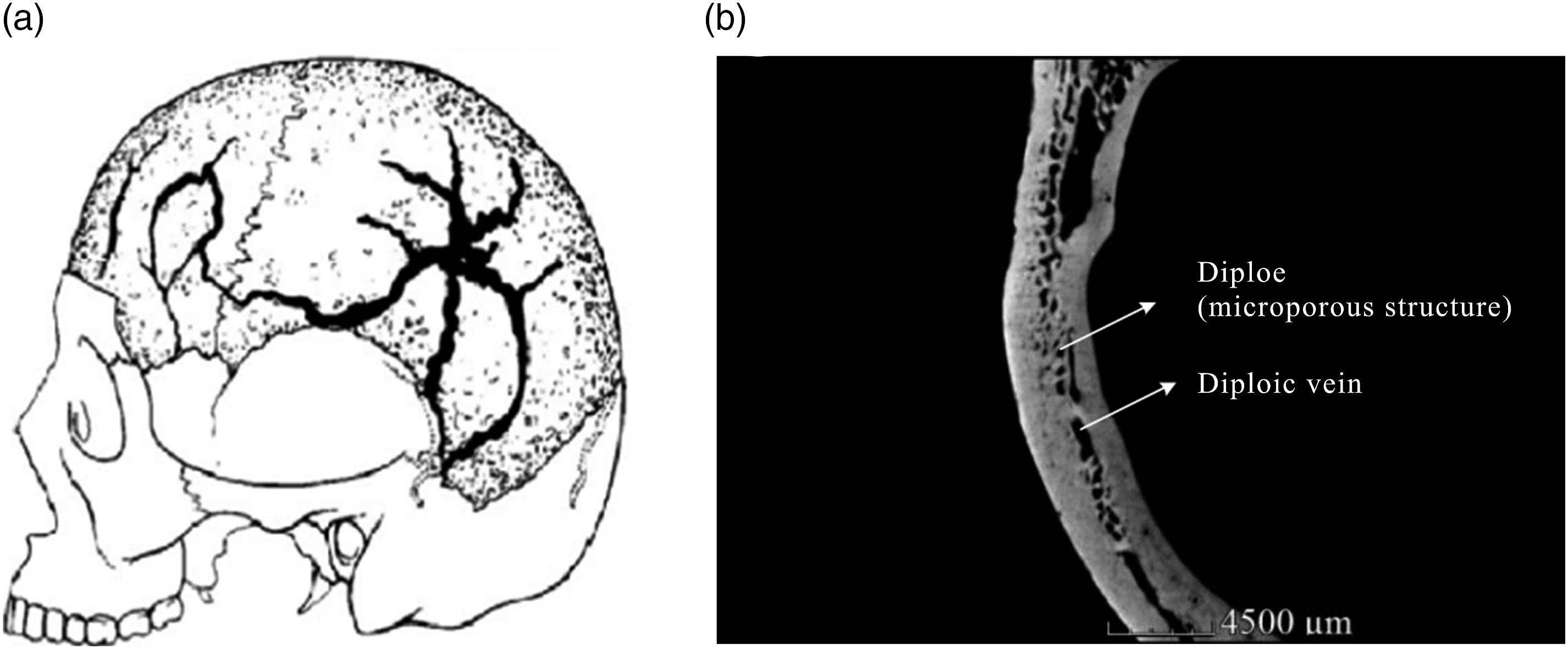
Recent research on engineered multi-scale microchannels such as vessel-on-chip models,5,12 3D blood vessels,10,13 and prefabricated scaffolds with vascular structures10,13–19 shows promise, but there are still challenges in the precise direct fabrication of hollow hierarchical vascular networks with biocompatible hydrogels. Direct manufacturing of complex spatial structures12,20 is more efficient and accurate than indirect manufacturing,10,13–19 which helps to simulate the real cell growth microenvironment.21–23 An effective and precise method for directly fabricating a 3D vascular network-wrapped cell is mainly based on lithography-based 3D bioprinting, including stereolithography (SLA) and digital light processing (DLP).24,25 SLA with its “line-by-line” printing pattern and DLP with its “layer-by-layer” printing pattern have higher printing resolution and speed to realize multi-scales.26,27 Compared with SLA, DLP is more suitable for cell-filled 3D manufacturing hydrogels because its production time is extremely short, and the wavelength of visible light can be adjusted to reduce cell damage.26,28
In addition to biocompatibility9,12 and mechanical properties, printing materials also have precision requirements.29,30 In DLP printing, the resolution is affected by the transmission and scattering 31 of light. When a transparent hydrogel is light penetrating, the excess visible energy will overcure the photocurable material of the cured layer and then affect the printing of the next layer 32 ; thus, it is difficult to accurately print graded networks and form lumens. The addition of some unreactive highly absorbing chemicals can successfully attenuate and limit light in the desired area.29,30,33 The choice of absorber for biomaterials depends on the solubility in the solvent, absorbance at the relevant wavelength, 34 and biocompatibility. Researchers have reported absorbers including brilliant blue,32,34,35 coloring-yellow, 36 Sudan I, 30 Rhod. B, 30 inert triazole dye,37,38 and others, which can also smooth the light intensity distribution on the width surface. 32 However, the use of absorbers causes uneven illumination in the depth direction, resulting in the poor crosslinking density of the material, which affects the cumulative material volume shrinkage, 29 strength, glass transition behavior, swelling capacity, and spatial changes in viscoelasticity. 39 These may result in a non-smooth surface formation, poor adhesion, and shape deformation such as curling and warping, 40 which affect cell adhesion. 41 The current research mainly reduces the final distortion by changing the printer program, 42 post-processing, 43 or optimizing the printing conditions, 29 which are cumbersome and do not solve the z-axis printing errors.44,45 Additionally, it has been verified that some food pigments, such as brilliant blue, have certain biocompatibility at low concentrations,32,36 but lower concentrations may not achieve a high enough absorbance to satisfy the formation of a hollow lumen at the transmission depth, 34 and the biocompatibility of most absorbers has not been verified.
In this study, absorbers were added to print a precise hollow vascular network with a poly(ethylene glycol) diacrylate (PEGDA)/gelatin-methacryloyl (GelMA) hydrogel by a DLP system. The printing effects of three dark absorbers, namely, nigrosin, brilliant black, and brilliant blue on the x-, y-, and z-axes were studied. A model of the difference in the curing width between the upper and lower surfaces of a unit-layer slice based on high-absorbance absorbers was established, and a reference value for slice thickness was proposed. Additionally, scanning electron microscopy (SEM), Fourier transform infrared (FTIR), tensile properties, swelling, degradation, and cytotoxicity of different concentrations of PEGDA-GelMA hydrogel and absorbers were tested. Human Umbilical Vein Endothelial Cells (HUVECs) were planted in the lumen to demonstrate the biocompatibility of the hydrogel by examining changes in the cell morphology.
Materials and methods
Materials
Compound bio-inks were prepared for direct vascular printing because of their composite index of biological and mechanical properties. 26 Poly(ethylene glycol) diacrylate (RHAWN, Yien Chemical Technology Co, Ltd, Shanghai, China), GelMA (SP, Beijing, China) and lithium phenyl-2,4,6-trimethylbenzoylphosphinate (Lithium phenyl-2,4,6-trimethylbenzoylphosphinate (LAP); Tokyo Chemical Industry, Shanghai, China), as a photoinitiator, were chosen to prepare the composite hydrogel. Poly(ethylene glycol) diacrylate can quickly prepare tissue engineering scaffolds with a certain mechanical strength, but lacks sufficient biological activity 46 ; GelMA can effectively promote cell adhesion and has a matrix metalloproteinase targeting sequence, which is conducive to cell remodeling47–49; and LAP has lower cytotoxicity and wider application conditions than other photoinitiators. 46 Dark-colored pigments were more efficient in the light absorption compared with the other colored pigments.25,34 Nigrosin (MACKLIN, Shanghai, China) is a water-soluble pigment with a strong coloring power and good light absorption. The absorption spectrum for nigrosin is broadband from the visible through the near infrared.50,51 Brilliant blue and brilliant black (MACKLIN, Shanghai, China) are food colorings, which are often used for dyeing.32,52 All other chemical reagents were of analytical grade and were commercially available.
Preparation of PEGDA-GelMA composite hydrogel
The PEGDA-GelMA-LAP ratio designs for the orthogonal experiment.
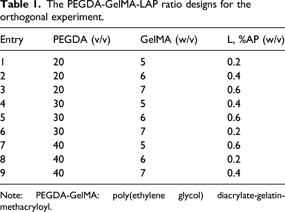
Note: PEGDA-GelMA: poly(ethylene glycol) diacrylate-gelatin-methacryloyl.
Establishment of DLP system
The printing principle of the DLP system is that the initial light is modulated by the Digital Micromirror Device (DMD) dynamic mask to generate a light pattern that is emitted by the projector and injected from the bottom of the material box. Then, the entire construction can be printed from bottom to top (Figure 2). In designing light source systems, it is necessary to consider the damage caused by the different wavelengths of light to cells. Studies have shown that medium and short wavelength (200–400 nm) ultraviolet light adversely affects the growth and development of cell genes
53
; therefore, 405 nm and 3700 lumens of visible light were selected. The principle and composition of the digital light processing system.
Evaluation of x- and y-axes printing accuracy
To reduce the curing of the edge area, dark pigments (nigrosin, brilliant blue, and brilliant black) were added to absorb photons to avoid polymerization of photons with the active LAP. 30 The influence of the additional printing width mainly depends on the absorbance of the solution, which is proportional to the dye concentration. 30 Therefore, 30% PEGDA-7% GelMA containing nigrosin (0.1%, 0.15%, 0.2%, and 0.25% (w/v)), brilliant blue (0, 0.1%, 0.2%, and 0.3% (w/v)), and brilliant black (0.05%, 0.1%, and 0.15% (w/v)) was printed separately as three groups of 5 mm × 1 mm × 1 mm, 5 mm × 2 mm × 1 mm, and 5 mm × 3 mm × 1 mm cuboids. The printed cuboids were imaged using optical microscopy (Leica, Germany) and compared to the designed dimensions.
Evaluation of z-axis printing accuracy
Adding absorbers can improve the difficulty of forming precise cavities with light-transmitting materials, because the latter layer of light easily irradiates the previous layer, and each layer can easily solidify the extra area of the edge.54–58 However, absorbers cause uneven formation along the z-axis.29,55 The light-receiving surface of the slice will receive strong light, and the backlit surface will be weakened, resulting in an uneven slice formation. If it is assumed that the laser irradiance distribution is Gaussian, according to the Beer-Lambert law,
33
the curing width of the front side
However, the first slice is solidified the earliest, and after the continuous n layers are solidified, each layer will be cured again by the light of the next layer (besides the last one). In this case, additional curing decreased as the light intensity decreased. The curing difference of each layer is
Curing depth and width measurement
To calculate the
Absorbance measurement
The absorbance of the PEGDA-GelMA precursor solution containing different concentrations of nigrosin, brilliant blue, and brilliant black was measured at 405 nm using a microplate reader (Thermo Multiskan GO, Thermo Fisher Scientific, USA). Then, 100 μL of the precursor solution was loaded into a 96-well plate to obtain the average absorbance.
Exposure time
Because the working curve of the curing width is nonlinear,
Designing and printing a hierarchical vascular network
The characteristics of the diploic veins were separated by micro-CT scan,8,59 and a hierarchical bifurcated vascular network with a spatial structure was obtained (Figure 3(a)). The first- and secondary-level blood vessels were extracted to perform nesting using a previously reported method,8,59 and a flat vein network was generated. Table 2 presents the design parameters of the vascular network, and Figure 3(b) shows the final designed of the skull vessel scaffold. (a) Characteristics of the diploic veins reconstruction by micro-CT. (b) The hierarchical vascular network design model of the skull. The design parameters of the vascular network.

Printing parameters.

Characterization of the composite hydrogel
A SEM (SIGMA300, ZEISS, USA) was used to acquire the microstructures of GelMA, PEGDA, and GelMA-PEGDA. Additionally, the FTIR spectra were recorded using an infrared spectrophotometer (Nicolet iS10, Thermo Fisher Scientific, USA). The FTIR spectra of GelMA, PEGDA, and GelMA-PEGDA were recorded in the range of 500–4000 cm−1.
The elastic membrane of the middle layer is composed of elastic fibers, collagen fibers, and smooth muscles. 8 The elastic modulus of elastic fiber is approximately 3×10⁴–6×10⁴ N/m26, which is the target tensile strength of the different proportions of bio-inks. Each group (nine groups; Table 1) was subjected to three tensile tests using the DDL-1 special loading system (Tianjin University), and the size of the tensile sample was 40 mm × 10 mm × 2 mm. The loading parameters were as follows: the displacement control mode was adopted, the speed was 0.5 mm/min, the target displacement was 2 mm, and the holding time was 60 s.
Water uptake can directly reflect the permeability of hydrogel materials, which is an important indicator for evaluating the biological performance of hydrogels.
60
The PEGDA-GelMA hydrogels (Table 1) were cured for 10 s, and after complete lyophilization, the resulting hydrogels were weighed as
To determine the degradation rate, nine groups (Table 1) were cured for 10 s, and after complete lyophilization, the resulting samples were weighed as
Cytotoxicity of composite hydrogels and tube planting
To verify the biocompatibility of the PEGDA-GelMA composite hydrogel, a NIH-3T3 cell (Procell, Wuhan, China) suspension with a cell density of 6 × 10⁵ cells/ml was seeded on the cured hydrogels. To verify the biocompatibility of nigrosin (0.05%, 0.1%, 0.2%, and 0.3%), brilliant blue (0.05%, 0.1%, 0.2%, and 0.3%), and brilliant black (0.05%, 0.1%, 0.2%, and 0.3%), the solidified hydrogel was first washed with alcohol and then a HUVEC cell (Procell, Wuhan, China) suspension with 6 × 10⁵ cells/ml was seeded on the cured 30% PEGDA-7% GelMA hydrogels containing absorbers. Cell Counting Kit-8 (CCK-8, DOJINDO, Japan) (10 vol%) was added to the cell culture medium at days 1, 3, 5, and 7. Cell culture medium samples (100 μL) were placed in a 96-well plate. A microplate reader was used to measure the absorbance value (optical density) to determine the number of cells.
To observe the growth of seeded cells in the tubular structure, a HUVEC cell suspension with a cell density of 6 × 10⁵ cells/ml was injected into the lumen of the vascular networks with 30% PEGDA-7% GelMA/0.1% brilliant black. Dead and live photos were randomly taken with a fluorescence microscope under different fields of view at 1, 3, 5, and 7 days, where live and dead cells being displayed in green and red, respectively. The CCK-8 kit was used to detect the cell viability on days 1, 3, 5, and 7.
Results and discussion
Improving printing accuracy by absorbers
Improved printing accuracy of x- and y-axes
The effects of the nigrosin, brilliant blue, and brilliant black contents on the x- and y-axes were evaluated by comparing the printed features with the intended features of 1000–3000 μm. The printing deviation with absorbers was clearly smaller than that without absorbers (Figure 4(a) and (b)). Absorbers were added to absorb the photons propagating in the solution to avoid photos to penetrate deeper or participate in chemical reactions.32,37 The printed features exhibited a narrow-edged shape as the concentration of the absorbers increased (Figure 4(a) and (b)). The PEGDA-GelMA containing 0.2% (w/v) nigrosin, 0.1% (w/v) brilliant black, or 0.3% (w/v) brilliant blue could print features highly correlated to the design size (Figure 4(a) and (b)) and had similar absorbance of 0.88–0.92. A similar absorbance may have a similar printing accuracy. The brightness at the same concentration of absorbers (Figure 4(c)) was ranked as follows: brilliant blue > nigrosin > brilliant black, which is similar to that of absorbance. As the concentration of absorbers increased in the hydrogel, the absorbance increased and the color darkened (Figure 4(b) and (c)). Brilliant blue is a widely used food coloring agent, and its biocompatibility at low concentrations has been verified,32,36 but the absorbance at the same concentration is lower than that of nigrosin and brilliant black (Figure 4(b)). (a) Optical microscope images of hydrogels 3D-printed with PEGDA-GelMA with varying concentrations of nigrosin, brilliant blue, and brilliant black. The beam irradiation width was 1000 μm. (b) Printing deviation of different nigrosin concentrations on x- and y-axes of 1000–3000 μm and the absorbance of different nigrosin concentrations at a 3 mm depth. (c) Photographs of PEGDA-GelMA with different concentrations of nigrosin, brilliant blue, and brilliant black at a 3 mm depth. Note: PEGDA-GelMA: poly(ethylene glycol) diacrylate-gelatin-methacryloyl.
Curing width and depth
Figure 4(b) shows that the absorptivity of 0.2% nigrosin, 0.1% brilliant black, and 0.3% brilliant blue through 3 mm reached 81–88%. According to Equation 4, the difference in the curing of the upper and lower surfaces of the slice is related to the slice thickness. It was verified that thick slices cause a large difference in the curing width on the slice (Figure 5(d)). To reduce unevenness, the prediction model corresponding to 0.2% nigrosin, 0.1% brilliant black, and 0.3% brilliant blue can be calculated to obtain the reference slice thickness. The curing size at the depth and width was recorded at 20, 40, 60, 80, 100, and 120 s, as shown in Figure 5(a) and (b). The curing width was limited by the size of the beam before reaching the beam size. Under long-term exposure, the tendency to increase the curing width slows down, similar to the effect of dyes on curing in other studies.
37
Because the irradiation depth experiment provides a solution with a depth of 3000 μm, the curing before 1500 μm can be approximately linearly changed, but as the curing depth increases, the growth rate with time changes more slowly, which may affect the values of (a) Curing depth at different concentrations of absorbers and fitted lines at a 3 mm depth. (b) Curing width at different concentrations of absorbers and fitted lines with a 1 mm × 1 mm beam width. (c) Curves of the curing width difference of the first and last layers. (d) Optical microscope images of the edge of the printed sample side-section and the cross-section of a semi-tubular tube of a 10, 50, and 100 μm slice.
Curing width difference model
The theoretical model of the Jacobs working curve is often discussed,32,36,61 and in this study, the relationship between exposure time
Characterization of composite hydrogel
Scanning electron microscopy imaging, water uptake, and degradation
The surface area to volume ratio and porosity of the hydrogel play an important role in suitable cells attachment, facile nutrients, waste exchange, and biological activity.63,64 The mechanical strength, swelling and degradation properties of hydrogels are often inseparable from their microstructures. Figure 6(a) shows that PEGDA has a tighter microstructure, low porosity, and a small void diameter, as compared to GelMA, resulting in the PEGDA having excellent mechanical properties but extremely low water uptake, poor biological properties, and a relatively slow degradation rate because of the smaller surface area, which can be confirmed by Figure 6(d) and (e). Gelatin-methacryloyl has a fine pore structure, high porosity, and transparent fibers which is conducive to cell adhesion, material exchange, and metabolism (Figure 6(a)). Similarly, the higher the GelMA concentration, the higher water uptake and the faster the degradation, as shown in Figure 6(d) and (e), similar to other studies.
65
(a) SEM images of PEGDA, GelMA, and PEGDA- GelMA. (b) FTIR spectra of PEGDA, GelMA, and PEGDA-GelMA. (c) The stress–strain curves of the hydrogel. (d) Comparison of the water absorption of the hydrogels. (e) Comparison of the degradation rates of the hydrogels. Error bars represent the standard deviation, and the asterisks denote the differences at p < 0.01. Note: SEM: scanning electron microscopy; PEGDA-GelMA: poly(ethylene glycol) diacrylate-gelatin-methacryloyl; FTIR: Fourier transform infrared.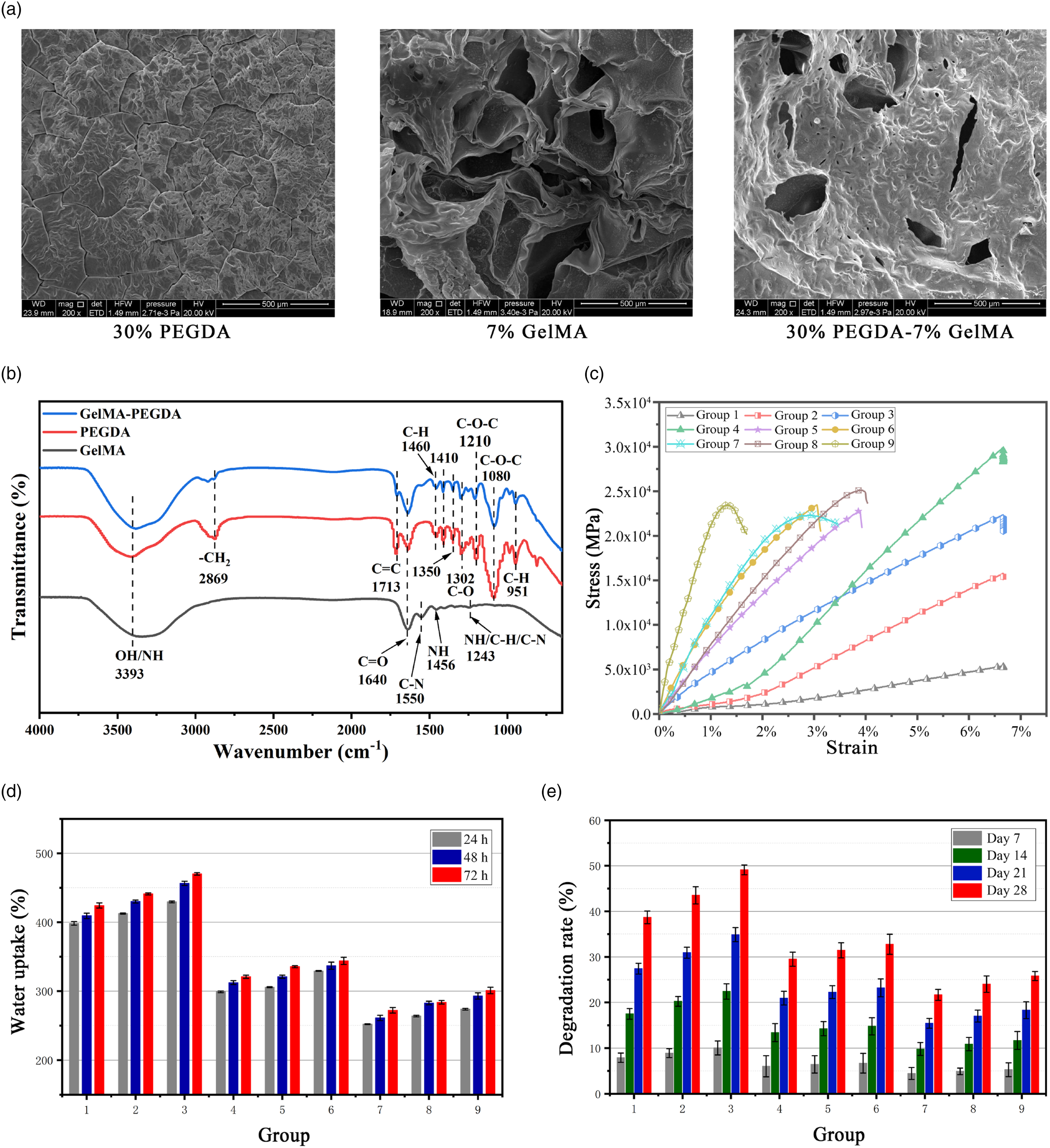
Fourier transform infrared analysis
Fourier transform infrared spectra were analyzed to confirm the crosslinking of GelMA-PEGDA. Figure 6(b) shows the infrared spectra of PEGDA, GelMA, and PEGDA-GelMA over the wavenumber range of 500 cm−1–4000 cm−1. Several characteristic bands of GelMA and PEGDA are visible, such as N-H band stretching vibration peak at 3393 cm−1; C = O band at 1640 cm−1; C-N stretching vibration and N-H bending vibration coupled at 1550 and 1456 cm−1, respectively; CH2 swing vibration, N-H bending vibration, and C-N stretching vibration superimposed peak at 1243 cm−1 for GelMA; and saturated C-H at 2869 cm−1, weak C-H bond at 1460 and 1410 cm−1; C = C bond at 1713 cm−1; C = O bond at 1640 cm−1, the symmetrical coupling of C-O bond at 1351 and 1302 cm−1; C-O-C bond at 1080 and 1210 cm−1; and the out-of-plane bending vibration of C-H at 951 cm−1 for PEGDA (Figure 6(b)). By comparison, it was found that the intensity of the stretching vibration peak of the C-H bond near 2869 cm−1 in GelMA-PEGDA was weakened, and the intensity of the C-H vibration absorption peak at 951 cm−1 decreased significantly, indicating that the -CH bond underwent significant deformation. The C-O-C asymmetric stretching vibration at 1210 cm−1 was significantly weaker than that of the PEGDA sample, which further indicates that the polymer has underwent a cross-linking reaction. More importantly, the tensile vibration peak intensity of the C = C bond at 1713 cm−1 in GelMA-PEGDA was significantly reduced. This is mainly because in the free radical polymerization reaction where the free radical A* attacks the PEGDA -C = C- double bond, the initiation center is generated and then the radical polymerization reaction is initiated, finally forming a 3D cross-linked network.
Mechanical characterization
Uniaxial tensile test was performed on PEGDA-GelMA (Table 1). As expected, a higher PEGDA content produced less strain and later yielded under the same stress, which is conducive to the generation of liquid flow, but lower swelling will inhibit cell growth due to less water storage; therefore, a 30% PEGDA content is more appropriate. As the content of GelMA increased, the strain limit decreased slightly, and the stress limit increased significantly, indicating that the content of GelMA can effectively improve the elastic properties of the scaffold. However, an increase in the PEGDA content greatly amplified the change in the strain limit. The stress limit did not significantly change with a further increase in the PEGDA content, but the strain limit exhibited a lower level, which indicates that when the content of the PEGDA component is too high, as compared to the GelMA component, the GelMA component can no longer significantly improve the elastic properties of the scaffold (Figure 7(b)). (a) The overall size of the vascular scaffold. (b) Images of the hollow structure.
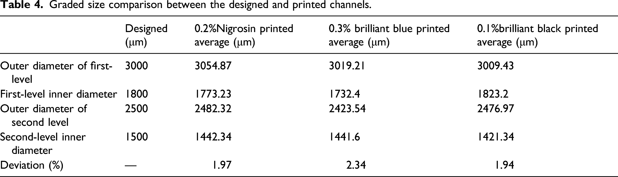
Printing of a hierarchical vascular network channels
Graded size comparison between the designed and printed channels.
Cell proliferation in scaffold
Cytotoxicity of composite hydrogel
To assess the biocompatibility of the PEGDA-GelMA composite hydrogel, NIH3T3 cells were seeded on the cured composite hydrogel, and the cell proliferation rate was evaluated using CCK-8 for 7 days. Figure 8(a) shows the effect of different concentrations of PEGDA-GelMA hydrogel on the survival rate of cells. The viability of the cells on the first day of the 20, 30, and 40% PEGDA groups was almost the same and within 3%. There was a significant gap on the third day, showing that 20% and 30% grew well, and the survival of 40% PEGDA decreased significantly, which means that the small pores in the hydrogel clearly affected the cell growth faster, mainly due to the transmission and exchange of nutrients and oxygen. From the fifth day to the seventh day, there was a difference between the 20% and 30% PEGDA. The survival of 30% PEGDA was generally higher than 20% probably because 30% PEGDA has a stronger and larger surface to attach in the micro-framework. The influence of the hydrogel pore size/number on cell growth may be related to the cell growth habits, that is, NIH3T3 cells are adherent cells similar to HUVECs. When GelMA content was relatively high, the cells were conducive to proliferation, but when the PEGDA content was much higher than GelMA, the growth of GelMA did not significantly promote the growth of cells. The content ratio of PEGDA and GelMA was maintained from 2 to 5:1 as this radio was more able to maintain cell growth. (a) Cytotoxicity of the PEGDA-GelMA hydrogel. (b) Cytotoxicity of PEGDA-GelMA containing nigrosin, brilliant black, and brilliant blue (0.05%, 0.1%, 0.2%, and 0.3%). (c) Fluorescence map of the dead and living cells in the inner cavity of the HUVEC-seeded vascular network with 30% PEGDA-7% GelMA/0.1% brilliant black. (d) Cell viability in the inner cavity of the HUVEC-seeded vascular network. Error bars represent the standard deviation, and the asterisks denote the differences at p < 0.01. Note: PEGDA-GelMA: poly(ethylene glycol) diacrylate-gelatin-methacryloyl; HUVEC: human umbilical vein endothelial cell.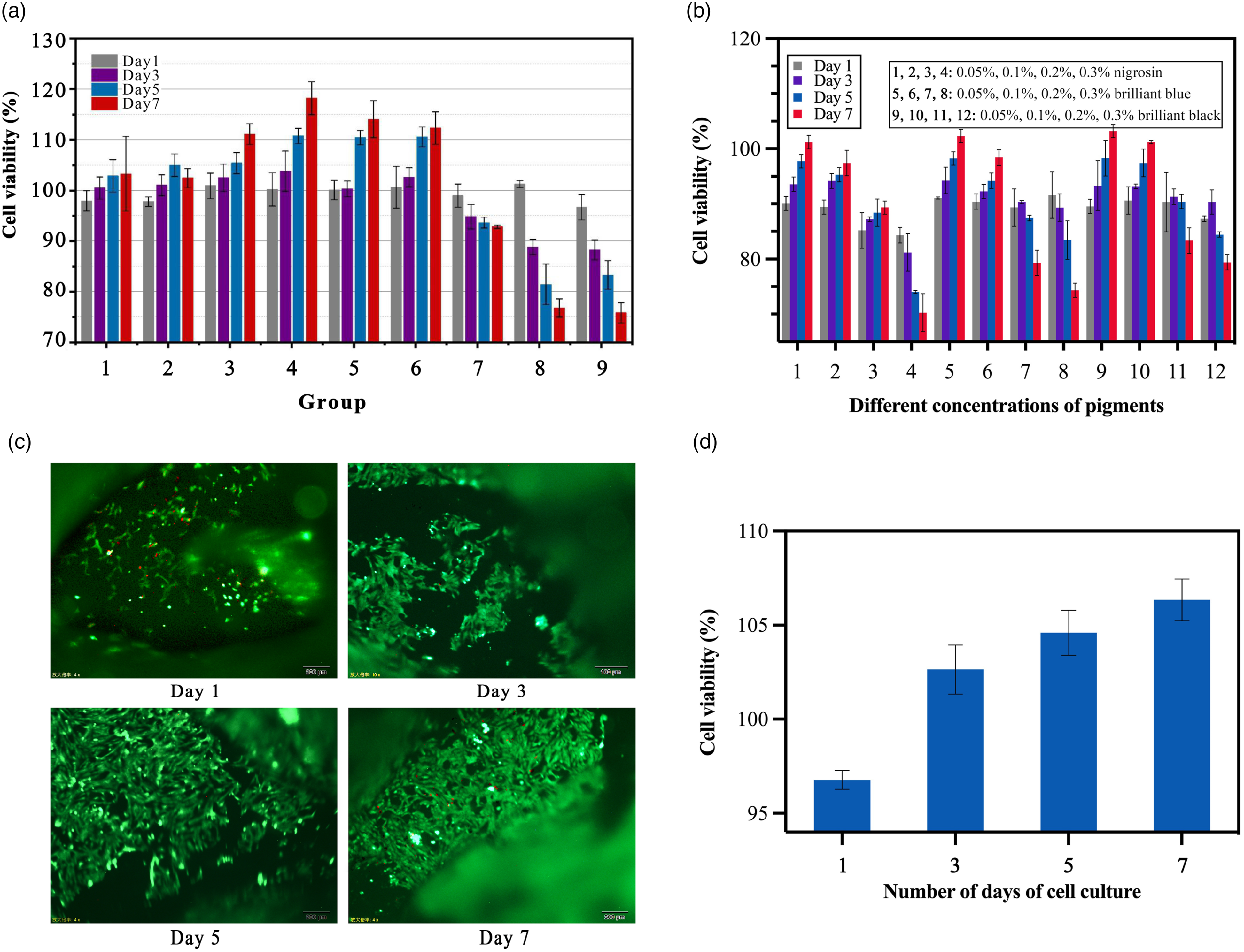
Cytotoxicity of absorbers
To assess the biocompatibility of the PEGDA-GelMA composite hydrogel with nigrosin, brilliant blue, and brilliant black, HUVEC cells were seeded on the cured hydrogel, and the cell proliferation rate was evaluated using CCK-8 for 7 days Figure 8(b) shows the effects of different concentrations of nigrosin, brilliant blue, and brilliant black on the cell viability. At a lower concentration (0.05%), all absorbers had good cell survival rates. When the concentration continues to increase, cell viability began to gradually weaken on the third day, which means that absorbers that achieve the required absorbance at lower concentrations are worth considering. Generally, food coloring, such as brilliant blue and black, has slightly better biocompatibility. Among the absorbers with the same printing accuracy, 0.1% brilliant black exhibited relatively high biocompatibility on days 5 and 7; therefore, brilliant black was used to complete the lumen implantation.
Cell lumen implantation
Human umbilical vein endothelial cells were evenly implanted into the cavity of the vascular network with 30% PEGDA-7% GelMA/0.1% brilliant black for 7 days to observe the intraluminal cell growth. Figures 8(c) and (d) show the growth status of the cells in the vascular scaffold on days1, 3, 5, and 7. On the first day, the HUVECs were polygonal, and cell aggregation and connection were apparent, indicating that the cells adhere well and the cell viability reached 96.78%. On the third day, the cells were growing rapidly and appeared in small areas connected into networks, and the cell viability reached 102.64%. On the fifth and seventh days, the morphology of the cells did not significantly change. They were all connected into networks, forming cell clusters and cell clumps, which filled the lumen of the vascular network. A small amount of cell death began to occur on the seventh day, and the survival rate reached 106%. This demonstrates that the cells grew well and had the conditions to continue growing on the hydrogel scaffold. Additionally, the number of cells at different positions in the vascular scaffold was slightly different. Under the action of gravity, the cells are more likely to accumulate at the end of the larger pores, resulting in an imbalance in the absorption of nutrients by the cells, leading to the death of a small number of cells.
Conclusions and future work
A DLP system was developed for the direct printing of a hierarchical vascular network with PEGDA-GelMA bio-ink. DLP leads to a higher printing efficiency and accuracy, and it is also a free-formed technology that can directly print vascular structures. 30% PEGDA-7% GelMA was selected to directly form a hierarchical vascular network, which was examined with SEM and FTIR, and underwent swelling, degradation, and toxicity tests. The results show that the 30% PEGDA-7% GelMA has excellent mechanical properties and biocompatibility. To print more precise and uniform pipes, absorbers, such as 0.2% nigrosin, 0.1% brilliant black, and 0.3% brilliant blue, were added to establish a model of the difference in curing width between the upper and lower surfaces of a unit slice to obtain a slice thickness. Because the biocompatibility is inversely proportional to the concentration of the light absorber, brilliant black achieved the required absorbance at low concentrations. Human umbilical vein endothelial cells were planted in the lumen of PEGDA-GelMA/brilliant black vascular networks to observe the cell morphology finally. Future work should focus on multi-cell printing. In terms of printing hardware, the ratio and resolution can be adjusted by changing the distance between the DMD chip, the lens and the aggregation plane to print even smaller complex 3D structures. Bio-inks with higher biocompatibility and better mechanical properties are still needed to be researched.
Footnotes
Declaration of conflicting interests
The author(s) declared no potential conflicts of interest with respect to the research, authorship, and/or publication of this article.
Funding
The author(s) disclosed receipt of the following financial support for the research, authorship, and/or publication of this article: This work was supported by the National Natural Science Foundation of China (No. 52175275 , 51875404).
