
Abstract
At present, commercial artificial biological valves are mostly prepared by crosslinking bovine or porcine pericardia with glutaraldehyde. Swim bladder has similar components and lower immunogenicity compared to bovine or porcine pericardium. In this study, we used a glycidyl methacrylate (GMA)–based radical polymerization method to crosslink decellularized swim bladders. Amino and carboxyl groups in the swim bladder were reacted with epoxy groups on GMA to introduce carbon–carbon double bonds to the swim bladder. The results showed that the platelet adhesion of GMA-crosslinked swim bladders (GMA-SBs) decreased by 35%, as compared to that of glutaraldehyde-crosslinked swim bladders (GLUT-SBs). Moreover, the superior anticoagulant property was further verified by the ex vivo arteriovenous shunt assay. Meanwhile, the subcutaneous implantation in rats showed that GMA-SBs were able to effectively inhibit the calcification compared with GLUT-SBs. In conclusion, GMA-SBs showed improved antithrombotic and anticalcification properties compared to GLUT-SBs.
Introduction
Although the most widely used biological valve material is bovine or porcine pericardium, there is immunogenicity associated with pericardia due to species differences. 1 Studies have shown that fish do not contain many immunogens, such as α-gal, and there is lower immunogenicity associated with biomaterials obtained from fish as compared to mammals. 2 In addition, unlike mammalian pathogens, fish pathogens are rarely infectious to humans, and therefore, materials derived from fish are safer than those derived from mammals. 3 . The swim bladder is similar in composition to the animal pericardium and has been reported as a promising cardiovascular and wound repair biomaterial.4,5
Thrombogenicity of biological valves is still an ongoing problem that has not yet been solved, and the clotting of valves is an important factor in the failure of biological valves. The reduced valve movement was found to be consistent with valve thrombosis in 40% of patients.6–8 Studies have shown that increased blood compatibility were observed with glutaraldehyde-crosslinked swim bladders when they were compared with glutaraldehyde-crosslinked pericardia, 5 which is expected to be utilized to replace animal pericardium with a new generation of cardiovascular biomaterials.
Glutaraldehyde is a crosslinking agent widely used in biological materials. However, it introduces the toxic aldehyde group, which causes cytotoxicity, inflammatory response, and accelerates calcification.9–11 Owing to the limitations of glutaraldehyde, there has been a great deal of research to develop new crosslinking methods using epoxies, polyphenols, and genipin, to find a low toxic crosslinking reagent that will enhance the durability of tissues and possesses more optimal anticalcification properties.12,13
Our lab developed glycidyl methacrylate (GMA)–based radical polymerization method to crosslink porcine pericardium and showed that GMA-crosslinked porcine pericardium could improve collagen and elastin stability, reduce inflammation and calcification compared to that of glutaraldehyde-crosslinked porcine pericardium. 14 In this study, GMA-based radical polymerization was used to crosslink the swim bladder. The swim bladder was first decellularized and then crosslinked with glycidyl methacrylate. The thermal shrinkage temperature, anti-enzyme degradation stability, cytotoxicity, hemocompatibility, and calcification properties of GMA-crosslinked swim bladders (GMA-SBs) were investigated and compared to that of glutaraldehyde-crosslinked swim bladders (GLUT-SBs).
Materials and methods
Materials
Grass carp (Ctenopharyngodon idella) swim bladders (SBs) were obtained from Sifan Co., Ltd (Hangzhou, China). Sodium deoxycholate (SD), sodium dodecylsulphate (SDS), and glutaraldehyde (GLUT) were purchased from Huaxia Reagent (Chengdu, China). Glycidyl methacrylate (GMA) was purchased from Aladdin Biotechnology Co., Ltd (Shanghai, China). Ammonium persulfate (APS) and sodium hydrogen sulfite (SHS) were purchased from Best-Reagent (Chengdu, China). Collagenase I and elastase were purchased from Sigma-Aldrich (St Louis, MO, USA). Cell Counting Kit-8 (CCK-8) was purchased from Beyotime Biotechnology Co., Ltd (Shanghai, China).
Swim bladder crosslinking treatment
The swim bladders (SBs) were washed three times with deionized water, and fatty tissues were gently removed. Briefly, swim bladders were incubated with 0.5% SDS for 2 h under vigorous stirring. The supernatant was discarded, and the swim bladders were then incubated with 0.5% SD for another 24 h. After rinsing 3 times with distilled water, SBs were treated with two different methods, as described below. Crosslinked SBs were soaked in an 80% ethanol plus 20% glycerol mixed solution for 4 h and then air-dried for preservation.
To prepare GLUT-modified SBs (GLUT-SBs): the decellularized SBs (DSBs) were incubated with 0.625% GLUT with shaking at 150 r/min on an orbital shaker (SK-O180-E, Dragonlab, Beijing, China) for 24 h at room temperature.
To prepare GMA-modified SBs (GMA-SBs): the decellularized SBs (DSBs) were reacted with 5% GMA in phosphate-buffered saline (PBS) buffer under constant stirring for 3 days at 37°C. The DSBs were subsequently crosslinked by incubating with PBS buffer containing 5 m Schematic illustrations of the crosslinking mechanism for GMA-modified SBs (GMA-SBs). APS, ammonium persulfate; SHS, sodium hydrogen sulfite.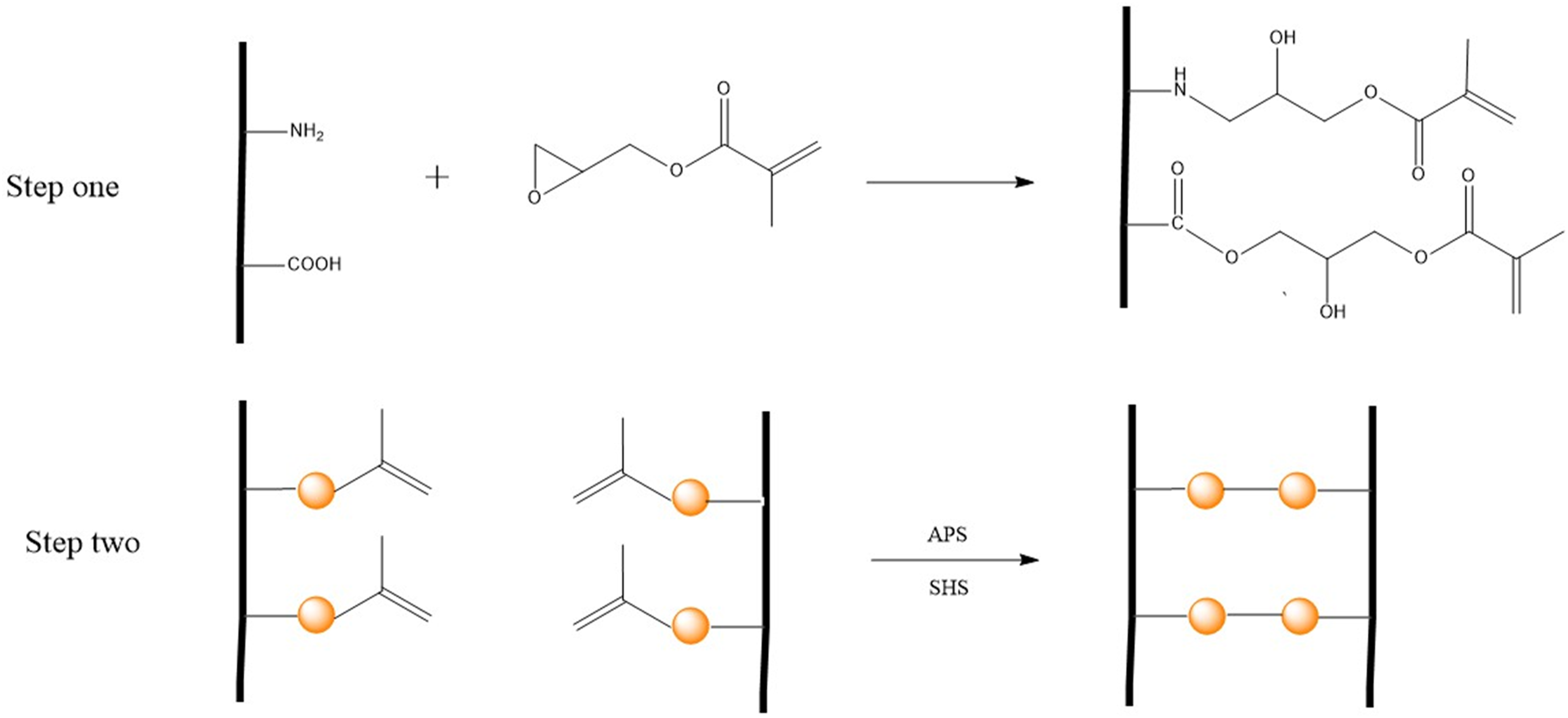
Relative amine and carboxyl content
The ninhydrin assay is commonly used to quantify the free amine groups present in xenogeneic tissue. In brief, ninhydrin was dissolved in citric acid aqueous solution (0.1

The concentration of the carboxyl groups was determined by the toluidine blue-O (TBO) method. For this test, 0.5 m
Determination of collagen content
Hydroxyproline is one of the main components of collagen, and hydroxyproline was measured using a hydroxyproline kit (Nanjing Jian-Cheng Biological Engineering Research Institute). The hydroxyproline in the swim bladders was quantitatively analyzed using the characteristic that the oxidized product of hydroxyproline can react with dimethylaminobenzaldehyde and produce a purplish red color. Swim bladders were hydrolyzed at 100°C in a water bath, and then, the working liquid was added. After centrifugation at 3500 r/min for 10 min, 200 L of supernatant was removed and transferred to a microplate, and the absorbance value was determined at the wavelength of 550 nm.
Determination of elastin content
The elastin content was determined using the Fastin kit (Biocolor Life Science, UK) according to the product instructions. First, elastin in tissue samples was extracted with 0.25
Determination of glycosaminoglycan (GAG) content
The amount of GAG was determined using a Blyscan kit (Biocolor Life Science, UK). GAG in tissues was first extracted with papain, and then, 1 mL of Blyscan dye reagent was added, with subsequent gentle shaking for 30 min. During this process, a glucosamine dye complex was formed, and the soluble dye precipitated. Then, 0.5 mL of dissociation reagent was added to the precipitate and it was re-dissolved. Finally, the solution was transferred to a 96-well plate, and the absorbance value was determined at the wavelength of 656 nm.
Fourier transform infrared (FTIR) spectroscopy
SB samples were punched into 6 mm diameter disks and then washed three times with deionized water. The disks were flattened and then freeze-dried. The FTIR spectrum across the wavelength range from 500 to 4000 cm−1 was measured using a Thermo Electron Nicolet 6700 (West Palm Beach, FL) in total reflection mode.
Enzymatic degradation in vitro
SB samples were cut into small pieces (1 × 1 cm2, n = 6) and then freeze-dried. The initial dry weight of the samples was recorded as W0. Then, the tissue was incubated in 1 mL of 50 U/mL type I collagenase or 50 U/mL elastase for 24 h at 37°C with shaking at 100 r/min on an orbital shaker (SK-O180-E, Dragonlab, Beijing, China). Digested tissue was washed in deionized water, freeze-dried, and weighed (Wt). The fraction of weight loss was calculated as weight loss ratio (%) = (W0 − Wt)/W0 × 100.
Differential scanning calorimetry (DSC)
Differential scanning calorimetry was used to measure the thermal shrinkage temperature of tissues. Small pieces of dry samples (2–5 mg for each, n = 6) were cut from tissues and tested using DSC 2920 under an N2 atmosphere. Samples were equilibrated at 40°C, and heated at 5°C/min up to 100°C. The thermal shrinkage temperature was recorded as the maximum value of the endotherm peak.
Uniaxial tensile test
Uniaxial tensile testing was performed on a universal testing machine (BioTester, 10N load cell; Cell Scale, Waterloo, Ontario, Canada). Tissues were cut using rectangular stamps (30 × 6 mm strips, n = 6). A thickness gauge was used to measure the thickness at three random locations, and the average thickness value was used for the stress calculation. The SBs were preloaded with approximately 0.1 N force and stretched at a constant extension speed of 10 mm/min until they fractured. The data where the fracture occurred at the clamping position of the fixture were not included in the statistical analysis.
Cytotoxicity test
The cytotoxicities of tissue samples were assessed according to ISO 10993-5 standard. L929 fibroblasts were cultured in Roswell Park Memorial Institute (RPMI) 1640 medium supplemented with 100 U/mL penicillin, 100 μg/mL streptomycin, and 10% fetal bovine serum. Samples were washed three times with sterile distilled water and sterilized in 75% ethanol overnight. The extracts were made by soaking the SBs in culture medium at 37°C for 48 h in a tissue weight-to-volume ratio of 0.2 g/mL. Untreated RPMI 1640 medium was used as a control. L929 fibroblasts were seeded in a 96-well plate for 24 h, the medium was replaced with 100 mL of extraction liquid, and the cells were cultured for another 1 and 3 days. The cell viability was tested using Cell Counting Kit-8 (CCK-8, Beyotime, Shanghai, China) reagent according to the instructions. The absorbance at 450 nm was measured on a microplate reader (Varioskan Flash, Thermo Scientific, Waltham, MA, USA).
Human umbilical vein endothelial cell (HUVEC) growth
SB samples were punched into 10 mm diameter disks and sterilized with 75% ethanol for 24 h. The sterilized samples were placed in a 48-well plate, and HUVECs were seeded on the samples at a density of 10,000 cells/well. After 3 days of incubation, the samples were transferred to a new 48-well plate, and 200 mL of CCK-8 working solution was added to measure the cell activity according to the supplier’s instruction. After the CCK-8 assay, the samples were rinsed with PBS, and then fixed with 4% paraformaldehyde. The cell nuclei and cytoskeleton were stained with 4′,6-diamidino-2-phenylindole (DAPI, Sigma-Aldrich, St Louis, MO, USA) and tetramethylrhodamine isothiocyanate (TRITC)-phalloidin (Solarbio, Beijing, China), respectively, and then visualized under a fluorescence microscope (Leica DMI 4000B).
Hemolysis rate and whole blood adhesion
Fresh rabbit blood was collected by using vacuum blood tube with sodium citrate anticoagulant. Samples were cut into 6 mm diameter disks and suspended in 600 μL (containing 200 μL red blood cell (RBC) suspension) of PBS buffer for 2 h at 37°C. H2O and PBS were used as the positive control and negative control, respectively. After incubation at 37°C for 2 h, the supernatant was centrifuged, and 100 μL of supernatant was added to a 96-well plate. The absorbance at 541 nm was measured on a microplate reader (Synergy H1, Bio-Tek Instruments Inc.). The hemolysis ratio was calculated as follows

For whole blood adhesion, each sample was cut into 10 mm diameter circles, which were incubated in rabbit whole blood for 2 h at 37°C. The samples were washed three times with saline and fixed with 2.5% glutaraldehyde. Finally, the samples were imaged with a scanning electron microscope (S4800, Hitachi, Japan).
Platelet adhesion tests
The thrombogenic properties of the tissues were assessed using platelet adhesion tests. SBs were cut into 6 mm diameter disks and incubated in rabbit platelet-rich plasma at 37°C for 1 h, and then rinsed three times with saline. For imaging, the tissue was dehydrated with 30%, 50%, 70%, and 100% ethanol for 10 min each. The tissue was then sputter-coated with gold, and images were obtained with a scanning electron microscope (JSM-7500F, Japan). Rhodamine B-labeled CD62p was added to the samples, which were incubated at 37°C for 1 h, and then washed with PBS. The activated platelets were observed under a fluorescence microscope (Leica DMI 4000B). Additionally, the lactate dehydrogenase (LDH) assay was performed for quantitative analysis of platelet adhesion.
Ex vivo blood circulation
The ex vivo arteriovenous shunt assay was used to demonstrate the anti-thrombogenicity of samples under actual blood flow. In this study, experimental animals were purchased from Chengdu Dashuo Experimental Animal Co., Ltd (Chengdu, China). New Zealand white rabbits (approximately 3.0 kg) were anesthetized by intraperitoneal injection with 10 mg/mL pentobarbital solution at a dose of 30 mg/kg. After arterial and venous isolation, and the left carotid artery and right external jugular vein were cannulated to connect the extracorporeal circuit of the PVC tubing. The specimens were cut into 1 cm × 1 cm pieces and placed into the external connection line. The blood flow in the circuit was monitored for 1 h. Then, the specimens were removed from the pipe and rinsed with saline. Finally, samples were imaged by scanning electron microscope (S4800, Hitachi, Japan).
In vivo rat subdermal implantation model
The conducted animal experiments were approved by an institutional review from the Medical Ethics Committee of Sichuan University. Square tissue SB samples (1 cm × 1 cm each) were rinsed in 75% ethanol for 24 h, and then washed with sterilized PBS. Male Sprague–Dawley (SD) rats (approximately 50g) were anesthetized by intraperitoneal injection with 4% chloral hydrate solution at a dose of 10 mL/kg. Two longitudinal surgical incisions were made on either side of the back of each rat to created pockets for a subcutaneous space, and one sample was placed into each pocket. After 30/60 days of implantation, samples together with fibrous capsules were explanted and fixed with formalin for further examination.
Quantitative calcium and histological analysis
Explanted SBs were washed with PBS, then lyophilized, weighed, and acid hydrolyzed in 6 N HCl for 24 h at 100°C. The supernatants were diluted 10 times, and the calcium content was detected by inductively coupled plasma optical emission spectrometry (ICP-OES). For histological analysis, the fixed explants were dehydrated and then embedded in paraffin. For each specimen, 6 μm sections were cut and stained for light microscopy analysis. Alizarin red stain was used to stain calcium deposition, and Masson’s trichrome stain was used to visualize the collagen of the explants.
Statistical analysis
The data were analyzed for statistical significance by Student’s t test for comparison of two groups, and one-way ANOVA with Bonferroni-corrected t test was used for comparison of multiple groups. Where indicated, the results are represented as the mean ± standard errors.
Results
Decellularization and ECM evaluation
The interior and external surface morphology of the sample was observed under a scanning electron microscope. The inner side of the swim bladder was smooth, and the outer side was slightly rough (Figure 2(a)). The thickness was 300–500 μm (Figure 2(d)). Furthermore, the content of collagen, GAG, and elastin was evaluated. Among them, collagen fibers accounted for approximately 70%, elastic fibers accounted for 20%, and GAG accounted for approximately 10% (Figure 2(c)). ECM evaluation of DSBs, GLUT-SBs, and GMA-SBs. (a) SEM images; scale bars are 10 μm. (b) Histological staining of fresh and decellularized swim bladder (scale bar = 50 μm). (c) Collagen, elastin, and GAG relative content (n = 6). (d) Swim bladder thickness was measured by thickness gauge (n = 6). ****p < 0.0001, ***p < 0.001, **p < 0.01, *p < 0.05, ns, no significant difference.
To decrease the immune response, decellularization was performed, and the acellular swim bladder was qualitatively analyzed by histological staining (Figure 2(b)). The collagen fibers and elastin structure were well maintained without any detectable damage after the acellular procedure. The swim bladder fibers were arranged in a neat direction with obvious anisotropy. Sirius red staining indicated that the collagen in the acellular swim bladder was mainly type I collagen fiber. Alcian blue staining indicated that the GAG was evenly distributed in the swim bladder fibers.
Chemical characterization
The degree of substitution of GMA was indirectly characterized in terms of conversion of amine and carboxyl groups. The results showed that amino and carboxyl residues in the swim bladder decreased with the increase in GMA concentration, and that the lowest amounts of amine and carboxyl groups were found in a 5% GMA-crosslinked swim bladder (Figure 3(a) and (b)). Chemical characterization of DSBs, GLUT-SBs, and GMA-SBs. (a) The relative amine content of SBs (n = 6). (b) The relative carboxyl content of SBs (n = 6). (c) Representative FTIR spectrum of SBs. ****p < 0.0001, ***p < 0.001, **p < 0.01, *p < 0.05, ns, no significant difference.
The FTIR spectra of DSBs and GMA-SBs exhibited obvious changes in their functional groups (Figure 3(c)). The FTIR spectrum of GMA exhibited an evident increase in the peak intensity at 1160 cm−1, which showed the signal of the stretch movement of C-O-C. The formation of a new ether bond group indicated successful polymerization of GMA. In addition, the peak shape changed at 3078 cm−1, which indicated that additional primary amines became secondary amines after crosslinking. Notably, the carbonyl peak at 1720 cm−1 appeared in the spectrum of GMA, perhaps because of the oxidation of amino acids by the redox initiators. 15
Componential stability
Componential stability was further evaluated by measuring the resistance of tissues to enzymatic degradation. The SBs were challenged with type I collagenase and elastase, and the higher the concentration of GMA treatment, the greater the stability of the collagen and elastin within a certain range (Figure 4(a) and (b)). The greatest protection for collagen and elastin was afforded by GMA with a feed ratio of 5%, which therefore was selected for further study. Componential stability of DSBs, GLUT-SBs, and GMA-SBs. (a) Weight loss ratio of SBs after incubating with collagenase for 24 h at 37°C (n = 6). (b) Weight loss ratio of SBs after incubating with elastase for 24 h at 37°C (n = 6). (c) Thermal shrinkage temperature of SBs (n = 4). ****p < 0.0001, ***p < 0.001, **p < 0.01, *p < 0.05, ns, no significant difference.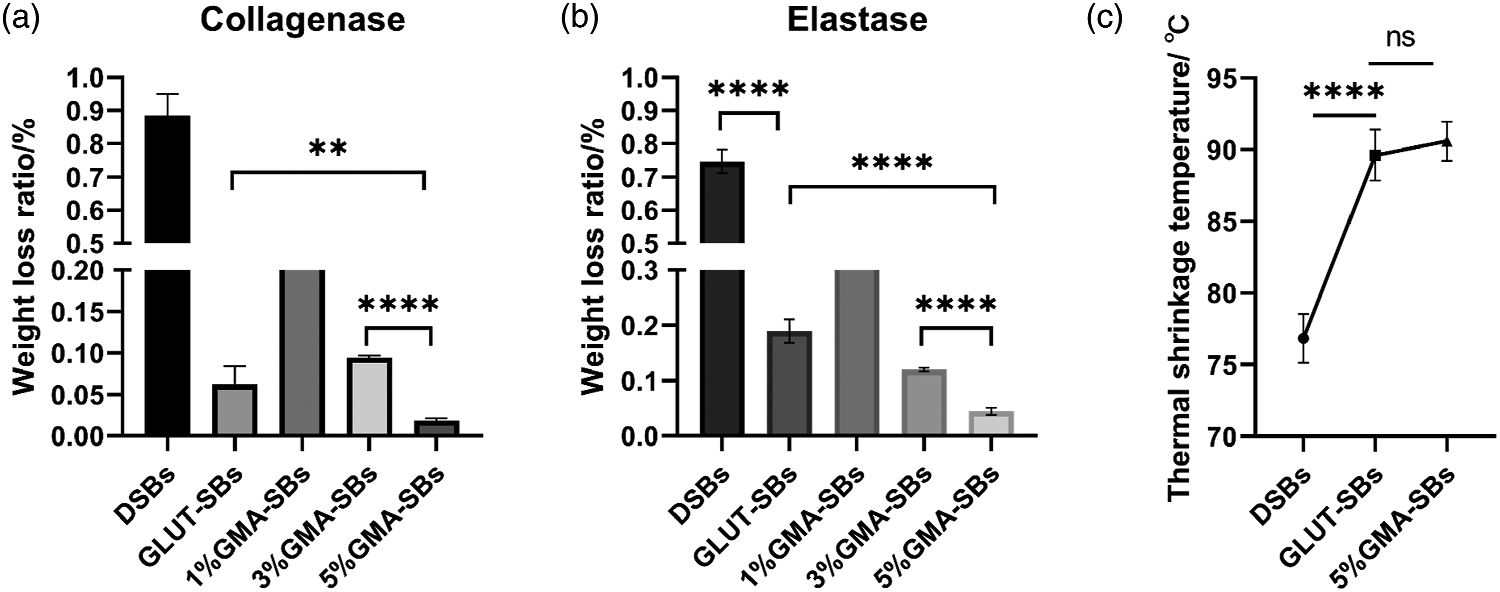
The temperature at which thermal shrinkage of SBs occurred was assessed by DSC to further determine whether GMA could increase collagen stability. The thermal shrinkage temperature of the GMA treatment group (90.60 ± 1.34°) was similar to that of the glutaraldehyde-crosslinking group (89.62 ± 1.78°), and both were significantly higher than that of untreated swim bladders (76.85 ± 1.70°) (Figure 4(c)). This result indicated that radical copolymerization could effectively crosslink the collagen protein and improves the collagen stability.
Biomechanics
As calculated from the stress–strain curve (Figure 5(a) and (b), Table 1), just like native heart valves, the swim bladder mostly revealed higher tensile stress in the circumferential direction rather than the axial direction and the crosslinking treatment had little effect on the tensile strength along the grain direction. In the circumferential direction, the ultimate tensile strength for GMA-crosslinked swim bladders (8.99 ± 0.597 MPa) was much higher than native heart valves (2.6 MPa) and might meet the requirements for bioprosthetic heart valves.
16
Biomechanics of DSBs, GLUT-SBs, and GMA-SBs. (a) Stress–strain curve derived from tensile tests that were conducted in the axial direction. (B) Stress–strain curve derived from tensile tests that were conducted in the circumferential direction. (c) Ultimate tensile strength at break (n = 6). (d) Elongation (n = 6). (e) Secant modulus in the axial direction (n = 6). (f) Secant modulus in the circumferential direction (n = 6). ****p < 0.0001, ***p < 0.001, **p < 0.01, *p < 0.05, ns, no significant difference. Biomechanics of DSBs, GLUT-SBs and GMA-SBs.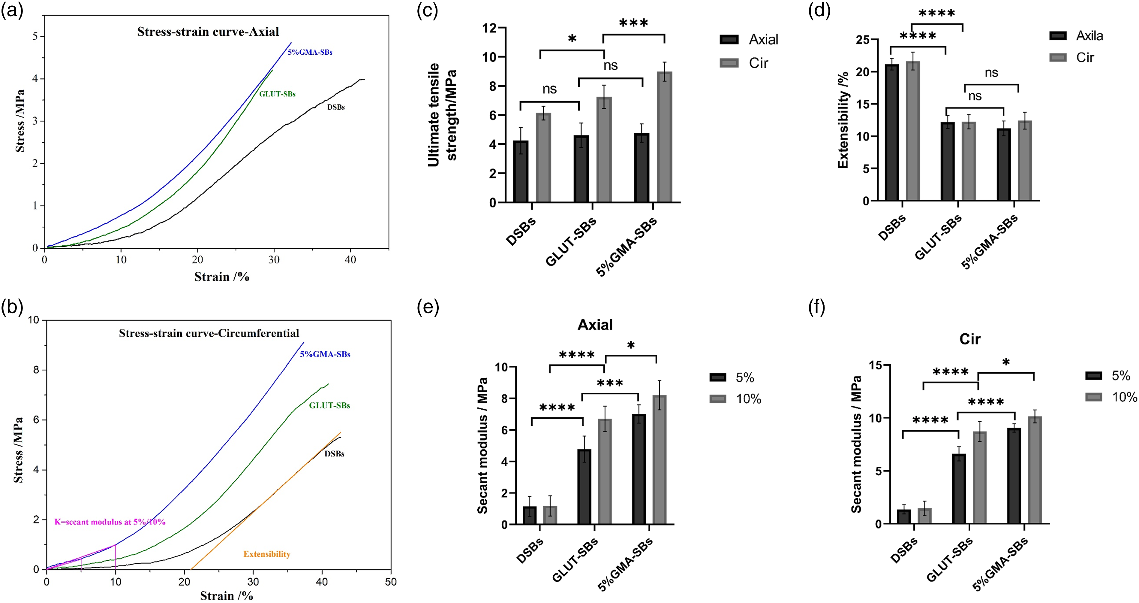
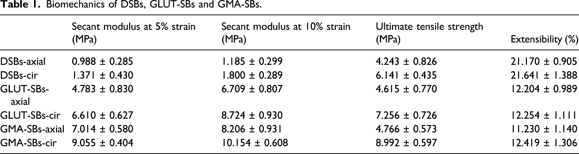
Extensibility was defined as the intercept of a linear fit to the high-modulus region against the strain. The extensibility of samples was between 11% and 32%, and no significant difference was observed between two directions (Figure 5(d)). The secant modulus of GMA-SBs was much higher than that of GLUT-SBs and DSBs at both 10% strain and 15% strain in two directions (Figure 5(e) and (f)). This result indicated that GMA-SBs were slightly stiffer than GLUT-SBs. 17
Cytotoxicity
The compatibility and proliferation of fibroblast L929 cells were evaluated using fluorescein diacetate staining and CCK-8 assay. As shown in Figure 6(a), there were a few L929 cells that grew in the GLUT-SBs group for 1 day and 3 days due to the cytotoxicity of GLUT. However, more cells were observed in the GMA-SBs and CONTROL groups. The CCK-8 quantitative results revealed lower absorbance for the GLUT-SBs group and higher absorbance for the GMA-SBs and CONTROL groups at 1 day and 3 days (Figure 6(b)). Compared to the control groups, the GMA treatment did not affect the cell morphology or the viability of L929 cells. The results suggested that GMA-SBs possessed low cytotoxicity. Cytotoxicity. (a) Live staining of L929 at 1 day and 3 days (scale bar = 100 μm). (b) The viability of L929 cells was determined by CCK-8 assay after culturing with extracts of the GLUT-SBs and GMA-SBs for 1 and 3 days. Normal RPMI 1640 medium was used as a negative control (n = 6). ****p < 0.0001, ***p < 0.001, **p < 0.01, *p < 0.05, ns, no significant difference.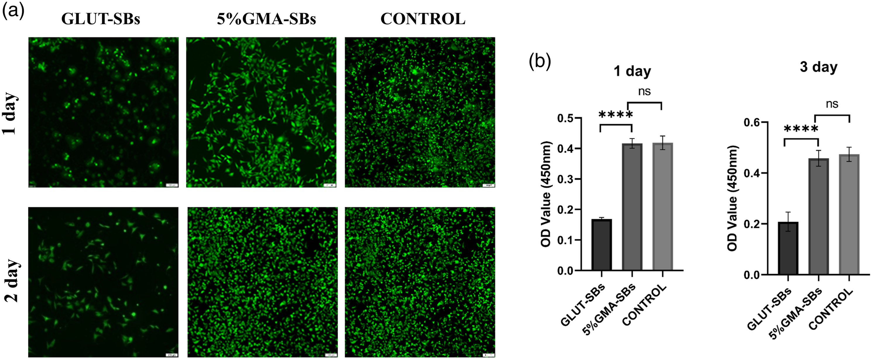
HUVEC growth on SBs
Fluorescence images of HUVECs on SBs also showed a growth pattern consistent with the quantitative results of the CCK-8 assay. As shown in Figure 7, the results indicated that few HUVECs attached and grew in the GLUT-SBs group after incubation for 3 days due to the cytotoxicity of GLUT. However, a greater number of cells was observed in the GMA-SBs and CONTROL groups. Normal expression of actin filaments was observed in the HUVECs growing on the surface of GMA-SBs, suggesting a correct morphological phenotype. These results demonstrated that GMA-SBs were more conducive to the growth of HUVECs compared with GLUT-SBs. HUVEC growth on the surface of GLUT-SBs and GMA-SBs. Normal Dulbecco’s modified Eagle’s medium (DMEM) was used as a control (n = 6). (a) The cells were visualized using fluorescent stains for nuclei (DAPI, blue) and F-actin (TRITC-phalloidin, red) under a fluorescence microscope. (b) Cell viability of different samples of HUVECs at 3 days by CCK-8 assay kit. ****p < 0.0001, ***p < 0.001, **p < 0.01, *p < 0.05, ns, no significant difference.
Whole blood adhesion and hemolysis rate
The adhesion of whole blood on different samples is shown in Figure 8(a). There was a greater amount of adhered blood cells and platelets on the surface of the GLUT group than that of the GMA group. These results indicated that there was more optimal blood compatibility in the GMA group. The hemolysis rate is an important consideration for surgical implantation of blood-contacting devices. The hemolysis rates of both samples were lower than the given safety value (5%) according to ISO 10993-4 (Figure 8(b)). (a) SEM of adhered blood cells and platelets on different SB samples after treatment with whole blood. (b) Hemolysis rates (n = 6). ****p < 0.0001, ***p < 0.001, **p < 0.01, *p < 0.05, ns, no significant difference.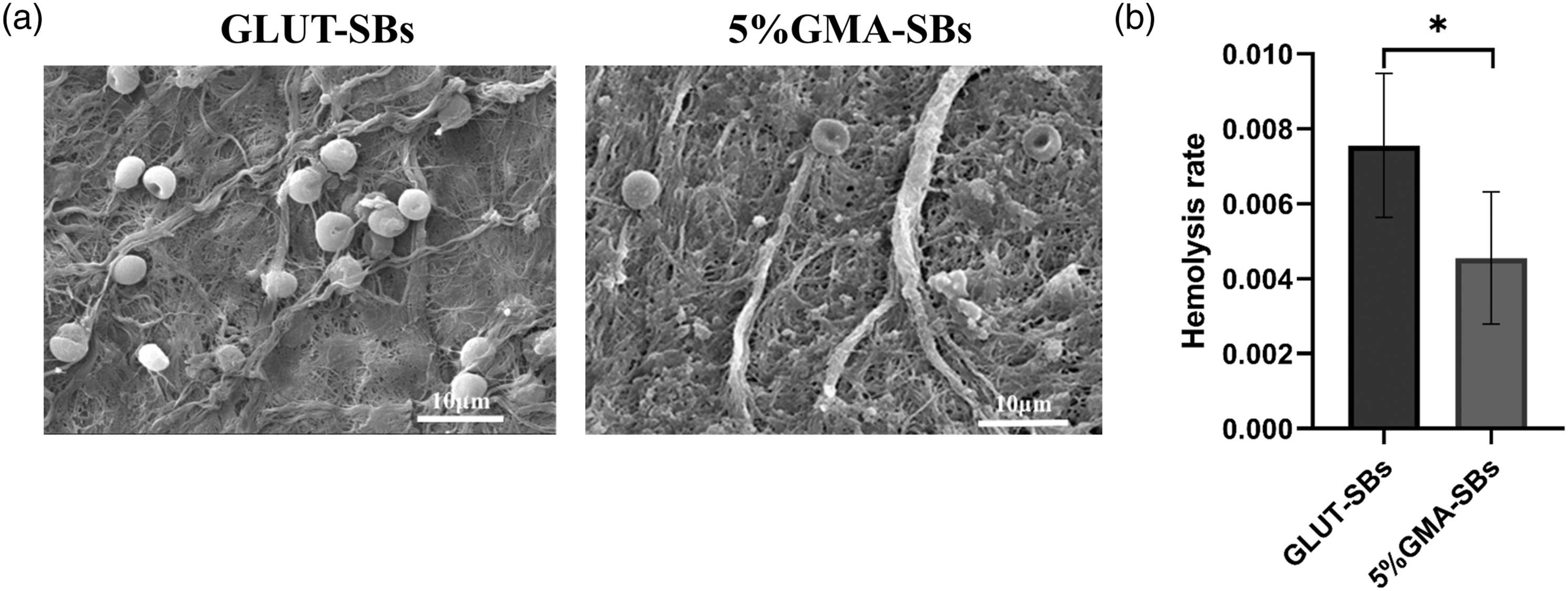
Platelet adhesion tests
In addition, platelet adhesion and activation tests were evaluated in the platelet-rich plasma solution. A significant number of platelets was adhered on the GLUT-SBs surfaces in a highly activated and spread state. However, only a limited number of platelets adhered to the surfaces of the GMA-SBs, and they exhibited a round shape without any extension of pseudopodia (Figure 9(a)). As shown in Figure 9(b), the LDH activity of GMA-SBs was decreased by 35%, as compared to that in the GLUT-SBs. Moreover, there was a clearly lower number of P-selectin (CD62P) positively activated platelets for GMA-SBs as compared to the GLUT-SB group. Only a limited number of platelets adhered to the surfaces of the GMA-SBs, and these remained in a resting, non-activated state (Figure 9(c)). (a) Representative SEM images of platelet adhesion in vitro. The red arrows indicate activated platelets. (b) Relative LDH activity of platelet-rich plasma incubated with GLUT-SBs and GMA-SBs for 1 h (n = 6). (c) CD62p (green) staining showed activated platelets; scale bar is 50 μm. ****p < 0.0001, ***p < 0.001, **p < 0.01, *p < 0.05, ns, no significant difference.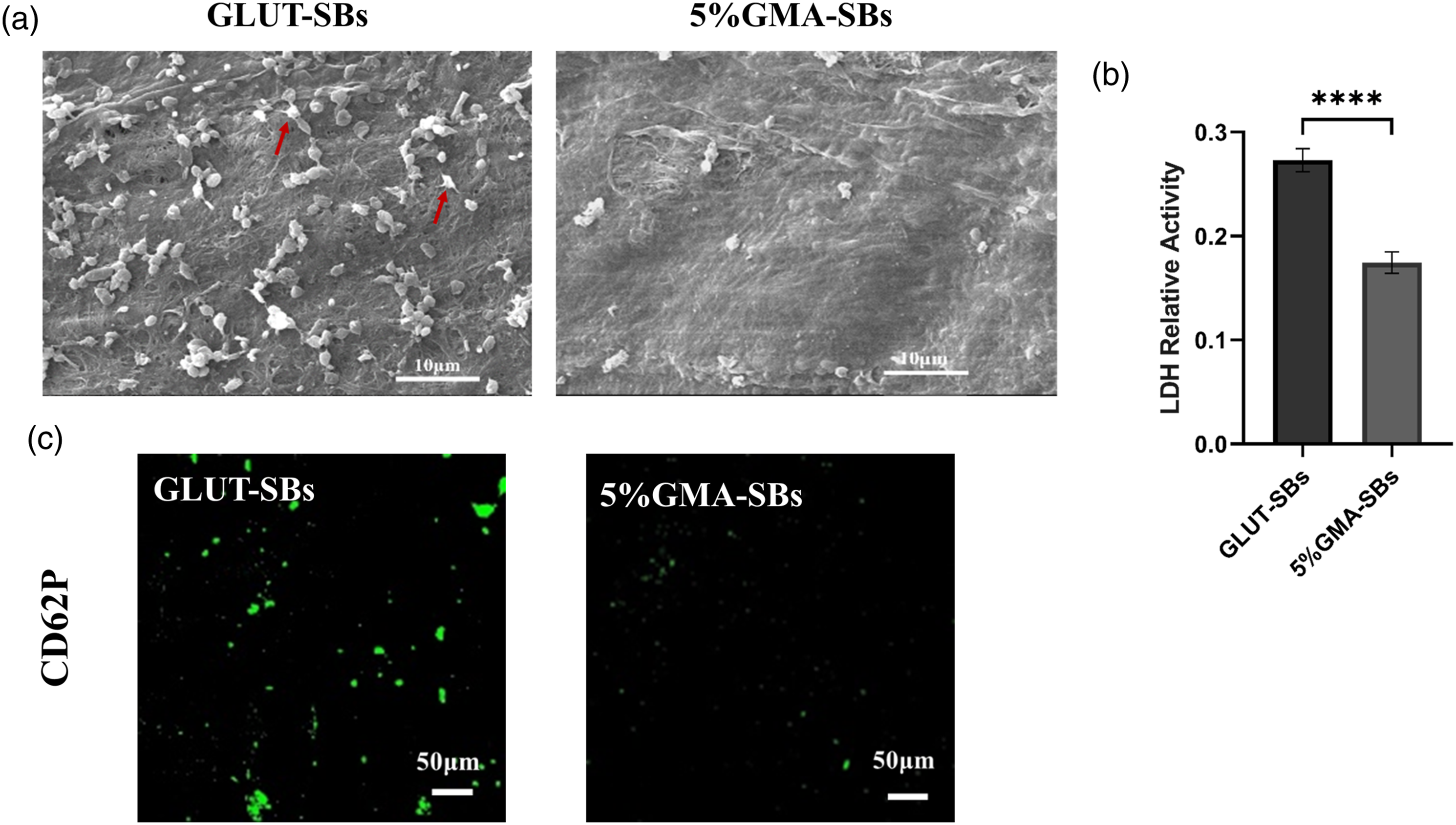
Ex vivo anti-thrombogenicity
The ex vivo arteriovenous shunt assay was used to demonstrate the anti-thrombogenicity of SBs under actual blood flow. After 1 h of ex vivo circulation in the arteriovenous shunts, the circuit of GLUT-SBs exhibited severe occlusion, whereas the GMA-SBs remained unblocked (Figure 10(a) and (b)). Moreover, the surface morphology was observed using SEM, which revealed that significant thrombus formation consisting of activated platelets was found on the GLUT-SB surfaces, as well as erythrocyte deposition. However, only a limited number of non-activated platelets adhered to the GMA-SBs surfaces. Anti-thrombogenicity analysis of the SBs with ex vivo arteriovenous shunt model. (a) Schematic diagram of rabbit arteriovenous shunt model and photos of the duplicate samples after testing. (b) SEM images of GLUT-SBs and GMA-SBs after exposure to blood flow for 1 h.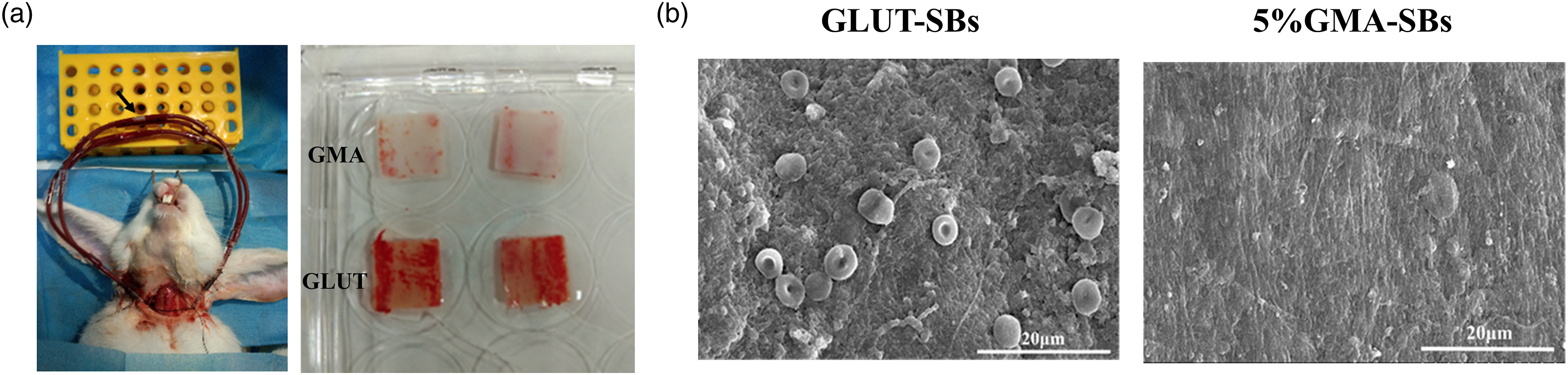
Calcification
The calcification tendency was assessed after 1 and 2 months of subcutaneous implantation of GLUT-SBs and GMA-SBs in rats. Alizarin red staining of implant sections was used to visualize calcium deposition (Figure 11(a)). Large and dense red spots were observed in GLUT-SBs, whereas only a few red spots were observed in GMA-SB sections, and the content of calcium in GLUT-SBs was much higher than that in GMA-SBs (Figure 11(b)). These results showed that GMA-SBs underwent mild calcification and exhibited more optimal anti-calcification properties than GLUT-SBs. Furthermore, to evaluate the degradation of swim bladders after subcutaneous implantation, explants were further analyzed by Masson’s trichrome staining. The results showed that there were more complete collagen fibers in GMA-SBs as compared to GLUT-SBs (Figure 11(c)). The results demonstrated that radical polymerization crosslinked swim bladders had a good protective effect on collagen in vivo. (a) Alizarin red staining. Calcium deposition was stained red (scale bar = 100 μm). (b) Calcium content of different SBs at 1 and 2 months (n = 6). (C) Masson’s trichrome staining. Collagen was stained blue (scale bar = 100 μm). ****p < 0.0001, ***p < 0.001, **p < 0.01, *p < 0.05, ns, no significant difference.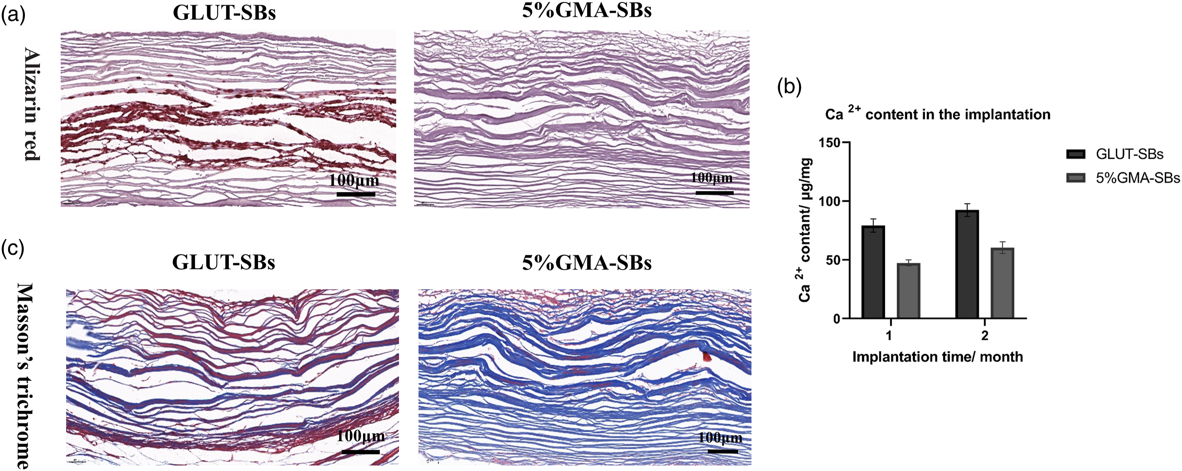
Discussion
It has been reported that less calcification occured in the glutaraldehyde-crosslinked swim bladder as compared to the glutaraldehyde-crosslinked pericardium, and thus, the GLUT-SBs may be an ideal cardiovascular replacement material. 5 However, some drawbacks remain with the glutaraldehyde-crosslinking method, such as high cytotoxicity, coagulation, and calcification.9–11
Owing to the limitations of glutaraldehyde, there have been studies on reducing the toxicity of residual aldehyde groups. Results showed that the amino-oleic acid treatment demonstrated good anti-calcification properties. The amino group in the amino-oleic acid bound covalently to the free aldehyde groups of the glutaraldehyde present in the fixed tissue. 18,19
In addition, developing new crosslinking method is another solution to the calcification problem. Radical polymerization was widely used to prepare functional biomaterials such as hydrogels due to the diversity of monomers and mild reaction conditions. 20 Our lab developed radical polymerization method to crosslink porcine pericardium and showed superior anti-calcification properties. This new method avoided the use of toxic aldehyde groups. 14 Besides, most commonly used crosslinkers including glutaraldehyde failed to enhance elastin stability, which also led to the calcification. 21 But in radical polymerization method, the polymer network and elastin could form hydrogen bonding interactions and improve the stability of elastin. 14
Our laboratory has preliminarily verified the feasibility of GMA-crosslinked pericardium as a biological valve material. Therefore, in this study, GMA radical polymerization was used to crosslink swim bladders, and glutaraldehyde-crosslinked swim bladders were used as the control.
Compared with the GMA-crosslinked porcine pericardium, GMA-crosslinked swim bladder has the following advantages: (1) Studies have shown that fish do not contain many immunogens, such as α-gal, and there is lower immunogenicity associated with biomaterials obtained from fish as compared to mammals.2. In addition, unlike mammalian pathogens, fish pathogens are rarely infectious to humans. 3 Therefore, the swim bladder would be safer than the bovine or porcine pericardium. (2) For the components, the glycosaminoglycans (GAGs) in fish were up to 10% (Figure 2(c)), which mainly included chondroitin sulfate and hyaluronic acid. 22,23 Compared with pericardium, swim bladders had much higher levels of GAGs. 24 Chondroitin sulfate has been reported to have anticoagulant activity due to the α-fucopyranosyl branches and sulfate groups. 25,26 Therefore, the swim bladder might possess better anticoagulant properties than the bovine or porcine pericardium.
The epoxy group of GMA was first reacted with the tissue’s reactive groups such as amine and carboxyl groups, and then crosslinked by radical polymerization to form multi-sites and varying-length connections across the proteins. The result of the amine and carboxyl content indicated that the content of the methacrylate group could be tuned by varying the concentration of GMA (Figure 3(a) and (b)). The creation of links between protein molecules and GMA was further confirmed by FTIR results (Figure 3(c)).
The stability of collagen is directly related to the structural integrity and mechanical function. The collagen stability can be characterized by thermal shrinkage temperature and resistance to collagenase degradation. The thermal shrinkage temperature of GMA-SBs was much higher than DSBs, indicating that the thermal stability of collagen was significantly improved after crosslinking. The collagen weight loss of GMA-SBs with a feed ratio of 5% was significantly lower than DSBs after the exposure to collagenase, even lower than GLUT-SBs (Figure 4(a) and (b)). Furthermore, the degradation of collagen was evaluated in vivo. After 30 days of implantation, explants were analyzed by Masson’s trichrome staining. As shown in Figure 11(c), the collagen structure in GMA-SBs was complete without obvious fracture. Therefore, the stability of the radical polymerization crosslinked swim bladders in vivo for 1 month can be predicted.
To assess the cytotoxicity of GMA-crosslinked swim bladders, the compatibility and proliferation of fibroblast L929 and endothelial cells were evaluated using fluorescein diacetate staining and CCK-8 assay. Compared to the control groups, the GMA-treated groups did not affect cell morphology, and had higher viability of L929 cells (Figure 6(a) and (b)). Endothelial cells can act as an native protective barrier between valves and blood that can prevent thrombosis and immune response. 27,28 In our study, we found that GMA-crosslinked swim bladders promoted endothelial cell proliferation in vitro with spreading morphology (Figure 7(a) and (b)), which suggested that GMA-SBs had the potential for re-endothelialization after being implanted. Sufficient re-endothelialization and adequate cell ingrowth are two crucial factors that govern fast tissue regeneration after implantation in situ, and these factors are positively.
Hemocompatibility is important for blood-contacting devices. To evaluate the blood compatibility of GMA-SBs, whole blood adherence, platelet adhesion and activation, and ex vivo arteriovenous shunt assay were conducted in this study. There was less whole blood adhesion, and less platelet adhesion/activation with GMA-SBs as compared to GLUT-SBs, and the superior anticoagulant ability of GMA-SBs was further verified by the ex vivo arteriovenous shunt assay. The superior anticoagulant effect of GMA-SBs might be due to the high content of chondroitin sulfate (CS) in swim bladder, while the poor blood compatibility of GLUT-SBs may be due to the toxicity of the aldehyde group. CS has been reported to have many bioactivities such as anticoagulation. 29 The anticoagulant activity is attributed to the α-fucopyranosyl branches and sulfate groups, and decreased or increased sulfo-groups in CS may change the anticoagulant activities. 25,26 In summary, these results indicated that GMA-crosslinked swim bladders possessed better anticoagulant properties than GLUT-crosslinked swim bladders.
Aldehyde group residue might be a potential binding site for calcium ion. Most of the non–glutaraldehyde-crosslinking system showed an anti-calcification potential due to the absence of aldehyde group residue. 30 The calcification of GMA-SBs was evaluated by the subcutaneous implantation. After 1 and 2 months of implantation, GMA-SBs underwent mild calcification and exhibited better anti-calcification properties than GLUT-SBs (Figure 11(b)). This result could be attributed that GMA-SBs eliminated toxic aldehyde group.
Conclusions
In this study, we focused on the feasibility of preparing artificial biological valves by means of using GMA-SBs in this study. The GMA-SBs showed high collagen, elastin stability, and thermal shrinkage temperature, ensuring that it could meet most of the criteria about the long-term durability of the implants. Moreover, GMA-SBs had better anticoagulant properties and endothelialization potential compared to GLUT-SBs, in the hope of reducing the risk of thrombosis. Meanwhile, the GMA-SBs showed a significant improvement in anti-calcification potential compared with GLUT-SBs. In conclusion, we provide a non-glutaraldehyde method to crosslink the swim bladders with improved antithrombotic and anticalcification properties compared to GLUT-SBs.
Footnotes
Acknowledgements
This work was supported by the Applied Basic Research of Science and Technology Projects of Sichuan Province (2018JY0538) and National Natural Science Foundation of China (31700833). We also appreciate Wang Hui from the Analytical and Testing Center of Sichuan University for her help with SEM characterization.
Declaration of conflicting interests
The author(s) declared no potential conflicts of interest with respect to the research, authorship, and/or publication of this article.
Funding
The author(s) disclosed receipt of the following financial support for the research, authorship, and/or publication of this article: This study is supported by Applied Basic Research of Science and Technology Projects of Sichuan Province (2018JY0538) and National Natural Science Foundation of China (31700833).