






Abstract
Resistance to apoptosis is a key mechanism underlying how cancer cells evade tumor therapy. Autophagy can prevent anticancer drug-induced apoptosis and promote tumor resistance. The purpose of this study was to improve the sensitivity and efficacy of chemotherapeutic drugs through the inhibition of autophagy. Hydrophobic doxorubicin–hydrazone–caproyl–maleimide (DOX-EMCH) and autophagy-inhibiting si-Beclin1 were simultaneously delivered via the amphiphilic peptide micelle system (Co-PMs) using poly(L-arginine)–poly(L-histidine)–DOX-EMCH as the copolymer building unit. The constructed micelle system promoted the escape of si-Beclin1 from endosomes and the release of DOX into the nucleus. The Co-PMs exhibited 2.7-fold higher cytotoxicity and proapoptotic ability in PC3 cells than DOX treatment alone, demonstrating that si-Beclin1 could inhibit the autophagic activity of prostate cancer (PCa) cells by targeting the type III PI3K pathway and enhance the sensitivity of the cells to the chemotherapeutic drug DOX. In addition, the peptide micelles successfully passively targeted DOX and si-Beclin1 to the tumor tissue. Compared with DOX or si-Beclin1 treatment alone, the Co-PMs showed a 3.4-fold greater tumor inhibitory potential in vivo, indicative of a significant antiproliferative effect. Our results suggested that the Co-PMs developed in this study have the potential to combine autophagy inhibition and chemotherapy in cancer treatment, especially for PCa.
Introduction
The rising incidence of prostate cancer (PCa) represents a serious risk to the health of men worldwide. In 2019, PCa was estimated to be the leading cause of cancer-related death in men in the United States. 1 Endocrine therapy is the mainstay of treatment for advanced PCa; however, most patients gradually regress to a hormone-refractory state after a median duration of 14–30 months and die due to the development of steroid-resistant tumors and distant metastasis.2–4 This highlights the difficulties associated with treating PCa effectively in clinical practice. Currently, doxorubicin (DOX)-based combination therapy is a first-line chemotherapy after complete failure of endocrine therapy5–7; however, the adverse effects of systemic chemotherapy and the development of drug resistance often lead to treatment failure,8–10 with several studies having implicated an abnormal increase in autophagic activity for these effects in PCa cells.11–14
Autophagy is an intracellular self-degradation process important for the regulation of metabolic stress and the maintenance of genome integrity and stability. Autophagy is closely associated with cancer and has a dual role in tumor cells. Although autophagy can suppress tumorigenesis, it can also prevent antitumor drug-induced apoptosis, thereby promoting resistance to treatment.15–17 Several studies have reported that treatment with docetaxel led to a significant upregulation of autophagy in lung cancer, and that autophagy inhibitors could help improve the efficacy of docetaxel.18–20 This suggests that inhibiting autophagy to enhance cancer sensitivity to chemotherapeutic drugs has potential as an alternative treatment for PCa.
The Beclin1 gene (BECN1) is a mammalian-specific homolog of yeast autophagy-related gene 6 (ATG6). Beclin1 regulates other ATG proteins, the localization of autophagy precursors, and autophagic activity mainly by forming a complex with class III PI3K.21–23 Additionally, it has been demonstrated that the upregulation of Beclin1 expression can stimulate autophagy in mammalian cells.24,25 Beclin1 was recently identified as a candidate tumor suppressor gene, with one study reporting that BECN1was monoallelic ally deleted in approximately 40–75% of ovarian, breast, and prostate cancers. 26 Furthermore, Beclin1 expression was significantly increased in A549 cells treated with paclitaxel, which protected the cells from apoptosis; however, the administration of autophagy inhibitors or small interfering RNAs could reverse this effect. 27 We assume that, si-Beclin1 was used to downregulate Beclin1 expression and inhibit autophagic activity in PCa by targeting the class III PI3K pathway which showed that si-Beclin1 could enhance the sensitivity of PCa to the chemotherapeutic drug docetaxel, which further suggests that targeting Beclin1 with siRNAs may represent a novel approach for the treatment of PCa. 28 However, whether the co-delivery of Beclin1 siRNA and the common chemotherapeutic agent DOX to tumor tissue can improve the sensitivity of advanced PCa to this drug remains unknown.
It is necessary to construct a gene-enveloping vector to prevent the disadvantages of siRNA instability, low cell uptake, and rapid clearance, and nano-delivery technology provides a good platform for this. Polymer micelles are a class of well-defined amphiphilic core-shell nanocarriers, which contribute to the encapsulation of hydrophobic chemotherapy drugs and the encapsulation of hydrophilic gene drugs.29,30 In addition, their nanosize allows them to accumulate passively in tumor tissues due to their enhanced permeation and retention (EPR) effect, thereby enhancing their antitumor effects.31,32 Efficient delivery systems can overcome various barriers to the delivery of drugs into the cells to produce anti-tumor effects. Therefore, these delivery systems must be multifunctional and must be long-term stable, specialized, and capable of enhancing endosomal escape. 33
The aim of the present study was to synthesize a pH-sensitive polymer—DOX–H3R6—and use R6 (arginine 6) as an intracellular delivery vector to determine whether they could work synergically to condense RNA through electrostatic interaction.34–36 Histidine can help nanomicelles to escape from endosomes through the proton sponge effect.37,38 DOX-EMCH is a 6-maleimidocaproylhydrazone DOX derivative. DOX-EMCH was conjugated with H3CR6C via the Michael addition reaction. DOX-H3CR6C was cross-linked by cysteine(DOX-HRss). The amide bond could break in the glutathione-reducing conditions present in tumor cells, thereby releasing the drug.39,40 To form the co-delivery nanomicelle (Co-PM), DOX-HRss was assembled into a nanomicelle in which DOX was encapsulated in the hydrophobic core, and si-Beclin1 was condensed on the hydrophilic layer (Scheme 1). The size, zeta potential, cellular uptake, autophagy inhibition potential, cytotoxicity, and proapoptotic ability of the Co-PMs were determined, and the in vivo distribution and antitumor effects were investigated in a PCa xenograft nude mouse model. We hypothesized that the co-delivery of DOX and si-Beclin1 would effectively inhibit autophagy, enhance chemotherapy-related cytotoxicity, promote cell apoptosis, and inhibit the growth of the cancer xenograft, with the hope that the co-delivery system would have the potential to help treat hormone-refractory PCa. The formation of Co-MPs and the in vitro/in vivo effect.
Materials and methods
Materials
All chemicals and organic solvents were of analytical grade. The following chemicals and instruments were used in this study: Doxorubicin hydrochloride, L-arginine, L-histidine hydrochloride, and L-cysteine hydrochloride (Sangon Biotech, Shanghai, China); Beclin1-targeting siRNA (Cell Signaling Technology, Danvers, MA, USA); Cell Counting Kit-8 (Dojindo Molecular Technologies, Tokyo, Japan); lipofectamine 2000(Invitrogen, Carlsbad, CA, USA); mRFP-EGFP-LC3 (Genomeditech, Shanghai, China); PC3 cells (Institute of Biochemistry and Cell Biology, Shanghai, China); Roswell Park Memorial Institute 1640 medium (RMPI 1640), penicillin–streptomycin solution (5 KU/mL), and fetal bovine serum (FBS) (Life Technologies, Grand Island, NE, USA); Gelred (Biotium, San Francisco, CA, USA); Matrigel (BD Biosciences, Sparks Glencoe, MD, USA); Annexin V-FITC/PI Apoptosis Analysis Kit (eBioscience, San Diego, CA, USA); antibodies against Beclin1, P62, LC3I/II, Ki67, and β-actin (Cell Signaling Technology); and an Autophagy Assay Kit (Sigma–Aldrich, Milwaukee, WI, USA).). All animal experiments were approved by the ethics committee of the Second Military Medical University (Shanghai, China).
Cell lines and plasmid transfection
Human PC3 cells (Institute of Biochemistry and Cell Biology, Shanghai, China) were cultured in RPMI 1640 with 10% FBS and 1% penicillin-streptomycin, and incubated under 5% CO2 atmosphere at 37°C.
mRFP-EGFP-LC3 plasmid was from Addgene. Cells were transiently transfected with the plasmid using lipofectamine 2000 (Invitrogen) according to the manufactures’ instructions.
Synthesis of DHRss
First, a histidine–arginine peptide (CH3CR6, HR) was synthesized using the F-moc-solid phase peptide synthesis (SPPS) method. 41 Second, DOX-EMCH was coupled to the terminal carboxyl group of the HR peptide to obtain DOX-HR (DHR) through a condensation reaction. The products were purified by reverse HPLC. Then, the DHR(50 mg) and L-cysteine hydrochloride (0.58 mg) were dissolved in 1.8 mL of distilled water (pH 7.0),0.2 mL of 5% hydrogen peroxide was added dropwise to the mixed solutions with stirring, followed by incubation for 12 h. The mixture was subsequently dialyzed in water for 12 h and freeze-dried for another 24 h to yield DHRss.
Preparation of DHRss polymer micelles (DOX-PMs) and si-Beclin1-loaded HRss polymer micelles (Co-PMs)
The DOX-conjugated DHRss polymer micelles (DOX-PMs) were prepared using probe-based ultrasonication. DHRss (5 mg) was dissolved in 8 mL of distilled water. Then, 2 mL of dichloromethane was injected into the DHRss solution dropwise, followed by ultrasonication at 200W for 1 min in an ice bath using a probe-based sonicator (JY92-IIN, Xinzhi Scientific Co., Ltd, Ningbo, China). The mixed solution was then stirred overnight at room temperature to eliminate the dichloromethane. Finally, an ultrafiltration membrane(MWCO = 3000; Millipore, Massachusetts, USA) was used to remove the monomer in the micellar dispersion (Figure 1). DOX-conjugated and si-Beclin1-loaded DHR polymer micelles (Co-PMs) were prepared by adding an appropriate amount of si-Beclin1 to the DOX-PMs at a molar mixing ratio (N/P)of 40. The Co-PMs were vortexed for 30s and then incubated for 30 min at room temperature before use. The structure, drug loading process and intracellular uptake of poly(L-arginine)-poly(L-histidine)-DOXO-EMCH.
Complex characterization
The particle size and zeta potential of the DOX-PMs (1 mg/mL) and Co-PMs (N/P = 40, 2 μg/mL si-Beclin1) were measured by dynamic light scattering (Zetasizer Nano ZS90, Malvern) at 25°C. The morphology of the Co-PMs was examined using TEM (Hitachi, Japan) at an acceleration voltage of 75 kV.
The condensation ability of the complexes was determined by agarose gel electrophoresis. The complexes were prepared at different N/P ratios (0.25–20). After 30 min of incubation, the complex (1 μg of si-Beclin1) was run at 100 V (30 min) in a 1% agarose gel prepared with Tris-acetate-EDTA (TAE). Complexes with an N/P ratio of 20 were prepared and incubated in 25 nM DTT at 37°C for 2 h. Samples were analyzed by agarose gel electrophoresis under the same above-mentioned conditions. The gel was subsequently photographed using a UV illuminator.
The pH-dependence of the drug release behavior of the Co-PMs was determined using a GloMax-Multi Jr Single Tube Multimode Reader. To examine the pH-dependent dye release, a 10% Co-PM solution was treated with disodium hydrogen phosphate citrate buffer at pH 5.5 or pH 7.4. The amount of DOX-EMCH released at each time point was determined using fluorescence detection.
Cellular uptake assay
Si-Beclin1 and DOX uptake by PC3 cells was analyzed by flow cytometry. PC3 cells were seeded into 12-well plates at 3×10⁵ cells per well and incubated for 24 h at 37°C in 5% CO2. The transfection reagent lipofectamine 2000 was used to protect si-Beclin1 from enzymatic degradation (lip2000/si-Beclin1, si-LIP). To determine the cellular uptake of si-Beclin1 by Co-PMs, FAM-labeled si-Beclin1(FAM-si-Beclin1) was complexed with DOX-PMs at N/P ratios of 10, 20, 40, and 80 to obtain the Co-PMs, followed by incubation for 30 min(100 nM si-Beclin1). For DOX uptake, a DOX-containing solution and DOX-PMs were added to PC3 cells at the final DOX concentrations of 0.5, 1.0, and 2.0 μg/mL. After 4 h of incubation, the cells were washed, trypsinized, centrifuged, resuspended in 300 μL of PBS, and finally analyzed on a FACScan flow cytometer (Becton Dickinson, SanJose, CA, USA). The experiment was repeated 3 times.
For confocal laser scanning microscopy (CLSM), PC3 cells were seeded into glass-bottom 24-well plates at a density of 1×10⁵cells per well and incubated for 24 h. After replacing the culture medium, the si-LIP, free DOX solution, DOX-PMs, and Co-PMs were added to PC3 cells with a final si-Beclin1 concentration of 100 nM and a final DOX concentration of 0.5 μg/mL. After 4 h of incubation, the medium was discarded, the cells were fixed in 4% paraformaldehyde, and the nucleus was stained with 4,6-diamidino-2-phenylindole dihydrochloride (DAPI). The cells were subsequently washed, sealed with mounting medium, and imaged using a CLSM.
Observation of autophagy
Flow cytometry
PC3 cells were seeded into glass-bottom 12-well plates at a density of 3×10⁵ cells per well and incubated for 24 h. After replacing the culture medium, the PC3 cells were incubated with si-LIP, free DOX solution, DOX-PMs, or Co-PMs (si-Beclin1: 100 nM; DOX: 0.5 μg/mL) for 24 h. After discarding the culture medium, the cells were washed twice with PBS and trypsinized. Then, 1 mL of a working solution of the autophagosome detection reagent was added to each well, and the cells were incubated at 37°C with 5% CO2 for 30 min. The cells were subsequently washed three times in wash buffer, resuspended in 300 μL of PBS, and finally analyzed on a FACScan flow cytometer (Becton Dickinson). The autophagosome fluorescence intensity was measured at λex = 360 nm/λ em = 520 nm. The experiment was repeated 3 times in triplicate.
CLSM
PC3 cells were transiently transfected with the mRFP-EGFP-LC3 plasmid using Lipofectamine 2000 (Invitrogen). After a 24 h incubation, PC3 cells were treated with si-LIP, free DOX, DOX-PMs, and CO-PMs for another 24 h. The mRFP-EGFP-LC3 puncta were observed byCLSM (Olympus).The experiment was repeated three times on more than 50 cells. The number of mREF-EGFP-LC3 puncta in each cell was counted.
TEM
PC3 cells were seeded into glass-bottom 6-well plates at a density of 5×10⁵ cells per well and incubated for 24 h. After replacing the culture medium, the PC3 cells were incubated with si-LIP, free DOX-containing solution, DOX-PMs, or Co-PMs (si-Beclin1: 100 nM; DOX: 0.5 μg/mL)for 24 h. The cells were then washed, trypsinized, centrifuged, and resuspended in precooled fixing liquid in 1.5 mL centrifuge tubes, fixed for 4–6 h at 4°C, dehydrated in increasing concentrations of ethanol and acetone, embedded in Araldite, sliced into 5–7 nm thick sections, post-stained with uranyl acetate and lead citrate, and finally examined under a Hitachi H7650 TEM.
Quantitative real-time PCR
The level of Beclin1 mRNA was analyzed by quantitative real-time reverse transcription-PCR (RT-qPCR). PC3 cells were seeded into glass-bottom 6-well plates at a density of 5×10⁵ cells per well and incubated for 24 h. After replacing the culture medium, the cells were further incubated for 24 h with si-LIP, free DOX solution, DOX-PMs, or Co-PMs (si-Beclin1: 100 nM; DOX: 0.5 μg/mL. Total RNA was extracted with Trizol (Invitrogen) following the manufacturer’s instructions. Reverse transcription of total RNA (2 μg) was performed using Transcriptor First Strand cDNA Synthesis Kit (Roche, Switzerland). QRT-PCR analysis was performed using FastStart Universal SYBR Green Master (ROX) (Roche, Switzerland) with the supplied LC3-specific primers. Each qRT-PCR reaction included 40 cycles of 10 min at 95°C, 15 s at 95°C, and 1 min at 60°C.RT-qPCR analysis was performed on an Applied Biosystems 7300(Thermo Fisher Scientific, Waltham, MA, USA). All data were analyzed using GAPDH as an internal standard. Primer sequences used were as follows: LC3 (forward: 5′-GTCCTGGACAAGACCAAGTTTT-3′, reverse: 5′-AGGCGTAGACCATATAGAGGAAG-3′). GAPDH (forward: 5′- ACTTTGGTATCGTGGAAGGACTCAT-3′, reverse: 5′-GTTTTTCTAGACGGCAGGTCAGG-3’).
Cytotoxicity assay
To evaluate the cytotoxicity of DOX and si-Beclin1, a CCK-8 assay was performed. Briefly, PC3 cells were seeded into 96-well plates at a density of 1×10⁴ cells per well and incubated for 24 h. The medium was then replaced with fresh culture medium containing various concentrations of the polymer. Untreated cells were used as a control. After 24 and 48 h of incubation, fresh medium containing a 10% CCK-8 solution was added to each well. The absorbance of each well was measured at 450 nm using a microplate reader (Thermo Fisher Scientific). The absorbance of the untreated cells was set at 100%, and cell viability was expressed as a percentage relative to the absorbance of the untreated cells. The experiment was repeated three times in triplicate.
Cell apoptosis
To determine the effect of Co-PMs on cell apoptosis, PC3 cells were seeded into 12-well plates (3×10⁵ cells/well) and treated with si-LIP, DOX, DOX-PMs, and Co-PMs (0.5 μg/mL DOX and 100 nM si-Beclin1) for 48 h. Untreated cells were used as a control. For the quantitative measurement of apoptosis, cells were harvested, washed twice with ice-cold PBS, and then stained with Annexin V–FITC and propidium iodide (PI) for 15 min at room temperature in the dark. Apoptosis was analyzed by flow cytometry (FACSCalibur; BD Biosciences, UK)
Biodistribution and in vivo antitumor effect
DOX fluorescence was used to assess micelle distribution in vivo. DOX absorbance was measured at 488 nm. A mouse xenograft tumor model was generated by subcutaneous injection of 0.1 mL of a PC3 cell suspension (1×10⁶) into the right axilla of nude mice. The tumors were allowed to grow to approximately 100 mm3 in size before the experiment. To determine the tissue distribution of DOX, 18 male nude mice bearing PC3 cell-derived prostatic cancer were equally and randomly distributed into three groups and injected with saline, DOX or DOX-PMs (5 mg/kg). The mice were euthanized 24 h later and the heart, liver, spleen, lungs, kidneys, and tumors were excised. The excised organs and tumors were washed with cold saline and imaged using the FX Pro in vivo imaging system (Caliper Life Sciences, Hopkinton, MA).
An in vivo antitumor effect assay was carried out as follows: 30 mice bearing visible PC3 cell-derived tumors were equally and randomly distributed into saline, si-LIP, DOX, DOX-PMs, and Co-PMs treatment groups. The mice were intravenously administered the respective formulations daily for three days at a dose of 5 mg/kg DOX and 2 mg/kg si-Beclin1. The body weight and tumor volumes ([major axis]×[minor axis]2/2, measured using calipers) were monitored and recorded twice a week for 21 days. Then, the mice were euthanized and the tumors were excised, weighed, and photographed. Tumor volume (V) was calculated as: V=A×B2/2.
TUNEL and immunohistochemical analysis
Paraffin-embedded tumor tissue sections (5 μm) were subjected to TUNEL staining and immunohistochemistry according to the manufacturers’ instructions. Apoptotic signals in tissue sections were visualized by microscopy. For immunohistochemical analyses of LC3II/I, Ki67, and paxillin, sections were permeabilized and incubated with antibodies targeting LC3, Ki67, and paxillin (Cell Signaling Technology) overnight at 4°C. After washing with PBS, the samples were incubated with horseradish peroxidase (HRP)-conjugated secondary antibody (Cell Signaling Technology). LC3-, Ki67-, and TUNEL-positive cells were imaged with a Nikon E-800 M microscope (Tokyo, Japan).
Histologic analysis
After euthanasia, the hearts, livers, spleens, lungs, and kidneys were collected from the mice, fixed in 4% paraformaldehyde for 24 h, and subsequently embedded in paraffin. Tissue sections (5 μm) were subjected to hematoxylin and eosin (H&E) staining.
In vivo TEM
Autophagy in tumor tissue sections (2–3 mm) was observed by TEM. Briefly, tissues were collected, fixed in a 2% glutaraldehyde solution overnight at 4°C, dehydrated in increasing concentrations of ethanol and acetone, embedded in Araldite, sliced into 5–7 nm-thick sections, post-stained with uranyl acetate and lead citrate, and finally examined under a Hitachi H7650 TEM.
Statistical analysis
All values are presented as the means ± SD. Each value is the mean of at least three repeated experiments in each group. Statistical significance was determined using analysis of variance (ANOVA) and student’s unpaired t-test. Differences were considered significant at p < 0.05.
Results and discussion
Preparation and characterization of DOX-conjugated micelles
The size, zeta potential, drug loading efficiency, and drug encapsulation efficiency of the micelles were investigated. DOX was conjugated into the micelles through hydrophobic interactions. As shown in Figure 2(a) and (b), as the N/P ratio increased, the particle size decreased and the zeta potential increased. At an N/P ratio of 40, the mean particle size was 129.9±2.5 nm, the polydispersity index (PDI) was 0.086±0.820, and the zeta potential was 25.8±1.65 mV. The reasonable size and zeta potential of the Co-PMs renders them suitable for systemic administration as a result of the enhanced permeability and retention (EPR) effect of the delivery system.42,43 TEM analysis showed that the Co-PMs were spherical shape well dispersed (Figure 2(c)). Characterization of the copolymer micelles (Co-PMs). (a) Particle size of the Co-PMs as determined by dynamic light scattering(DLS). (b) The zeta potential of the Co-PMs as determined by DLS. (c) Transmission electron micrographs of the Co-PMs at a molar mixing (N/P) ratio of 40. (d) Agarose gel electrophoresis resultsforCo-PMs. (e) The profiles of doxorubicin (DOX) release from Co-PMs in PBS at pH 5.5 and pH 7.4 at 37°C.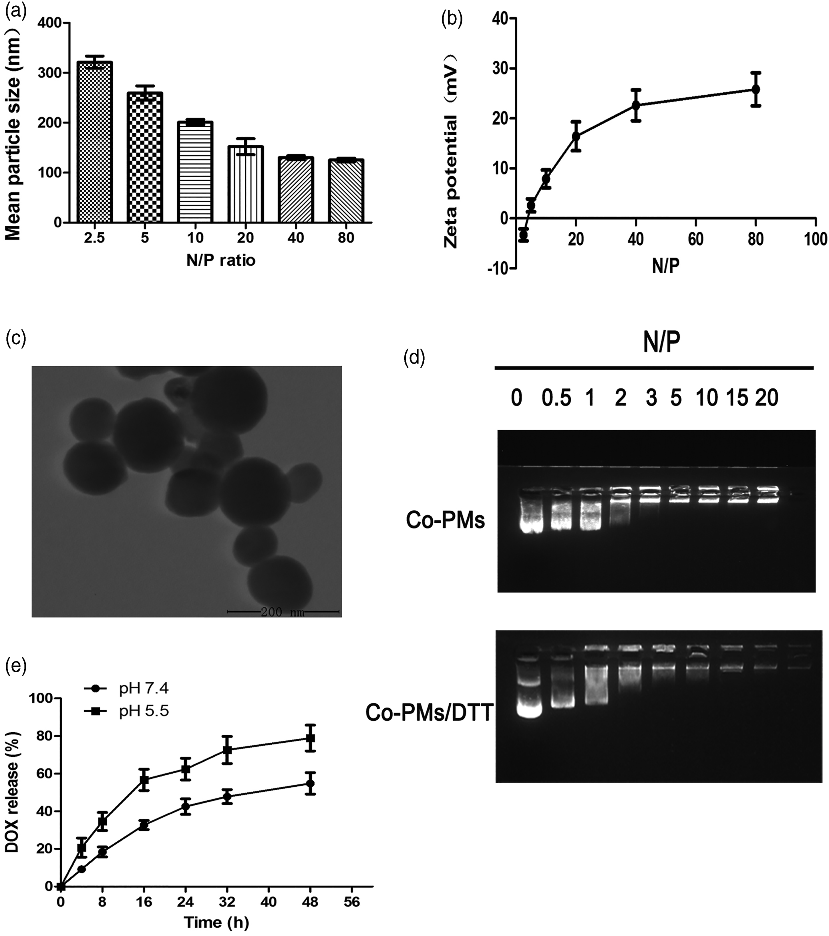
The condensing ability of the complexes was determined by agarose gel electrophoresis. As shown in Figure 2(d), when the N/P ratio was increased from 0 to 20, the condensing ability of DOX-conjugated micelles was gradually enhanced. At N/P ratios greater than 10, si-Beclin1 was completely condensed, indicating that the compression ability of DOX-conjugated micelles was improved by electrostatic interaction. 44 To confirm this finding, DTT, a reducing agent, was used to break this disulfide bond. As shown in Figure 2(d), the Co-PMs showed a weaker si-Beclin1 binding affinity in the presence of DTT, likely as a result of their depolymerization. Based on the above results, we hypothesized that the breaking of the intermolecular disulfide bond would lead to the release of DOX and si-Beclin1 by reducing the condition in the cytoplasm, thus reducing the affinity of the Co-PMs. This reductive-sensitive drug delivery system remains stable extracellularly, but not in the cytoplasm, which can maintain effective release.
DOX release at different pHs
The DOX release profiles of the Co-PMs were investigated at pH 5.5 and pH 7.4 at 37°C. As shown in Figure 2(e), DOX release from the micelles was pH-sensitive. For example, DOX release from the Co-PMs reached 78.9% at pH 5.5, but reached only 54.8% at pH 7.4 (p < 0.01) when observed for 48 h, most likely due to the release of the DOX from the disulfide bond under acidic conditions and the protonation of the histidine within the structure of the micelles at endolysosomal pH (∼5.0).45,46 The proton sponge effect of histidine can destroy the internal structure of micelles, thereby promoting DOX release. 47
Cellular uptake
Because efficient cellular uptake is essential for drug delivery,
48
we labeled si-Beclin1 with a FAM probe and incubated PC3 cells with si-LIP, DOX, DOX-PMs, and Co-PMs for 4 h. FAM-positive cells were quantified by flow cytometry after 4 h of incubation. As shown in Figure 3(a) and (b), the FAM fluorescence signal increased with increasing N/P ratio, and at an N/P ratio of 80, the number of FAM-positive cells was 1.79-fold and 1.32-fold that at the N/P ratios of 20 and 40, respectively (p < 0.05), and was also substantially higher than si-LIP in PC3 cells. DOX uptake in PC3 cells was dose-dependent, and, at the DOX concentrations of 0.5, 1.0, and 2.0 μg/mL, DOX-positive cells accounted for 45.57%, 54.11%, and 88.66% of the total, respectively. However, cellular uptake of DOX-PMs was highest at the lowest DOX concentration. At a DOX concentration of 0.5 μg/mL, the percentage of positive cells was 93.01% (Figure 3(c) and (d)). These results indicated that DOX modified with the membrane-penetrating peptide could more easily enter the cell when encapsulated in micelles. Notably, Co-PMs exhibited optimal uptake efficiency at an N/P ratio of 40. The cellular uptake of doxorubicin (DOX) and FAM-labeled si-Beclin1 (FAM-si-Beclin1). (a) Quantitative analysis of FAM-si-Beclin1 uptake in PC3 cells after treatment with copolymer micelles (Co-PMs) for 4 h at different molar mixing (N/P) ratios. (b) Flow cytometry results for FAM expression in PC3 cells treated with Co-PMs at different N/P ratios. (c) Quantitative analysis of doxorubicin (DOX) uptake in PC3 cells after 4 h of treatment with DOX and DOX-PMs at different DOX concentrations. (d) Flow cytometry results for DOX uptake by PC3 cells after treatment with DOX and DOX-PMs at different DOX concentrations. (e) Confocal microscopic images of PC3 cells incubated with si-LIP (Lipofectamine2000+si-Beclin1), DOX, DOX-PMs, or Co-PMs for 4 h. Green fluorescencere presents FAM-labeled si-Beclin1, red fluorescence represents DOX, and blue fluorescence represents the cell nucleus.Scale bar is 20 μm.
To determine the intercellular location of si-Beclin1, DOX, DOX-PMs, and Co-PMs, PC3 cells were observed under a CLSM. The nuclei were counterstained with DAPI. Figure 3(e) shows the intracellular distribution of the micelles in PC3 cells 4 h after transfection. In PC3 cells incubated with the free DOX, red fluorescence was distributed in the nucleus and cytoplasm. In the DOX-PM group, red fluorescence was also distributed in the nucleus and cytoplasm, and the fluorescence intensity was brighter than that of the free DOX group, indicating that the DOX could enter the nucleus to exert its cytotoxic effect by nanomicelles. In PC3 cells treated with Co-PMs, the red and green fluorescence were uniformly distributed in the nucleus and cytoplasm, indicating that DOX and si-Beclin1 were successfully transported into the cells by the nanomicelles, and that FAM-si-Beclin1 had escaped from the endosome and entered the nucleus.41,49 These results suggested that the co-delivery system could promote endosomal escape and carry the DNA and the chemotherapeutic drug into the nucleus. This finding was in good agreement with the flow cytometry data.
In vitro evaluation of autophagy inhibition
To evaluate the autophagy-inhibiting capability of Co-PMs in PC3 cells, autophagy was monitored by flow cytometry and confocal microscopy. As shown in Figure 4(a), at a DOX concentration of 0.5 μg/mL and a si-Beclin1 concentration of 100 nM, the level of autophagy in cells treated with free DOX was significantly higher than that of cells treated with DOX-PMs or Co-PMs. Si-LIP-containing cells displayed the lowest level of autophagy(0.52%). The percentage of autophagy-positive cells in the Co-PM group was lower than that in the free DOX group (6.77% vs 18.00%; p < 0.05), indicating that si-Beclin1 could be effectively delivered by Co-PMs and inhibit the nanomaterial-mediated autophagy. Autophagy assay. (a) Quantitative analysis of the percentages of autophagy-positive PC3 cells treated for 24 h with si-LIP (Lipofectamine2000+si-Beclin1), doxorubicin (DOX), DOX-polymer micelles (DOX-PMs),and Co-PMs at the DOX concentration of 0.5 μg/mL and a si-Beclin1 concentration of 100 nM. Data are expressed as means ± SD (n = 3). (b) PC3 cells stably expressing mRFP-EGFP-LC3 were treated with si-LIP, DOX, DOX-PMs, and Co-PMs for 24 h. Puncta formation was imaged using a confocal microscope; representative images are shown. The number of EGFP-LC3-positive and REF-LC3-positive puncta per cell was calculated. For counting the number of mRFP-LC3 or EGFR-LC3 puncta in each treatment group, at least 20 cells per experiment were randomly selected. Data are representative of three independent experiments. *p< 0.05, **p < 0.01. (c) Transmission electron micrographs of PC3 cells treated with si-LIP, DOX, DOX-PMs, and Co-PMs (DOX: 0.5 μg/mL; si-Beclin1: 100 nM) for 24 h. Scale bar = 1 μm. (d) RT-qPCR quantification of LC3II/I levels in PC3 cells. *p < 0.05, **p < 0.01.
In addition to autophagosomes, autophagic flux was monitored by observing red and green fluorescent spots in the cells by confocal microscopy. Tandem fluorescent-tagged LC3 (mRFP-EGFP-LC3) was used to monitor autophagic flux based on the different sensitivities of EGFP and mRFP fluorescent proteins to acidic pH. 50 A weakening of the GFP signal and a strengthening of the RFP signal would be indicative oflysosome/autophagosome fusion to form autophagolysosomes. As shown in Figure 4(b), the GFP fluorescence intensity gradually increased in the free DOX group as compared with that in the control group, and punctate red fluorescence distribution was also increased, suggesting that autophagy had been activated in PC3 cells. The red fluorescence intensity in the DOX-PM group was decreased when compared with that of the free DOX group, and was weakest in the Co-PM group, indicating that DOX-enhanced autophagy in the Co-PM group, which contained si-Beclin1, was inhibited.
We also examined the dynamic process of autophagy by TEM. The presence of autophagic vacuoles in the cytoplasm is indicative of the emergence of autophagy. 51 Figure 4(c) shows the cells treated with si-LIP, free DOX, DOX-PMs, and Co-PMs (DOX:0.5 μg/mL; si-Beclin1: 100 nM). In the control and si-LIP groups, the cell structure was clear, the nuclear membrane was smooth, and abundant cytoplasmic organelles could be seen. In contrast, the mitochondrial cristae in the DOX-PM and free DOX groups were ruptured, swollen, and denatured, and more autophagosomes were present when compared with that in the si-LIP and Co-PM groups. This indicated that DOX could induce a greater number of autophagic vacuoles, but low autophagic vacuoles were produced in nanomicelles of DOX.
The results of the RT-qPCR analysis of LC3II/I showed that the LC3II/I expression level in the Co-PMs group was41.3% and 10.3% lower than that in the free DOX and DOX-PM groups, respectively (Figure 4(d)). Compared with the control and si-LIP groups, DOX treatment induced a marked increase in the expression of the autophagy marker protein LC3II. Additionally, the expression of LC3II was weak in the Co-PM group, which indicating that DOX could induce autophagy in PC3 cells, and that DOX/si-Beclin1 co-loading could counteract this effect.
Cell viability assay
We next assessed the cytotoxicity of the different treatments against PC3 cells. Figure 5(a) and (b) show the viability of PC3 cells treated for 24 and 48 h with si-LIP, free DOX, DOX-PMs, or Co-PMs at the DOX concentration range of 0–2 μg/mL and the si-Beclin1 concentration of 100 nM. The viability of PC3 cells was not significantly affected by si-LIP treatment (si-Beclin1 concentration:100 nM), indicating that si-Beclin1 exerts minimal cytotoxicity. However, the proliferation of PC3 cells treated with free DOX, DOX-PMs, or Co-PMs was inhibited in a DOX concentration-dependent manner. The results of the CCK-8 assay demonstrated that the IC50 values for free DOX, DOX-PMs, and Co-PMs against PC3 cells at 48 h were 0.95, 0.70, and 0.45 μg/mL, respectively. The IC50 value for the Co-PMs was approximately 2.1-fold lower than that for free DOX (p < 0.05), indicating that co-delivery of DOX and si-Beclin1 can accelerate intracellular DOX release and improve the endo/lysosomal escape and release of si-Beclin1. This suggests that the Co-PMs constitute an efficient co-delivery system with a good synergy between DOX and si-beclin1 that leads to enhanced DOX cytotoxicity. In vitro antitumor effect of the copolymer micelles (Co-PMs). (a) The viability of PC3 cells after treatment with si-LIP (Lipofectamine2000+si-Beclin1), doxorubicin (DOX), DOX-PMs, and Co-PMs at different DOX concentrations for 24 h. (b) The viability of PC3 cells after treatment with si-LIP, DOX, DOXPMs, and Co-PMs at different DOX concentrations for 48 h. (c) Cell apoptosis assay. Cell apoptosis analysis of PC3 cells treated as above for 48 h was measured by flow cytometry using Annexin V–FITC and propidium iodide (PI) staining (DOX: 0.5 μg/mL; si-Beclin1: 100 nM). Data are expressed as the mean ± SD (n = 3).
Cell apoptosis
Figure 5(c) shows the level of PC3 cell apoptosis in each treatment group. No significant apoptosis was observed in PC3 cells exposed to si-LIP after 48 h of treatment, which was consistent with the cell viability data. Compared with the control group, cell apoptosis was reduced by 19.34% in the DOX-PM group, which was approximately 1.32-fold higher than that in the free DOX treatment group (14.67%).In the Co-PM group, cell apoptosis was reduced by 39.77%, which was approximately 2.0- and 2.7-fold higher than that in the DOX-PM and free DOX groups, respectively. These results suggested that Co-PMs could silence the Beclin1 gene and inhibit DOX-induced autophagy.
In vivo distribution
The investigation of in vivo distribution is essential for the evaluation of the safety and effectiveness of nanomicellar delivery.
52
DOX autofluorescence can be used as an indicator to reduce the interference of animal-related autofluorescence.53,54 To assess the biodistribution of DOX and DOX-PMs, the tumors and major organs were excised from the treated mice at 4, 8, 12, 24-h post-administration and then imaged (Figure 6(a)). In the free DOX group, a fluorescence signal was observed in the liver, lung, kidney, and tumors. In the DOX-PM group, a significant fluorescence signal was observed in the tumor site as a result of the EPR effect. Quantitative analysis indicated that DOX signal intensity in the tumor tissue of mice from the DOX-PM group was 5-fold higher than that of mice from the free DOX treatment group (p < 0.01) and 3.2-fold lower than that in the liver of mice in the free DOX group (p < 0.01). This could be explained by the EPR effect and the avoidance of the reticuloendothelial system (RES) owing to the nanosize of the micelles. Combined, these results indicated that DHRss micelles can efficiently deliver drugs into tumor tissues, likely via the EPR effect.31,55 (a) Ex vivo imaging of tumors and organs collected from mice administered doxorubicin (DOX) or DOX-polymer micelles (DOX-PMs) for 4, 8, 12, and 24 h(DOX: 0.5 μg/mL; si-Beclin1: 100 nM). (b) In vivo evaluation of the efficacy of the Co-PMs through intravenous injection of the micelles into nude mice bearing PC3 cell-derived tumor xenografts. Tumor growth curve for mice treated with phosphate-buffered saline (PBS), si-LIP (Lipofectamine2000+si-Beclin1), DOX, DOX-PMs, or Co-PMs. (c) The weight of the excised tumor tissues from all groups. Data are expressed as means ± SD (n = 6). (d) The body weight of tumor-bearing mice treated with PBS, si-LIP, DOX, DOX-PMs,or Co-PMs. Data are given as the mean±SD (n = 6). (**p < 0.01, vs PBS).
In vivo antitumor effect
The in vivo antitumor effect of the Co-PMs and the effects of DOX and si-Beclin1 were investigated in PC3-derived tumor-bearing nude mice. The tumor volume and body weight of the nude mice were monitored at regular intervals. As shown in Figure 6(b), no inhibitory effect on tumor growth was observed in the normal saline group or the si-LIP group. The tumor volume was smaller and the inhibitory effect greater in the Co-PM group than in the other treatment groups (p < 0.01). On day 21, the tumors were 4.8-, 3.4-, and 2.1-fold smaller in the Co-PM group than in the groups treated with si-LIP, DOX, and DOX-PMs, respectively, thereby confirming the effect of DOX and si-Beclin1 against PC3 cell-derived solid tumors in vivo. A similar result was obtained for the weight of the isolated tumors on day 21(Figure 6(c)) (p < 0.05). This is consistent with the antitumor effects observed in vitro.
The safety of Co-PMs was evaluated by body weight changes before and after DOX administration in the nude mice. 56 As shown in Figure 6(d), the body weight of the nude mice in the free DOX group first increased slowly, and then decreased in the whole process, indicating that DOX-PMs, si-LIP, and Co-PMs did not induce significant systemic toxicity during the experimental period. However, a significant weight loss was observed in the free DOX group due to the systemic toxicity associated with DOX (p < 0.01). 57
TUNEL and immunohistochemical analysis
A TUNEL staining assay was used to determine whether the Co-PMs could induce cancer cell apoptosis, which is a key factor in the inhibition of cell proliferation. As shown in Figure 7(a), stronger brown staining could be observed in the Co-PM treatment group, suggesting that the antiproliferative mechanism underlying the effects of the Co-PMs was associated with the induction of cell apoptosis. The immunohistochemical results showed that Co-PMs significantly inhibited the protein expression of LC3, which was represented by a reduction in the expression of Ki67 (Figure 7(b)), a key marker of cell proliferation. The above results indicated that the co-loaded micelles enhanced the antitumor activity of the chemotherapeutic drug DOX, and that this was related to the inhibition of autophagy in tumor cells. (a) TUNEL staining of tumor tissue (×100). (b) Immunohistochemistry to detect LC3 and Ki67 in tumors. Images were acquired with a Leica microscope at high magnification (×100). (c) The histological characteristics of PC3 tumor tissue and organ histology after treatment with phosphate-buffered saline (PBS), si-LIP (Lipofectamine2000+si-Beclin1), doxorubicin (DOX), DOX-polymer micelles (DOX-PMs),or Co-PMs. (d)Transmission electron micrographs of PC3 cell-derived tumors following treatment with si-Beclin1, DOX, DOX-PMs, or Co-PMs. Scale bar is 1 μm.
Histological analysis
Histology was performed to further evaluate the in vivo safety of the Co-PMs. The H&E staining results (Figure 7(c)) for tissues and organs showed that, in the free DOX group, the myocardial cells were damaged, the myocardial fibers were ruptured, and the intermuscular space was wider. However, no significant organ-related toxicity was observed in the si-LIP, DOX-PM, and Co-PM groups. These results showed that DOX delivered by Co-PMs could markedly reduce cardiotoxicity compared with that observed with free DOX treatment.
Evaluation of in vivo autophagy inhibition
The process of autophagy was observed in vivo by TEM. The presence of autophagic vacuoles in the cytoplasm is indicative of the emergence of autophagy. As shown in Figure 7(d), the number of autophagosomes was significantly higher in the DOX- and DOX-PM-treated groups than in the si-LIP and Co-PM groups. This was consistent with the in vitro cytology results, indicating that Beclin1, a key autophagy inhibitor, could reduce autophagy levels in tumor tissues.
Conclusion
Autophagy is an important mechanism used by cells to adapt to environmental changes, prevent invasion by pathogenic microorganisms, and maintain the stability of the internal environment. Autophagic activity is altered in a variety of human tumors. Autophagy plays a dual role in promoting and inhibiting tumor development. In the case of tumorigenesis, cancer cells confer tolerance to stressors such as an acidic environment, chemotherapy, hypoxia, and nutrient deficiency by utilizing autophagy. Additionally, cancer cells can also activate autophagy in response to various chemotherapeutic drugs, which inhibits cell death and reduces the curative effects of the drugs.20,58
In this study, the PC3 cell line was used as a model to investigate the combined efficacy of chemotherapy and autophagy inhibition by nanomicelle-mediated co-delivery. We synthesized an amphiphilic peptide micelle system for the co-delivery of si-Beclin1 and DOX aiming to silence the Beclin1 gene and thereby suppress DOX-induced autophagy. Our co-delivery system exerted a clear tumor-killing effect. The TEM results showed that the co-delivery system significantly reduced the number of autophagosomes, while CLSM results indicated that DOX and si-Beclin1 had been successfully transported into the cells by the nanomicelles, and that si-Beclin1 had escaped from the endosome and entered the nucleus. In addition, the si-Beclin1/DOX combination significantly induced the apoptosis and necrosis of tumor cells. The in vivo antitumor effects of si-Beclin1 and DOX showed a significant synergistic effect.
Footnotes
Declaration of conflicting interests
The author(s) declared no potential conflicts of interest with respect to the research, authorship, and/or publication of this article.
Funding
The author(s) disclosed receipt of the following financial support for the research, authorship, and/or publication of this article: This research was funded by from the National Natural Science Foundation of China (Grants 81672516, 81672545); the Science and Technology Project of Jiaxing, Zhejiang, China(Grants 2019AY32013); and the Research Fund for Academician Lin He New Medicine(Grants 199331309).
