
Abstract
In this study by considering the advantages of bredigite (Br) and titanium dioxide (TiO2) bioceramics, composite scaffolds of bredigite/titanium dioxide were prepared by the gelcasting method, then, to improve the mechanical, biological and antibacterial properties, scaffolds were coated with chitosan (Ch) polymer phase. The phase structure, fundamental groups, chemical composition, and elemental distribution analysis, morphology and the form of porosity were respectively characterized by XRD, FTIR, EDS, and SEM. Mechanical properties and porosity percentage of scaffolds were also measured by the compressive strength test and Archimedean method, respectively. In order to verify the cell compatibility, MG63 bone marrow cells were cultured on the surface of the specimens. The results showed that the addition of titanium dioxide to the scaffold of bredigite resulted in decrease of porosity and increase of compressive strength of scaffolds from 0.299 to 0.687 MPa. Furthermore, the coated scaffold with chitosan polymer reduced porosity from 83 to 63 percent and a remarkable improvement in compressive strength from 0.585 to 2.339 MPa. The results of the antibacterial test showed that in composite scaffolds, The diameter of the inhibition zone is 22 and 29 mm, in the culture media of Escherichia coli (Gram-negative) and Staphylococcus aureus (Gram-positive), respectively. On the other hand, the results of cell compatibility and cell adhesion tests showed that the scaffolds had no toxicity and the growth, proliferation, and adhesion of MG63 bone cells adjacent to the scaffolds was desirable. Therefore, the scaffold in this study can be used as an ideal scaffold for use in bone tissue engineering.
Introduction
Tissue engineering is a newly emerging field aimed at rebuilding damaged tissue or contributing to the growth of a new tissue using biomaterial, cell, and signaling molecules. In this regard reconstruction of hard tissues such as bones is of particular importance. 1 In bone tissue engineering, a porous and biodegradable scaffold is used to support the cells cultivated in the bone regeneration process and three-dimensional guidance for the formation of new bone tissue. 2 Porous scaffolds in bone tissue engineering play a key role in converting cells to bone and angiogenesis. 3 In this way, a degradable scaffold after implanting in the affected area of the bone begins to degrade after a while, and at the same time, the cultured cells in the scaffold grow and multiply until the new bone tissue is finally replaced. 4 Bredigite (Br) is one of the most well-known silicone biochemicals that has a high potential for the release of silicon ions, which results in the growth of osteoblasts and cellular differentiation. Ionic products resulting from the dissolution of bredigite promote cell proliferation and show good biocompatibility that highlights the use of these materials in tissue engineering applications.5–7 Titanium dioxide in nature has three crystalline phases, including rutile, anatase, and bruxite. According to studies, the anatase phase crystallizes in a column and thus results in more adhesion of bone cells, which makes it suitable for use in bone tissue engineering. Titanium dioxide ceramic is widely used in tissue engineering due to its mechanical properties, corrosion resistance, hydrophilicity, and proper chemical properties, antibacterial properties, and non-toxicity.8–10
The natural polymer of chitosan has been used extensively in the field of tissue engineering due to its biocompatibility, biodegradability, non-toxicity and antibacterial properties.11,12
There are various techniques for constructing engineering scaffolds. In this study, the method of gelcasting was used to construct scaffolds. In this method, the ceramic slurry containing the gelling agent and the foaming agent is poured into the mold after intense stirring and bubbling. Due to the gelation of the slurry at a given temperature, the ceramic particles are stacked together and the bubbles inside the structure stabilized, and the sample is molded. The samples after leaving the mold are dried and sintered. 13 The gelcasting method has advantages, including, construction of scaffolds with high mechanical strength, production of complex shapes and uniform porous structure of produced scaffolds. Considering the advantages of ceramic matrix scaffolds and the similarity of these scaffolds with natural bones, as well as the advantages of the gelcasting method, the aim of this study was the construction of a composite scaffold of bredigite/titanium dioxide by gelcasting. Finally, to use antibacterial properties of chitosan, as well as improving mechanical properties, scaffolds were coated with chitosan polymer phase. The scaffolds were evaluated for structural, antibacterial, bioactivity and biocompatibility properties. Then the effect of chitosan coating on mechanical properties, pore size, and porosity of the scaffolds was evaluated.
Materials and methods
Materials
The chemicals used in this study are including, tetraethylorthosilicate (TEOS, Merck), calcium nitrate tetrahydrate (Ca(NO3)2.4H2O, Merck), magnesium nitrate hexahydrate (Mg (NO3)2.6H2O, Merck), nitric acid (HNO3, Merck), acetic acid (Arman Sina CH3COOH,), titanium dioxide (TiO2, Merck), chitosan (C8H13NO5, Sigma), agarose powder (Merck), Tergitol (Aldrich), ethanol (C2H5OH, Merck), and deionized water.
Preparation of bredigite ceramic powder
In order to produce bredigite powder by sol-gel method, certain amounts of TEOS were dissolved in water and 2 N nitric acid. Then Ca (NO3)2.4H2O was added to the solution, After complete dissolution Mg (NO3)2.6H2O was added to the solution and stirred for a period of 24 hours by a magnetic stirrer at 60°C. Finally, the gel was dried in the oven. For calcination, the powder was poured into a dish, then placed in an electric furnace at 1150°C for two hours at a heating rate of 5°C. In order to produce bredigite powder, it was ball-milled by zirconia cup and balls with ball to powder ratio of 5, for 1 hour.
Synthesis of ceramic scaffold by gelcasting method
The following steps were carried out to make ceramic scaffold by gelcasting method:
In order to produce ceramic slurry, the amount of 0.05 g of sodium polyacrylate (dispersant agent to prevent slurry settling) was dissolved in 4 ml of distilled water. After complete dissolution, the amount of 4.67 g of bredigite powder (or Br powder with 15 wt% of TiO2) was added gradually to the above solution and stirred until uniformly mixed with the magnetic stirrer at the ambient temperature. At the same time, as ceramic slurry production, 0.135 g of powdered agarose (as a gelling agent) was dissolved in 2 ml of water at 90°C. After complete dissolution, the solution was added to the ceramic slurry at high temperature and stirred for 5 minutes with a magnetic stirrer for uniformity. In the end, a value of 0.5 ml of Tergitol (as a surface activator) was added to the slurry and bubbled by a vigorous mixing by a mechanical stirrer. The slurry was then poured into the mold and placed in a freezer at a temperature below 0°C to stabilize the resulting bubble gel agent gelatinization. The prepared scaffolds after leaving the mold and drying at the ambient temperature were sintered in a furnace at a temperature of 1200°C for 3 hours.
Scaffold coating with the chitosan polymer solution
In order to cover the scaffolds with the chitosan polymer solution, 1.5 g of medium-molecular weight chitosan powder was dissolved in 1 ml of acetic acid and 99 ml of distilled water. Then 2 ml of 2% glutaraldehyde as cross linker was added to 100 ml of chitosan solution drop wise and the solution was placed on a magnetic stirrer for 2 hours at room temperature. The produced scaffolds were immersed in chitosan solution under vacuum to allow the solution to penetrate the porosity of the scaffolds. The scaffolds were dried under vacuum after leaving the solution and removing excess solution.
The materials characterization
X-ray diffraction analysis (XRD) was used to investigate and identify the phases of the synthesized bredigite powder and bredigite/titanium dioxide composite scaffold. For XRD analysis, CuKα radiation was used. The applied voltage was 30 kV, the current was 30 mA, the wavelength was 1.5406 angstrom, and the angle of diffraction (2θ) was selected from 10 to 90 degrees with a step size of 0.05. Fourier transform infrared spectroscopy (FTIR) test was used to investigate the bonds in powder and composite scaffolds and to determine the functional groups and chemical compositions.
Scanning electron microscopy (SEM) was used to study the pore size distribution, pore morphology, and pore interconnectivity of the scaffolds. SEM was also used for measuring the thickness of the chitosan coating on the scaffold and investigating the stability and durability of the coating after different immersion times in simulated body fluid (SBF) solution. Energy dispersive X-ray Spectroscopy (EDS) was used to identify chemical compositions and to examine the uniform distribution of TiO2 in composite scaffolds. To calculate the porosity of scaffolds, the fluid displacement method (Archimedes law) was used. Compressive strength test was performed for all samples synthesized with a diameter of 15 mm and a height of 20 mm, by a Universal Testing Machine (HOUNSFIELD: H50KS), at a strain rate of 0.5 mm/min.
Antibacterial properties of scaffolds on two types of Escherichia coli bacteria (Gram-negative) and Staphylococcus aureus bacteria (Gram-positive mass) were studied. In order to evaluate the cellular behavior of the scaffolds, the MG63 osteoblast cells on the surface of pure bredigite scaffolds (Br), bredigite scaffolds with chitosan coating (Br-Ch), as well as composite scaffolds of bredigite/titanium dioxide with chitosan coating (Br-15% TiO2-Ch) were cultured for 5 days. The survival, growth, and proliferation of cells were measured using MTT test with three repetitions. Also, cell adhesion after cell culture on scaffolds was evaluated by scanning electron microscopy.
Results and discussion
The X-ray diffraction pattern of bredigite powder, titanium dioxide powder and Br-15% TiO2 composite scaffold is presented in (Figure 1). According to phase evaluation conducted with Expert Highscore software, It was found that the characteristic peaks of the Br powder are at the 26/585°, 31/215°, 32/716°, 33/655°, 40/474°, 43/607°, 47/293°, 59/305°, and 70/441° angle corresponds to the standard card number for X-ray diffraction of Br ceramic (JCDPS Card No.036-0399).
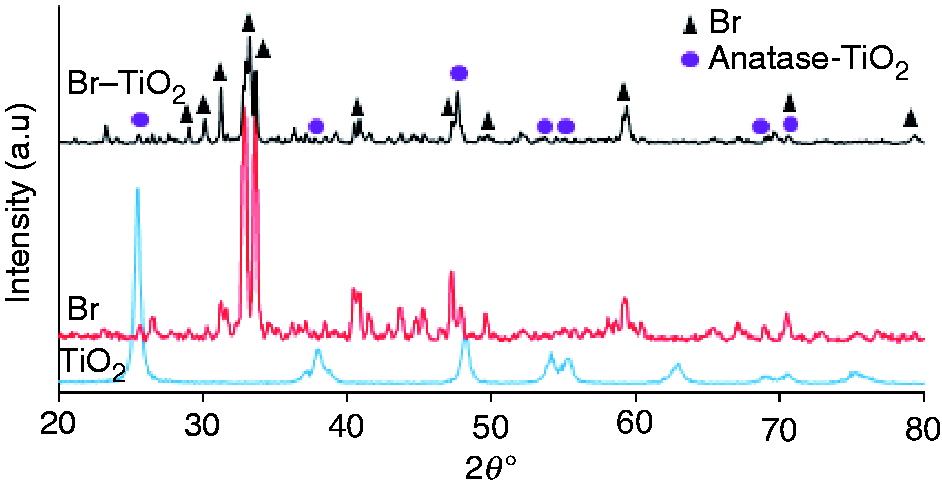
X-ray diffraction pattern of titanium dioxide powder, bredigite powder and Br/TiO2 composite scaffold.
Investigation of TiO2 X-ray diffraction pattern showed that the phase structure of the TiO2 is anatase and the peaks formed at 25.5°, 37.96°, 48.20°, 54.143°, and 62.87°, are very consistent with the diffraction pattern of anatase phase with card number of (JCPDS Card No. 21-1272). As can be seen, the major peaks of TiO2 and Br are available in the XRD spectrum of composite scaffold, which indicates the presence of bredigite and titanium dioxide in the structure of composite scaffolds.
The FTIR spectra of chitosan powder and bredigite/titanium dioxide composite scaffold with and without chitosan coating in 400–4000 cm−1 were shown in Figure 2. In the FTIR spectrum of chitosan (Figure 2), the absorption bands range from 1000 to 1150 due to the presence of vibrations of C–O–C, CH–OH, C–C, and N–H in the chitosan structure. The observed absorption peak at 1650 cm−1 belongs to the C=O bond of amide. A broad absorption peak in the region of 3200–3500 cm−1 is related to the stretching vibration of the O–H bonds. The FTIR spectrum of two-component bredigite/titanium dioxide scaffold without chitosan shows the absorption peaks of Si-O bond in SiO4 tetrahedral, indicating the presence of bredigite. The absorption bands of the bredigite characteristic are in the region of 890, 980 and 1018 cm−1, which are related to the stretching vibration of SiO4, and the stretching vibration associated with the Ca–O and Ti–O–Ti bonds are in 455 and 571 cm−1, respectively. The broad-adsorption peak at 3423 cm−1 is related to O–H groups on the surface of TiO2 nanoparticles. The spectrum obtained from the composite scaffold with chitosan coating shows that, in addition to the presence of the peaks relative to Br and TiO2, the increase in absorption peak in the 3400 cm−1 area, which is not observed in the uncoated scaffold spectrum, is related to the presence of chitosan in the structure. Also, an increase in the absorption peak in the 1629 cm−1 region resulting from the C=O-amide bond confirms the presence of chitosan in the scaffold. A strong absorption peak in the region of 571 cm−1 is related to the Ti-O-Ti bond vibration.14–16
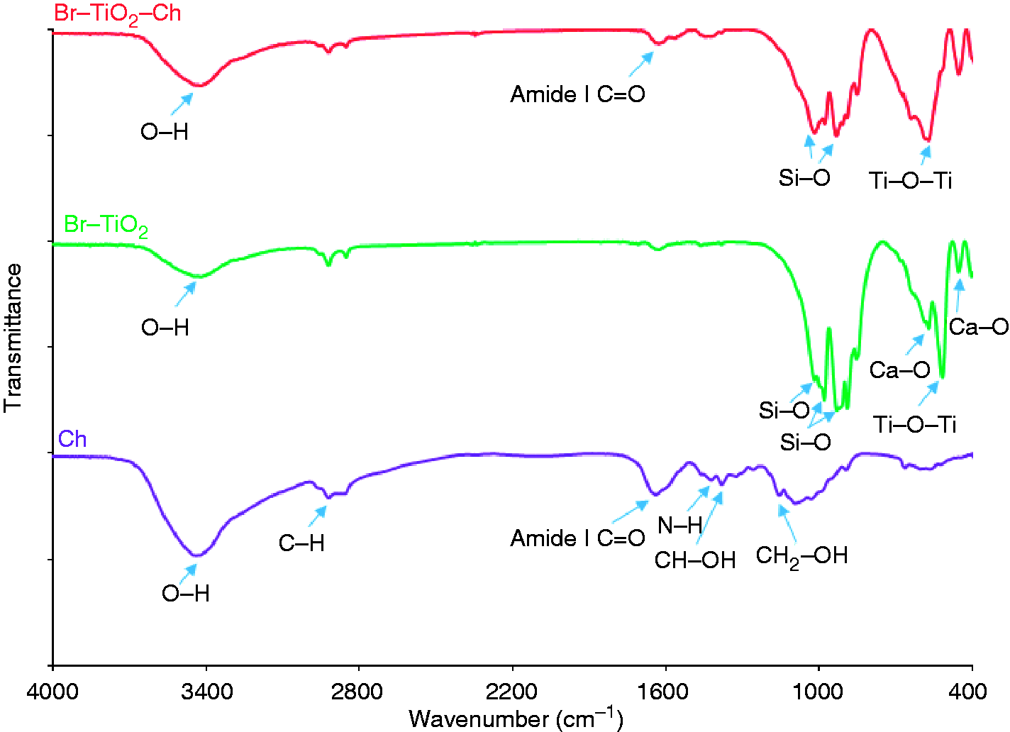
The FTIR spectra of chitosan powder and bredigite/titanium dioxide composite scaffold with and without chitosan coating.
The EDS spectrum and its results are presented in in (Figure 3). As can be seen, the calcium, magnesium, silicon, and oxygen peaks indicating the presence of Ca7MgSi4O16 bredigite ceramic . Furthermore, The titanium and oxygen peaks, indicating the presence of TiO2 in the synthesized scaffold.
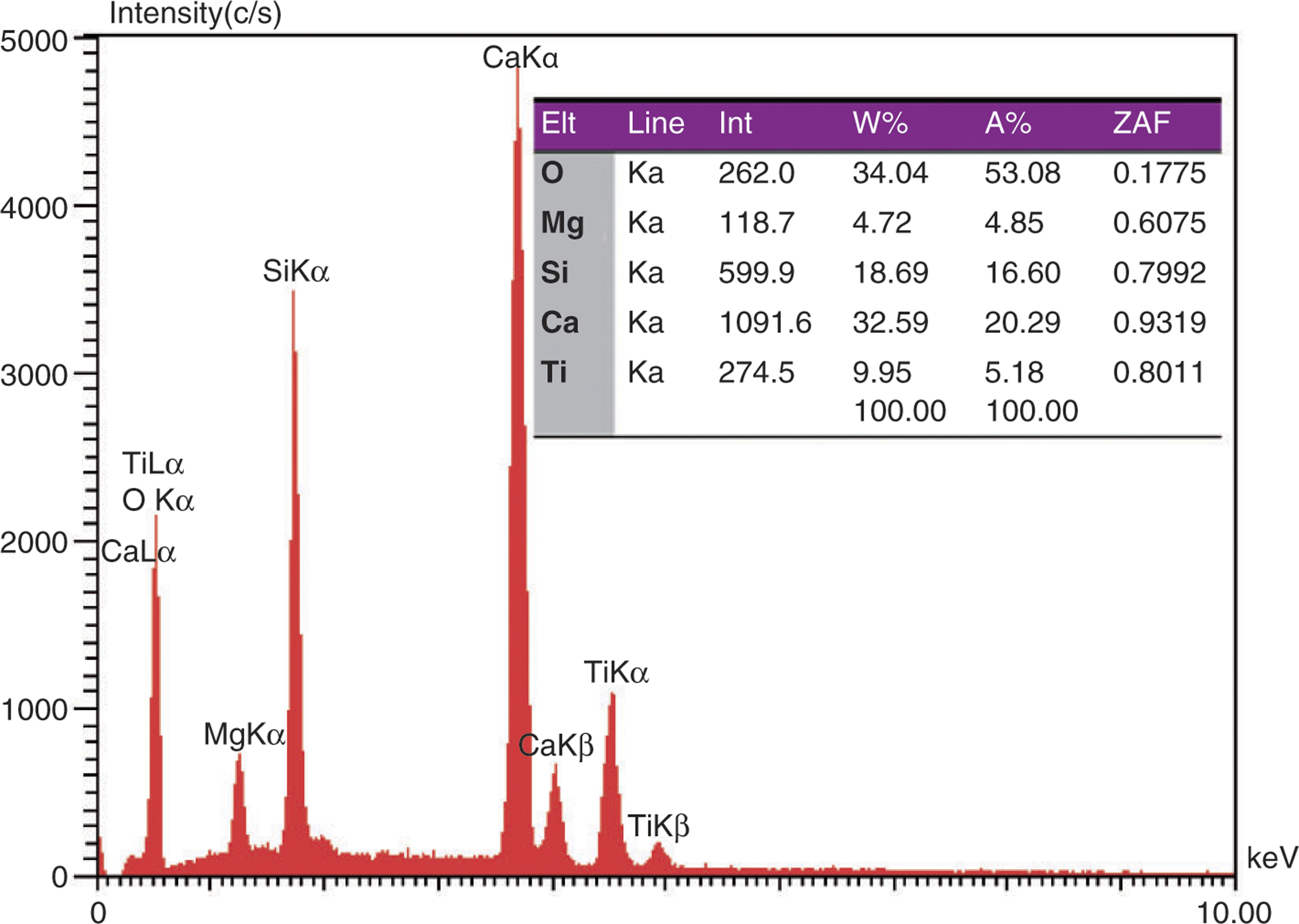
The EDS spectrum and quantitative results from the EDS spectrum (representing the weight and atomic percent of the constituent elements) of the bredigite/titanium dioxide composite scaffold.
To evaluate the uniform distribution of titanium dioxide particles in composite scaffolds, MAP analysis was performed. FE-SEM image and images from the MAP analysis of the Br-15% TiO2 composite scaffold, which represent the frequency distribution of the constructive elements of the scaffold, are shown in (Figure 4). According to the images, elements of calcium, magnesium, and silicon are related to bredigite, and titanium element is related to titanium dioxide particles, which is distributed uniformly throughout the image and represents the uniform distribution of TiO2 particles in the scaffold matrix.

(a and b) FE-SEM and (c to f) MAP images obtained from a cross-section of the composite scaffold representing the distribution of calcium, magnesium, silicon, and titanium.
To identify the scaffold surface morphology and porosity checking, SEM images were taken from composite scaffolds before and after applying chitosan coating. SEM images of composite scaffolds before and after applying the chitosan coating are shown in Figure 5.
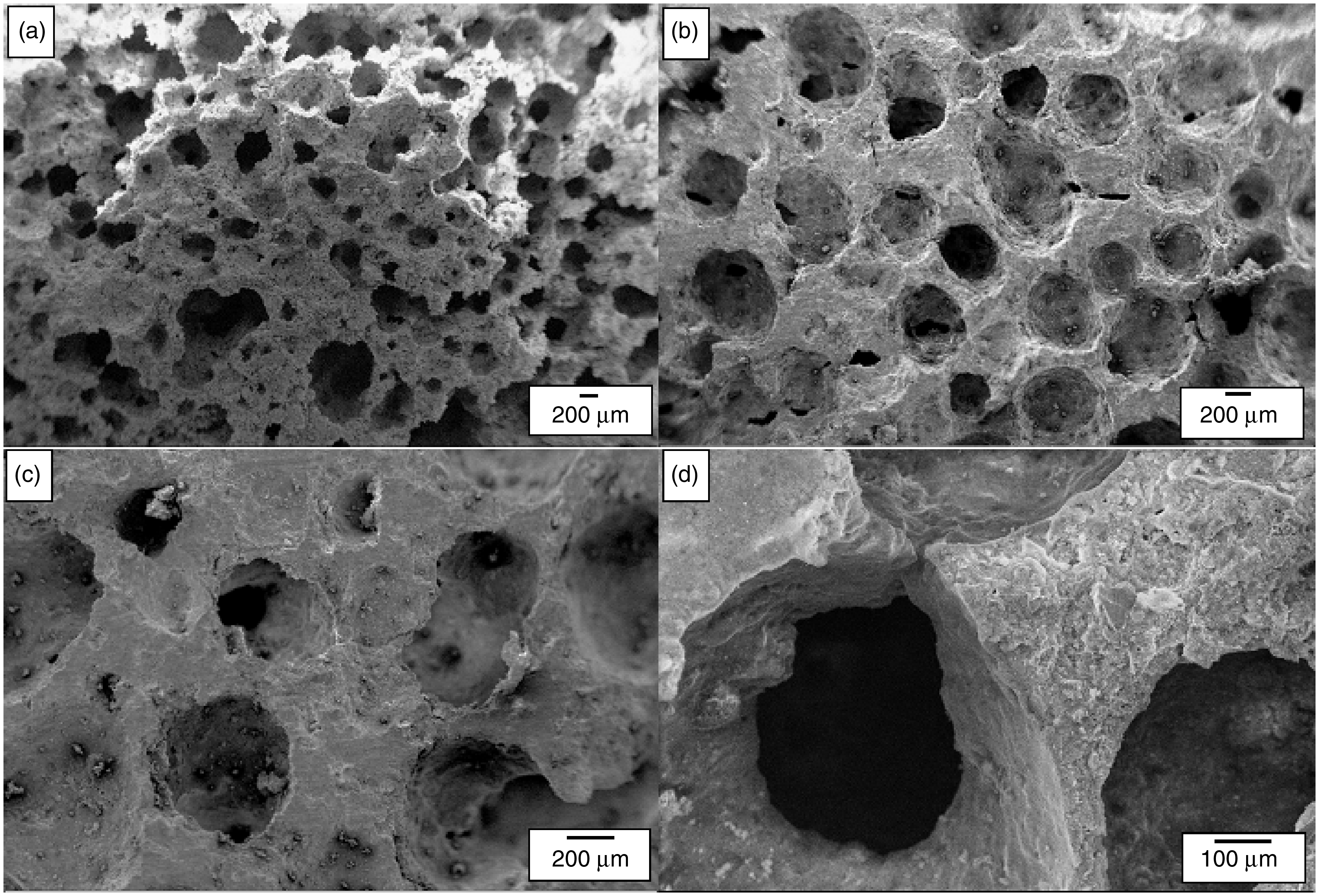
SEM images of composite scaffold (a and b) before and (c and d) after coating with chitosan.
The suitable pore size for use in bone tissue engineering is recommended between 200 and 600 μm. In general, the size of the pores greater than 300 micrometers, improves the bone formation and develop the capillary network. Furthermore, the presence of porosity and the interconnection between them leads to the penetration of the cell into the pores, tissue ingrowth, vascular formation and the arrival of protein substances to the tissue which is being restored. 17
The obtained images of the prepared scaffolds represent high porosity with regular pores, suitable spherical morphology and the existence of communication paths between the pores. The porosity measurements on the scaffolds were carried out by the Digimizer software in the range of 150 to 600 μm. The average porosity of the scaffolds was calculated as 375 ± 133 micrometers. The scanning electron microscopy of the scaffolds after applying the chitosan polymer coating showed that the porosity morphology did not change significantly compared to the images taken from the scaffolds before applying the chitosan coating. According to the results, the interconnection between pores after the coating is preserved, and the addition of titanium dioxide does not significantly change the size of the pores. The coating of chitosan polymer in the scaffolds only created a thin layer of polymeric phase in the porosities wall, and some of the micro-porosity was filled in the scaffold, and full pore filling was rarely seen. Figure 6(a) and (b) shows the cross-section SEM images of the pore wall of the scaffold showing the thickness of chitosan coating layers. As can be seen from the figure, the thickness of the chitosan coatings was below 20 µm. In order to evaluate the stability of the chitosan coatings, the coated composite scaffolds were immersed in the simulated body fluid (SBF) solution at 37°C for 28 days. Figure 6(c) shows the presence of coatings on the surface of the scaffold. Also, the presence of coatings on parts of the scaffold surface can still be seen after 28 days of immersion. It should be noted that the chitosan polymer used, Due to high degradability after placing inside the body reopens the closed pores again. Due to the suitable size of the porosities and the internal connection of porosity with each other, before and after coating, it can be said that scaffolds synthesized with gelcasting method, have the characteristics of an ideal scaffold for bone tissue engineering to a large extent.
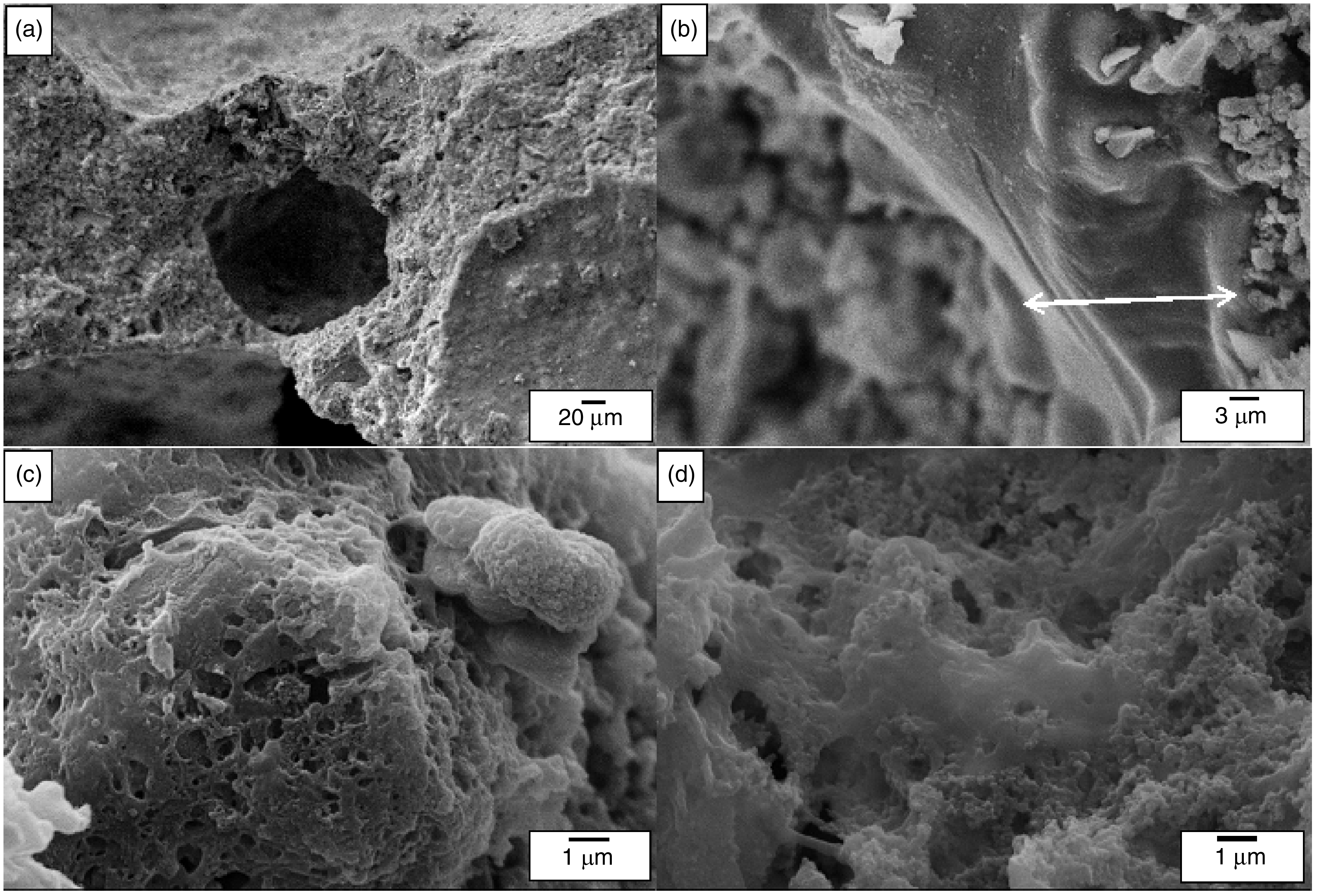
SEM images of chitosan coating on composite scaffold (a and b) Cross-section images of pore wall of scaffold showing the thickness of coating layers (c), after 14 days, and (d) after 28 days of immersion in SBF solution.
Results of porosity and compressive strength of scaffolds along with the standard deviations from three repetitions before and after applying the chitosan coating is presented in (Table 1). The average porosity percentage of the scaffold was calculated in the range of 63–87%. The coating of scaffolds with chitosan polymer has reduced porosity, which indicates the closure of the scaffold pores by chitosan polymer. The addition of TiO2 also increases the slurry concentration and reduces the porosity of the scaffold in a small amount compared with the single-component bredigite scaffold.
The average porosity and compressive strength values of synthesized scaffolds before and after applying chitosan coating.
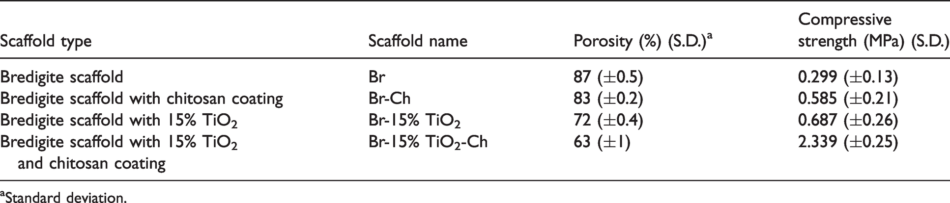
aStandard deviation.
According to the results of the compressive strength test (Table 1), the compressive strength of the scaffold was increased after applying the chitosan coating. Also, by adding TiO2 to the pure Br scaffold, the compressive strength of the scaffold increased, so that the Br-15% TiO2 scaffold had strength of about 2.3 times higher than that of the Br scaffold. After coating the composite scaffold with chitosan polymer, the compressive strength of the scaffolds increased substantially. The highest compressive strength is related to the composite scaffold with chitosan coating, which is 2.339 MPa and has a strength of about 8 times higher than the strength of the pure bredigite scaffold without chitosan coating. According to studies, the compressive strength for cancellous bone is in the range of 2–12 MPa. 18 The compressive strength for composite scaffolds synthesized in this study is also within the range of compressive strength of spongy bone that is suitable for use in bone tissue engineering.
In a study, the composite scaffolds of nano-bioglass-poly-3-hydroxybutyrate (nBG/P3HB) with 3, 6 and 9 wt% of titanium dioxide synthesized by polyurethane foam replication method. The results showed that the addition of titanium dioxide resulted in a reduction of porosity of scaffolds and, consequently, increasing the compressive strength of the scaffold. According to the results of this study, the addition of different TiO2 percentages in general does not differ significantly in the structural form of the scaffolds, and modifies the porosity of the scaffold in a modest way, which is an advantage. 19 The electrostatic interaction between bredigite (Mg+2) band=O sites of chitosan as well as hydrogen bonding provided by OH groups of TiO2 leads to great bonding between the bredigite/TiO2 scaffold (substrate) and chitosan (coated layer). This is supported by the EDS results that show the presence of Mg in the bredigite. Also, it was reported 20 that it is clear that the addition of TiO2 further reduces the movement of the chitosan chain which may result in higher bonding strength between the coating layer and scaffold as a substrate. Similarly, it is suggested that 21 most of the positive effects such as mechanical (tensile strength and compressive strength) increased via the encapsulation of TiO2 on the starch coatings/films were related to the strong interfacial interaction between TiO2 and the amorphous region of the starch chain. 21 Amin et al. reported that incorporating TiO2 into corn starch films improved their tensile strength. In this perspective, Sreekumar et al. the presence of TiO2 in corn starch films can act as a reinforcement agent, improving its mechanical (tensile strength). Similarly, a maize starch/PVA composite film with TiO2 incorporated had an increase of 100% in the tensile strength. The other study demonstrated that water and oxygen permeability values of CS films depend on various interrelated factors such as the structural characteristics of the polymeric chains, hydrogen bonding features, and other intermolecular interactions, degree of cross-linking, and dispersion of nanoparticles into the polymeric matrix. In this respect, Lian et al. stated that the presence of TiO2 significantly improves the film functionality by reducing the exchange of oxygen through the CS–TiO2 composite film.21,22
The results of the antibacterial test showed that Br scaffolds in the culture media of Escherichia coli (Gram-negative) and Staphylococcus aureus (Gram-positive) do not cause any inhibition zone, which means that bredigite alone does not have antibacterial properties. While the Br-Ch scaffold has antibacterial properties and, on the Escherichia coli and Staphylococcus aureus bacteria, the diameter of the inhibition zone was about 18 and 26 mm, respectively. The Br-15% TiO2-Ch composite scaffold had antibacterial properties in contact with both Escherichia coli and Staphylococcus aureus and the diameter of the inhibition growth zone was 22 and 29 mm, respectively. According to the results of the antibacterial test (Figure 7), the chitosan polymeric coating resulted in the creation and improvement of antibacterial properties for both Escherichia coli and Staphylococcus aureus bacteria. Research on TiO2-containing composites shows that this structure has antibacterial activity, and bacteria disappear shortly after exposure to titanium dioxide. 23
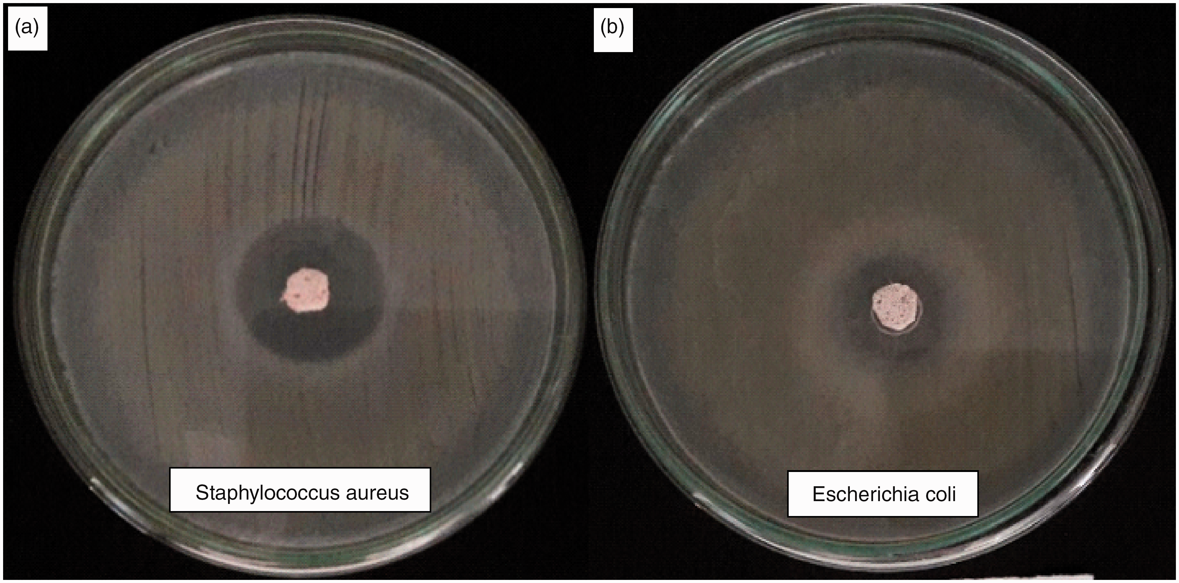
Composite scaffolds with chitosan coating in bacterial culture (a) Staphylococcus aureus (b) Escherichia coli.
According to the results of this study and the research carried out in this field, the diameter of the non-growth zone measured in the culture media of Staphylococcus aureus was higher than that of Escherichia coli, indicating a lower resistance of Staphylococcus bacteria to chitosan and titanium dioxide compared to Escherichia coli bacteria. In a study, it has been shown that chitosan with a wide range of antibacterial activity exhibits a different inhibitory effect on fungi, Gram-positive, and -negative bacteria. The results show that chitosan generally has a stronger bactericidal effect for Gram-positive bacteria than Gram-negative bacteria, which is due to the outer membrane barrier of Gram-negative bacteria. Furthermore, in comparison with common antibiotics, chitosan prevents bacterial growth better, in other words, bacteria resistant to common antibiotics are susceptible to chitosan.24,25
MTT test was performed to determine the cell compatibility and non-toxicity of synthesized scaffolds. Figure 8 represents the optical density changes of samples in the MTT test, after culture of MG63 cells in different periods at the surface of the samples, with three repetitions at each time. In this test, tri-calcium phosphate was used as the control sample.
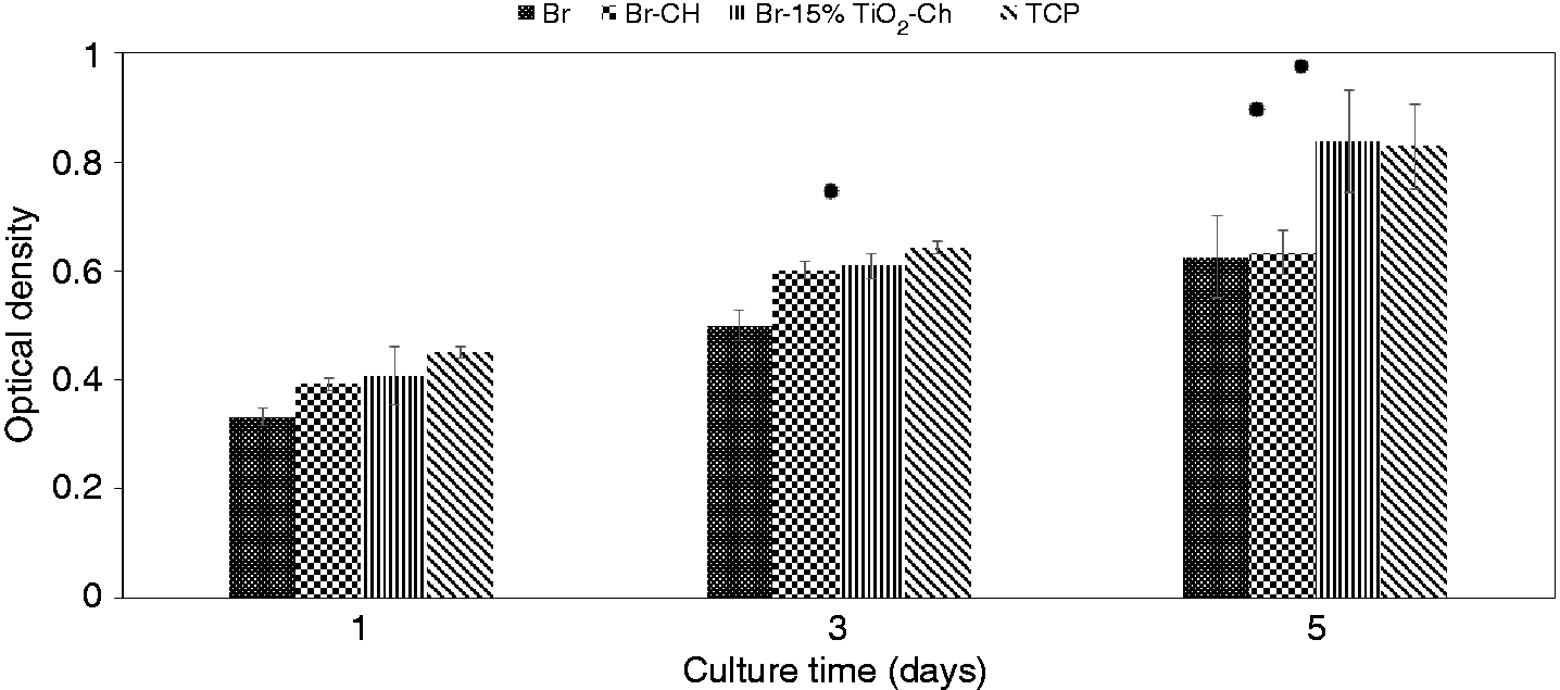
Specimen optical density changes in the MTT test after different culture time.
The results of MTT test showed that increasing cell culture time in surface of all the samples increased the growth and proliferation of MG63 bone cells, and none of the samples had a negative effect on cell growth, indicating no scaffold toxicity and shows cell compatibility. On the first day, there was no significant difference between the cells cultured on all samples and the control sample (TCP). Increasing culturing time up to five days significantly increased cell growth and proliferation on the surface of Br-15% TiO2-Ch optimal composite scaffolds compared to the Br scaffold, which indicates the positive effect of titanium dioxide and polymeric chitosan coating on cell growth in the scaffold surfaces. Meanwhile, in comparison with the control sample, growth of the cell at the surface of the Br scaffold was lower. Therefore, the presence of titanium dioxide and chitosan improves cellular interaction. Furthermore, compared to the Br-Ch scaffold, the number of cells increased at the Br-15% TiO2-Ch sample surface. Accordingly, the presence of titanium dioxide in the scaffold increases the growth and proliferation of cells after five days of culture. It has been proven in numerous studies that magnesium silicate bioceramics, such as bredigite, by releasing ions such as silicon and magnesium in cell culture media, stimulate the growth and proliferation of bone cells, form an extracellular matrix, and ultimately repair and regenerate bone tissue.26–29
Wu et al. concluded that the potential for osteogenic and angiogenesis, as well as the concentration of released ions from bredigite, was higher than other magnesium silicate systems, such as akermanite and diopside. In another study, they demonstrated the high bioactivity of bredigite, and also showed that osteoblastic bone cells had a better adhesion to the bredigite bioceramic.30–32
Cell adhesion test results have been investigated by the scanning electron microscope (SEM) which is presented in Figure 9. Figure 9 shows SEM images of MG63 cells cultured on control Br scaffolds after five days of culture with and without chitosan coating.
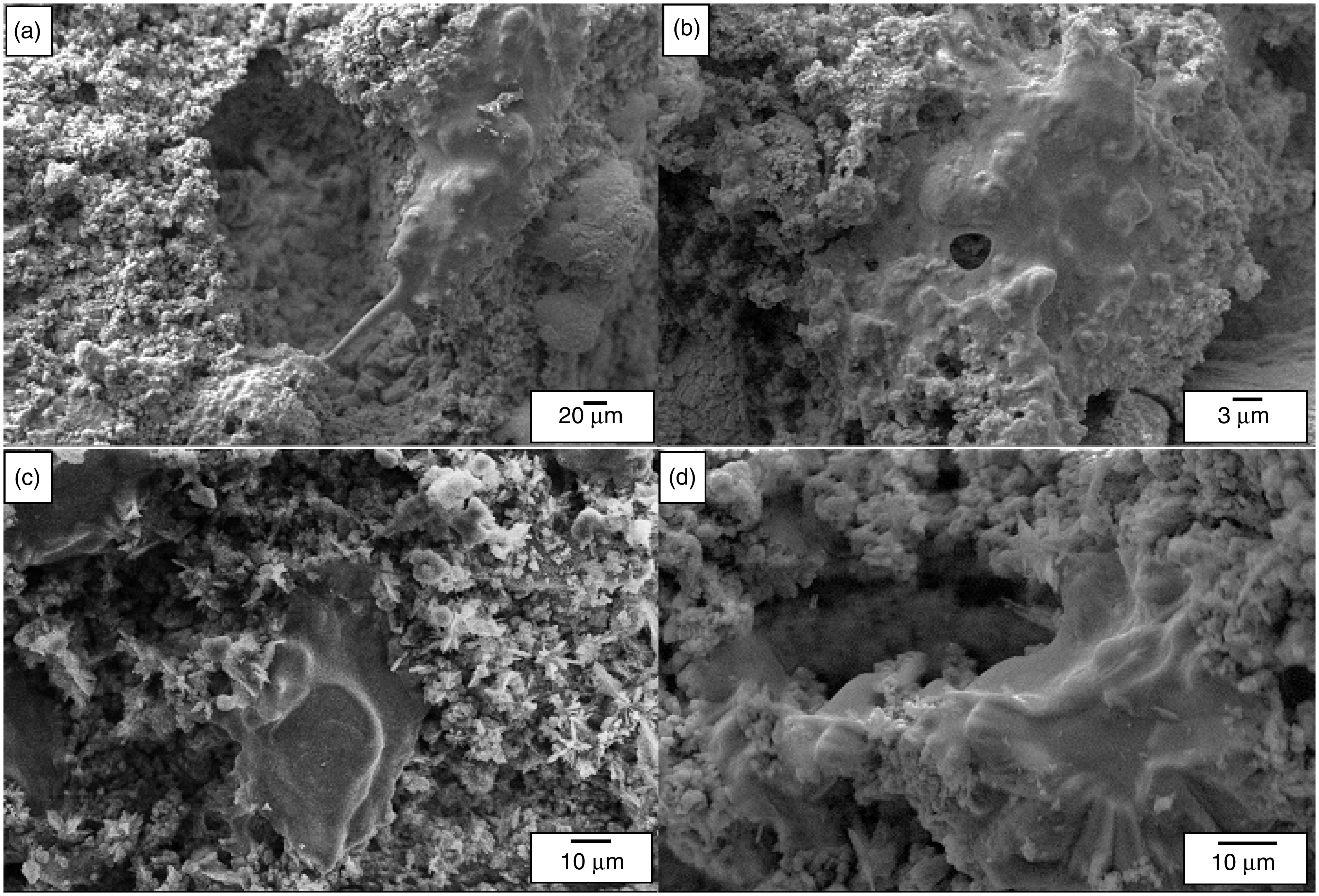
SEM images of MG63 cells grown on the Br scaffold (a and b) without and (c and d) with chitosan coating.
According to the results of the cell culture assay, the Br scaffolds with and without chitosan coating, as well as the Br/TiO2 composite scaffold with chitosan coating, showed excellent biocompatibility. However, according to SEM images, the scaffolds with chitosan coating showed better cell compatibility to the scaffolds without chitosan coating. As seen in Figure 10(c) and (d), the behavior of the cells in terms of growth and adhesion is better than that of the uncoated Br scaffolds observed in Figure 10(a) and (b), which indicates more suitable physical and chemical properties of scaffolds with chitosan coating.
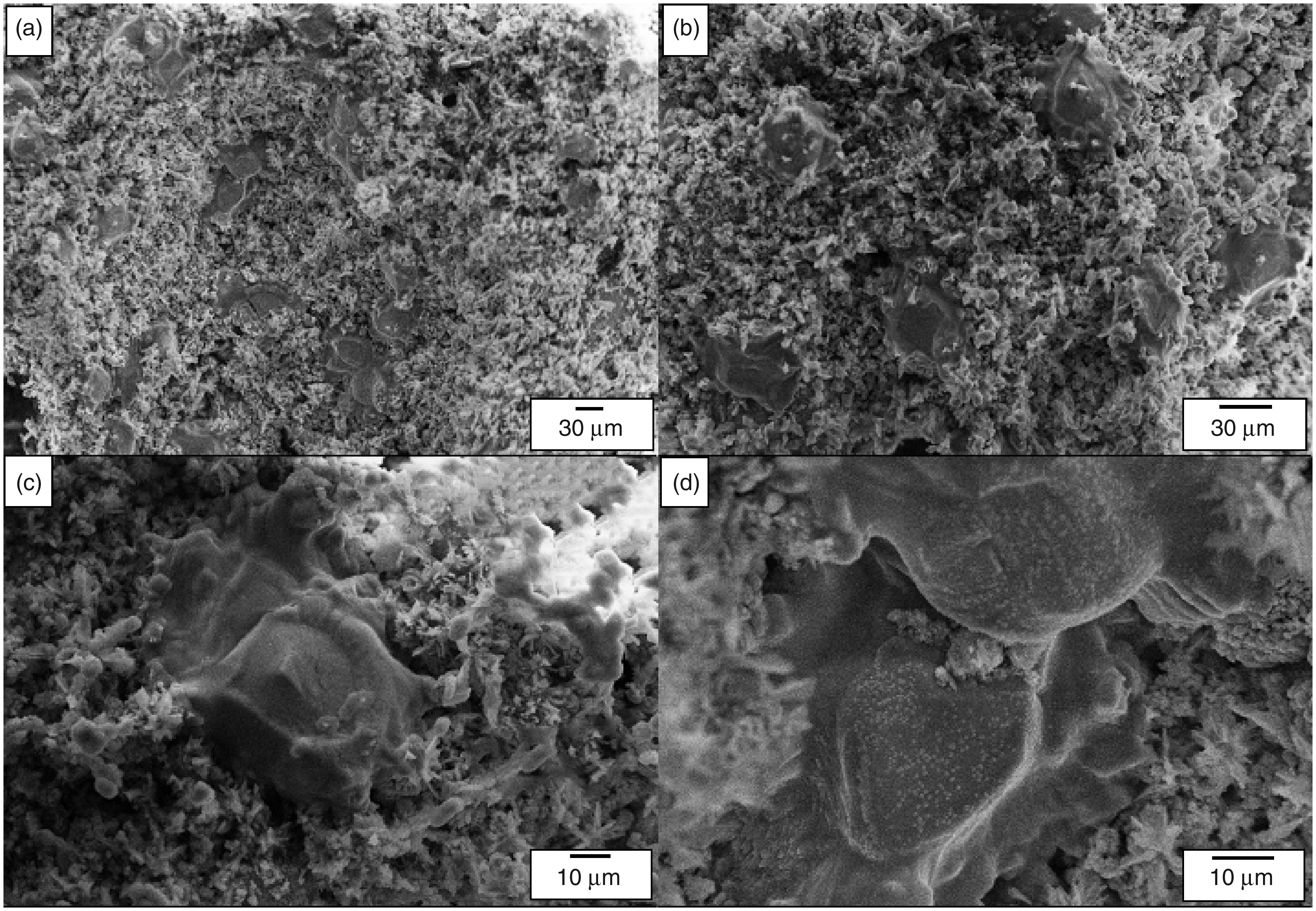
SEM images of MG63 cells grown on optimum Br-15% TiO2-Ch composite scaffolds.
Figure 10 shows SEM images of MG63 cells cultured on a Br-15% TiO2-Ch scaffold after five days of culture. The SEM images show significant proliferation of MG63 bone marrow on optimum bredigite/titanium dioxide composite scaffold with chitosan polymer coating.
Adding titanium dioxide to scaffolds has led to improved adhesion, growth, and proliferation of the cells. The suitability of Br-15%TiO2-Ch scaffolds for bone tissue engineering was determined by observing cellular adhesion and significant cell proliferation by SEM images. As seen in (Figure 10), significant proliferation of MG63 cells, as well as better adhesion of cells than Br scaffold, are shown on the surface of the composite scaffolds with the polymer coating. Generally, the results of the cell adhesion test indicate that Br-15% TiO2-Ch scaffold provides a very good environment for the growth of bone cells. Therefore, these scaffolds are suitable candidates for bone tissue engineering.
In a study, composite scaffolds of chitin and chitosan polymers with nanometric titanium dioxide were prepared and bioactivity and biocompatibility of chitin and chitosan samples with and without TiO2 were investigated. The results showed that samples containing TiO2 particles had higher potential for the formation of apatite than chitin and chitosan samples without TiO2. They showed that TiO2 ceramics promote the formation and nucleation of more apatite crystals on the scaffold surfaces. To verify the cell compatibility of the scaffolds, osteoblast-like MG63 cells, L929 fibroblast cells, and human mesenchymal stem cells (hMSCs) were cultured on scaffolds. MTT results showed that none of the samples was toxic and also, all cells in the TiO2-containing specimens were more involved in growth and proliferation and cellular attachment. 33 The results of the mentioned study are consistent with the results of MG63 cell culture in the present study on better bone marrow cell growth and proliferation and cellular attachment in titanium dioxide containing scaffolds and chitosan coatings shown in Figure 10.
Conclusion
According to the results, it was found that gelcasting method is a suitable method for synthesis of scaffolds with high porosity (63–87%), regular pore size distribution (150–600 μm with a mean pore size of 375 μm) with spherical and interrelated morphology, as well as mechanical properties accepted in bone tissue engineering. The application of the chitosan polymer coating in the scaffolds only created a thin layer of polymeric phase in the porosity wall, and some of the micro-porosity were filled in the scaffold, and full pore filling was rarely observed. The addition of titanium dioxide to the scaffold of bredigite resulted in a decrease in porosity from 87 to 72 percent and an increase in compressive strength of scaffolds from 0.299 to 0.687 MPa. Furthermore, coating scaffolds with chitosan polymer reduced porosity from 83 to 63 percent, and significantly improved the mechanical properties of scaffolds from 0.585 to 2.339 MPa. Furthermore, according to the map images, the uniform dispersion and distribution of titanium dioxide particles in the composite scaffold of bredigite/titanium dioxide were determined. According to the antibacterial test results, Br-15% TiO2-Ch composite scaffold had the highest diameter of inhibition zone in Escherichia coli and Staphylococcus aureus bacteria culture medium which was 22 and 29 mm, respectively. Due to the antibacterial properties of scaffolds and, on the other hand, due to the side effects of antibiotics, the Br-15% TiO2-Ch scaffold can be used as a substitute for the use of antibiotics, especially in the field of bone tissue engineering and in the field of drug delivery. Biocompatibility (non-toxicity) of the samples was proved by the MTT test. The results of the cell adhesion test also indicated that adding TiO2 to the Br scaffold and coating the scaffolds with the chitosan polymer would improve the surface properties of the scaffold, including the proliferation and adhesion of the bone cells. The produced scaffolds because of the high and interconnected porosity, good mechanical properties, high antibacterial properties, and suitable cell compatibility behavior, can be used as an ideal scaffold for use in bone tissue engineering and bone loss repair.
Footnotes
Acknowledgements
The authors thank the Islamic Azad University (Najafabad Branch) for providing the facilities for this research.
Declaration of conflicting interests
The author(s) declared no potential conflicts of interest with respect to the research, authorship, and/or publication of this article.
Funding
The author(s) received no financial support for the research, authorship, and/or publication of this article.