
Abstract
Three-dimensional (3D)-printed scaffolds are a new strategy to fabricate biomaterials for treating bone defects. Here, using a 3D-printing technique, we fabricated scaffolds consisting of gelatin (Gel), sodium alginate (SA), and 58S bioactive glass (58S BG). To evaluate mechanical properties and biocompatibility of Gel/SA/58S BG scaffolds, the degradation test, compressive strength test, and cytotoxicity test were performed. The effect of the scaffolds on cell proliferation in vitro was determined by 4′,6-diamidino-2-phenylindole (DAPI) staining. To evaluate osteoinductive properties, rBMSCs were cultured on the scaffolds for 7, 14, and 21 days and the expression of osteogenesis-related genes was analyzed using qRT-PCR. To examine the bone healing properties of Gel/SA/58S BG scaffolds in vivo, we used a rat mandibular critical-size defect bone model. The scaffolds were implanted into the defect area of rat mandible and bone regeneration and new tissue formation were assessed using microcomputed tomography (microCT) and hematoxylin and eosin (H&E) staining. The results showed that Gel/SA/58S BG scaffolds had appropriate mechanical strength as a filling material for bone defects. Furthermore, the scaffolds could be compressed within certain limits and then could recover their shape. The extract of the Gel/SA/58S BG scaffold showed no cytotoxicity. In vitro, the expression levels of Bmp2, Runx2, and OCN were increased in rBMSCs cultured on the scaffolds. In vivo, microCT and H&E staining demonstrated that scaffolds induced the formation of new bone at the mandibular defect area. These results indicated that Gel/SA/58S BG scaffolds have excellent mechanical characteristics, biocompatibility, and osteoinductive properties, suggesting that it could be a promising biomaterial for the repair of bone defects.
Introduction
Bone defects are defined as the absence of bone due to infection, trauma, tumor, or a congenital disease. 1 If the defect is larger than the limit of self-healing, these bones cannot be repaired without using a substitute: a bone graft or an implant. 2 Currently, autografts are the best treatment for critical-sized bone defects; however, their application is limited due to possibility of infection, insufficient source, immune rejection, and secondary trauma after surgery.3,4 As an alternative approach to bone defect treatment, a variety of effective and safe biomaterials was developed. 5 In our previous study, we fabricated 3D-printed Gel/SA/80S BG scaffolds and demonstrated that these scaffolds had excellent biocompatibility, cell adhesion, and biodegradability. We demonstrated that the extract derived from Gelatin/Sodium alginate/80S BG scaffolds induced proliferation and osteogenic differentiation of MG-63 cells. 6 In this study, we adjusted the 3D-printing process and used 58S bioactive glass (58S BG) to improve the bone healing ability of the scaffolds. Here, we evaluated mechanical properties of the new scaffold, as well as its biocompatibility and osteoinduction, both in vivo and in vitro. qRT-PCR was used to detect the expression level of osteogenic genes to evaluate the promoting function of osteogenesis in vitro. Moreover, to verify the osteogenic function of the scaffolds in vivo, a critical bone defect was made at the angle of the mandible of rat. The scaffolds were implanted into the defect area. Micro CT and HE staining were used to survey the new bone tissue formation. The results indicated that the Gel/SA/58S BG scaffolds have good mechanical property, appropriate rate of degradation and property of promoting osteogenesis.
Methods
Fabrication of 3D-printed Gel/SA/58S BG scaffolds
58S BG composed of tetraethyl orthosilicate TEOS (6.6 mL), triethyl phosphate TEP (0.86 mL) and Ca(NO3)2· 4H2O (4.25 g) (Aladdin, Shanghai, China) was prepared using an evaporation-induced self-assembly (EISA) method as previously reported. 7 Ca, P, and Si were measured by inductively coupled plasma-optical emission spectrometry (ICP-OES) (Spectro Arcos, SPECTRO Analytical Instruments GmbH, Boschstr, Kleve, Germany). 4 mL of each storage solution were sucked into ICE-MS directly from falcon centrifuge tube of each group with a hyphenated autosampler (Cetac ASX 520, USA). Before measurement, the instrument was calibrated using calcium, standard, phosphorous standard, and silicon standard solutions (Sigma-Aldrich, USA), which were diluted to prepare a set of diluted standards. The standard curves were obtained by plotting the intensity (counts) for each standard versus its concentration in mg/L. The concen-tration of each unknown sample was calculated form the standard curves. The measurements were triplicated.
To prepare the ink, 58S BG (10.5%), Gel (15%), and SA (6%) were added to deionized water (all of the concentrations were w/v%) at 55°C and mixed using a magnetic and mechanical stirrer for 30 min. Next, the ink was transferred into the barrel of the 3D bio-printer (Regenovo, Hangzhou, China). The experimental parameters of the printing process were set as follows: the needle diameter was 0.4 mm, the extrusion pressure was 0.38 MPa, the printing speed was 15 mm/s, the adjacent filaments were 0.8 mm, and the extrusion temperature was 28°C. By adjusting the ratio of two materials and improving the mixing process, we observed that the optimal fluidity and plasticity were at the Gel and SA ratio of 10:4. The temperature of composite and extrusion pressure was also adjusted during printing. We found that the fiber could be extruded continuously and formed as a regular cylinder at the temperature of 28°C and the extrusion pressure of 0.38 MPa. To stabilize the shape of scaffolds, the temperature of print table was set at 10°C. Next, the scaffolds were soaked in 10% CaCl2 solution, crosslinked for 10 min, and then further crosslinked in 0.25% glutaraldehyde (GTA) solution for 30 min. After crosslinking, scaffolds were soaked in absolute alcohol, washed with distilled water, frozen at −80°C, and then lyophilized for 24 h in the freeze-dryer (CHRIST, Germany) for further use. There were 10 layers in every scaffolds and the height of the scaffolds was 4.5 mm in average.
Degradation tests
Gel/SA/58S BG scaffolds were weighed and incubated in simulated body fluid (SBF) at 37°C (each set of three parallel samples) for 16 weeks. Every week, the scaffolds were taken out SBF, blotted dry with a filter paper, and weighed. Week 0 was set as a baseline. The ratio of weight loss and volume change was calculated as follows: Weight loss (%) = (W0 − Wt)/W0 × 100%; W0 = the weight of waterlogged scaffolds and Wt = the weight of the scaffolds after surface moisture was removed using the filter paper.
Compressive strength test
The scaffolds were incubated in SBF for 3 h before the compressive strength test. Next, the scaffolds were placed onto the universal mechanics tester and then stressed at 1 mm/min until the load reached 10 kN. The Young’s modulus was defined as the slope of the stress–strain curve in the linear phase of the material during compression and calculated using the software.
Elasticity evaluation
Since bone defects often have an irregular shape, scaffolds require certain elasticity so that they can return to their original shape after passing through a narrow opening. To simulate the implantation of the scaffolds into the irregular-shaped bone defect area, models with narrow slots were made with glass slides (Figure 1(d)). Next, the scaffolds were compressed and pushed through different size slots. After the scaffolds returned to their original shape, the breakage and deformation of the scaffolds were evaluated and the elasticity of the scaffolds was determined. Material characterization and biocompatibility of Gel/SA/58S BG scaffolds. (a) Printability evaluation. Filament testing: the bioink is extruded over the pillar support with different spacing. (b) Images of top and cross-sectional views of Gel/SA/58S BG scaffolds. (c) The surface topography of the scaffolds was evaluated using scanning electron microscopy (SEM); representative images at 50× (scale bar = 100 μm) and 300× (scale bar = 10 μm). magnifications; the red arrows indicate 58S BG particles; (d) Scaffolds were passed through the slots with different widths and then allowed to recover; black arrows indicate the fractured fibers. (e) Swelling ratio of the scaffolds. (f) The degradation rate of the scaffolds. (g) The load curve of the compressive strength test.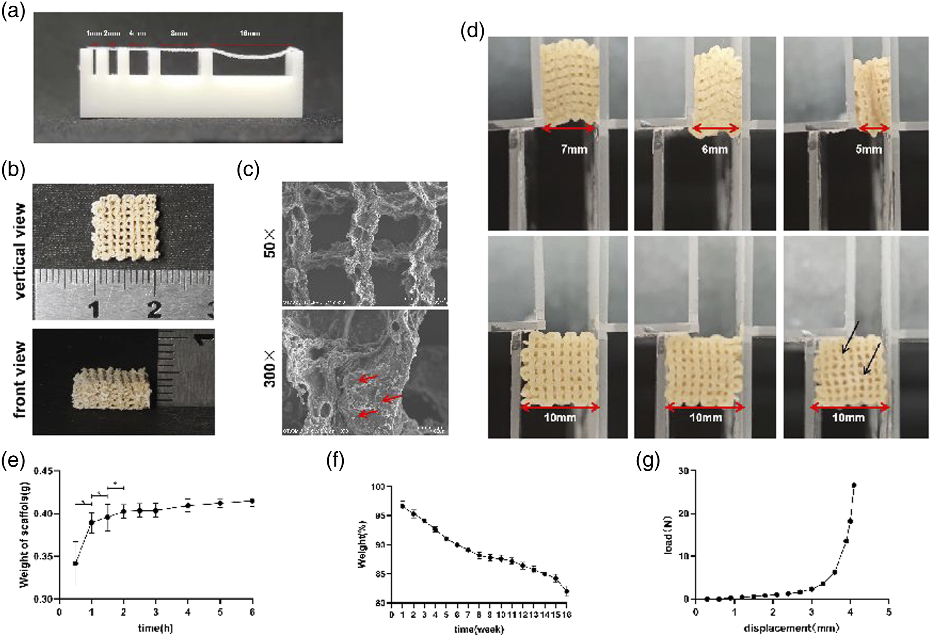
Cytotoxicity test
L929 cells (Shanghai Cell Bank, Chinese Academy of Sciences) were cultured with Dulbecco’s Modified Eagle Medium (DMEM) supplemented with 10% fetal bovine serum (Gibco, Thermo Fisher Scientific, Inc, USA). The cells were maintained at 37°C in a humidified atmosphere at 5% CO2. The manufactured scaffolds were incubated in absolute alcohol (Guangzhou Chemical Reagent Factory, China) for 10 min and then removed. After the alcohol completely evaporated, the scaffolds were washed for 3 min in 5 mL distilled water in a culture dish and then transferred into another culture dish. Next, the scaffolds were divided into 5 groups depending on the number of washes: 1, 3, 5, 7, and 10 washes. Finally, the washed scaffolds were freeze-dried. For experiments, the scaffolds were incubated in Minimum Essential Medium (MEM) supplemented with 10% FBS for 24 h and the extracts were collected. L929 cells were seeded in 6-well plates at 1 × 10⁵ cells/well and cultured with the 100% extracts for 3 days. L929 cells were stained with calcein for 10 min and observed under an inverted fluorescence microscope, cell morphology was evaluated, and the number of live and dead cells was counted using the ImageJ software. Shrinken or broken cells were identified as deformed cells.
In vitro evaluation of the scaffolds
rBMSCs (Shanghai Cell Bank, Chinese Academy of Sciences) were cultured in DMEM supplemented with 10% fetal bovine serum (Gibco, Thermo Fisher Scientific, Inc., USA). The cells were maintained at 37°C in a humidified atmosphere at 5% CO2. Cells were seeded onto the scaffolds placed in 6-well plates at 4 × 10⁵ cells/well and incubated at 37°C for 4 h to allow cell attachment. Next, the scaffolds were transferred to another set of 6-well plates and cultured for 7, 14, and 21 days. At the end of the incubation period, the scaffolds were removed and washed three times with PBS. The cells were fixed in 3.7% (vol/vol) formaldehyde in PBS for 30 min and then washed three times with cold PBS. The scaffolds were permeabilized with ice-cold 0.25% Triton X-100 for 5 min and then washed again in cold PBS. Finally, the cells on the scaffold were incubated with 0.25% 4′,6-diamidino-2-phenylindole (DAPI) for 10 min, washed again with cold PBS, and then examined using a fluorescent microscope (LEICA DMI3000 B, Germany).
Quantitative real-time PCR
The sequences of primers used for RT-PCR.
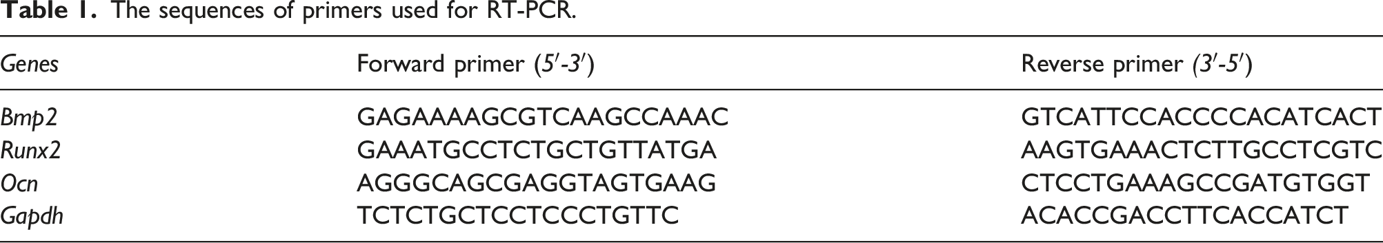
was 25 ll and the RT-qPCR was performed using 40cycles with a 10 min denaturation at 95°C, 15 s annealing at 95°C and a 1 min extension at 60°C. The relative expression level for each gene was normalized to GAPDH and relative quantification of gene expression was performed using the 2−ΔΔCq method.
Animal model and experimental design
Male Sprague Dawley rats (6–8 weeks old) were purchased from the Guangdong Medical Laboratory Animal Center (Guangzhou, China) and maintained in a common facility at Laboratory Animal Center of Guangzhou University of Chinese Medicine (Guangzhou, China). All procedures in this animal study were approved by the institutional animal care and use committee of Guangzhou University of Chinese Medicine. The principles of laboratory animal care, as well as national laws for animal experimentation, were followed. Critical-size 5-mm mandible defects were prepared using a burr drill as previously described. Briefly, rats were anesthetized by intraperitoneal injection of 60 mg/kg ketamine hydrochloride (Ketalar, Trustech Pharma Care, Bayern, Germany). After the induction of general anesthesia, the rats were fixed on the operating table in a supine position. After skin and mucous membranes were cut, a cylindrical perforated defect (5 mm in diameter) was made on both sides of the mandible near the mandibular angle under copious irrigation with cold saline solution. Rats were randomly assigned to two groups (5 rats every group): blank control and implant group. The control group did not contain any implant material, while Gel/SA/58S BG scaffolds were placed into the bone defects of the implant group.
Micro-computed tomography analysis
Rats were sacrificed 4 and 8 weeks after the surgery. The entire mandibles of the rats were excised, the defect area and the surrounding tissues were resected together, and then fixed with 1% paraformaldehyde for 5 days. The samples were scanned using micro-CT (Bruker Corporation, Billerica, USA). The 3D images were reconstructed using the NRecon software and the images of mandibular angle areas were transformed into 2D images. Bone tissue/tissue volume ratio (BV/TV) was measured.
Hematoxylin and eosin staining
For histological evaluation, the specimens were decalcified in 10% Ethylene Diamine Tetraacetic Acid (EDTA) (pH 7.4) for 30 days. The obtained samples were dehydrated with alcohol gradients and embedded in paraffin. Then 5-μm paraffin section were cut and then stained with H&E. Images were acquired using a light microscope at 100× magnification (Leica Microsystems, Germany).
Statistical analysis
Data are presented as the mean ± standard error. Statistical analysis was performed using the SPSS statistical software v22.0 (International Business Machines, Corp. Armonk, NY, USA). Data were evaluated using the analysis of variance (ANOVA) followed by the least significant difference test or Bonferroni’s post hoc test. Statistical significance was set at p < 0.05.
Results
Fabrication and characterization of scaffolds
The results of ICP-MS revealed that the mass fraction of each chemical element are: Si (32.24%), Ca (24.35%), O (36.56%), P (6.85%). The filament fusion tests showed that a maximum inter-fiber spacing of 8 mm could be achieved for this bioink (Figure 1(a)). The scaffolds had a porous structure (Figure 1(b)), which was further confirmed by SEM as the presence of pits and multi-level pore structures on the surfaces of the fibers. SEM images also demonstrated that 58S BG particles were evenly distributed in the scaffold (Figure 1(c)). The elasticity test showed that the scaffolds were compressed when passing through the slots and then were restored to their original form (10 mm) in the wider area. The video of the process of experiments was in supplementary data. We observed that after passing through 6-mm and 7-mm slots, the scaffolds were restored to their original form without any damage to the fibers; however, in the 5-mm group, several fibers were broken after passing through the narrow slot (Figure 1(d)).
Next, we evaluated the swelling ratio and showed that the scaffolds had a fast speed of water absorption during the first hour and then had a stable swelling rate of approximately 4.58 after 2 h (Figure 1(e)). At the same time, the degradation rate of the scaffolds was 17% after incubation in SBF for 16 weeks (Figure 1(f)). The compressive strength test demonstrated that the load was significantly increased when displacement reached 3 mm (Figure 1(g)), and that the average Young’s modulus of the scaffolds was 265.80 Mpa.
Cytotoxicity of the scaffolds
To confirm the removal of GTA, the cytotoxicity test was performed using L929 cells as measured by the number of “deformed” cells. The percentage of “deformed” cells in the control group was 6.5%; however, the percentage of “deformed” cells in the PBS-washed groups was 43.8% (1 wash), 34.5% (3 washes), 19.5% (5 washes), 10.9% (7 washes), and 8.6% (10 washes) (Figure 2(a) and (b)). Our results showed that, as the number of washes increased, the percentage of “deformed” cells was significantly decreased. It has been proposed that scaffolds could be identified as non-cytotoxic if the percentage of “deformed” cells was less than 20%.
8
Therefore, the scaffolds that were washed for more than 10 times were used in rBMSC experiments. We observed that a large number of rBMSCs was adhered to the surface of scaffolds after seeding. Furthermore, DAPI staining confirmed that the number of rBMSCs had expanded after culturing cells on scaffolds for 7 days. On days 14 and 21, the number of fluorescent cells further significantly increased compared with day 7 (Figure 2(c)). The evaluation of Gel/SA/58S BG scaffold biocompatibility in vitro. (a) L929 cells were cultured with conventional medium for 3 days (negative control, scaffold extracts washed 1, 3, 5, 7, and 10 times) and cell morphology was evaluated; “lightspots” depict the “deformed” cells; scale bar = 10 μm. (b) The quantification of experiments described in (a); the percentage of “deformed” cells was 6.5% (negative control), 43.8% (1 wash), 34.5% (3 washes), 19.5% (5 washes), 10.9% (7 washes), and 8.6% (10 washes). (n = 3, *p < .05, *p < .01 vs. control group). (c) rBMSCs were cultured on the Gel/SA/58S BG scaffolds for 7, 14, and 21 days and then stained with DAPI; blue fluorescent dots represent cell nuclei; scale bar = 200 μm.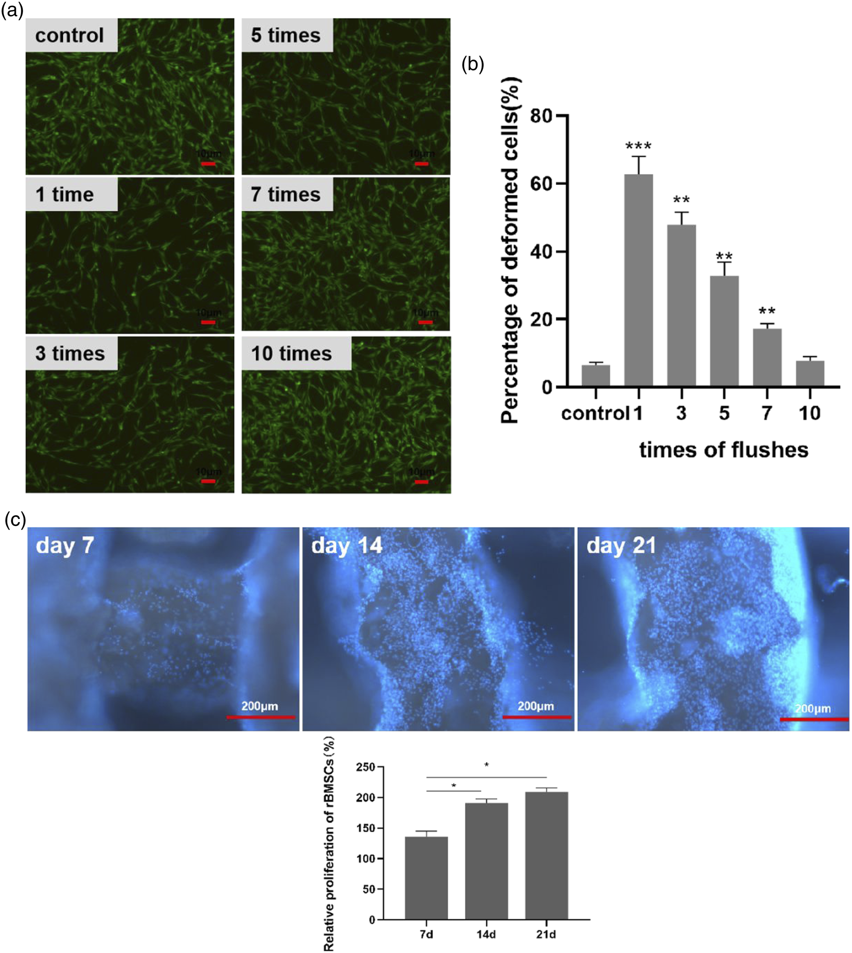
Assessment of osteogenesis-associated genes
To examine the effect of Gel/SA/58S BG scaffolds on the induction of osteogenesis, qRT-PCR was performed and the expression of osteogenesis-related genes (Bmp2, Runx2, and OCN) on days 7, 14, and 21 was evaluated. Our results showed that the expression of osteogenesis-related genes was significantly increased in the scaffold groups compared with the control group. The expression level of Bmp2 was increased on day 7 (Figure 3(a)), while the levels of Runx2 and OCN were upregulated on day 14 (Figure 3(b) and (c)). The expression of osteogenesis-related genes in rBMSCs cultured on Gel/SA/58S BG scaffolds. rBMSCs were cultured on Gel/SA/58S BG scaffolds for 7, 14, and 21 days and the expression levels of (a) Bmp2, (b) OCN, and (c) Runx2 were determined by qRT-PCR; all results were normalized to Gapdh; (n = 3, *p < .05, *p < .01 vs. control group).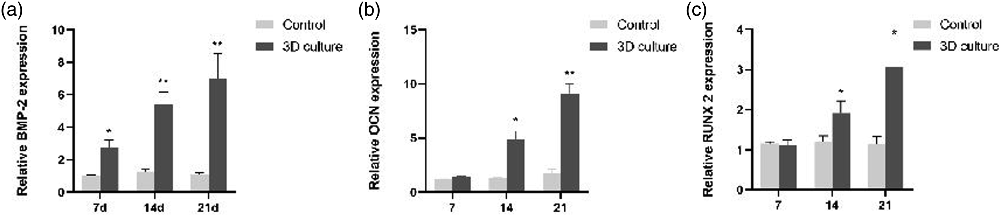
The scaffolds promoted the osteogenesis in vivo
After the critical-size 5-mm mandible defects were prepared using a burr drill as previously described, the scaffolds of the same shape as the defect were implanted into the defect area(Figure 4(a)). Micro-CT results demonstrated that the new bone tissue was formed at the site of the defect at 4 and 8 weeks, while the scaffolds maintained their structure; however, there was no obvious bone formation in the defect area of the blank control group, indicating that the defect did not heal on its own (Figure 4(b) and (c)). The quantification of bone volume fraction showed that the BV/TV ratio was significantly higher in the implant group than in the control group (Figure 4(d)). H&E staining showed that the fibrous tissue filled the entire defect area, while new bone tissue formed from the edge of the defect (Figure 4(e)). Gel/SA/58S BG scaffolds induced osteogenesis in vivo. (a) The preparation of critical-size mandible defect in rats and the implantation of Gel/SA/58S BG scaffolds. (Blue arrows show the bone defect area). (b) Micro-CT images of bone defect areas after the scaffold implantation at 4 and 8 weeks (c) 3D-reconstruction of the samples, the red-colored areas represent the scaffolds. (d) Micro-CT quantification; bone volume/tissue volume (BV/TV) of the samples at 4 and 8 weeks was determined. (n = 3, *p < .05, **p < .01 vs. control group). (e) H&E staining of newly formed tissues in the bone defect area; black circles: regenerated new bone; black square: scaffold; asterisk: rat bone; black triangle: fibrous tissue; scale bar = 50 μm. Red-dashed lines represent the demarcation of different tissues.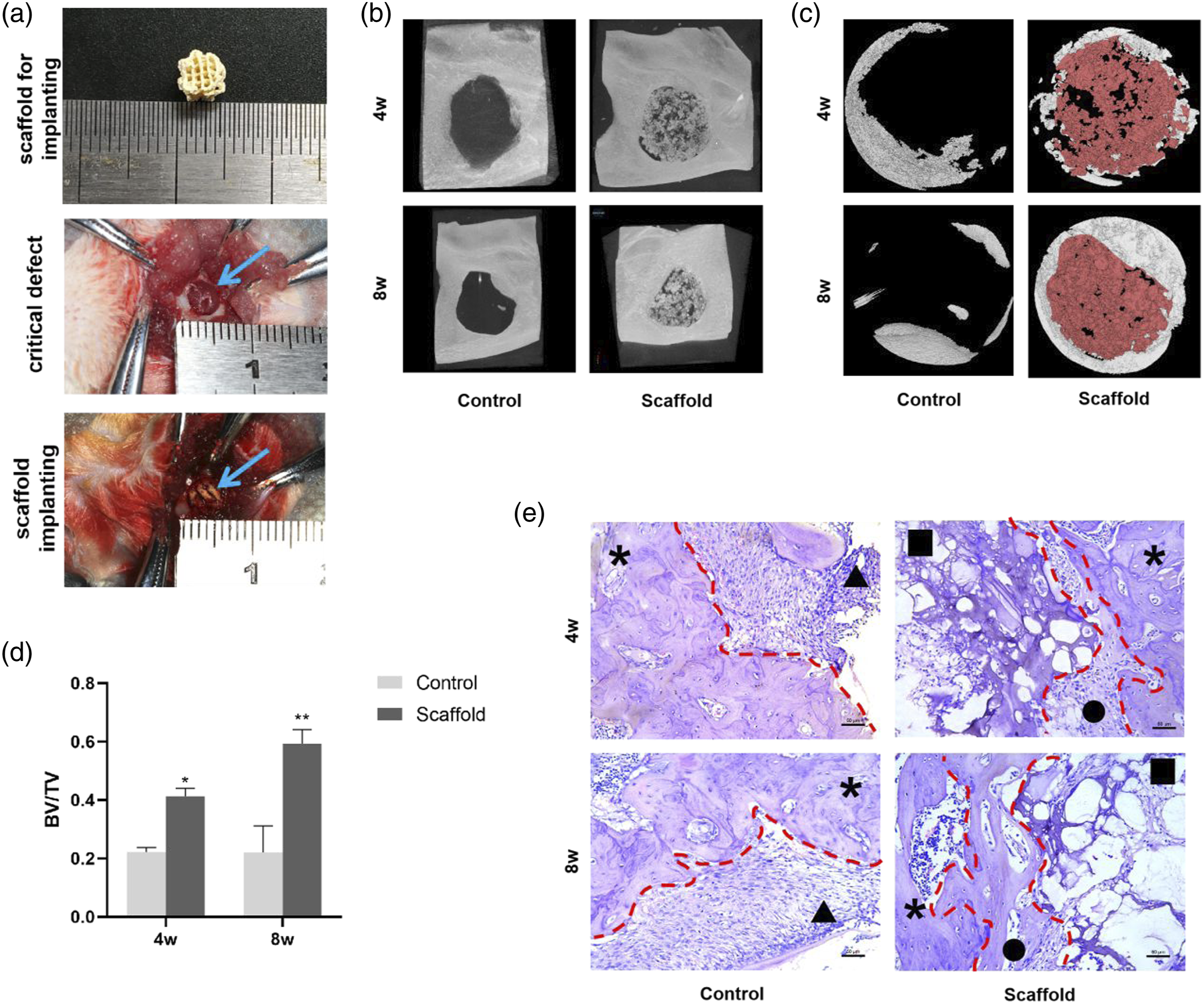
Discussion
Gel, SA and 58S BG were chosen as the host material of the scaffolds. As a protein-based material, Gel has excellent biocompatibility, low antigenicity, and facilitates cell adhesion via cell-surface interactions. However, as a single component, gelatin is hard to shape due to its temperature-dependent fluidity, i.e., a significant change in liquidity in response to a small change in temperature.9,10 To improve the printing performance of the material, SA was selected to increase the viscosity of the slurry. SA is considered to be an efficient option for enzyme immobilization, since it is nontoxic, hydrophilic, biodegradable, and biocompatible. It can stabilize the formation of biocompatible hydrogels and provide an ideal cell microenvironment. 11 At the same time, 58S BG can form local alkaline microenvironment and release free calcium ions to provide favorable conditions for bone tissue formation. Furthermore, it has been reported that 58S BG also improves the anti-inflammatory properties of the tissue. 12 Moreover, Gel and SA had been used in food and drug industry for several decades. It had been verified that there was no toxic degradation products after Gel or SA entered the human body. For these reasons, Gel, SA, and 58S BG were selected as the matrix materials for the scaffold in this study. Compared with 80S BG, the pore size of 58S BG is 2∼50 nm, which enhances the ion exchange rate of the BG particle surface. 13 As the ratio of CaO/P2O5 in 58S BG is higher than in other types of BG, 58S BG can release higher levels of SiO2 ions when in contact with body fluids, 14 promoting the formation of hydroxyapatite layer on the surface of BG particles. 15 The rigidity of Gel varies with temperature, while SA influences the liquidity of the matrix. 58S BG is insoluble, therefore, the excess of BG would make the matrix too viscous. After a series of extrusion tests, the 58S BG, Gel, and SA were mixed in the 7:10:4 ratio, ensuring that the matrix could be smoothly extruded and the scaffolds would remain rigid after printing. 3D-printing technology can create complex structures based on medical imaging and computer-aided design; however, it requires the composite materials to simultaneously have moderate plasticity, mobility, and rigidity. 16 Traditional hydrogel materials generally have relatively poor mechanical properties, resulting in materials that cannot be accurately shaped or maintain a stable shape after implantation into the area of defect. In this study, the ratios of these three constituents were carefully adjusted to allow the slurry to be easily printed into required forms under controllable print parameters. Moreover, the morphology of the scaffold remained stable before the crosslinking treatment.
As the slurry had sufficient plasticity and stability, the shapes and structure of scaffolds could be controlled. It has been reported that the pore size of biomaterial scaffolds larger than 300 μm and a porosity higher than 50% are preferred in bone substitute implants because of their good function of nutrient transport.17,18 Based on SEM results in this study, it was demonstrated that the pore size was approximately 600 μm, allowing cells to attach to the scaffolds to provide a suitable microenvironment. 19 The ideal scaffold should maintain sufficient strength and stiffness to function for a period of time it takes for a tissue to replace the slowly diminishing artificial matrix. To simulate the process of implant degradation, we evaluated the weight loss of scaffolds incubated in SBF. Our results showed that the scaffold maintained its structure at least 4 weeks and lost approximately 15% weight after 16 weeks. These results indicated that the rate of scaffold degradation was slower than the rate of new bone formation, suggesting that the defect would be filled with scaffold until it was repaired.
We observed that the scaffolds remained rigid after they were freeze-dried. Furthermore, after absorbing SBF, the scaffolds maintained good compressive strength, and the average Young’s modulus was 265.80 Mpa (while the Young’s modulus of cancellous bone is 2–12 Gpa). 20 These results showed that the compressive strength of Gel/SA/58S BG scaffolds corresponded to the required mechanical strength of bone defect filling material. As the Young’s modulus of the scaffolds was less than the cancellous bone, the scaffolds would be less stressed due to the stress shielding effect, providing more suitable stress condition for jaw restoration Since jaw defects have irregular shapes, requiring bone filling materials to be elastic, to simulate the process of implantation we tested the ability of the scaffolds to maintain the shape using 5, 6, and 7-mm wide slots. We observed that the scaffolds could be compressed down to at least 60% of the width and then could be restored to their original form, without breaking the fibers. These results indicated that these scaffolds were suitable for variously-shaped and sized bone defects.
Glutaraldehyde (GTA) was chosen as the crosslinking reagent in this study due to its excellent crosslinking performance GTA is a common crosslinking agent that improves the chemical and biological performance of gelatin; however, the traces of GTA remaining in the scaffolds are toxic to the cells. 21 To eliminate the residual GTA cytotoxicity, the scaffolds were washed 1, 3, 5, 7, and 10 times with distilled water, for at least 5 min each wash. Based on previous reports, the toxicity of GTA is removed after more than 5 washes, without producing a histotoxic reaction after subcutaneous implantation.22,23 These results suggested that the scaffolds provided a favorable microenvironment for cell proliferation. These patterns of osteogenic gene expression levels correspond to the expected timelines of osteogenic differentiation. Compared with the control group, our results showed that scaffolds induced osteogenic differentiation in rBMSCs, highlighting the osteoinductive potential of these scaffolds.
Next, we evaluated the scaffolds in vivo using a rat mandibular critical-size defect model. In this model, the defect should be large enough to be exposed to considerable loads, without risking a fracture of the condyles. 24 The mandibular defect in our rat model was designed as a cylinder with a diameter of 5 mm penetrating the mandible, as previously reported. 25 The mandible defect may heal entirely on its own or at least to an extent with intrinsic healing capacity by this design.26,27 The scaffold must maintain sufficient structural integrity during bone tissue growth and remodeling so that the newly formed tissues would have the support necessary to repair the defect area.28,29 Histological analysis of the samples in the implant group showed that the scaffolds were integrated with the surrounding bone tissue, with the newly formed bone tissue forming toward the center of the defect. H&E staining indicated that there was increased bone-like structure formation in the area of the defect. The boundary of defect area in the implant group was not evenly shaped. Under physiological conditions, extracellular matrix fills the bone defect area, gradually mineralizing and forming a new bone tissue.2,22 H&E staining showed that the fibrous tissue filled the entire defect area, while new bone tissue formed from the edge of the defect, similar to the process of physiological bone defect repair. 30 This result indicated that the implanted scaffolds integrated with the bone tissue peripheral to the defect and suggested that this type of bone repair was different from filling repair. Traditional bone defect filling materials were usually filled into the defect area in order to occupy the space, which result in that the bone defect could not heal and the function of the bone could not recover. Ideal bone defect filling materials should present the feature that guiding the bone tissue regeneration so that newly formed bone tissue could integrate with the original tissue and the function of the bone would recover. In this study, the scaffolds preliminarily demonstrated this feature. We expect that newly formed tissues would fill the whole defect area after scaffolds completely degrade. In summary, our results suggested that Gel/SA/58S BG scaffolds have the potential of inducing osteogenesis and bone tissue regeneration both in vivo and in vitro.
Conclusion
Here, we fabricated and characterized novel Gel/SA/58S BG scaffolds. Our results showed that these scaffolds have ideal plasticity and stability, as well as sufficient mechanical strength and appropriate rate of degradation. Furthermore, these scaffolds induced osteogenesis both in vitro and in vivo, indicating that Gel/SA/58S BG has the properties suitable for a bone grafting material.
Footnotes
Declaration of conflicting interests
The author(s) declared no potential conflicts of interest with respect to the research, authorship, and/or publication of this article.
Funding
The author(s) disclosed receipt of the following financial support for the research, authorship, and/or publication of this article: Science & Technology Bureau of Guangdong Province; 2018B050502012.