
Abstract
Bone regeneration can be accomplished through osteogenesis, osteoinduction, and osteoconduction mechanisms. This study aimed to investigate the properties of the PRF scaffold with tricalcium phosphate nanoparticles in socket preservation in an animal model. Fabrication of PRF performed. In this experimental study, 18 rats were divided into three negative control, PRF, and PRF/TCP groups. The mechanical and chemical tests including swelling rate, degradation time, and MTT tests were applied to the scaffolds. In each animal, the first maxillary right molar was extracted, and extraction sites of test groups were filled with a resorbable biocompatible biomaterial in situ hardening bone substitute. After 2 and 4 weeks all animals were sacrificed and examined histopathologically and with qRT-PCR. Histological results showed TCP in combination with PRF accelerates bone regeneration with the highest amount of lamellar bone and collagen formation compared to the control and PRF alone. Mechanical and chemical tests on the scaffolds showed the addition of TCP to the PRF scaffold decreases the swelling rate and increases the degradation time. qRT-PCR showed expression of osteogenic genes increased significantly (p < 0.05) in PRF/TCP and PRF, respectively. In conclusion, the gelatin hydrogel containing PRF/TCP scaffold led to more bone formation after tooth extraction. Therefore, the injectable PRF\TCP hydrogel is a promising candidate for bone repair and regeneration.
Introduction
Bone is a highly complex structure that constantly undergoes tissue changes, which can self-repair and adapt to new forces. Tooth extraction triggers disuse atrophy. Approximately 50% of the ridge width is reduced during the first year after tooth extraction. 1 chewing forces in form of tensile and compressive strains influence the mechanosensitive cells of the periodontal ligament (PdL), which is closely attached to the alveolar bone, leading to changes in PdL cell vitality, proliferation, and differentiation, this can lead to ridge atrophy of the surrounding alveolar bone, especially in the first year after extraction.1,2 Rehabilitation of structures and functions of bone can occur by multiple methods. Ridge preservation is a recent form of tissue engineering that helps to maintain the ridge dimension, reduces the possibility for bone graft application, eases the implant placement, and almost prevents marginal bone loss and improves the success rate of implants. 1 Human healing socket process occurs in 6 weeks. 3 Histologic evidence of active bone formation in the bottom of the socket can be seen 2 weeks after the tooth extraction and the new mature bone progressively fills the socket in about 6 months. 4
Osteogenesis, osteoinduction, and osteoconduction can be accomplished as three bone regeneration mechanisms: Osteogenesis is the natural formation of the bone, without mesenchymal stem cell differentiation. Osteoinduction is the use of growth factors that exist in the living bone to promote the differentiation of mesenchymal stem cells into osteoblasts or chondroblasts. Osteoconduction is the process that provides a bio-inert scaffold, to manage the deposition of new bone from the surrounding bone or guide the differentiated mesenchymal cells' migration to growth along the graft surface. 5 Currently, it is still unclear which material or surgical method is the most effective in limiting post-extraction resorption while assisting in regenerating adequate vital bone. 6 The main characteristics of an ideal bone grafting material are to be osteoconductive, osteoinductive and biocompatible, gradually replaced by newly formed bone, exhibit controlled breakdown and resorption, the coordination scaffold degradation time and the bone healing process should be able to maintain the ridge contour in the augmented site. Moreover, it should have satisfactory mechanical properties and no risk of disease transmission. 7 Alloplasts represent a group of synthetic and highly biocompatible bone grafting materials. 8 Alloplastic biomaterials do not pose a risk of host reaction in the donor site, and there is no limitation for preparation the vast amount of alloplastic graft material 9 Calcium phosphate ceramics are bioactive osteoconductive materials and there is strong experimental evidence that they also have osteoinductive properties while promoting neovascularization. Among ceramics, beta-tricalcium phosphate (β-TCP) is widely used in orthopedics and dentistry. 7 It has a chemical composition of natural bone that releases ions to favor bone regeneration. 10 Previous studies of the bone regeneration of β-TCP focused on bulk-sized β-TCP products such as granules, blocks, or wedges with size ≥100 nm. Little is known about the bone regeneration and biocompatibility of nano-sized β-TCP (nβ-TCP) particles, which may be an alternative to bulk-sized β-TCP. The use of nβ-TCP may dramatically increase surface area, surface roughness, and surface area to volume ratios, leading to superior physicochemical properties. Lin et al. 11 reported that scaffolds fabricated from nβ-TCP had more than two times the Vickers hardness and compressive strength compared to the scaffold fabricated from micro-sized β-TCP. The size of nβ-TCP may better mimic the nanocomposite of the natural bone, which consists mainly of nanocrystalline hydroxyapatite (Ca5(PO4)3). 12
PRF (platelet-rich fibrin) is a second-generation platelet concentrate obtained after centrifugation of the blood. The protocol was first presented in France by Choukroun et al. 13 The collected clot (or biomaterial) is stable, elastic, adhesive, and flexible. It can be used in combination with bone replacement materials or as a sole graft material. PRF has hemostatic, angiogenic, osteogenic, anti-inflammatory, antimicrobial, analgesic, and healing properties. 14 PRF contains cytokines, platelets, leukocytes, and circulating stem cell. 15
This experimental study aimed to test the hypothesis that filling intact extraction sockets with a resorbable, biocompatible, alloplastic biomaterial, consisting of β-TCP nanoparticles and PRF (in a hydrogel phase), will assist in the regeneration of new bone, compared to sites subjected to spontaneous healing, in a rat model.
Materials and methods
The study protocol was approved by the ethics committee of Shiraz University of Medical Sciences (IR.SUMS.AEC.1401.042).
Fabrication of PRF
Blood samples (9 mL) were collected from healthy volunteer humans (aged 17–40 years). The test tubes used did not contain any anticoagulants. The blood samples were extracted in strict accordance with the PRF manufacturing process. The venous blood is rapidly centrifuged at 400 × g at room temperature for 10 min using a specific table centrifuge (Andreas Hettich GmbH and Co. KG, Tuttlingen, Germany) and will be kept aside for 35 min. The sample was divided into three layers: The bottom layer of red blood cell (RBC) debris, the middle layer of PRF gel, and the top layer of supernatant. The middle layer was removed using sterile tweezers, and the RBC layer was removed from the PRF gel. Finally, the PRF gel was placed in the PRF box for 10 s under light pressure. As a result, the rigid and elastic PRF membrane obtained the PRF.
16
Then, for decellularization; the samples on a shaker plate were washed with PBS for 24h. Hematoxylin and eosin staining was used to evaluate the success of decellularization. For lyophilization the concentrated gel products were first frozen for 2h at −80°C. Then the frozen gels were lyophilized in a freeze drier 3 days at −50°C to obtain the porous scaffold. Then lyophilized PRF was completely pulverized with scissors and a mill. Re-material in the freezer at
Water uptake test
To evaluate the swelling capacity of the scaffold, PRF and PRF/TCP hydrogels were poured into 2 mL falcons. Three samples of hydrogels were dried using a freeze drier until a constant weight of the gel was obtained and primary weight of each sample (
Weight loss test
To evaluate degradation time, the cross-linked scaffolds were cut into the square samples (10 × 10 mm2) and then immersed in 5 mL PBS (pH = 7.4) and incubated at 37°C for 42 days And the primary weight of each sample (
MTT cell viability
MTT (3-(4,5-dimethylthiazol-2-yl)-2,5-diphenyltetrazoliumbromide) colorimetric assay was used to investigate cell viability 1, 3 and 6 days after seeding. First, the cells were seeded in 12-well plates in DMEM, containing 10% FBS and 1% penicillin-streptomycin at 37°C. 5000 seeded cells were used for each assy. Cell-seeded scaffolds were rinsed with PBS and MTT solution at 0.5 mg/mL was added to each well and incubated for a period of 4 h. The yellow MTT dye was reduced by the mitochondrial reductase enzyme in living cells to purple formazan after the incubation period; the formazan was destained with 400 μL of DMSO and its absorbance read with the spectrophotometer at 590 nm. 17
Fourier transform infrared analysis
Fourier Transform Infrared Spectroscopy (FTIR) identifies chemical bonds in a molecule by producing an infrared absorption spectrum. The spectra produce a profile of the sample, a distinctive molecular fingerprint that can be used to screen and scan samples for many different components. Samples were cut into 10 × 10 mm square pieces. Which was used to process the FTIR analysis in reflection mode. All spectra were obtained between 3500 and 500 cm − 1 at a resolution of 1 cm – 1.
Experimental design
18 female Sprague Dawley rats ranging from 190 to 270 g and with an age range of 10–12 weeks were selected for the study. The animals were housed in separate cages in a room with a 12 h day-night cycle, with a temperature of 24–28°C, and a humidity of 45–64%. All animals were fed with a semi-purified diet and water ad libitum before the start of the experiment. The experimental protocol used was approved by the Shiraz University ethics committee. Prior to the surgery, the rats received general anesthesia by xylazine, 2% (Alfasan Woerden, Hollan) and Ketamine, 10% (Alfasan Woerden, Hollan) with 1; 1 ratio and by intraperitoneal injection by an expert animal lab technician. The right maxillary first molar was extracted. Extraction was performed using a curved hemostat with gentle buccolingual movements. For the buccal soft tissue and tongue retraction infant mouth retractor was used which was so helpful for appropriate visibility and access. After the surgical procedure, an amount of 5 μl of each scaffold was injected into the socket area of experimental groups and preserved with a suture. The 18 rats were arranged to be put into three groups including negative control, PRF and PRF/TCP nanoparticle applications groups (Figure 1). (a) Infant mouth retractor and curved hemostat for appropriate tongue and soft tissue retraction and tooth extraction. (b) Extracted rat’s first molar tooth.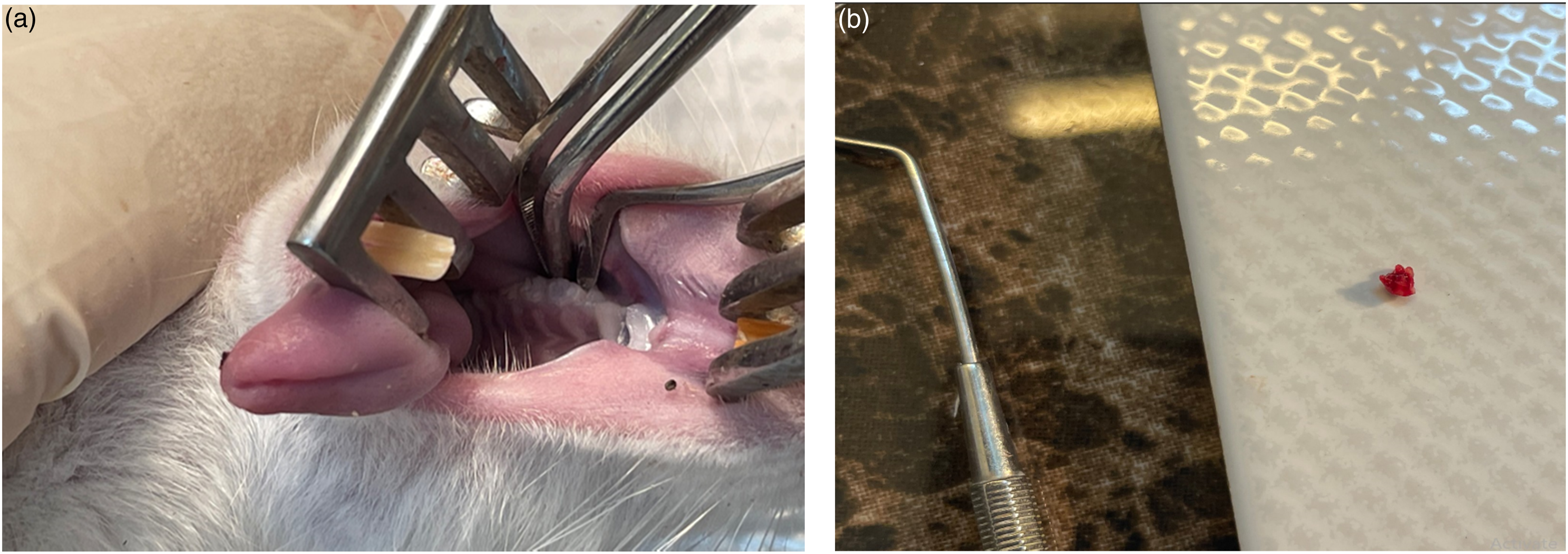
The rats are fed a soft diet for 2 days and were given ampicillin sodium for three consecutive days (60 mg/kg). At 2 and 4 weeks after the surgical procedure, two animals in each group were randomly selected and sacrificed by overdose of Carbon dioxide 70% (in the Carbon dioxide box).
For the histological study, the specimens were fixed using 4% paraformaldehyde. After complete fixation, the maxilla was separated and immersed in the 4% paraformaldehyde for 24 h. Afterward, specimens were fixed in 10% formaldehyde for 10 days, then, washed and soaked in 10% EDTA for decalcification for 4 weeks, and then rinsed in distilled water. Specimens were dehydrated in ascending grades of alcohol and embedded in paraffin. Finally,5 µ Sagital (mesiodistal) sections by microtome were prepared and H&E and Masson trichrome stainings were performed.
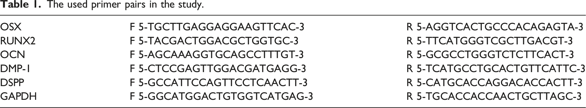
qRT-PCR
The used primer pairs in the study.
Statistical analysis
All experimental samples were collected in triplicates and all data were expressed as the mean ± standard deviation. Differences between experimental data sets were assessed by the one-way analysis of variance (ANOVA) using the SPSS 19.0 statistics software. Values of p < 0.05 were considered statistically significant.
Results
Water uptake test
The results of scaffold swelling behavior are shown in Figure 2 in all groups, water absorption increased in the first hour and it seems to have a linear slope but we need more points to prove it. Afterward, the graph stays steady till the end of 20 h. As can be seen with the addition of TCP Nanoparticles the maximum amount of water uptake decreases, that the PRF/TCP 5% scaffold shows the lowest swelling rate. The swelling rate of the scaffolds in different experimental groups over a period of 20h.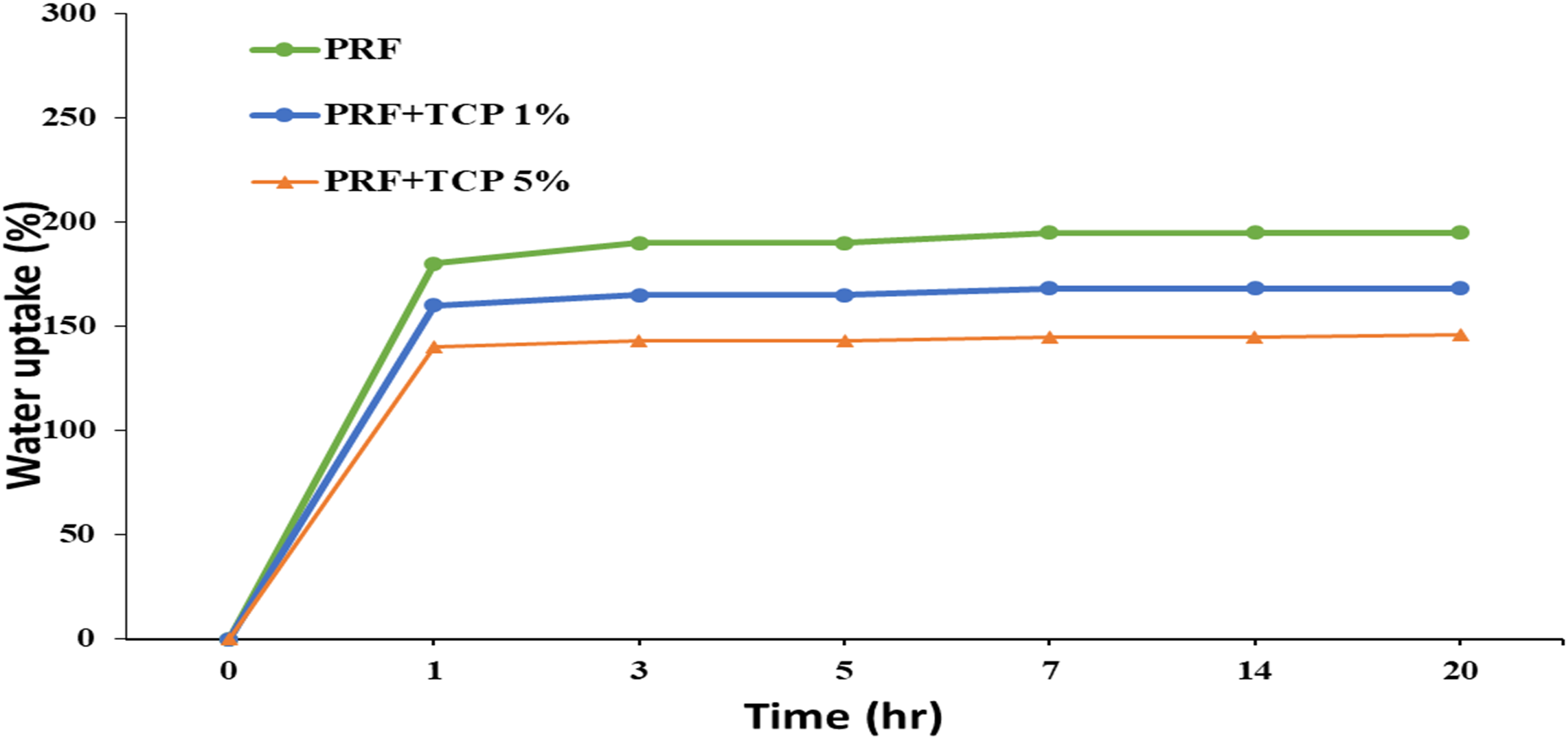
Weight loss test
All samples showed a relatively constant degradation rate over a period of 7 days (Figure 3). The slope of the graph is linear; after the seventh day, the amount of weight loss was 40%, 30%, and 2% in PRF, PRF/TCP1%, and PRF/TCP5% groups respectively. The degradation rate of the scaffolds in different experimental groups over a period of 7 days.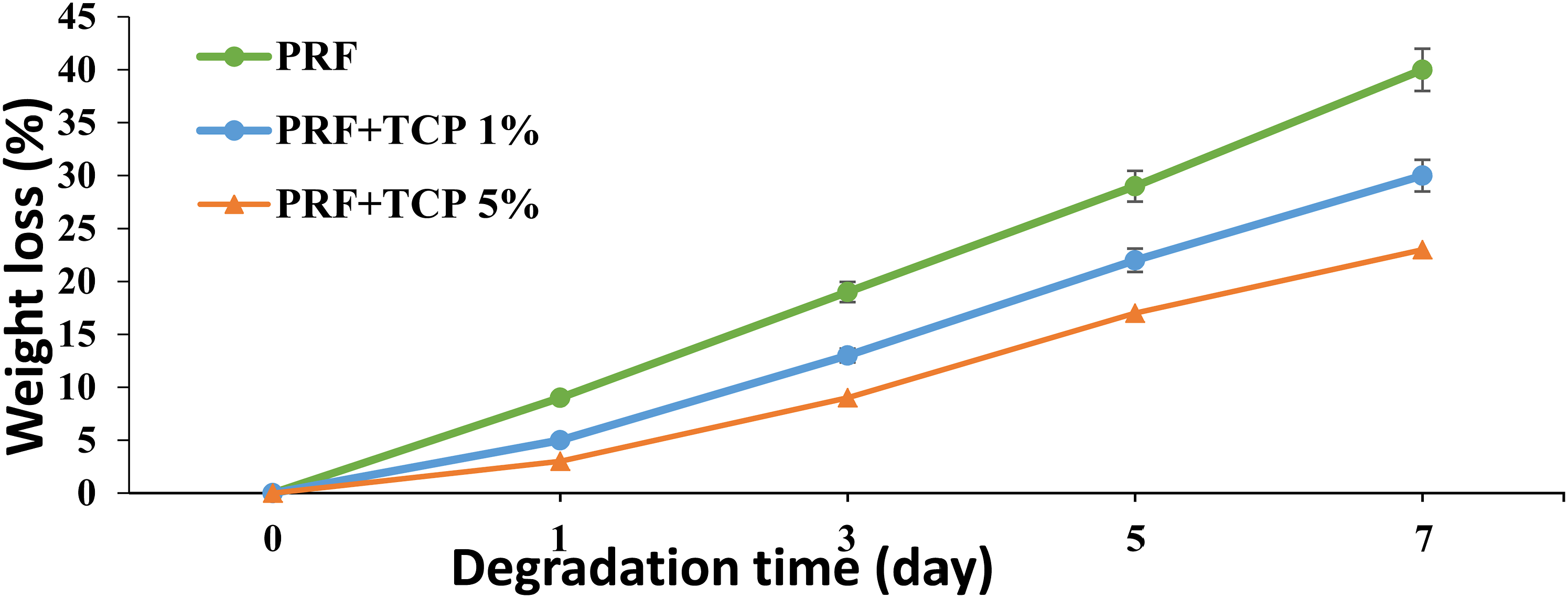
MTT cell viability
Biocompatibility evaluation of dental pulp stem cells on scaffolds of experimental groups and control by MTT test showed the viability of cells in the scaffold increased over time in all groups (Figure 4). On each day three types of scaffolds that are compared to the control group show almost the same amount of viable cells and according to the results, no significant relation is found regarding the scaffold type or scaffold existence in cell viability (p > 0/05). Biocompatibility evaluation of dental pulp stem cells on scaffolds of experimental groups and control by MTT test, * indicates p < 0.05 and the absence of asterisk indicates no significant relation between data.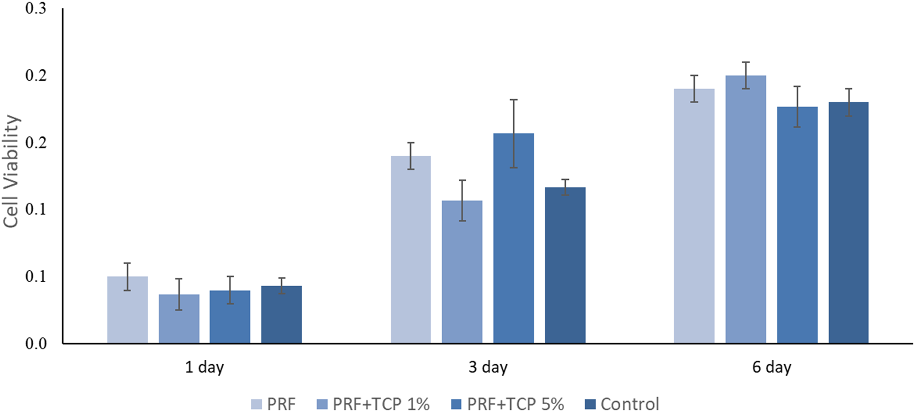
Fourier transform infrared analysis
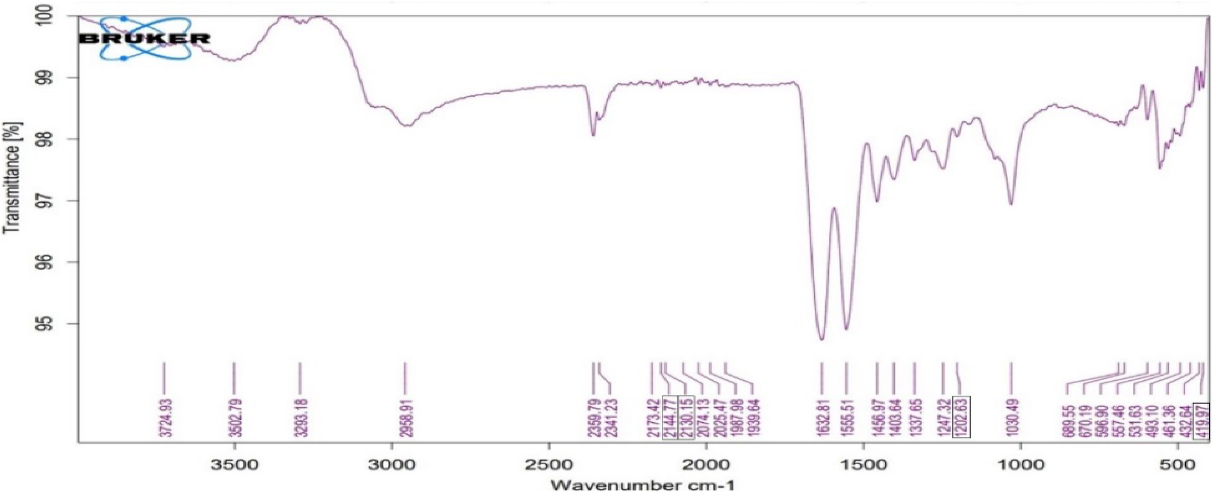
FTIR spectrum was used to confirm the chemical structure of the scaffolds. Figures 5 and 6 shows the FTIR spectrum of scaffolds in absorbance mode. In confirmation of the presence of PRF, there are fibrin index peaks as follows: around 1632 cm− 1 (amide I: C = O tensile vibrations), around 1554 cm− 1 (amide II: N–H flexural vibrations in the plane and C–N tensile vibrations) and around 1458 cm− 1 (amide III: C–N tensile vibrations). In confirmation of the presence of TCP nanoparticles in the PRF/TCP sample, the prominent bands at around 2144.77 cm−1 and 2130.15 cm−1 correspond to the structural O–H stretching of the nanomaterials. The absorption peaks at around 419 cm−1 and 1202 cm−1 in PRF/TCP which are in good agreement with literature values.
18
FTIR spectrum of PRF. FTIR spectrum of PRF/TCP hydrogel.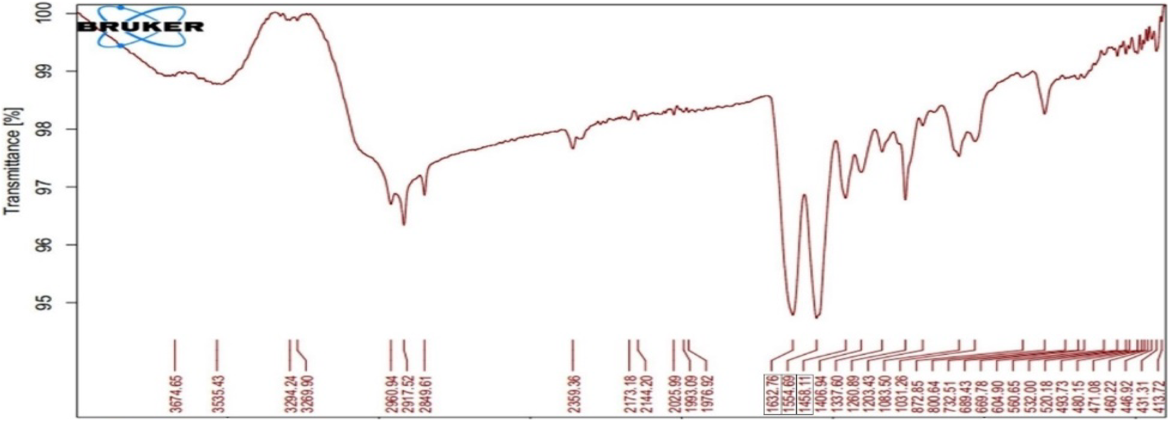
Histological evaluation for cell removal in decellularized PRF
Analysis of cell removal was done using H&E staining. Based on Figure 7 there was an absence of stained cell nuclei in decellularized PRF. Evaluation of cell removal from decellularized PRF by H&E staining with 10X magnification.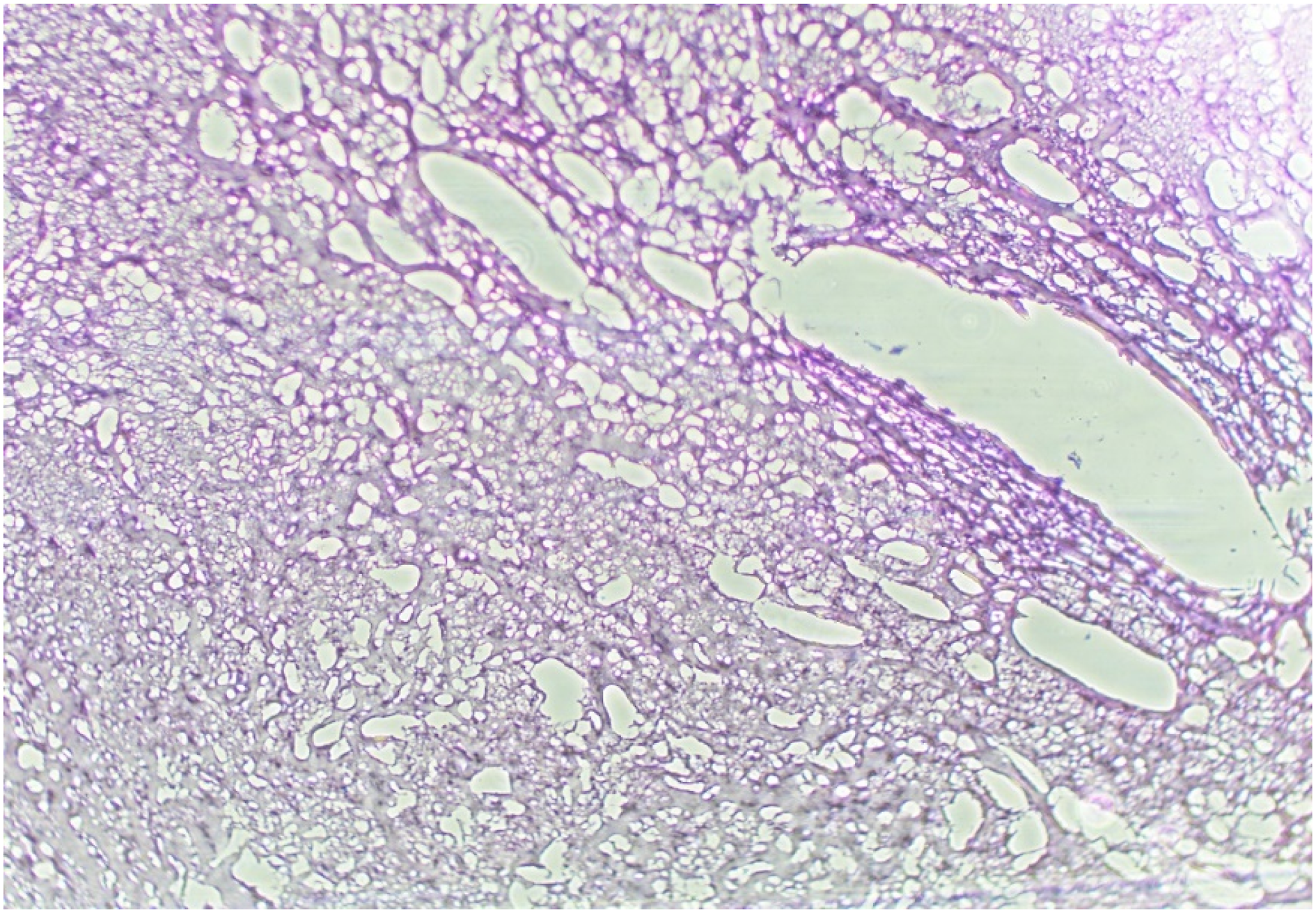
Hematoxylin and eosin staining
Histological images are showing Hematoxylin and eosin staining of PRF, PRF/TCP scaffolds and negative control group after a period of 2 and 4 weeks (Figure 8). No signs of inflammation or infection were observed in the affected area. In the second week woven and lamellar bones in the PRF and PRF/TCP were observed, while in the control group, the lesion area often contained connective tissue and osteoid. After 4 weeks, the area occupied by bone increased in all groups compared to the second week. H&E staining images in groups treated with PRF, PRF/TCP scaffolds and negative control group in the second and fourth weeks after treatment with scaffolding.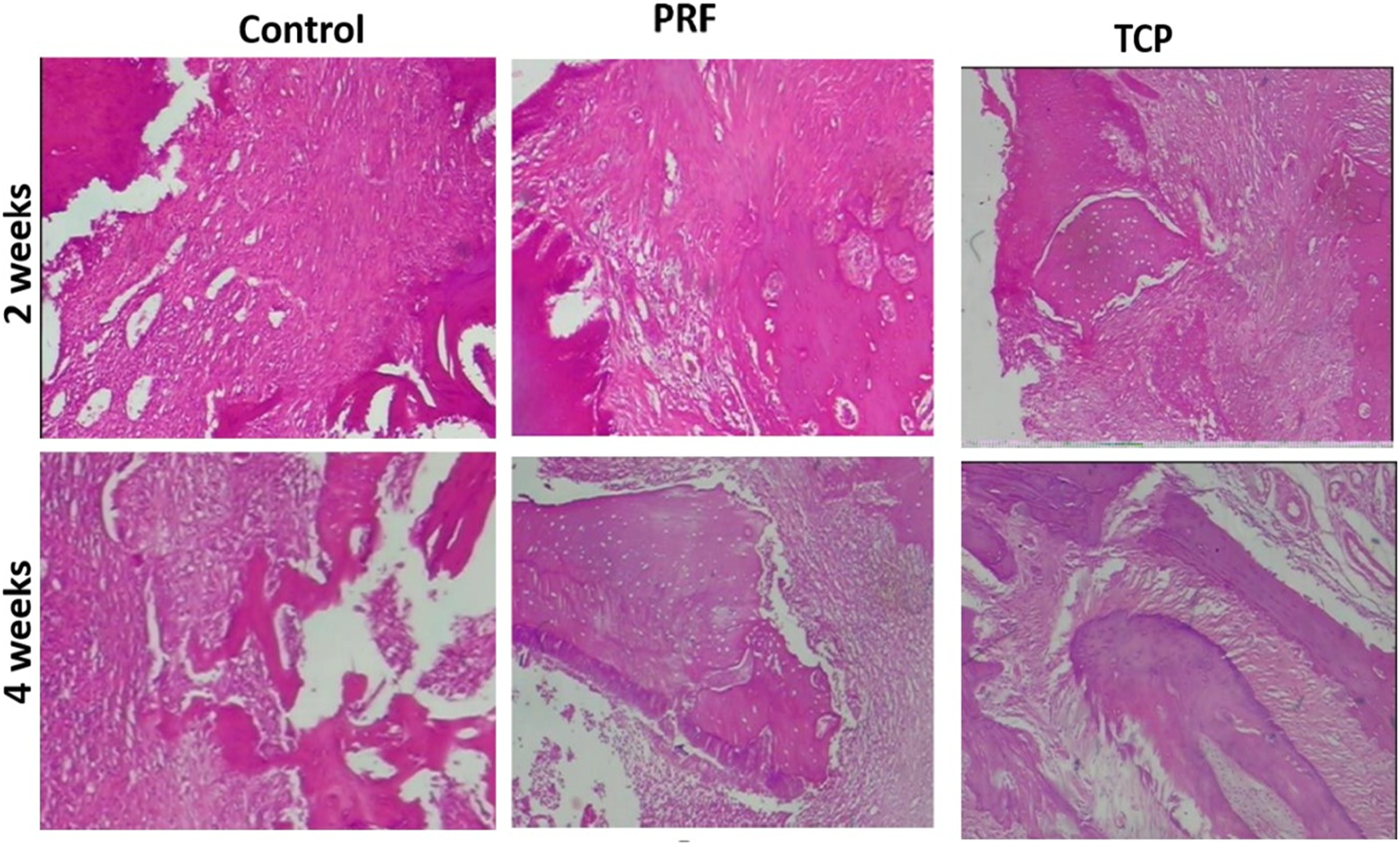
Also due to the production of extracellular matrix by bone cells, the percentage of area occupied by connective tissue is reduced in the fourth week compared to the second week, especially in the control group.
Scientifically, the amount of bone formation on day 28 in the TCP/PRF group is more than the negative control group.
Trichrome masson staining
Figure 9 shows Mason trichrome staining of the TCP group and the negative control at 14 and 28 days. As can be seen in the figure, on day 14, although there was no significant bone regeneration in both groups, the amount of regeneration in the TCP group was higher than the control group. Trichrome Masson staining images in groups treated with TCP scaffolds and negative control group in the second and fourth weeks after treatment with scaffolding.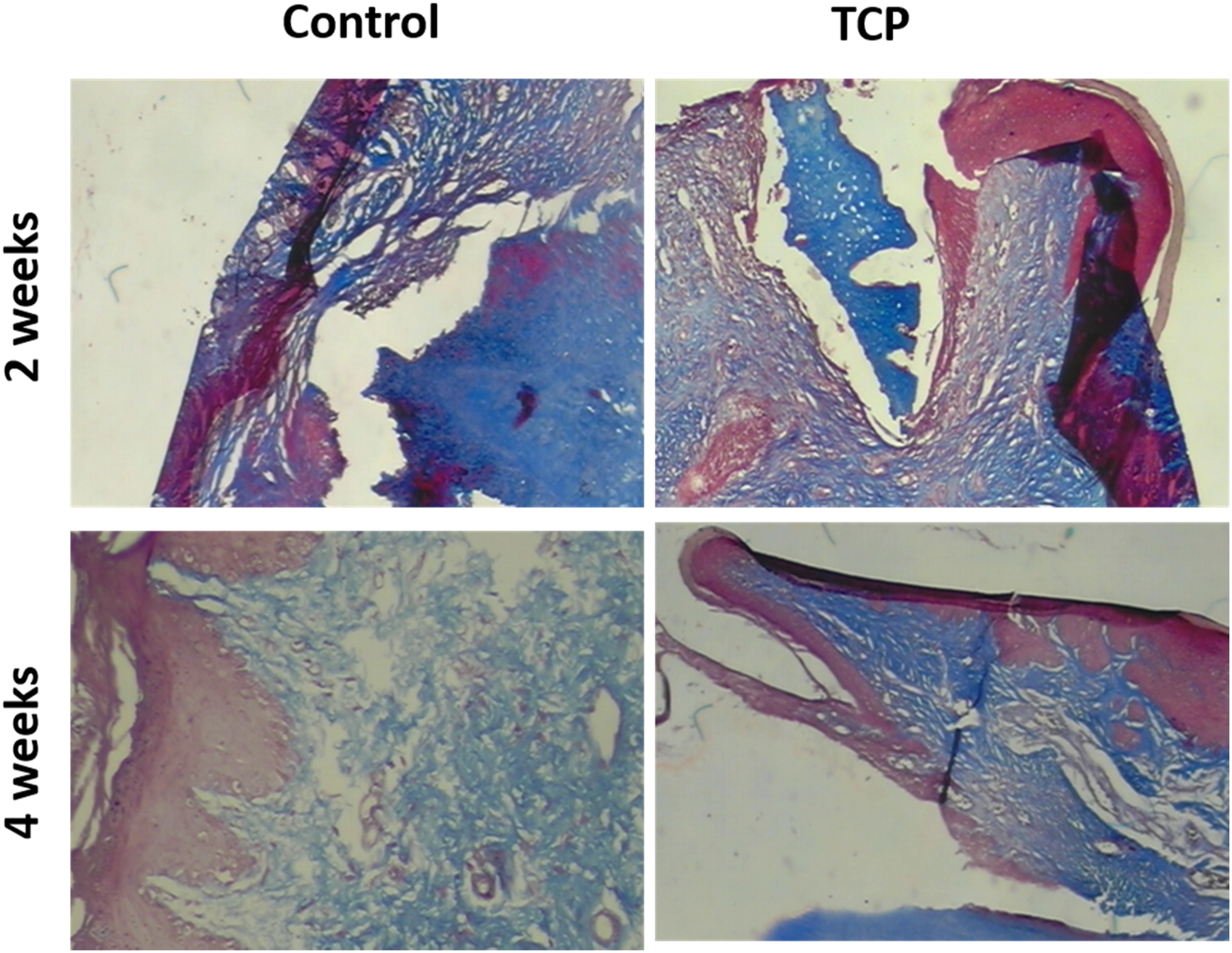
In the images of Masson’s trichrome staining on day 28, the abnormal and discontinuous pattern of collagen I fibers without alignment can be recognized in some areas of the control sample. In contrast, collagen I fibers showed proper organization in almost parallel patterns similar to normal tissue after 28 days in the TCP group (Figure 9). Therefore, it seems that on the 28th day, The arrangement and regeneration of collagen I fibers in the TCP group are better than the control group.
qRT- PCR analysis
As is shown in Figure 10: The expression of genes that are involved in bone formation and remodeling significantly increase in PRF/TCP compared to the two other groups. The OSX and RUNX2 genes express almost two times more in PRF/TCP group in comparison with the negative control group (p < 0.001). The Osteocalcin (OCN) expression in PRF/TCP is also 1.5 times more compared to the negative control group (p < 0.01). Evaluation of osteogenic gene expression in experimental groups and control by PCR test, *p < 0.05, **p < 0.01, ***p < 0.001 and the absence of asterisk indicates no significant relation between data.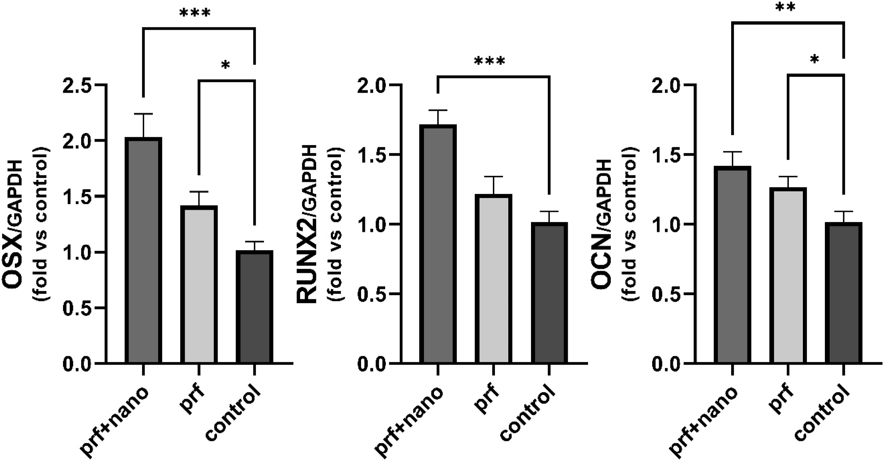
PCR analysis of two dominant genes in dentin formation (DMP-1 and DSSP) is shown in Figure 11. As can be seen, there is no reasonable relation in dentinogenesis gene expression between the three examined groups (p > 0.05). Evaluation of expression of genes that stimulate dentin formation in experimental groups and control by PCR test, *p < 0.05 and the absence of asterisk indicates no significant relation between data.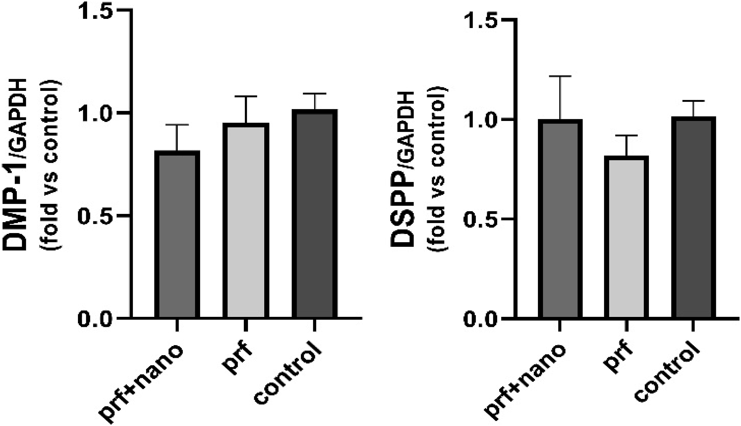
Discussion
This study was performed to determine the efficacy of PRF and β-TCP Nanoparticles in preserving and repairing extraction sockets. After tooth extraction, different grafting materials are used in socket repair to obtain optimal alveolar ridge volume, which is essential for an esthetic and functional outcome of implant placement. Although autogenous bone is considered the gold standard graft among other graft materials, the surgical procedure is required and cannot be prepared in an unlimited amount. To avoid such complications, the focus is being placed on other graft materials. 19 Also finding the synergic effect of two different types of bone graft materials can benefit us with the advantages of both and also facilitate bone remodeling and bone repair, even more, when they are used together.
In this study, we converted the β-TCP Nanoparticles and PRF into an injectable hydrogel phase and evaluated the histological changes in bone healing after 2 and 4 weeks following socket preservation using a rat first maxillary molar extraction socket model. The excellent injectability of the hydrogel system facilitated its easy fitting into any irregular extraction socket through a syringe. In 2006, Boix et al. 20 assessed the efficacy of a ready-to-use injectable bone substitute on the prevention of alveolar ridge resorption after tooth extraction. After a 3 months healing period, resorption in filled maxillary sites with biphasic calcium phosphate (BCP) was significantly lower than in negative control sites in the dog animal model. Interestingly, alveolar ridge augmentation was measured in mandibular-filled sockets including 30% of newly-formed bone. It was concluded that an injectable bone substitute composed of a polymeric carrier and calcium phosphate can significantly increase alveolar ridge preservation after tooth extraction.
The degradation of the scaffolding materials left space to allow new bone tissue ingrowth. The long-term presence of residual non-resorbable or slowly resorbable particles of the graft might interfere with the bone healing mechanism. 21 The main degredaion mechanism of the scaffolds are hydrolysis so scaffolds must therefore absorb sufficient water so as to have a proper degradation rate inside the body. In addition, water absorption improves cell nutrition and proliferation and prevents the scaffold from collapsing. Although the PRF scaffolding alone has a faster degradation rate than we expect to be appropriate for socket preservation in our animal model, by adding TCP nanoparticles the degradation rate will decrease which would be more acceptable in comparison with PRF alone. The weight loss of PRF/TCP 5% after 1 week is about 20% as our scaffolding material degrades with approximately the linear slope, we can assume that after 4 weeks almost 80% of the grafting material will degrade, which is suitable for the rat model because bone remodeling is faster than human beings in rats.
In 2021, Sareethammanuwat et al. 22 supported our findings and hypothesized that a high concentration of β-TCP was related to a low breakdown rate of the chitosan/collagen hydrogel scaffold. It can be explained that increasing ionic crosslinks between positively charged ions in the β-TCP and other components, particularly the negative charge of the bGP-chitosan/collagen matrix, cause extensive electrostatic attraction. 22
Based on our results the addition of nanoparticles to the scaffolds reduces the swelling rate; In explanation, it can be said that the application of nanoparticles increases the surface roughness and thus increases the contact angle, thus the surface acquires hydrophobic properties. 23
To investigate the possible interactions between PRF and TCP, FTIR analysis was applied. The results show that the FTIR analysis of TCP/PRF in comparison with the analysis of PRF shows almost the same picks which means the mixture of TCP and PRF shows no reaction and they work as two individual substances.
Evaluating the TCP and the PRF cytotoxicity and cell viability and proliferation on these scaffolds are important aspects to investigate their biocompatibility and effectiveness of them in bone regeneration. For the proper evaluation the MTT analysis has been conducted; our results demonstrate the increase in cell proliferation in all experimental and control groups over time and almost similar cell proliferation on each day which are positive criteria that show that PRF and the different concentrations of TCP have no reasonable effect on cell viability.
The right maxillary first molar of each rat was extracted and the sockets were filled with TCP/PRF and PRF scaffold alone. After 14 and 28 days, the maxillary bones were fixed and histologically processed for Mason’s trichrome and H&E staining.
According to the histological images, H&E images show that the amount of ossification and the surface occupied by bone on weeks two and four in the PRF/TCP 5% group were higher than the PRF group. These results can be due to the effect of TCP nanoparticles on the ossification process. So far, the application of TCP-based biomimetic scaffolds has been widely researched to promote new bone tissue growth and has shown positive results. Woven and lamellar bones as well as fibrin residues in the PRF and PRF/TCP were observed, while in the control group, the lesion area often contained connective tissue and steroids. On day 28, the area occupied by bone increased in all groups compared to the second week. Also the reason for the production of extracellular matrix by bone cells, is the percentage of the area occupied by connective tissue. On the 28th day, compared to the 14th day, it decreased and it was observed more in the control group. Scientifically, the amount of bone formation on day 28 in the TCP group is more than in the negative control group.
Images of Masson’s trichrome staining on day 28 shows the abnormal and discontinuous pattern of collagen I fibers without alignment in some areas of the control sample. In contrast, collagen I fibers showed proper organization in almost parallel patterns similar to normal tissue after 28 days in the TCP group. Therefore, it seems that on the 28th day, the organization and regeneration of collagen I fibers in the TCP group is better than the control group.
PCR is a highly accurate way to diagnose genetic changes. 24 The tests work by finding the DNA or RNA of cells in the samples. The control group is the empty socket and the experimental groups are the sockets in which PRF and PRF/TCP have been injected.
qRT PCR was performed to evaluate the activity of proteins that play an important role in osteogenic activity from the osteoblast differentiation stage (OSX &RUNX2), to its mineralization (OCN). 25 OSX, OCN and RUNX-2 were expressed significantly higher in the TCP/PRF and PRF groups in sequence compared to the control group. On the other hand, the expression of the most dominant protein for the initiation of dentin formation (Dentin matrix protein 1) and maintaining dentin formation procedure (DSPP) were observed. There was no significant relationship between the dentinogenesis gene expression and our scaffolding materials which shows the correct guidance of cell differentiation in dental socket with the presence of TCP/PRF.26, 27
Autogenous bone is considered the gold standard for bone augmentation. To approve the osteogenic properties of a scaffold it can be an appropriate way to compare it to autogenous bone. Three following studies are evaluating the β-TCP scaffold as so; in horizontal bone, augmentation simultaneously to implant placement using β-TCP or bovine bone, histological evidence concluded that no significant differences were evident at 6 months post surgery. 28 The formation of new bone in maxillary sinuses in which β-TCP was grafted was compared to sinuses regenerated with autologous bone at 6 months, and no statistical differences were found regarding the new bone density. 29 Similarly, another RCT did not observe significant differences between the β-TCP and autologous bone groups at 6 months. 30 In 2022 Putri et al. 25 compared the effectiveness of alveolar reconstruction utilizing human cancellous freeze-dried graft (HCG), beta tricalcium phosphate (β-TCP) and autogenous bone graft. A 5 × 5 mm alveolar defect in 36 male Wistar rats were treated One week after surgery, β-TCP was higher in RUNX2, OSX, ALP, and BMP2 than autogenous bone graft and HCG. Micro-CT revealed that H2 had a higher trabecular thickness of bone than two other scaffolds 8 weeks after surgery. (p < 0.05).
According to our results TCP in combination with PRF is suitable for socket reconstruction because they: 1) have the similar properties of autogenous bone without the need for surgery in the donor site; 2) are available in unlimited quantity and consistent quality; 3) have a highly porous architecture that facilitates the infiltration and sediment of new osteoid and bone trabeculae; 4) have considerably biocompatible characteristics 5) the hydrophilicity properties helps to dispose of the waste materials and nutrition absorption; 6) have a degradation rate coordinate to the natural socket healing process; 5) do not evoke an undesirable prolonged inflammatory response; and 6) lead to osteogenic gene expression. 31
Limitations and suggestions
1. Undeniably by extending the time of the study, subtle alterations in the process of osteogenesis can be determined more definitely. The period in which the histological evaluation has been checked out was relatively short which was due to ethical considerations. To precisely determine the newly formed mature bone 4–6 months study is suggested. 2. We need a more developed animal model in future studies in which the bone healing process is more similar to human bone healing, in the rat model the bone regeneration is notably faster and the criteria might be different from what is suitable for human socket preservation such as scaffold degradation time. 3. As the PRF was extracted from humans, the decellularization process is necessary and following this procedure the osteoinductive and mechanical properties might differ from the autograft which is going to be used in the human model in the future. 4. Mechanical tests including tensile and compressive strength is suggested to be done in future studies. Although using β-TCP in Nanoscale has an extreme improvement in mechanical properties, according to other studies, the main disadvantage of this graft material is its poor mechanical strength.
28
Biphasic calcium phosphates with different HA/β-TCP ratios, may improve the mechanical properties and maintain a good reabsorption capacity which is suggested to be used as an alteration for β-TCP alone in the highly loaded sites.
32
5. Finally, in vivo human studies for therapeutic purposes, elimination of ridge resorption and higher success rate for implant placement can be intended.
Conclusion
The present study demonstrated that the gelatin hydrogel containing TCP nanoparticles and fibrin scaffold led to more bone formation than that of the PRF scaffold and negative control group after tooth extraction. Therefore, the injectable PRF\TCP hydrogel is a promising candidate for bone repair and regeneration.
Footnotes
Declaration of conflicting interests
The author(s) declared no potential conflicts of interest with respect to the research, authorship, and/or publication of this article.
Funding
The author(s) received no financial support for the research, authorship, and/or publication of this article.