
Abstract
Background
Lenvatinib (LEN) is a first-line therapy for patients with hepatocellular carcinoma (HCC), but has a larger adverse effect profile. In this study, we developed a liposome with drug-carrying function and magnetic resonance imaging (MRI) imaging function to investigate the targeted drug-carrying function and MRI tracing ability of liposome for HCC.
Methods
Magnetic nano-liposomes (MNL) with dual targeting function of epithelial cell adhesion molecule (EpCAM) and vimentin and capable of encapsulating LEN drugs were prepared. The characterization performance, drug loading efficiency and cytotoxicity of EpCAM/vimentin-LEN-MNL were tested, and the dual-targeting slow release drug loading function and MRI tracing ability were investigated in cellular and animal models.
Results
EpCAM/vimentin-LEN-MNL has a mean particle size of 218.37 ± 5.13 nm and a mean potential of 32.86 ± 4.62 mV, and is spherical in shape and can be uniformly dispersed in solution. The encapsulation rate was 92.66 ± 0.73% and the drug loading rate was 9.35 ± 0.16%. It has low cytotoxicity, can effectively inhibit HCC cell proliferation and promote HCC cell apoptosis, and has specific targeting function and MRI tracing ability for HCC cells.
Conclusions
In this study, an HCC-specific dual-targeted sustained-release drug delivery liposome with dual-targeted recognition and sensitive MRI tracer was successfully prepared, which provides an important scientific basis for maximizing the multiple effects of nano-carriers in tumor diagnosis and treatment.
Introduction
Hepatocellular carcinoma (HCC) is one of the most common cancers worldwide, and the third leading cause of cancer-related death globally, and the healing outcome of advanced HCC is particularly poor.1,2 Therefore, the development of a targeted drug for HCC, especially for advanced HCC, has significant social and economic benefits. Lenvatinib (LEN) is a multi-targeted tyrosine kinase inhibitor that exhibits potent anti-angiogenic and tumor proliferation inhibiting abilities in vitro and in vivo.3,4 LEN clinical remission rate was 327% of sorafenib (SOR) (40.6% vs. 12.4%) and progression-free survival was 203% of SOR (7.3 months vs. 3.6 months). Therefore, LEN is significantly superior to the previous generation of HCC targeting drugs SOR. 5 Despite the excellent clinical results of LEN, it still has some adverse effects.6,7 Some patients experienced adverse effects such as hypertension and arterial thromboembolism with LEN. 8 In phase III clinical studies of patients with HCC treated with LEN, the incidence of adverse reactions with LEN was 99% in all cases, 3 leaving only the option of reducing the frequency of dosing or changing to another drug. Therefore, there is an urgent need to develop a novel drug formulation with good biocompatibility and high tumor targeting to deliver effective drugs directly and safely to tumor lesions.
Liposomes are closed vesicles constructed from phospholipids and/or cholesterol as the basic membrane material and have a bilayer structure similar to that of biological membranes. 9 Liposomes have been widely used in drug delivery systems with the advantages of good biocompatibility, biodegradability, non-toxicity and non-immunogenicity.10,11 By encapsulating the drug in liposomes, the liposomes prevent dilution and degradation or inactivation of the drug in the bloodstream. 12 Fe3O4 nano-particles modified by polyethylene glycol (PEG) can increase the water solubility and biocompatibility of drug-loaded materials, avoid the recognition of reticuloendothelial system, prolong the circulation time of drug-loaded materials in vivo, etc. 7 Fe3O4 nano-particles can be metabolized in vivo, and the material itself can be used to treat anemia, such as the “Feraheme” drug, which can be applied in vivo.13,14 Studies have shown that when magnetic nano-materials are used as contrast agents for MRI, they can form multifunctional drug delivery and imaging systems, and take advantage of the special microenvironment of tumor tissues to design and prepare “smart” nano-drug release systems to improve the therapeutic and imaging effects at the lesion site.15,16 The combination of two materials with their respective advantages into composite materials has become a current research hotspot, and can be applied to magnetic targeting drug transport and bioimaging, to achieve the integration of diagnosis and treatment.
In recent years, it has been found that tumor cells undergo epithelial-mesenchymal transition (EMT) during their entry into the peripheral blood circulation. 17 Tumor cells under the regulation of EMT-related transcription factors cause down-regulation of epithelial cell adhesion molecule (EpCAM) protein expression and up-regulation of vimentin protein expression,18,19 suggesting that the conversion of EMT from epithelial to mesenchymal phenotype leads to high expression of vimentin and loss of EpCAM. Therefore, the combined use of EpCAM and vimentin as HCC capture targets provides a more comprehensive capture of HCC tumor cells. In this study, we designed to combine EpCAM and vimentin as targets for the modification of magnetic nano-liposomes (MNL) to jointly construct dual-targeted functional MNL encapsulating LEN drugs (EpCAM/vimentin-LEN-MNL), with the aim of not only improving drug efficacy in HCC, but also serving as MRI contrast agents for the localization and diagnosis of malignant tumors.
Methods
Cells and reagents
Human HCC cell lines (Hep3B and Huh-7), human umbilical vein endothelial HUVEC cells and human biliary epithelial HIBEC cells were obtained from the Cell Bank of the Chinese Academy of Sciences (Shanghai, China). RPMI-1640 and DMEM medium, N-Hydroxysuccinimide (NHS), 1-Ethyl-3-(3-dimethyllaminopropyl) carbodiimide hydrochloride (EDC.HCl), fetal bovine serum and trypsin were obtained from the Thermo Fisher Scientific (China) Co., Ltd Distearoyl phosphatidyl ethanolamine-polyethylene glycol (DSPE-PEG) were obtained from the Shanghai Koether Chemistry Technology Co., Ltd Glycidyl hexadecyl dimethylammonium chloride (GHDC) and 1,2-dioleoyl-sn-glycero-3-phosphocholine (DOPC) were obtained from the TCI (SHANGHAI) Development Co., Ltd Octadecy l quaternized carbox ymethyl chitosan (OQCMC) were obtained from the Jukang (Shanghai) Biotechnology Co., Ltd Cholesterol, methylene chloride and other common reagents were obtained from the Sinopharm Chemical Reagent Co., Ltd
Preparation of drug-carrying microspheres
56.8 mg Fe3O4, 10 mg DSPE-PEG, 15 mg cholesterol, 10 mg DOPC, 15 mg HQCMC, 15 mg GHDC and 15 mg LEN were added to the pear-shaped vial, along with 4 ml dichloromethane. The solution was sonicated using a probe sonicator and sonication was paused after 30 s and 12 ml of water was added to the mixed solution. Ultrasound was continued for the emulsification reaction, with a pause of 1 s after each ultrasound for 2 s, and the reaction was continued for 6 min. The solution was rotary evaporated by rotary evaporator for 30 min at 0.8 MPa and room temperature to remove dichloromethane to obtain 12 ml of MNL solution encapsulated with LEN drug (LEN-MNL). Take 1 ml of LEN-MNL solution, add 2.1 mg NHS and 2.1 mg EDC-HCl coupling agent and mix thoroughly. Finally, 60 μg of EpCAM and 60 μg of vimentin antibody were added to the mixed solution and the chemical coupling reaction was carried out at 4°C. The EpCAM/vimentin-LEN-MNL was obtained after 12 h of reaction.
Characterization test and microstructure analysis of drug-carrying microspheres
The 10 μL of the sample was diluted in 1 mL of distilled water, and the particle size and potential of the sample were detected by particle size analyzer. The infrared spectra of the samples were detected by fourier transform infrared spectroscopy (FITR). The crystalline properties of the samples were examined by X-ray diffraction (XRD). The magnetization curve of the sample is detected by a vibrating sample magnetometer (VSM). The microsphere morphology of the samples was observed by atomic force microscopy (AFM), scanning electron microscopy (SEM) and transmission electron microscopy (TEM).
Encapsulation rate, drug loading rate and in vitro release assay
The UV detection wavelength of LEN was determined by scanning the full wavelength from 200 to 600 nm for 0.1mg/ml of LEN drug solution using a UV spectrophotometer. A standard solution of 0.01 mg/ml LEN was prepared and diluted to concentrations of 0.1 μg/ml, 0.5 μg/ml, 1 μg/ml, 5 μg/ml and 10 μg/ml, respectively. UV tests were performed on these solutions using a UV-Vis spectrophotometer at 481 nm to plot the standard curve for LEN. The encapsulation rate and drug loading were measured by standard curve: actual loading rate (%) = (total drug mass - unencapsulated drug mass)/total mass of drug-laden liposomes × 100%. Drug encapsulation rate (%) = (total drug mass - unencapsulated drug mass)/total drug mass × 100%. Using PBS as the release medium, dialysis bags containing 2 ml of PBS were placed in the release medium and equilibrated for 12 h. 200 μl of EpCAM/vimentin-LEN-MNL was added to the dialysis bag and the amount of free LEN in the release medium was measured at a fixed time point.
Cellular uptake experiments
Hep3B and HUVEC cells were inoculated in 6-well culture dishes (1 × 10⁵ cells/well) and incubated for 24 h. 10 μl of EpCAM/vimentin-LEN-MNL and LEN-MNL were added, respectively. After continuing the incubation for 8 h, staining was performed using Prussian blue staining kit (nuclear solid red method) and the specific binding effect of the material to the cells was observed under the microscope.
Cytotoxicity, proliferation, and apoptosis tests
HUVEC and HIBEC cells were inoculated in 96-well (1 × 10⁴ cells/well) plates for 24 h. 50 μL of different concentrations of EpCAM/vimentin-LEN-MNL, LEN-MNL, MNL and LEN were added, respectively. After incubation for 24 h, 15 μl of CCK8 solution was added to each well separately. The absorbance was measured at 490 nm by an enzyme marker. Cell survival rate (%) = (OD (materials) − OD (blank))/(OD (untreated) - OD (blank))×100%. Logarithmic growth phase Hep3B and Huh-7 cells were inoculated in 24-well (1 × 10⁵ cells/well) plates for 12h. EpCAM/vimentin-LEN-MNL, LEN-MNL, MNL and LEN were added at optimal concentrations of 30 μl, respectively. After 24 h of incubation, the cells were digested by adding trypsin and the cell suspension was collected by centrifugation at 3000 rpm for 3 min. Add 195 μl of annexin V-FITC conjugate to resuspend the cells and 5 μl of propidium iodide (PI) staining solution, respectively. Incubate for 20 min at low light and detect apoptosis by flow cytometry.
Cell enrichment efficiency test
Hep3B and Huh-7 cells were added to 2 ml of PBS solution and blood and mixed well (50 cells/ml), and 20 μl of EpCAM/vimentin-LEN-MNL, EpCAM-LEN-MNL, vimentin-LEN-MNL, and LEN-MNL were added, respectively. Incubate for 20 min and then place on a magnetic separation rack, magnetically separate for 15 min and aspirate the solution. Add 20 μl CK8/18/19-FITC, 20 μl DAPI and 20 μl CD45-PE, respectively, mix well and stain for 15 min protected from light. Wash twice with double distilled (dd) H2O, finally add 20 μl dd H2O and mix well, spread evenly on slides, observe and count under fluorescence microscope.
MRI contrast analysis
Hep3B cells were cultured in 6-well (1 × 10⁵ cells/well) plates for 24 h. 50 μl of EpCAM/vimentin-LEN-MNL and LEN-MNL were added, respectively. The concentration gradients were set at 10 μg/ml, 50 μg/ml, 80 μg/ml and 100 μg/ml. After 1, 3 and 5 days of culture, respectively, the medium was aspirated and discarded, 50 μl of fresh medium was added to mix the cells, and the cell solution was transferred to 8-lined PCR tubes for MRI testing. A Siemens 3.0 T magnetic resonance machine was used, with a human shoulder coil, normal cross-section, and coronal sweep. A SET1WI sequence was used. Specific parameters: T1WI (TR/TE300 ms/14 ms), echo chain length: 20–40, scan parameters: layer thickness 2 mm, layer spacing 2 mm, matrix 384 × 256, FOV 14 cm × 14 cm, NEX 2–4 times.
In vivo functional experiments
Hep3B cells (5 × 10⁷) were injected directly into the axilla of the right foreleg of 5-6 week old BALB/c female nude mice. Starting from day 7, two nude mice with similar tumor size were randomly selected every 5 days and injected with 100 μl EpCAM/vimentin-LEN-MNL and LEN-MNL via tail vein, respectively. After 3 h of injection, 500 μl of blood was taken through the eye, followed by execution of the mice and removal of the subcutaneous tumors. Measure the tumor length and width, weigh the tumor and take pictures, tumor volume = length × width2/2.
Statistical methods
GraphPad Prism 8.2.1 statistical software was used to process and analyze the experimental data, and each experiment was repeated at least three times, and the data were presented in Mean ± SD. p < 0.05 was considered as statistically significant (*), and p < 0.01 as extremely different (**).
Results
Material preparation process and experimental flow chart
Liposomes were prepared by degradable biomaterials such as DSPE-PEG, Fe3O4 particles were encapsulated in the outer layer of liposomes, and LEN drugs were encapsulated in the inner layer of liposomes. Subsequently, EpCAM/vimentin-LEN-MNL encapsulated with LEN drug was prepared by co-coupling EpCAM and vimentin antibodies on the surface of LEN-MNL (Figure 1(a), concentration of 15.72 mg/ml). The targeting performance, drug delivery performance and MRI tracer ability of EpCAM/vimentin-LEN-MNL were investigated by cellular experiments, and the in vivo function of EpCAM/vimentin-LEN-MNL was studied by animal experiments (Figure 1(b)). Preparation process and experimental flow of EpCAM/vimentin-LEN-MNL. (a) Flow chart of EpCAM/vimentin-LEN-MNL preparation; (b) Flowchart of experimental design.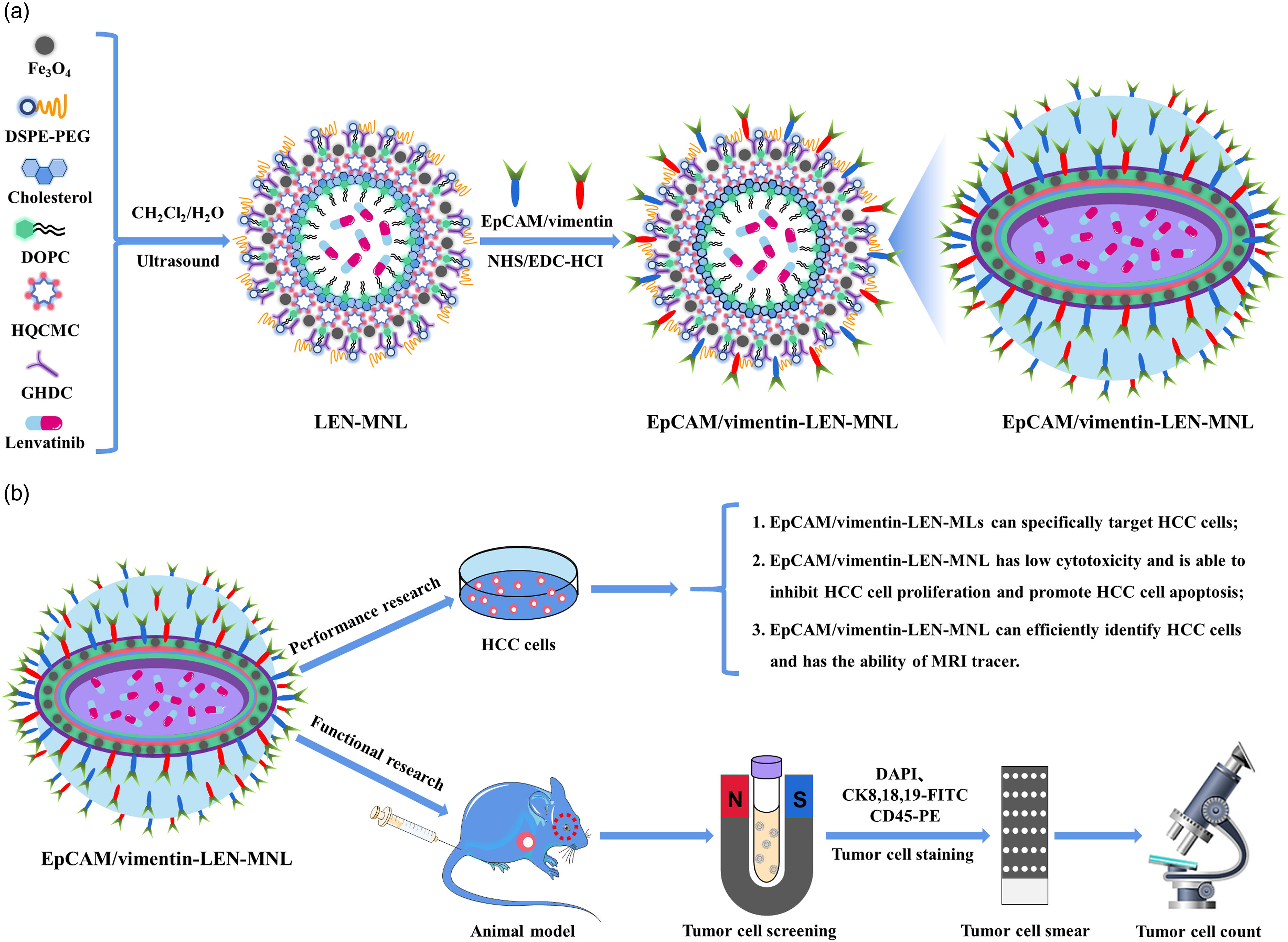
Characterization test and microstructure analysis of EpCAM/vimentin-LEN-MNL
The results showed that the average particle size of EpCAM/vimentin-LEN-MNL was 218.37 ± 5.13 nm, and the particle size was mainly distributed between 122.4 nm–396.1 nm with a polydispersity index (PDI) of 0.216 (Figure 2(a)). EpCAM/vimentin-LEN-MNL was positively charged with an average potential of 32.86 ± 4.62 mV (Figure 2(b)). EpCAM/vimentin-LEN-MNL, LEN-MNL, and MNL have characteristic absorption peaks at 1652.8 cm−1, 1714.1 cm−1, and 2849.55 cm−1 of FITR, respectively, which are the absorption peaks of the stretching vibrations of -CO-NH- in the amide bond and -C = O and -CH- in the ester group, respectively (Figure 2(c)). The test results of XRD showed that the typical diffraction peaks of Fe3O4 appeared at 2θ of 30.1°, 35.5°, 43.1°, 53.7°, 57.2° and 62.5°, corresponding to (220), (311), (400), (422), (511) and (440) crystal planes, respectively, which are consistent with the positions in the standard mapping of Fe3O4, indicating that the material contains Fe3O4 and has the potential function as an MRI contrast agent (Figure 2(d)). The saturation magnetization intensity of Fe3O4 is 58.58 A.m.2/Kg, and the saturation magnetization intensity of MNL, LEN-MNL and EpCAM/vimentin-LEN-MNL are 43.28 A.m.2/Kg, 41.37 A.m.2/Kg and 40.19 A.m.2/Kg, respectively (Figure 2(e)). The saturation magnetization intensity of Fe3O4 decreases significantly after being encapsulated, but still has a strong saturation magnetization intensity, indicating superparamagnetic properties. AFM, SEM and TEM all observed that EpCAM/vimentin-LEN-MNL was irregularly sphere-like with a diameter of about 200 nm and had the characteristics of liposomal vesicles. EpCAM/vimentin-LEN-MNL characterization tests and microstructure analysis. (a) Particle size distribution; (b) Potential distribution.; (c) FITR test; (d) XRD test; (e) VSM testing; (f) AFM observation morphology; (g) SEM observation morphology. (h) TEM observation morphology.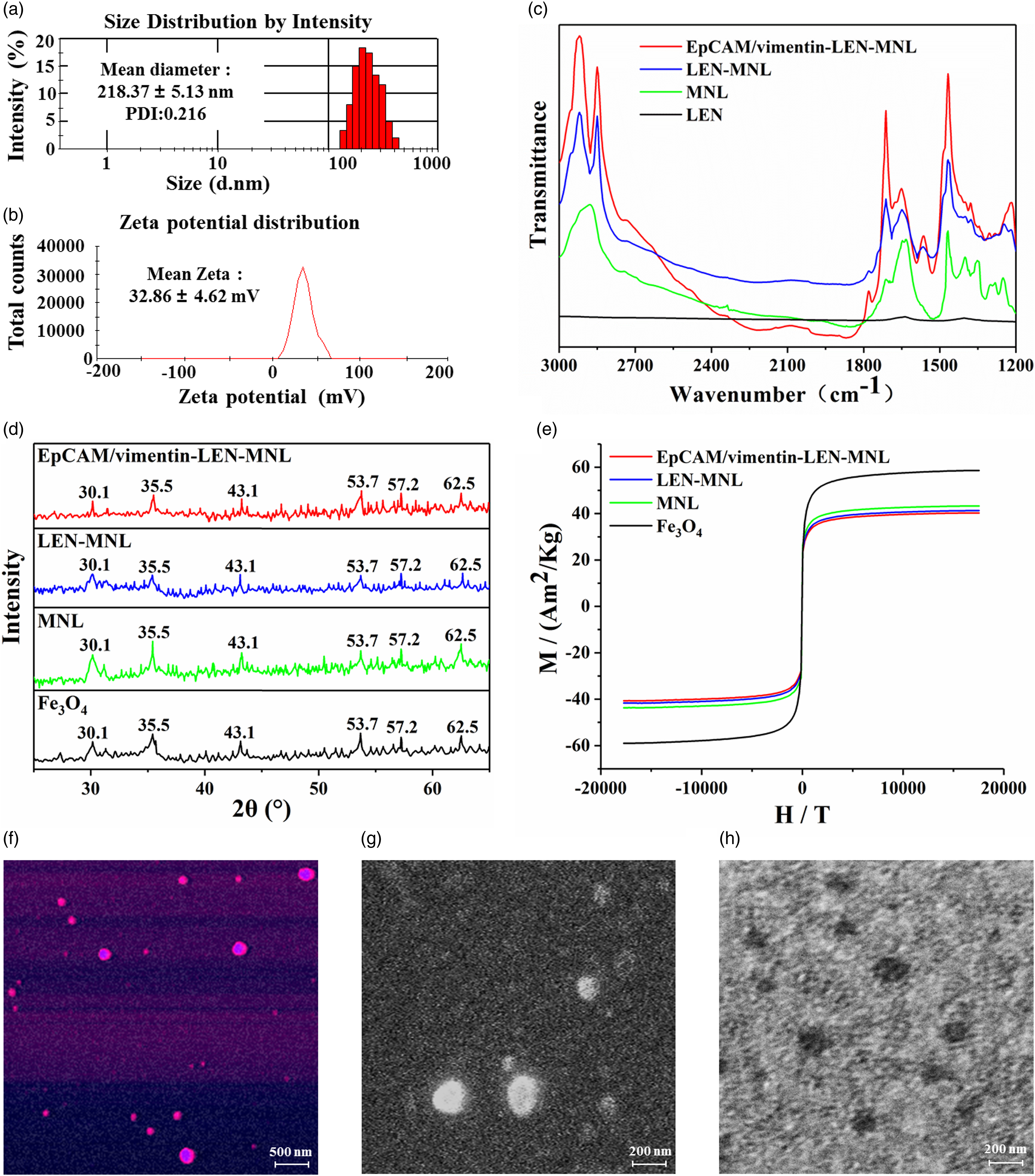
Encapsulation rate, drug loading rate and in vitro release efficiency
The absorption peaks of LEN were found at 279 nm, 342 nm, 437 nm and 481 nm, respectively, but there might be interference from the drug excipients within 280 nm. Therefore, the relatively high peak at 481 nm was chosen as the characteristic absorption peak of LEN (Figure 3(a)). The equation of the standard curve for LEN is: y = 0.11756 × −0.1377, R2 = 0.9992 (Figure 3(b)). The measured EpCAM/vimentin-LEN-MNL encapsulation rate was 92.66 ± 0.73% and the drug loading rate was 9.35 ± 0.16% (Figure 3(c)). At a pH of 7.4, the LEN drug was released at a rate of 84% within 24 h. Both EpCAM/vimentin-LEN-MNL and LEN-MNL had a release rate of about 30% within 24 h (Figure 3(e)). The drug was encapsulated by liposomes with a significant slow release effect, and the drug release rate increased in the acidic (pH = 5.3) release medium (Figure 3(d)).
EpCAM/vimentin-LEN-MNL can specifically target HCC cells
Prussian blue staining showed that in HUVEC cells, liposomes were distributed freely around the cells in both EpCAM/vimentin-LEN-MNL and LEN-MNL groups (Figure 3(f)). In Hep3B cells, the liposomes in the LEN-MNL group were all distributed freely around the cells, whereas in the EpCAM/vimentin-LEN-MNL group, the liposomes were adsorbed on the surface of Hep3B cells with only a small amount of free distribution (Figure 3(g)). It was shown that EpCAM/vimentin-LEN-MNL can specifically target and recognize Hep3B cells. Detection of the encapsulation rate, in vitro release efficiency and specific targeting ability of EpCAM/vimentin-LEN-MNL. (a) UV scan curve of LEN; (b) Standard curve of LEN; (c) Encapsulation rate and drug loading capacity of EpCAM/vimentin-LEN-MNL; (d) In vitro release profile of LEN (pH = 5.3); (e) In vitro release profile of LEN (pH = 7.4); (f) Prussian blue staining of EpCAM/vimentin-LEN-MNL after binding to HUVEC cells; (g) Prussian blue staining of EpCAM/vimentin-LEN-MNL after binding to Hep3B cells.
EpCAM/vimentin-LEN-MNL has low cytotoxicity and is able to inhibit HCC cell proliferation and promote HCC cell apoptosis
The cell survival rate gradually decreased with increasing concentration of EpCAM/vimentin-LEN-MNL. EpCAM/vimentin-LEN-MNL is safe to use at a concentration of 100 μg/ml and has a low cytotoxicity with cell survival rates above 85% when used in this concentration range (Figure 4(a) and (b)). EpCAM/vimentin-LEN-MNL is capable of slow release of LEN drug and has a long-lasting value-added inhibitory effect on both Hep3B and Huh-7 cells (Figure 4(c) and (d)). EpCAM/vimentin-LEN-MNL significantly promoted apoptosis in Hep3B and Huh-7 cells (Figure 4(e) and (f)). These results suggest that EpCAM/vimentin-LEN-MNL can efficiently inhibit HCC cell proliferation and promote HCC cell apoptosis with a slow drug release function. Cytotoxicity study of EpCAM/vimentin-LEN-MNL and the effect on proliferation and apoptosis of HCC cells. (a) Toxicity study of EpCAM/vimentin-LEN-MNL on HUVEC cells; (b) Toxicity study of EpCAM/vimentin-LEN-MNL on HIBEC cells; (c) Toxicity study of EpCAM/vimentin-LEN-MNL on the value-added of Hep3B cells; (d) Study of EpCAM/vimentin-LEN-MNL on the value-added of Huh-7 cells; (e) Study of EpCAM/vimentin-LEN-MNL on apoptosis of Hep3B cells; (f) Study of EpCAM/vimentin-LEN-MNL on apoptosis of Huh-7 cells.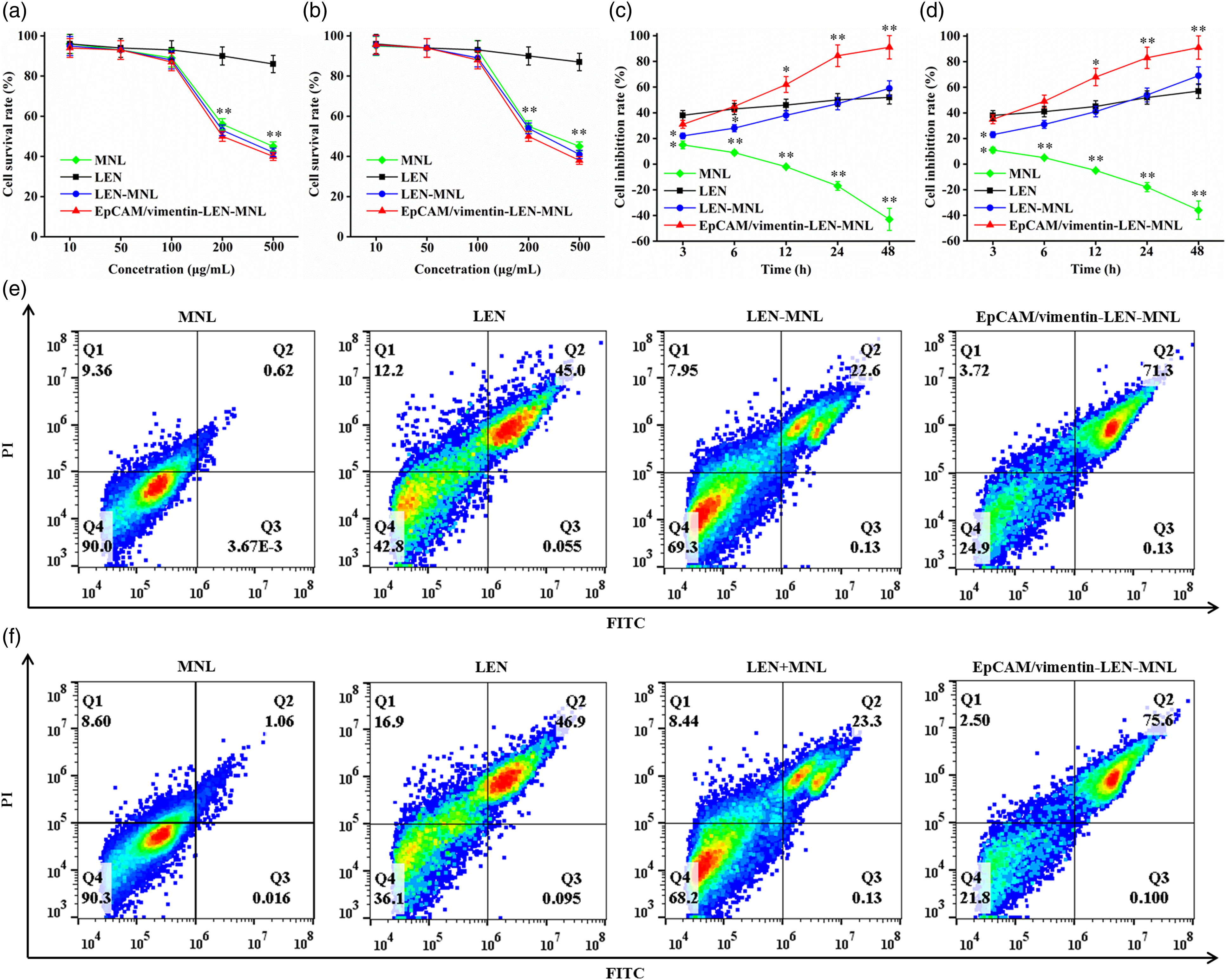
EpCAM/vimentin-LEN-MNL has dual targeting recognition function
To further confirm the dual target recognition function of EpCAM/vimentin-LEN-MNL, we comparatively investigated the target recognition ability of EpCAM-LEN-MNL and vimentin-LEN-MNL. The results showed that the specific capture efficiency of EpCAM/vimentin-LEN-MNL in PBS solution for Hep3B cells was significantly higher than that of EpCAM-LEN-MNL and vimentin-LEN-MNL (Figure 5(a)), and the results were verified in the blood simulation system (Figure 5(b)). The specific capture efficiency of EpCAM/vimentin-LEN-MNL in PBS solution for Huh-7 cells was significantly higher than that of EpCAM-LEN-MNL and vimentin-LEN-MNL (Figure 5(c)), and the results were verified in the blood simulation system (Figure 5(d)). In contrast, LEN-MNL has only a small non-specific adsorption capacity. Dual target recognition function study of EpCAM/vimentin-LEN-MNL. (a) Capture efficiency of Hep3B cells in the PBS system; (b) Capture efficiency of Hep3B cells in the blood simulation system; (c) In the PBS system, the capture efficiency of Huh-7 cells; (d) In the blood simulation system, the capture efficiency of Huh-7 cells.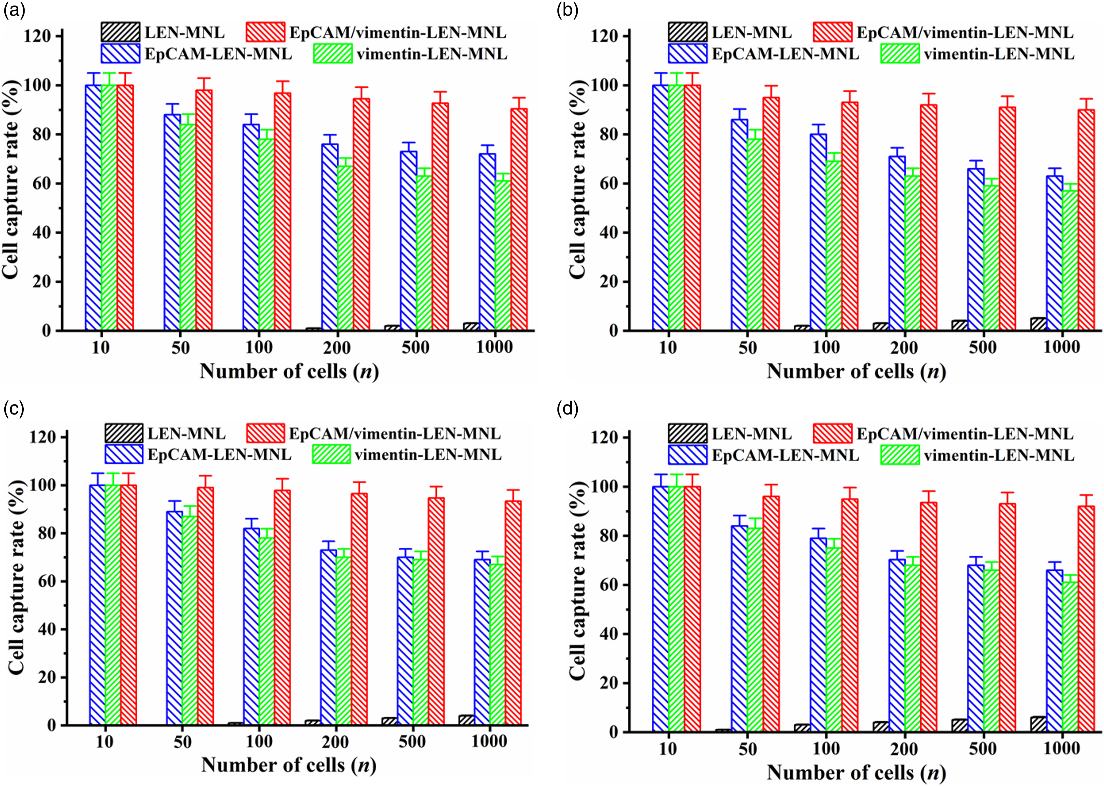
EpCAM/vimentin-LEN-MNL has MRI tracer ability and can effectively identify tumor cells in vivo
To further investigate whether dual-targeted nano-liposomes loaded with Fe3O4 could be used as an MRI targeted contrast agent for biomedical diagnostic applications, we incubated EpCAM/vimentin-LEN-MNL and LEN-MNL with Hep3B cells for different times, respectively. The results showed that EpCAM/vimentin-LEN-MNL co-cultured with Hep3B cells at a concentration of 80 μg/mL still had a significant MRI signal on day 5, and the MRI signal was gradually enhanced as the concentration of EpCAM/vimentin-LEN-MNL increased. Due to the Hep3B cell-targeting function of EpCAM/vimentin-LEN-MNL, the MRI signal of EpCAM/vimentin-LEN-MNL was attenuated compared with LEN-MNL (Figure 6(a)). Animal experiments showed that the tumors were increasing in size with time (Figure 6(b)). EpCAM/vimentin-LEN-MNL was injected into nude mice at different time points, and tumor volume and weight were measured after execution of the nude mice, and tumor cells were detected in the blood of the nude mice, respectively (Figure 6(c)). The number of tumor cells specifically recognized by EpCAM/vimentin-LEN-MNL was found to increase progressively with increasing tumor volume and body weight, while LEN-MNL only non-specifically adsorbed a very small number of tumor cells (Figure 6(d) and (e)). MRI tracer ability of EpCAM/vimentin-LEN-MNL and in vivo targeting ability to identify tumor cells were studied. (a) MRI test; (b) Tumor changes; (c) Identification of tumor cells in the blood of nude mice; (d) Relationship between tumor volume and the number of tumor cells in vivo; (e) Relationship between tumor weight and the number of tumor cells in vivo.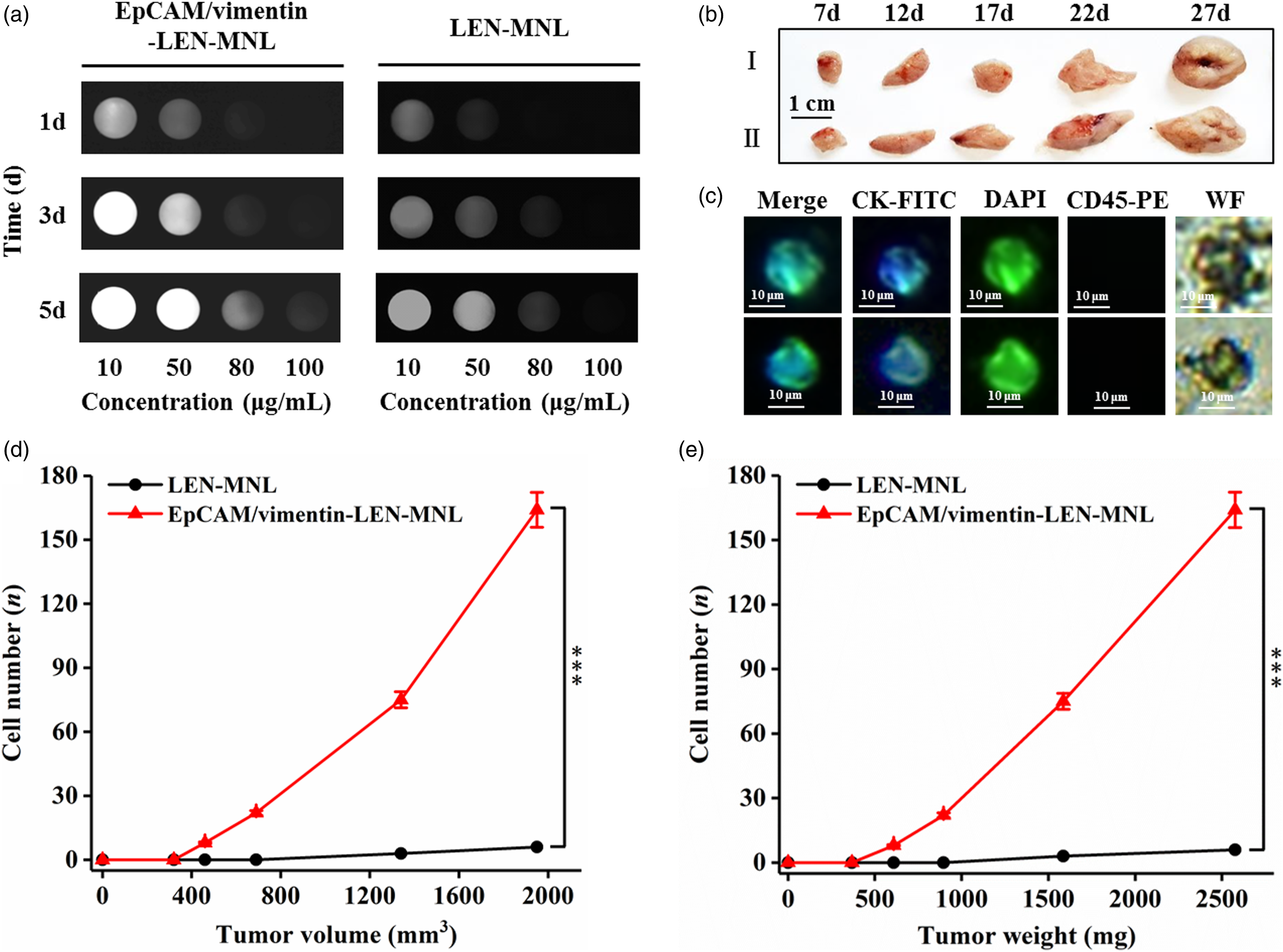
Discussion
Because of the insidious onset of HCC, most patients are diagnosed at an advanced stage.20,21 Due to the lack of selectivity of conventional antitumor drugs to tumor cells, most of the current oncology drugs suffer from the defects of lack of targeting, multi-drug resistance and low bioavailability, which largely limit their clinical use, thus novel drug delivery systems that deliver effective drugs directly to tumor sites through targeted transport have been a hot research topic in the medical field.22–26 Due to the advantages of good biocompatibility and biodegradability, easy loading of lipophilic and hydrophilic drugs, MRI imaging and therapeutic drugs can be loaded inside the bilayer lipid shell or lipid core, or they can be attached to the surface of liposomes, which has great advantages in the drug delivery process.27,28 Compared to free drugs, liposome-encapsulated nano-drug delivery systems are effective in enhancing bioavailability and reducing the toxic effects of antitumor effects. 29 In this study, based on the characteristic that tumor cells undergo EMT during the process of entering the peripheral blood circulation, we coupled EpCAM and vimentin antibodies with drug-loaded liposomes to prepare dual-specific targeted drug-loaded liposomes, which not only can precisely identify the molecular characteristics of HCC cells, but also can further improve the precision and imaging quality of HCC tumor lesion diagnosis.
Nano-drug-loaded liposomes are able to load anti-tumor drugs and deliver effective drugs precisely to tumor sites, enabling slow and stable release of tumor drugs, which can prolong the stagnation time of anti-cancer drugs in cancerous tissues, resulting in a reduced risk of systemic exposure.30–34 In addition, it is also able to be used as an MRI contrast agent for the diagnosis of tumors. 35 Previous studies have shown that a multifunctional nano-particle can carry both adriamycin and sodium bicarbonate and has good anti-HCC tumor effects. 36 Vandetanib DEBs have suitable characteristics for intra-arterial delivery and site-specific sustained release of drug into liver tumours. 37 A dual-targeted slow-release delivery microsphere for Glypican-3 (GPC3) and EpCAM not only specifically targets HCC cells, but also serves as a potential MRI contrast agent. 38 In addition, IOE particles containing iron oxide were detectable by dedicated MRI imaging during intra-arterial injection in an animal model of HCC. 39 Dual-specific MRI molecular probes for HCC, results of in vitro studies demonstrated higher targeting efficiency and MRI sensitivity of dual-specific probes for HCC cells compared to single-targeted or non-targeted molecular probes. 40 Based on these studies, the EpCAM/vimentin-LEN-MNL prepared in this study has a dual targeting effect and can efficiently identify tumor cells in the blood before and after the occurrence of EMT changes. After the specific targeting recognition of tumor cells, it can effectively slow down the release of LEN drug, thus inhibiting the proliferation and promoting the apoptosis of HCC cells. In addition, it can also be used as an MRI-targeted contrast agent in the field of biomedical diagnosis.
The results of this study showed that EpCAM/vimentin-LEN-MNL has small particle size and positive charge, no obvious agglomeration, and has Fe3O4 characteristics with good super paramagnetic properties, indicating that EpCAM/vimentin-LEN-MNL has the characteristics of drug-laden liposomes and the potential function of MRI contrast agent. EpCAM/vimentin-LEN-MNL showed significantly higher cell value-added inhibition and apoptosis efficiency than LEN-MNL with good biosafety, and EpCAM/vimentin-LEN-MNL showed significantly higher capture efficiency for HCC cells, indicating that EpCAM/vimentin-LEN-MNL has slow-release drug delivery function and dual-targeting recognition function. In addition, EpCAM/vimentin-LEN-MNL could still be used as an MRI contrast agent after co-culture with HCC cells for 5 days, and the magnetic response function and properties of EpCAM/vimentin-LEN-MNL did not change significantly, and the MRI signal was attenuated compared with LEN-MNL, indicating that EpCAM/vimentin LEN-MNL targets HCC cells and accelerates the uptake of EpCAM/vimentin-LEN-MNL by HCC cells, indicating a stronger MRI targeting contrast ability for EpCAM/vimentin-LEN-MNL on HCC cells. In vivo functional assays have shown that EpCAM/vimentin-LEN-MNL has the ability to specifically target and identify tumor cells in vivo. In conclusion, EpCAM/vimentin-LEN-MNL with dual targeting function of EpCAM/vimentin and MRI contrast function was prepared in this study, which is expected to provide a low-toxic and efficient drug delivery system for the highly toxic antitumor drug LEN, which can target HCC cells or other tumor cells and slowly release the drug, and is expected to achieve drug-targeted delivery and malignancy-specific MRI diagnostic function. The current study provides preliminary research data to support future in vivo or clinical studies.
Conclusions
The novel EpCAM/vimentin-LEN-MNL nano-therapeutic agent prepared in this study has the function of integrated MRI imaging and diagnosis and treatment. Preliminary studies confirmed that EpCAM/vimentin-LEN-MNL can specifically target HCC cells and can efficiently inhibit HCC cell proliferation and promote HCC cell apoptosis, and has good biosafety. It has a good application prospect as MRI contrast agent. It provides an important scientific basis for realizing the multiple efficacies of nano-carriers in tumor diagnosis and treatment. It opens a new path for the biomedical application and clinical translation of the integrated system of targeted therapy based on magnetic nano-particles.
Footnotes
Declaration of conflicting interests
The author(s) declared no potential conflicts of interest with respect to the research, authorship, and/or publication of this article.
Funding
The author(s) disclosed receipt of the following financial support for the research, authorship, and/or publication of this article: This work was supported by the Key Discipline Program of the Shanghai Pudong New Area Health Commission (No. PWZxk2022-13) and Talents Training Program of the Seventh People’s Hospital, Shanghai University of Traditional Chinese Medicine (No. XX2021-03).