
Abstract
Addressing fracture-related infections (FRI) and impaired bone healing remains a significant challenge in orthopedics and stomatology. Researchers aim to address this issue by utilizing biodegradable biomaterials, such as magnesium/poly lactic acid (Mg/PLA) composites, to offer antibacterial properties during the degradation of biodegradable implants. Existing Mg/PLA composites often lack sufficient Mg content, hindering their ability to achieve the desired antibacterial effect. Additionally, research on the anti-inflammatory effects of these composites during late-stage degradation is limited. To strengthen mechanical properties, bolster antibacterial efficacy, and enhance anti-inflammatory capabilities during degradation, we incorporated elevated Mg content into PLA to yield Mg/PLA composites. These composites underwent in vitro degradation studies, cellular assays, bacterial tests, and simulation of the PLA degradation microenvironment. 20 wt% and 40 wt% Mg/PLA composites displayed significant antibacterial properties, with three composites exhibiting notable anti-inflammatory effects. In contrast, elevated Mg content detrimentally impacted mechanical properties. The findings suggest that Mg/PLA composites hold promise in augmenting antibacterial and anti-inflammatory attributes within polymers, potentially serving as temporary regenerative materials for treating bone tissue defects complicated by infections.
Introduction
Bone injuries resulting from trauma, inflammation, or tumors present a substantial challenge in orthopedics, stomatology, and traumatology. Among these challenges, fracture-related infections (FRIs) remain a critical concern. 1 Pathogenic bacteria infiltrating and proliferating within the wound not only impede normal bone healing but can also trigger severe deep tissue inflammation, potentially leading to permanent disability. 2 While advancements have been made in addressing infection in orthopedic procedures, such as localized drug delivery, 3 the limited spectrum of antibiotics and the risks of inducing host antibiotic resistance have prompted research into enhancing the antibacterial properties of orthopedic implants and exploring biodegradable materials. 4
Biodegradable materials have garnered attention due to their potential to reduce the risks of secondary surgeries for implant removal and associated infection risks. 5 Within this domain, polymers stand out, particularly polylactic acid (PLA), renowned for its biodegradability, mechanical properties, 6 and FDA approval for biomedical applications. 7 Despite its advantages, PLA’s acidic byproducts can accelerate degradation, causing aseptic inflammatory reactions and foreign body responses in vivo. 8 To address these limitations, the integration of functional fillers has been explored to augment PLA’s performance. 9 Magnesium (Mg), known for its bioabsorbable nature and essential role in biochemical processes, 10 has shown promise in mitigating the adverse effects of PLA degradation. 11 The incorporation of magnesium into PLA matrices enhances biological activity, 12 promotes osteoblast differentiation and proliferation, and exhibits broad-spectrum antibacterial properties. 13
In view of the deficiency of the existing biodegradable implant materials, we propose introducing a higher proportion of magnesium particles into polylactic acid to improve the antibacterial properties of the composite materials and meet the corresponding clinical needs. Specifically, PLA-based composites containing different mass percentages of magnesium microparticles (MgMPs) were prepared in this study, and their morphology, mechanical properties, antibacterial function, and immune environment regulation were characterized (Figure 1). Schematic diagram of antibacterial and inflammatory inhibition of temporary Mg/PLA composite implant.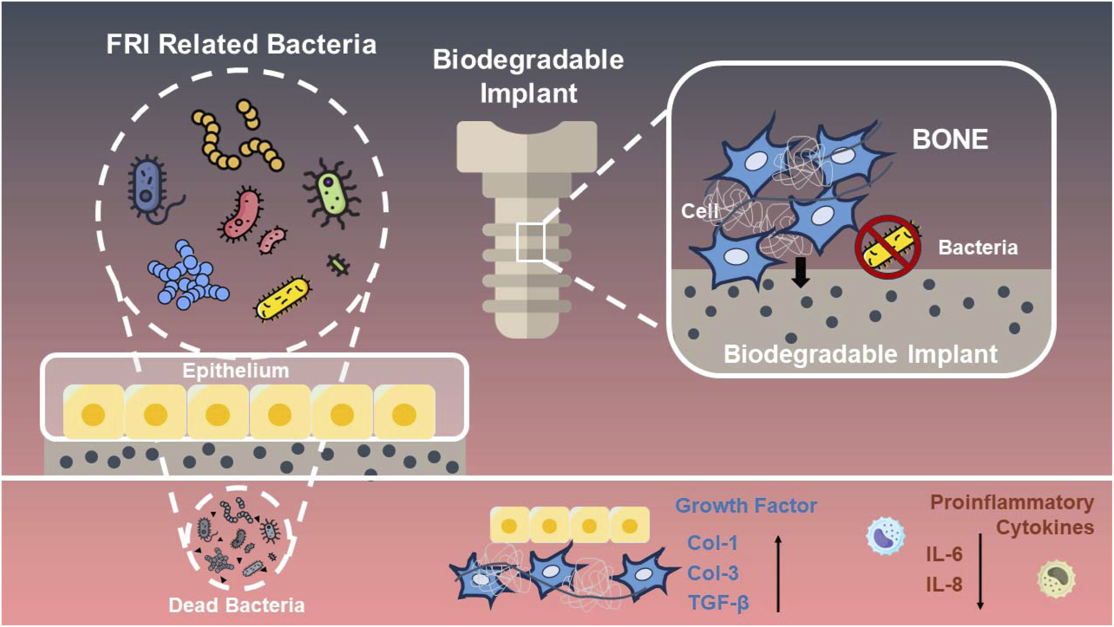
Mg2+ produced by the degradation of composite materials can regulate the activity and immune response of immune cells, reduce the level of oxidative stress, alleviate inflammatory damage, and promote the repair process. Co-culture of the samples with L929 cells revealed that Mg/PLA composites showed more significant effects of down-regulating proinflammatory factors and up-regulating anti-inflammatory and repairing cytokines compared to pure PLA. Additionally, Mg2+ can play an antibacterial role by affecting the stability of bacterial cell membranes and interfering with the metabolic pathway and growth process of bacteria. However, the introduction of higher mass percentage MgMPs not only improves the antibacterial and anti-inflammatory properties of the composites but also has some negative effects on the mechanical properties. When 10 wt% MgMPs were added to PLA, they could be evenly dispersed in the matrix and form a good interface with the matrix, enhancing the overall mechanical properties of the composite. However, excessive addition of MgMPs may lead to increased density, weakened interface bonding, and increased crack sensitivity of the composites, ultimately reducing their mechanical properties.
In conclusion, this study highlights the potential of biodegradable materials, particularly Mg/PLA composites, in addressing complex bone tissue defects caused by infections. By comprehensively considering both antibacterial properties and mechanical properties of composites, we aim to provide valuable insights for the development of effective treatment strategies in orthopedics and related fields.
Materials and methods
Materials preparation
Mg/PLA composites were prepared by dissolving PLA pellets (number-average molecular weight ≈ 80,000 g/mol, viscosity = 2.66 dL/g) in dichloromethane, followed by thorough mixing with spherical Mg powder (D50 = 27 μm) at varying weight percentages (0%, 10%, 20%, and 40%). The resulting suspension was cast into 150 mm diameter glassware at 25°C until dichloromethane volatilization was complete, yielding disk-shaped membranes of 0.3 mm thickness, subsequently dried at 50°C for 24 h. Ethylene oxide sterilization was applied to obtain samples of pure PLA and the three composite types in different shapes. Square samples with 10 mm in side length underwent immersion tests in culture medium to monitor pH value and Mg2+ concentration changes in the leachate. I-shaped samples (50 mm × 10 mm) were used for tensile tests, while round samples (15 mm diameter) were prepared to assess antibacterial and anti-inflammatory properties.”
Characterization
The samples were examined for surface morphology using scanning electron microscopy (SEM; LEO 1530VP, LEO, Germany), and their crystalline structure was analyzed via X-ray diffraction (XRD, Bruker D8-Discover). Mg content was quantified using energy dispersive spectrometry (EDS; INCA300, Oxford Instruments, Oxford, UK). Atomic force microscopy (AFM, Dimension ICON, Bruker, Germany) facilitated surface imaging and topographical examination. Differential scanning calorimetry (DSC, DSC8000, Perkin, USA) was employed to assess the thermal behavior of PLA and Mg/PLA composites.
Mechanical properties
In order to investigate the basic mechanical property of different samples, a tensile test was made with an electronic universal testing machine (Zwick Roell Z1.0, Germany), and the ultimate tensile strength and breaking strain of the samples were calculated by using the stress-strain curve.
In vitro degradation test
The PLA and Mg/PLA samples underwent degradation testing by immersion in complete α-MEM culture medium (Hyclone; SH30265.01) supplemented with 10% fetal bovine serum (Life Technologies; 10099-141) and 1% penicillin-streptomycin, with an extraction rate set at 1.25 cm2/ml. Time points of day 0, day 5, and day 10 were selected for sample collection, and the distribution of magnesium particles was observed in cross-sectional images using an optical microscope (GX51 F, OLYMPUS, Japan). The remaining samples were subjected to an 18-days degradation test at 37°C, with the solution changed every 3 days, maintaining a consistent degradation medium. Throughout the in vitro degradation test, the pH value of the incubated culture medium was monitored using a pH meter (PT-11, INESA, China), while Mg2+ concentration was determined through inductively coupled plasma optical emission spectrometry (ICP-OES, 710-ES, Varian, America). The samples were taken out and vacuum freeze-dried for weight loss measurement. The graph is plotted based on the Mg2+ concentration and pH value of the solution, and the corrosion rate is calculated and depicted using the weight loss data.
Cell viability
L929 and MC3T3-E1 cells were used to evaluate membrane cytocompatibility for viability and proliferation. Conditioned culture medium was obtained by extracting degradation medium from PLA or Mg/PLA samples immersed in a 1:1 ratio with complete culture medium. Cell proliferation was assessed after 1, 3, and 5 days using the Cell Counting Kit-8 (CCK-8, Dojindo, Japan), measuring absorbance at 450 nm following a 1.5h incubation with CCK-8 reagent using a microplate reader (BioTek, Winooski, USA).
Antibacterial properties
Gram-positive bacteria Staphylococcus aureus (S. aureus, ATCC 25923) and Gram-negative bacteria Escherichia coli (E. coli, ATCC 25922) served as representatives for evaluating the antibacterial efficacy of the composites. Co-cultivation of the materials with bacteria in a liquid environment facilitated the determination of bacterial growth curves, enabling an assessment of the material’s impact on bacterial growth dynamics. Utilizing scanning electron microscopy enhanced the visual observation of morphological changes in bacterial cell membranes on the co-cultured materials. Moreover, a comprehensive quantitative evaluation of the material’s antibacterial effects was conducted using the agar plate technique. One experiment involved co-culturing bacteria directly with the material, while another utilized the material’s extract for testing purposes.
Bacterial biofilm inhibition
In assessing the potential inhibitory effect against bacterial biofilms, bacteria and materials were co-cultivated. Crystal violet staining facilitated the observation of the remaining biofilm area, while measuring the optical density (OD) values at 570 nm post-crystal violet dissolution provided a quantitative assessment of the sample’s potential for biofilm inhibition. Additionally, Confocal Laser Scanning Microscopy (CLSM) via fluorescence confocal imaging was employed to further characterize the inhibitory effect on the biofilm.
Inflammatory response assays
The impact of degradation medium on inflammation was assessed utilizing L929 cells. After 24 h of cell adhesion, the experimental group received conditioned medium, while the control group maintained complete DMEM medium. Cells were then cultured at 37°C with 95% humidity and 5% CO2 for 48 h. Total RNA was extracted using TRIzol reagent (Takara, Tokyo, Japan), followed by reverse transcription using the PrimeScript RT kit (Takara, Tokyo, Japan). Subsequent RT-PCR analysis employed SYBR Premix Ex Taq II (TaKaRa, Tokyo, Japan) on the QuantStudio 7 system (Applied Biosystems, USA).
Statistical analysis
The experiments were conducted in triplicate, and the results are expressed as the mean ± standard deviation (SD). Statistical comparisons among multiple groups were performed using one-way analysis of variance (ANOVA) followed by Tukey’s post hoc test.
Results and discussion
Characterization
The SEM images of the samples are presented in Figure 2(a), revealing distinct differences between the pure PLA and PLA/Mg composite samples. The PLA/Mg composite exhibits a noticeably coarser and grainier texture compared to pure PLA, with the particles appearing larger as the Mg content increases. (a) Morphology of specimens SEM images (scale bar 100 μm) at 500× magnification and (b) EDS elements mapping (scale bar 100 μm) of the PLA and the composites with different contents of Mg powder. (c) Three-dimensional surface profiles (20 μm × 20 μm).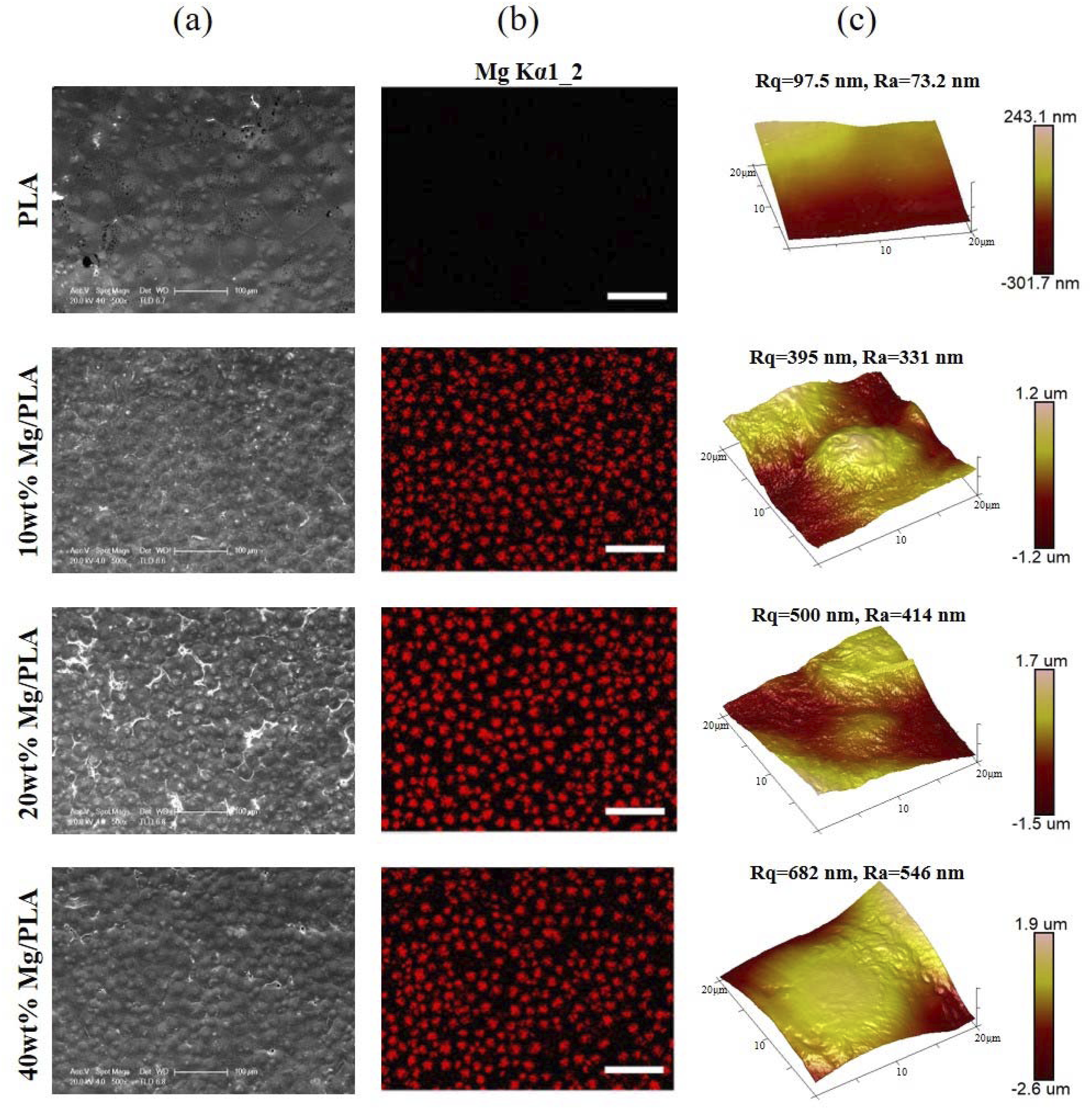
Figure 2(b) displays the EDS elements mapping of samples. The red color represents the presence of Mg, allowing for a clearer visualization of Mg distribution within the material. In the pure PLA sample, the absence of red color indicates the absence of Mg. In contrast, the PLA/Mg composites exhibit varying degrees of Mg distribution. In the 10 and 20 wt% PLA/Mg composites, the red particles are evenly distributed against a black background, indicating a uniform dispersion of magnesium particles within the composite matrix. However, in the 40 wt% PLA/Mg composite, a slight uneven distribution of red particles is observed, suggesting potential agglomeration or clustering of MgMPs at higher concentrations.
In Figure 2(c), the topographical surface features of each sample are depicted under AFM observation, providing a detailed representation of peaks and valleys via 3D surface mapping. The root mean square roughness (Rq) for pure PLA is quantified at 97.5 nm, with an average roughness (Ra) of 73.2 nm. Upon incorporation of 10 wt% MgMPs, the Rq increases to 395 nm, accompanied by an Ra of 331 nm. Remarkably, with the magnesium content rising to 40%, the Rq of the composite experiences a notable surge to 682 nm, alongside an Ra of 546 nm. These findings underscore a significant increase in surface roughness induced by the incorporation of MgMPs.
These observations suggest that the addition of magnesium particles influences both the surface morphology and elemental distribution of the composite material. The observed coarser texture and larger particle size in the PLA/Mg composites indicate changes in material microstructure induced by the presence of magnesium.
Figure 3 depicts the XRD patterns and DSC curves, offering insights into the structural alterations and crystallinity changes of the materials, while Table S1 (Supporting Information) provides data on crystallinity and melting points for the various samples. (a) XRD patterns of the PLA and Mg/PLA composites with different Mg powder contents. (b) DSC curves of PLA and Mg/PLA composites.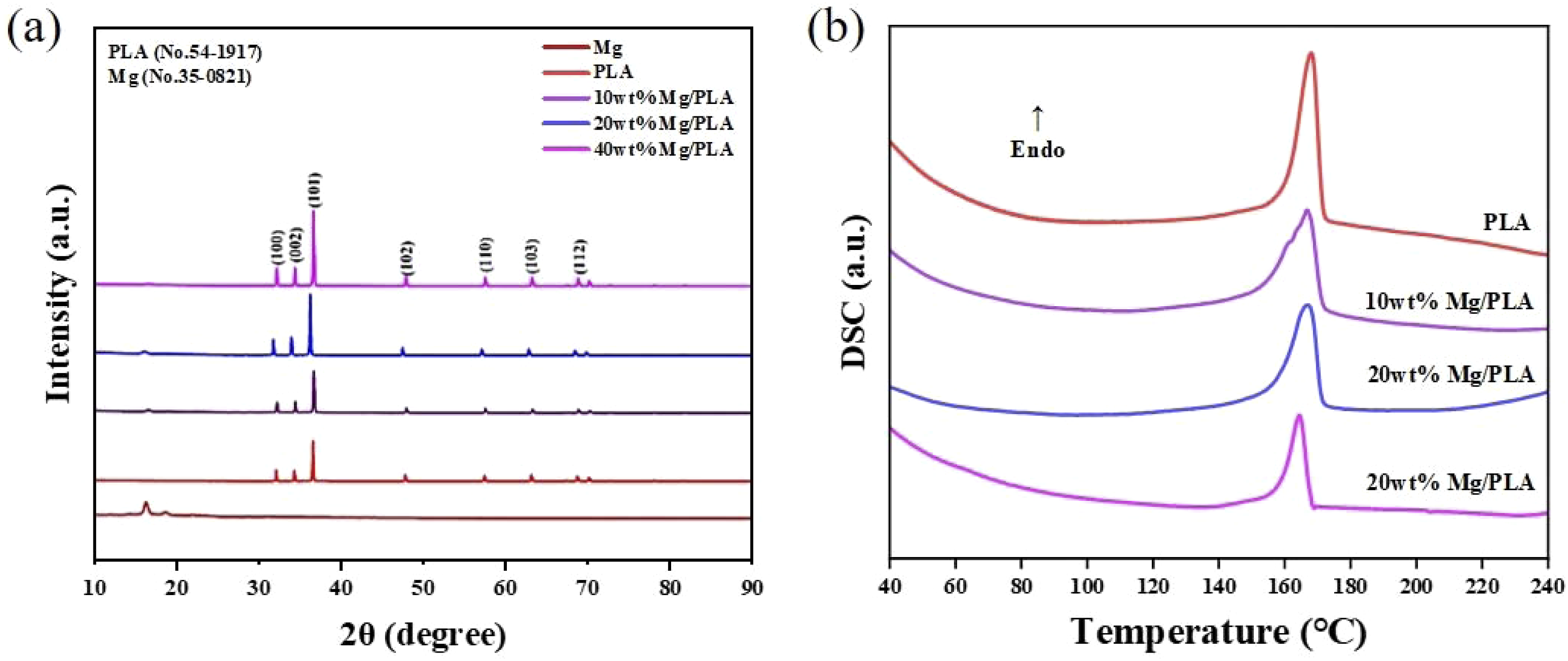
With a slight increase by 10%, MgMPs potentially serve as nucleating agents within the PLA matrix, facilitating crystallization and marginally enhancing crystallinity. However, further escalation to 40% reveals apparent MgMPs aggregation, impeding the PLA molecular crystallization process and inducing a decline in crystallinity.
The augmentation of MgMPs from 0 wt% to 20 wt% does not elicit significantly alter the thermal stability of the composite material. Nevertheless, a notable reduction in the composite material’s melting point is observed upon reaching 40 wt%. The substantial decreases in crystallinity and melting point at 40 wt% suggest that the composite material’s stability may diminish.
Mechanical properties
Figure 4 shows the tensile stress-strain curves and detailed mechanical properties of the specimens. The mechanical parameters display a consistent trend across varying Mg content in the composites, initially exhibiting a notable increase followed by a gradual decline. For instance, the introduction of 10 wt% MgMPs significantly boosts tensile strength from 37.6 ± 2.1 MPa to 52.3 ± 1.5 MPa. However, as Mg content increases, tensile strength subsequently diminishes to 34.1 ± 0.5 MPa and 22.7 ± 3.4 MPa, respectively. Moreover, the incorporation of 10 wt% or 20 wt% MgMPs enhances failure strain from 3.4% ± 0.06% to 4.6% ± 0.5% and 5.9% ± 0.2%, respectively. (a) Typical stress-strain curves, (b) tensile (ultimate) strength and (c) breaking strain of four prepared specimens. p < .05 (*), p < .01 (**), p < .001 (***).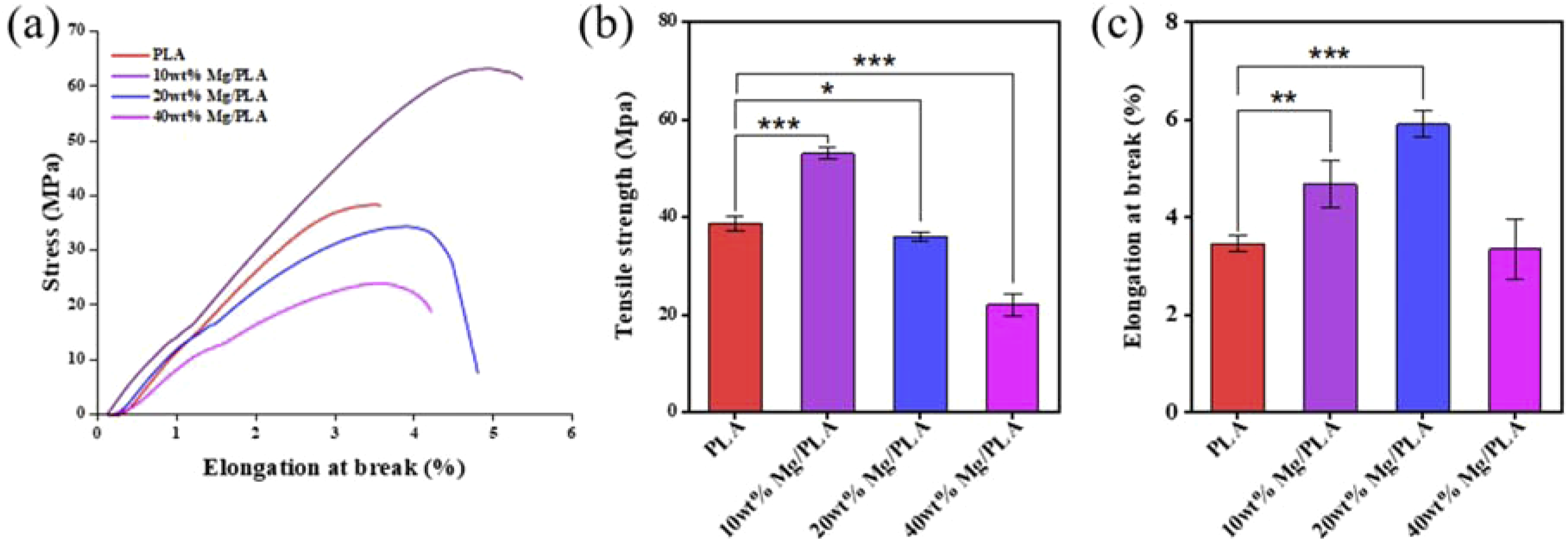
Consistent with prior studies, our findings reaffirm that an optimal Mg content enhances the mechanical strength of base PLA, while higher content (40 wt%) compromises this enhancement. 14 With a slight increase, MgMPs potentially serve as nucleating agents within the PLA matrix, thereby facilitating PLA crystallization and marginally enhancing its crystallinity. However, with further escalation of Mg content to 40%, MgMPs aggregation becomes apparent, impeding the PLA molecular crystallization process and inducing a decline in crystallinity. 15
Regarding thermal stability, the augmentation of MgMPs from 0 wt% to 20 wt% does not induce significant alterations in the composite material. Nonetheless, a notable reduction in the composite material’s melting point is observed upon MgMPs reaching 40 wt%. This significant decrease in crystallinity and melting point at 40 wt% Mg/PLA composite suggests a potential compromise in the composite material’s stability within biological systems. Consequently, this degradation in stability could lead to modifications in pore structure and surface properties, ultimately influencing the release rate and behavior of MgMPs.
Degradation behavior
Figure 5 shows the progression of cross-sectional images of samples observed over varying soaking durations, providing insight into the degradation behavior of the different specimens. The degradation of MgMPs was visualized using a metallographic microscope, wherein Mg exhibited a distinct bright yellow hue while PLA retained its characteristic brown coloration. With increasing Mg content, a higher concentration of magnesium particles became evident within the cross-sections. Over time, extended soaking led to the gradual erosion of magnesium particles, resulting in irregular contour shapes. Due to direct contact with the solution, magnesium particles on the material’s surface were preferentially degraded. By the 10th day, only a sparse scattering of magnesium particles remained within the 10 wt% Mg/PLA composite. Notably, the 40 wt% Mg/PLA composite still retained a significant number of magnesium particles. Metallographic microscope images of cross sections of PLA and composites samples show the distribution of magnesium particles.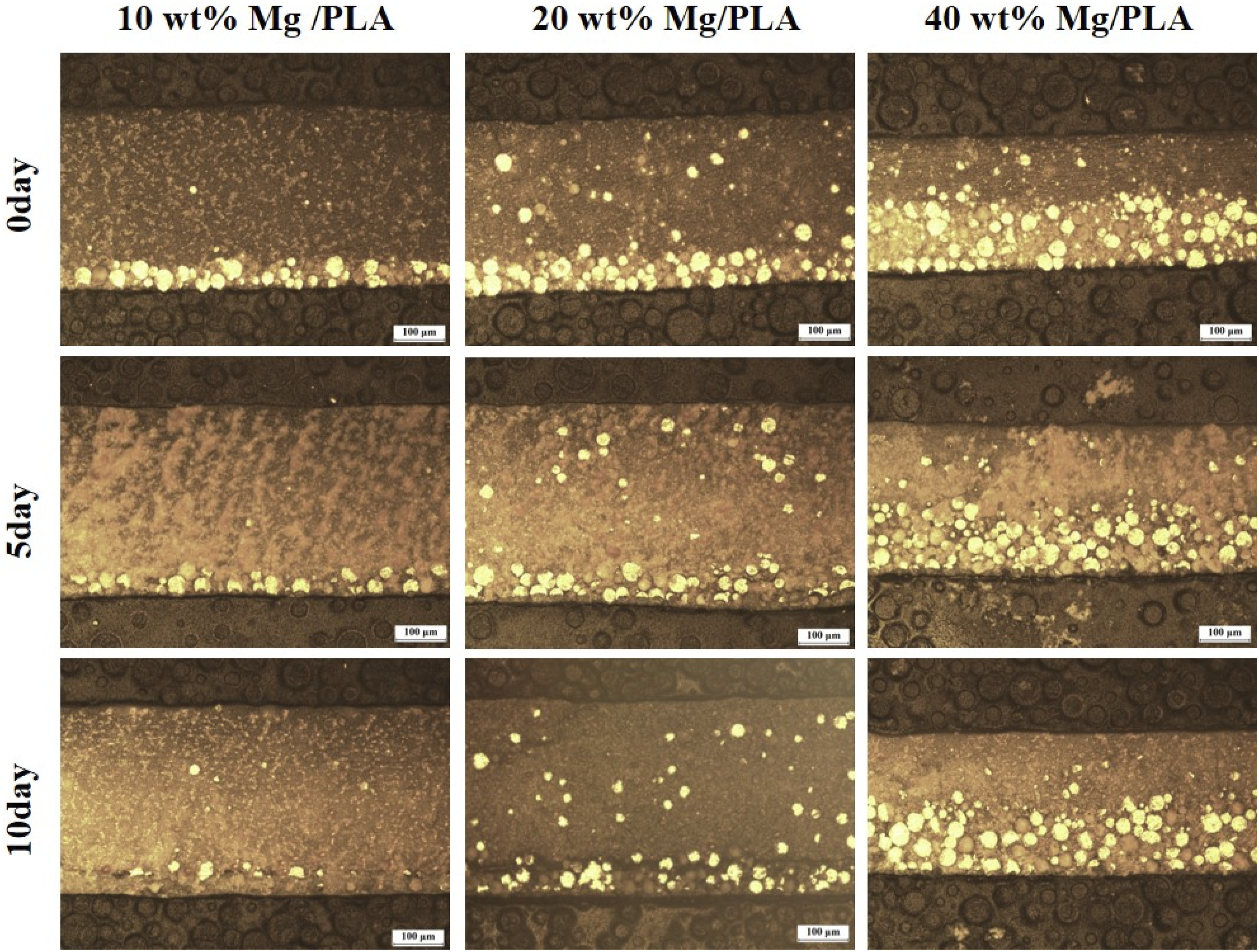
We further assessed composite degradation through the quantification of Mg2+ concentration, pH value, and corrosion rate of degradation products in the solution. Since MgMPs located near the material’s surface come into quicker and fuller contact with the solution, they exhibit rapid degradation upon re-soaking initiation, leading to an initial surge in magnesium ion concentration. Subsequently, this concentration increase stabilizes over time (Figure 6(a)). PLA degradation progresses relatively slowly, resulting in a stable pH value of the soaked solution over the 18-day period. However, the degraded solution displays slight acidity due to weak acids generated during PLA degradation. Conversely, in the composite material group, degradation by magnesium particles generates Mg(OH)2, elevating the solution’s pH value (Figure 6(b)). In the degradation experiment, sample mass loss escalates with time. Notably, PLA exhibits a gradual mass loss, whereas composites containing magnesium particles experience more pronounced mass loss, with higher magnesium content correlating with a greater proportion of mass loss (Figure 6(c)). (a) Mg2+ concentration, (b) pH value, and (c) Sample mass loss over 18 days of immersion experiment.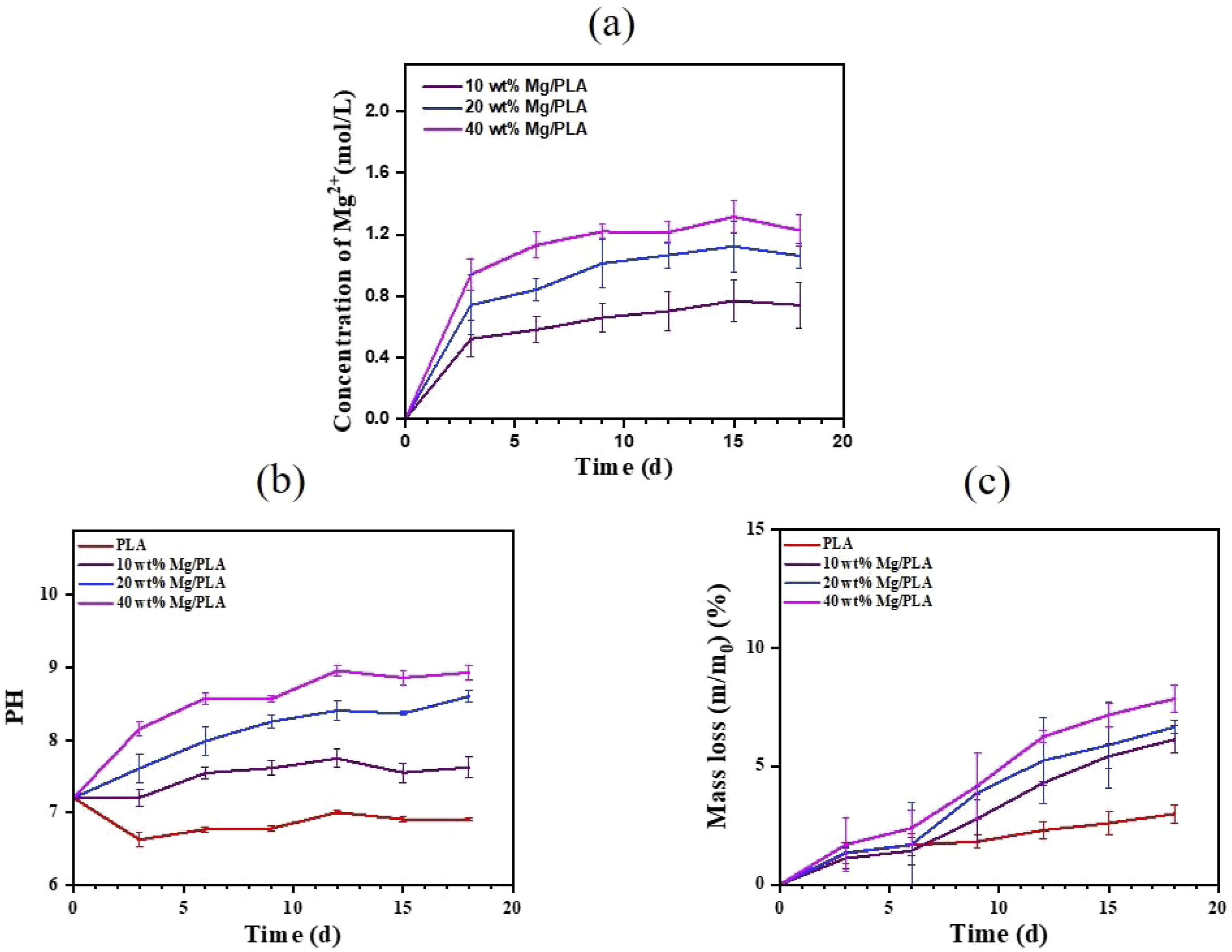
Biocompatibility and antibacterial properties
The biocompatibility evaluation with MC3T3-E1 and L929 cells using the CCK8 kit revealed that PLA and low Mg content composite degradation mediums displayed minimal cytotoxicity compared to the blank control group. However, the 40 wt% Mg/PLA composite exhibited adverse effects on cell growth (Figure 7). Evaluation of the cytocompatibility of specimens. The proliferation of (a) MC3T3-E1 cells, (b) L929 cells. p < .05 (*), p < .01 (**), p < .001 (***).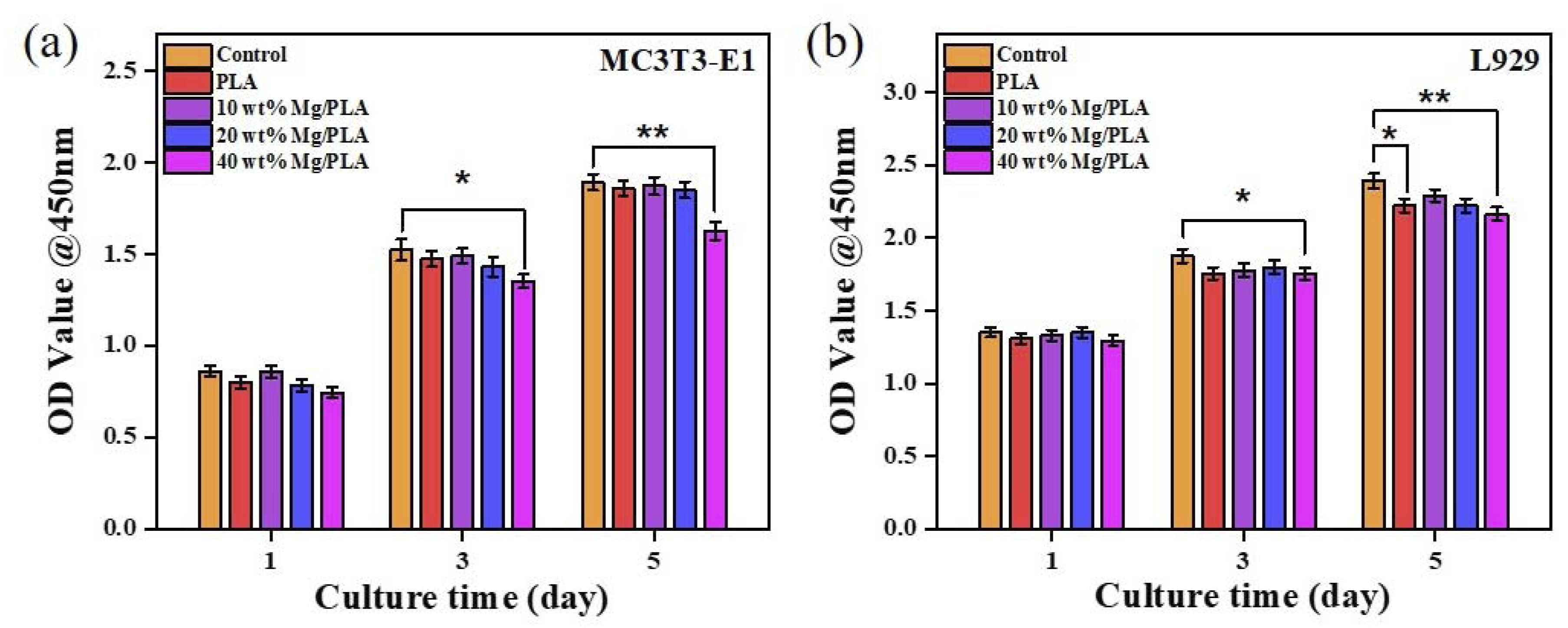
Considering the complexity of the oral flora and the possibility of “non-oral microorganisms” settling in the oral cavity, the antimicrobial properties of composites were evaluated with gram-negative E. coli and gram-positive S. aureus (Figure 8). Bacterial growth curve (OD/hours) of (a) S. aureus and (b) E. coli in Luria Bertain liquid medium with different samples.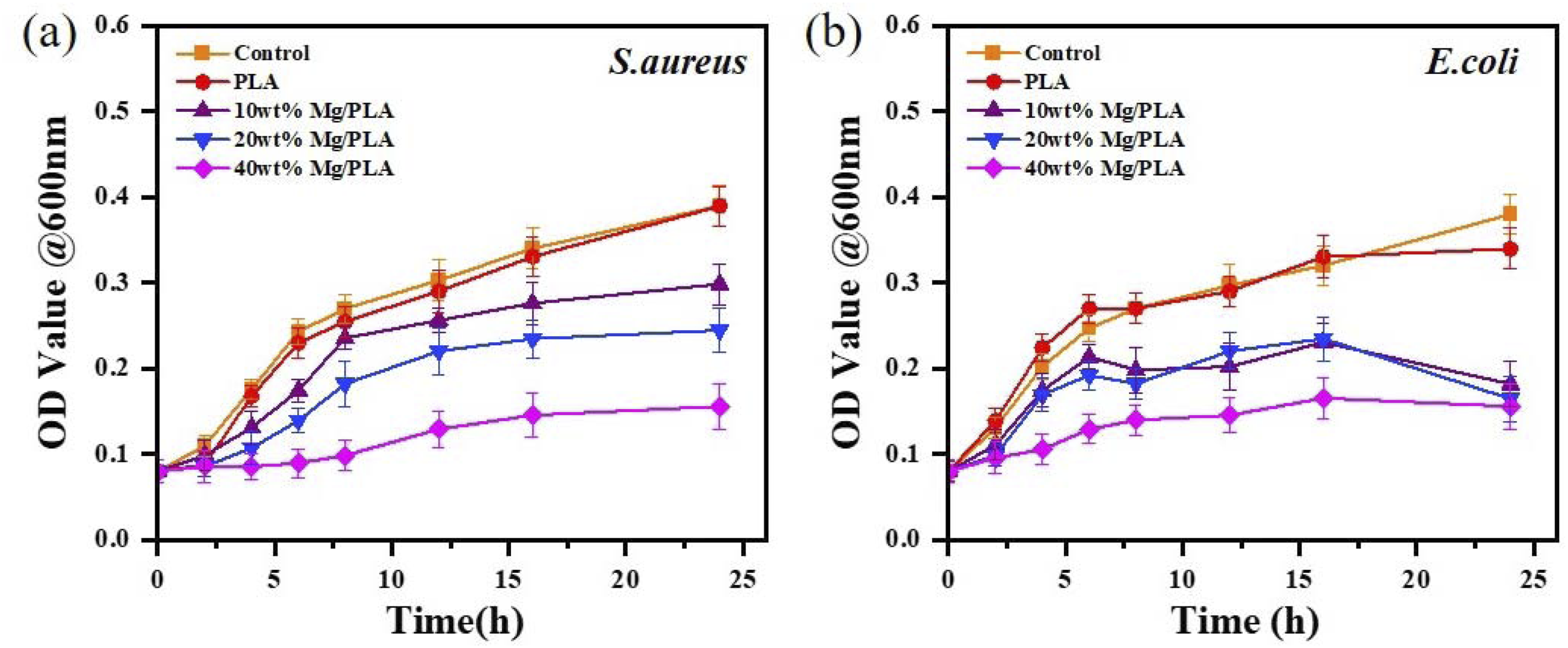
Assessment of antimicrobial properties against E. coli and S. aureus demonstrated negligible antibacterial performance in the blank control and PLA groups, suggesting limited inherent antibacterial activity. However, the 20 wt% and 40 wt% Mg/PLA groups exhibited notable growth delay in the initial logarithmic growth phase, particularly inhibiting E. coli growth significantly.
Quantitative antibacterial assessments (Figure 9) demonstrated a substantial decrease in bacterial colony count with increasing Mg content. The 40 wt% Mg/PLA showed the highest antibacterial rates against S. aureus (94.33% ± 6.27%) and E. coli (94.92% ± 5.03%). Photographs of LB-agar plates coated with colonies of S. aureus and E. coli of (a) different samples and (b) 24h extract, corresponding quantitative analysis of antibacterial effect against (c) S. aureus and (d) E. coli on the samples and (e, f) in the extract. p < .05 (*), p < .01 (**), p < .001 (***).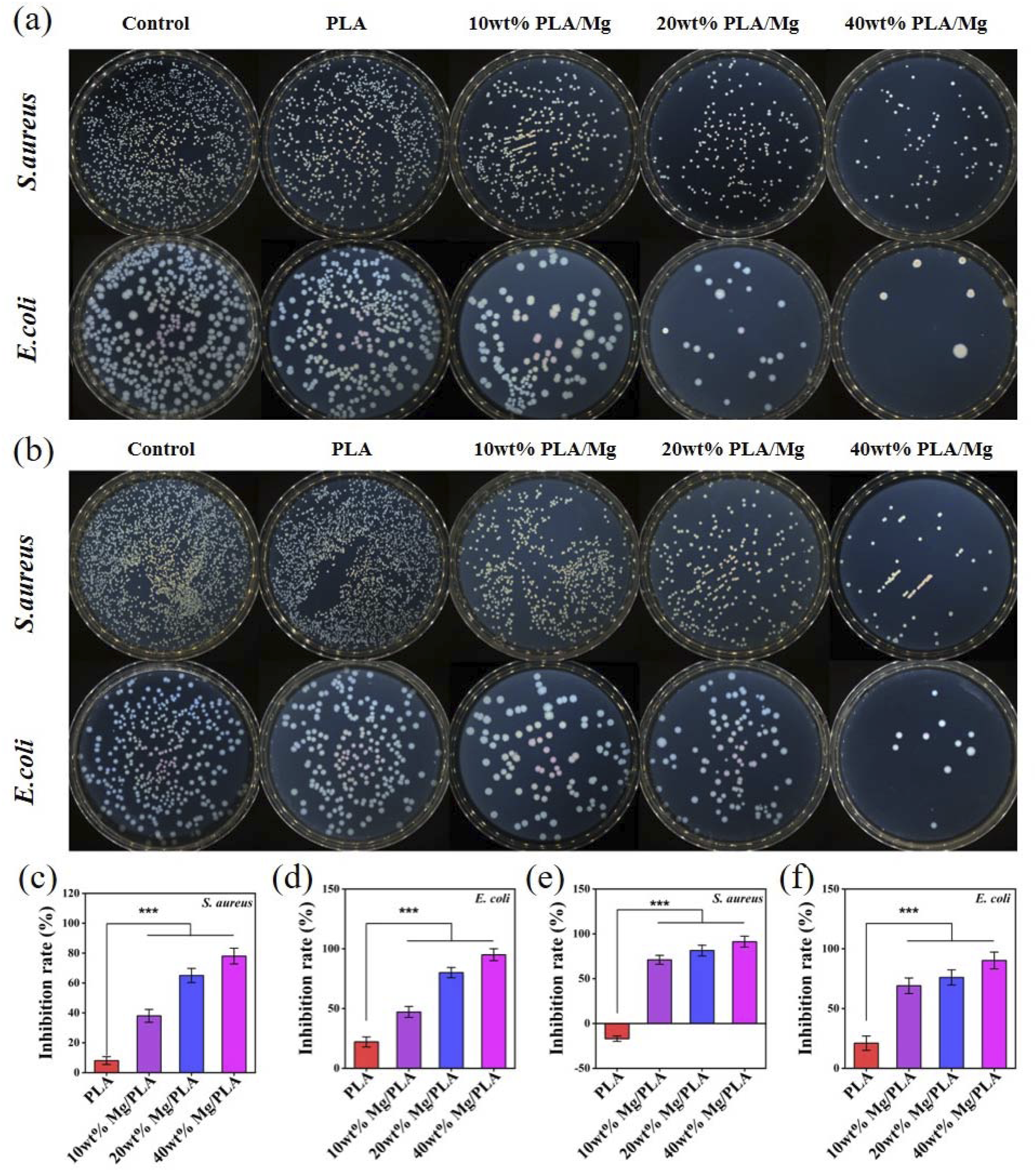
The proposed antimicrobial mechanisms involved alterations in bacterial membrane permeability caused by the interaction of antimicrobial Mg2+ with the bacterial outer membrane, as well as changes in the acid-base microenvironment. 16 These modifications resulted in irregularities or distortions on the bacterial outer membrane, impacting permeability and ultimately resulting in bacterial death.
In additionally, SEM observations suggested decreased bacterial adhesion with increased Mg content, potentially attributed to H2 release from partial dissolution of MgMPs. Higher Mg supplementation (>10 wt%) effectively reduced bacterial initial affinity to the material surface (Figure 10). SEM images of S. aureus and E. coli after 24 h of incubation with PLA and Mg/PLA composites, scale bar 5 μm.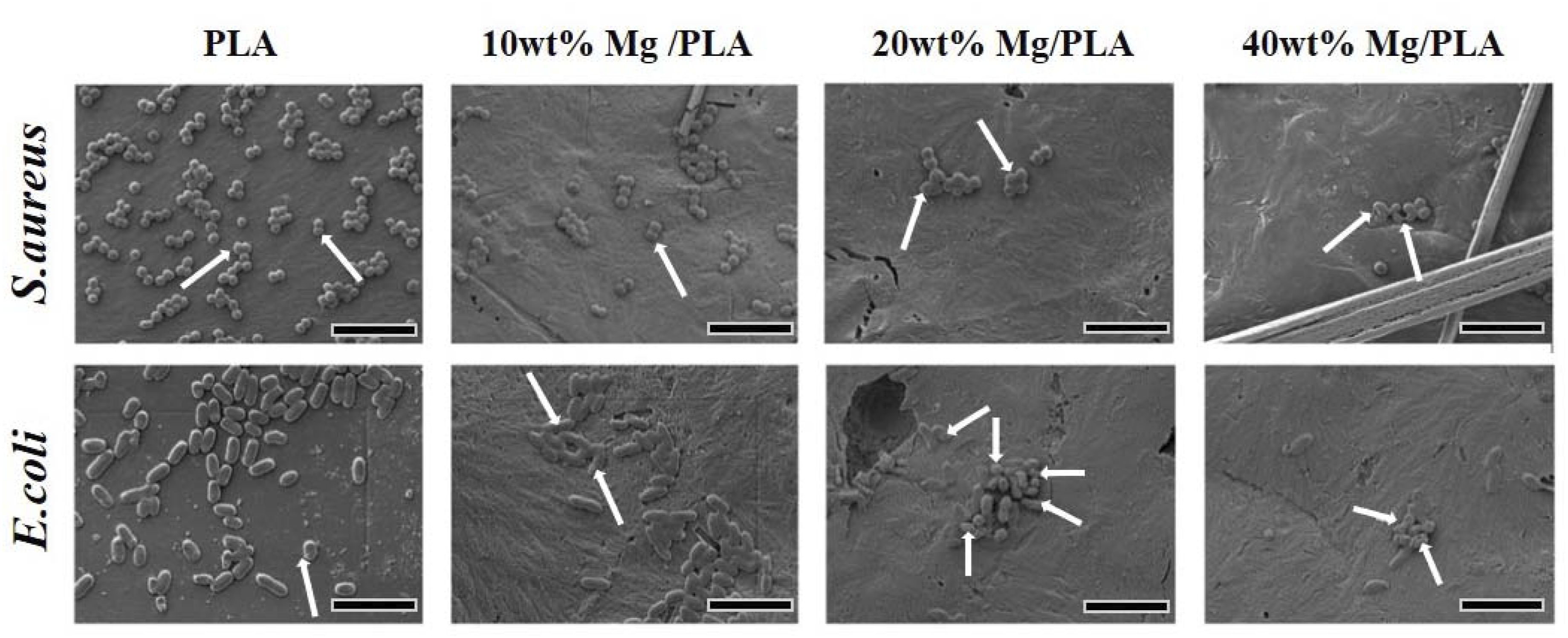
We examined the material’s ability to scavenge bacterial biofilm using crystal violet staining (Figure 11). It is evident that the biofilm formed in the PLA group appears dense and prominently deep purple. However, as the magnesium content in the composite increases, the purple color notably lightens, and the biofilm at the bottom of the well plate is visibly disrupted. Crystal violet staining of biofilms of (a) S. aureus and (b) E. coli Quantitative analysis of biofilms of (c) S. aureus and (d) E. coli and (e, f) corresponding crystal violet staining of biofilms on PLA and composites incubated with LB liquid medium. p < .05 (*), p < .01 (**), p < .001 (***).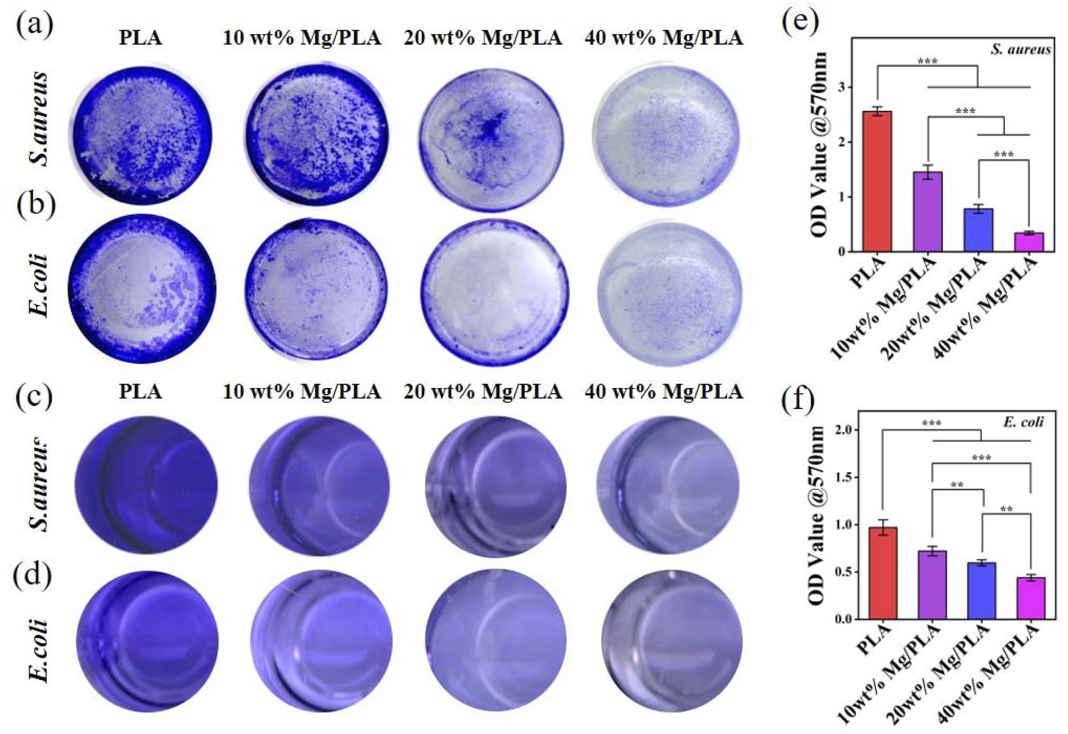
Live/dead bacteria fluorescence imaging further substantiated the bacteriostatic effects of the composites (Figure 11). The decrease in biofilm mass and reduced viable bacteria confirmed the effective mitigation of biofilm formation and inhibition of bacterial growth on the composite surface. 17 The inhibition of biofilm formation, crucial for bacterial resistance to antibiotics and host defenses, was observed on the composite surface, indicating potential resistance against the development of resistant bacteria.
The observed reduction in biofilm mass and viable bacteria further confirmed the composite’s effectiveness in inhibiting biofilm formation and reducing bacterial growth on the surface. These findings are crucial as biofilm formation significantly contributes to bacterial resistance against antibiotics and host defenses. 18
The relationship between increased Mg content and reduced bacterial adhesion to the material surface suggests a potential deterrent effect attributed to H2 release from the partial dissolution of MgMPs. 19 This aligns with previous studies and emphasizes the role of Mg supplementation in discouraging initial bacterial adhesion. 20
In summary, the Mg/PLA composites exhibited promising antibacterial properties by inhibiting biofilm formation and curtailing bacterial growth and reproduction. However, to optimize these composites for biomedical applications, further research is required to fine-tune their composition for enhanced biocompatibility without compromising their antimicrobial efficacy. Moreover, understanding the precise mechanisms governing these antibacterial effects will aid in developing superior materials against bacterial proliferation and biofilm formation (Figure 12). (a, b) CLSM images of live/dead bacteria staining, live bacteria stained green and dead bacteria stained red (scale bar 20 μm). Quantitative analysis of (c, d) S. aureus and (e, f) E. coli of green fluorescence intensity and red fluorescence intensity. p < .05 (*), p < .01 (**), p < .001 (***).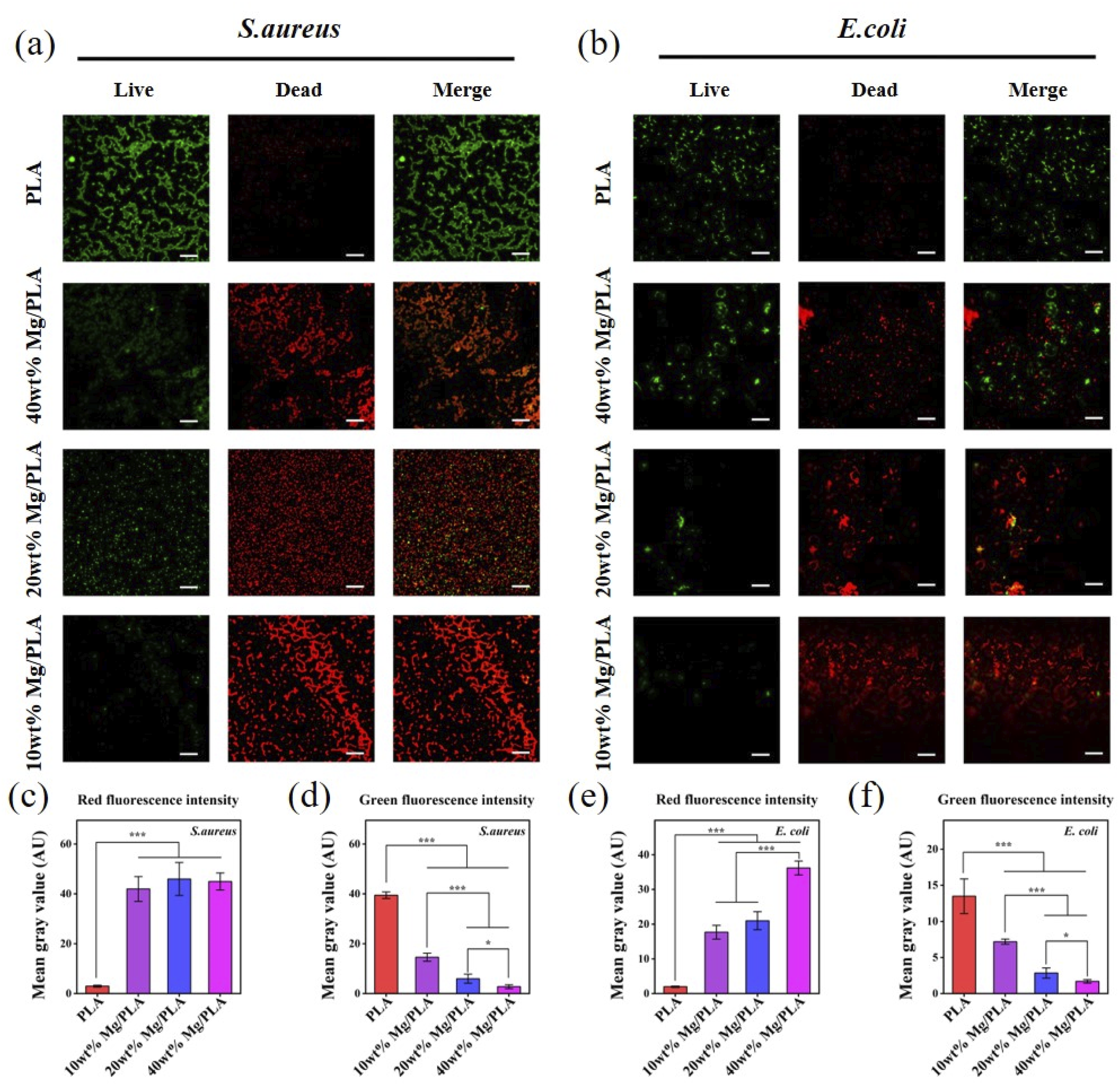
Inflammatory response
The study investigated how the introduction of magnesium powder into PLA affects the pH microenvironment induced by PLA degradation and its impact on L929 cells. To understand the cellular response to acid degradation products in vitro, RT-qPCR analysis was conducted to detect the expression of inflammatory factors at the gene level.
The results in Figure 13 demonstrated an upregulation of COL-1, COL-3, and TGF-β expression in the composite groups, while only the 10 wt% Mg/PLA group showed no significant difference in COL-3 expression from the PLA group. In contrast, COL-1, COL-3, and TGF-β expression were downregulated in the PLA group. Moreover, the gene expression levels of IL-6 and IL-8, proinflammatory cytokines, were significantly lower in the Mg/PLA groups compared to the PLA group. These findings suggest that Mg/PLA composite degradation products alleviate the inflammatory response caused by PLA acidic degradation products. They promote the secretion of extracellular matrix components, potentially facilitating wound healing. Gene expression of (a) COL-1, (b) COL-3, (c) TGF-β, (d) IL-6, and (e) IL-8 by RT-qPCR test. p < .05 (*), p < .01 (**), p < .001 (***).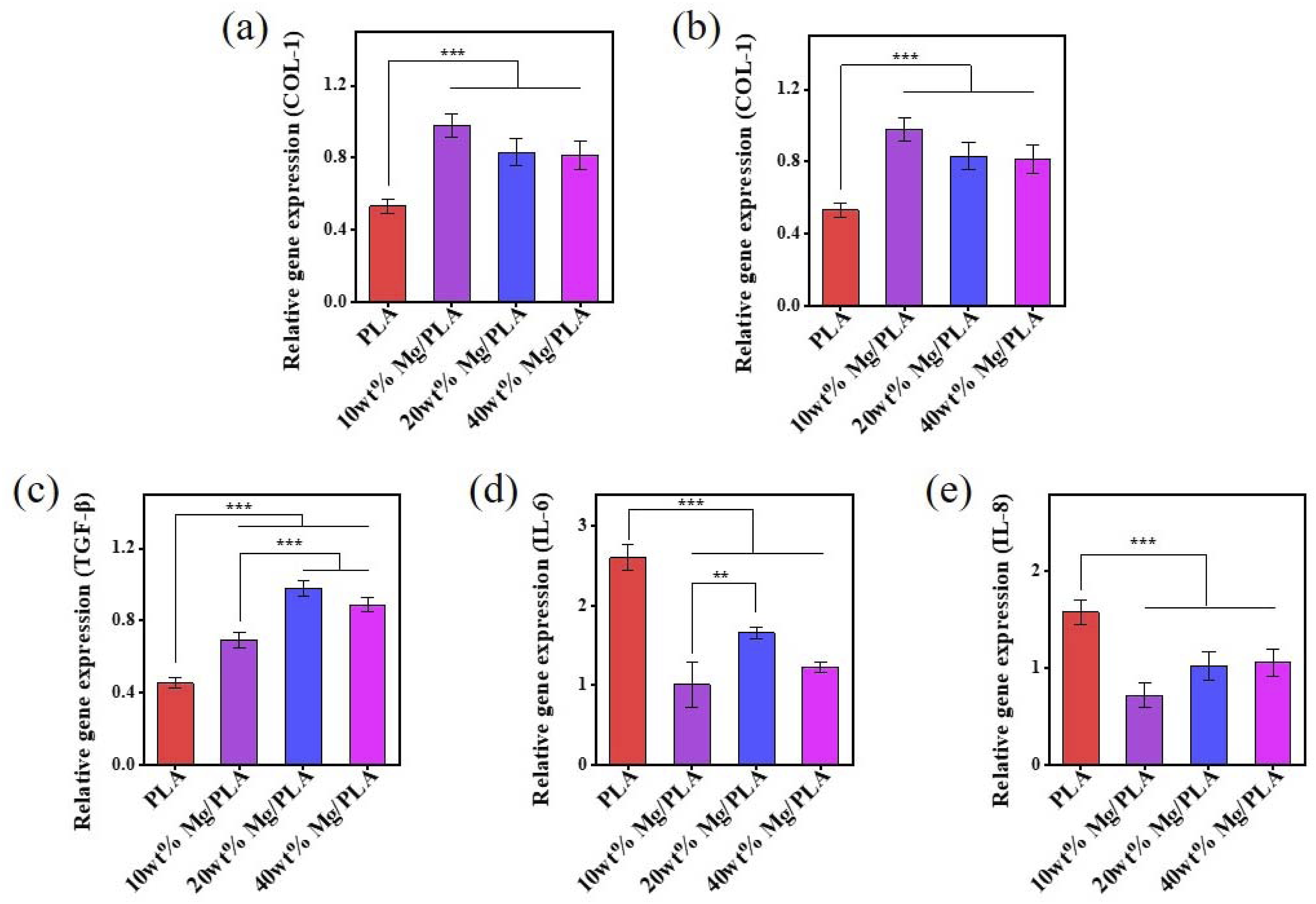
Tissue repair mechanisms involve cellular infiltration, collagen production, and normal inflammatory responses. 21 TGF-β plays a crucial role in attracting and activating fibroblasts, facilitating tissue restoration. 22 However, an acidic environment caused by PLA degradation may impair cellular function and tissue repair by activating inflammatory responses. 23
Initial degradation of MgMPs in Mg/PLA composites might hamper initial cell adhesion. However, subsequent experiments using composite extracts displayed good cytocompatibility without observable adverse effects after contact with the liquid. Additionally, simulated acidic environments from degradation products exhibited decreased inflammatory factor expression in the composite groups compared to the PLA group. These observations align with previous studies showing that Mg2+ induces down regulation of M1 subtype marker expression and upregulation of M2 subtype marker expression. 24
This research underscores the potential of Mg/PLA composites in mitigating inflammatory responses caused by PLA degradation, highlighting their promise in promoting tissue repair and wound healing. This study emphasizes the potential of Mg/PLA composites in reducing the inflammatory reaction caused by PLA degradation, and highlights their prospects in promoting tissue repair and wound healing.
Conclusion
This study successfully developed Mg/PLA composites exhibiting dual antibacterial and anti-inflammatory properties, showcasing promise as temporary implants for bone loss-related infections. The 10 wt% and 20 wt% Mg/PLA composites demonstrated significant biocompatibility, preserved mechanical strength, and robust antibacterial and anti-inflammatory effects. However, the 40 wt% Mg/PLA composite, while highly effective in antimicrobial properties, compromised mechanical integrity. The Mg/PLA composites effectively neutralized the acidic environment caused by PLA degradation, showing potential in mitigating inflammation through the downregulation of IL-6 and IL-8 genes and upregulation of TGF-β, COL-1, and COL-3 genes. This underscores their anti-inflammatory potential and support for tissue repair processes. Despite these promising findings, further validation through in vivo experiments is crucial to understand the material’s actual effectiveness in mitigating inflammation and promoting tissue repair. Assessing osteogenic effects is also essential due to uncertainties regarding high magnesium environments’ impact on bone formation. While the study confirmed the non-toxic nature of higher magnesium content to cell growth, future research will delve into evaluating osteogenic effects and optimizing biocompatibility, antibacterial, and anti-inflammatory functions. Achieving a delicate balance between mechanical strength and biological efficacy remains a critical goal for advancing Mg/PLA composite applications in diverse biomedical settings.
Supplemental Material
Supplemental Material - Biodegradable implant of magnesium/polylactic acid composite with enhanced antibacterial and anti-inflammatory properties
Supplemental Material for Biodegradable implant of magnesium/polylactic acid composite with enhanced antibacterial and anti-inflammatory properties by Yuxin Qian, Xianli Wang, Ping Wang, Jin Wu, Yue Shen, Kunzhan Cai, Jing Bai, Mengmeng Lu and Chunbo Tang in Journal of Biomaterials Applications
Footnotes
Declaration of conflicting interests
The author(s) declared no potential conflicts of interest with respect to the research, authorship, and/or publication of this article.
Funding
The author(s) disclosed receipt of the following financial support for the research, authorship, and/or publication of this article: This work was supported by the Natural Science Foundation of the Higher Education Institutions of Jiangsu Province (BK20190649), National Natural Science Foundation of China, (81901056 and 82170993).
Supplemental Material
Supplemental material for this article is available online.
References
Supplementary Material
Please find the following supplemental material available below.
For Open Access articles published under a Creative Commons License, all supplemental material carries the same license as the article it is associated with.
For non-Open Access articles published, all supplemental material carries a non-exclusive license, and permission requests for re-use of supplemental material or any part of supplemental material shall be sent directly to the copyright owner as specified in the copyright notice associated with the article.