
Abstract
Osteoporosis treatment using alendronate (ALN) is limited by poor oral bioavailability and gastrointestinal side effects. To address this limitation, this study developed a graphene oxide (GO)-enhanced soluble microneedle (MN) system for transdermal administration of ALN to treat osteoporosis. The MNs were fabricated from polyvinyl alcohol (PVA) and polyvinylpyrrolidone (PVP) using a two-step casting method, forming a drug-loaded core–barrier outer layer. The incorporation of GO significantly improved mechanical strength, with penetration efficiency reaching 66∼88%, and enhanced swelling capacity (PP/GO-ALN swelling ratio: 299%). In vitro release studies showed no significant difference between PP-ALN and PP/GO-ALN MNs in a dialysis bag model, while ex vivo skin permeation demonstrated that PP/GO-ALN MNs achieved significantly higher cumulative drug permeation (1069.53 µg/cm2) over 24 h compared to PP-ALN MNs (712.89 µg/cm2). Furthermore, GO conferred notable antibacterial activity, and the PP/GO-ALN formulation synergistically promoted osteoblast proliferation (cell viability: 107.42%, p < 0.05). These findings demonstrate that the GO-ALN MN system possesses mechanical properties, transdermal delivery, antibacterial effects, and biocompatibility. It provides a highly promising non-invasive strategy for the treatment of osteoporosis.
Keywords
Introduction
Osteoporosis is a metabolic bone disease caused by an imbalance between bone resorption and bone formation, characterized by decreased bone density and destruction of bone microstructure.1,2 Alendronate sodium (ALN), a first line clinical drug, inhibits osteoclast activity. 3 But suffers from extremely low oral bioavailability and gastrointestinal irritation. 4 Intravenous administration causes injection site reactions and hypocalcemia.5,6 Hence, a safer and more efficient delivery system is urgently needed.
Transdermal drug delivery (TDD) avoids first pass metabolism and improves compliance,7,8 yet is limited by the stratum corneum barrier.9,10 Microneedles (MNs) can penetrate this barrier and create microchannels, offering a feasible approach for ALN transdermal delivery.11,12 Soluble MNs based on Polyvinyl alcohol (PVA) and Polyvinyl pyrroidone (PVP) have shown progress due to their water solubility and molding properties.13–15 However, pure PVA/PVP MNs suffer from low mechanical strength and uncontrolled burst release, making it difficult to balance puncture efficiency and long term treatment requirements.
Graphene oxide (GO) possesses high mechanical strength, large specific surface area, and abundant oxygen containing groups.16–18 When dispersed in a polymer matrix, GO can synergistically enhance mechanical properties and structural stability. 19 Recent studies have applied GO polymer composites in biomedical fields, such as nanofiber scaffolds with antibacterial activity 20 and hydrogels for cartilage regeneration. 21 Nevertheless, the use of GO reinforced PVA/PVP soluble MNs for transdermal ALN delivery in osteoporosis has not been reported.
Based on the above background, the present study aims to construct ALN loaded soluble MNs using a PVA/PVP/GO composite. The core objective is to elucidate the influence of the PVA to GO ratio on the mechanical properties, swelling behavior, drug release kinetics, and skin insertion capability of the resulting MNs. Compared with pure PVA/PVP MNs and other polymer based ALN systems, our work has clear advantages: water solubility, enhanced mechanical strength, controlled release, and applicability to osteoporosis therapy.
Materials and methods
Samples preparation
Materials
PVA (type 1799, 98∼99% alcoholysis) was purchased from Aladdin Biochemical Technology Co., Ltd (Shanghai, China). ALN (97%, MW = 325.12) was obtained from Yuanye Biotechnology Co., Ltd (Shenyang, China). PVP (K30, Analysis of purity) was bought from Zonglian Chemical Reagent Co., Ltd (Tianjin, China). Sodium bicarbonate (NaHCO3 ≥ 99.5%, MW = 84.01) was from Ruijinte Chemical Co., Ltd (Tianjin, China). GO (Diameter range: 0.2∼10 μm) was from Tuling Evolution Technology Co., Ltd (Shenzhen, China). Benzaldehyde pentatriene ketone (C9H4O3·H2O ≥ 95.0%, FW = 178.14), Anhydrous ethanol (≥ 99.7%), Agar powder (Purification) and Peptone were provided by Sinopharm Chemical Reagent Co., Ltd (Shanghai, China). Yeast extract came from the British Thermo Fisher Scientific Group. DMEM (High Glucose), Fetal Bovine Serum, Penicillin-Streptomycin Solution (100×), and Trypsin-EDTA solution (0.25% trypsin, containing phenol red) were all purchased from Bio-Channel. Alizarin Red S Solution (0.2%, pH = 8.2) was obtained from Solarbio. Cell Counting kit-8, Enhanced RIPA lysis buffer, L-Ascorbic acid, and 4% Paraformaldehyde solution (4%PFA) were supplied by Psaitong, Alkaline Phosphatase Assay Kit was purchased from Beyotime. β-Glycerophosphate disodium salt hydrate was purchased from the Sigma-Aldrich.
Preparation of composite gel solutions
The PVA-PVP (referred to as PP) composite gels were prepared respectively with PVA mass fractions of 5%, 8%, and 12% (w/w), and the PVP mass fraction was 8% (w/w). PP gel preparation: Precisely weigh 1.25 g, 2.00 g, and 3.00 g of PVA powder, respectively, and place them in beakers containing 21.75 mL, 21.00 mL, and 20.00 mL of deionized water, respectively. Then, stir and heat them in a 90°C oil bath for 4 h until the PVA is completely dissolved. Subsequently, add 2.00 g of PVP powder to each beaker and continue stirring for 1 h. At this point, the PVP and PVA solutions are fully mixed, resulting in three different PVA concentration PP composite solutions.
For PP/GO gels, GO dispersion was added to achieve a final GO concentration of 0.03% (w/w) in the 8% PVA formulation. (GO dispersion: 15 mg of GO powder were weighed and added to 6 mL of pure water. Mix the mixture under the conditions of 25°C and 40 kHz for 1 h to obtain a uniform GO solution with a concentration of 2.5 mg/mL 22 ). For drug-loaded gels (PP-ALN and PP/GO-ALN), 1% (w/w) ALN was incorporated. All gels were prepared under controlled heating and stirring, then cooled to room temperature to remove bubbles.
Preparation of MNs
The polydimethylsiloxane (PDMS) molds used for fabricating micro needles were purchased from Anhui Zhongding Yuxuan New Materials Technology Co., Ltd. (1 cm × 1 cm, 600 μm needle length, 300 μm base diameter, 600 μm pitch, 10 × 10 array).
To construct a controlled-release system with a “drug-loaded core–barrier outer layer” structure, drug-loaded MNs were prepared using a two-step sequential casting method
23
(Figure 1(a)). Drug-loaded gel was first filled into the mold tips under vacuum degassing (−0.1 MPa, 5 min per cycle, three times). After removing excess solution, a blank PP or PP/GO gel was cast as the barrier layer. The mold was then refrigerated at 4°C for 12 h, followed by air-drying at room temperature and demolding. (a) Schematic diagram of the components of the GO MNs for drug loading. (b) Scheme for releasing ALN using the MNs.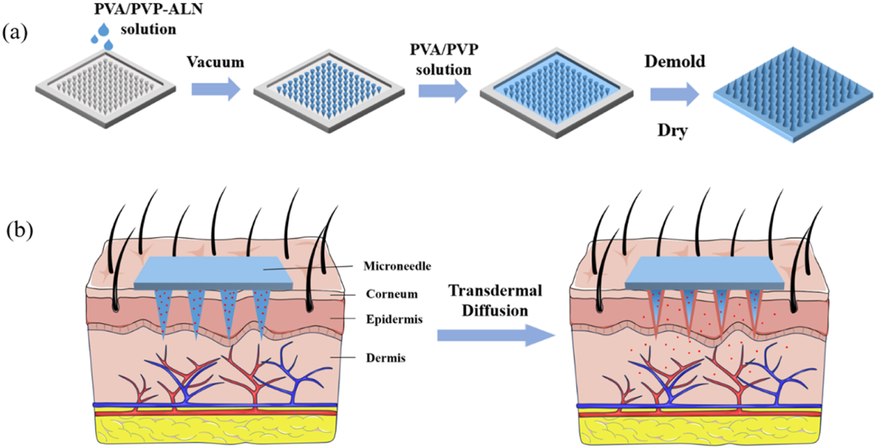
Swelling ratio
Systematic analysis of the macroscopic array and structural morphology of the MNs body was conducted using visual inspection and optical microscopy in sequence. 24 The dried MNs (mass W0) were immersed in PBS and retrieved at predetermined time points. Surface moisture was quickly absorbed with filter paper, followed by weighing (mass Wt). The swelling ratio was calculated using the formula S = [(Wt – W0)/W0] × 100% to evaluate their water absorption capacity and dissolution characteristics.
Mechanical properties
To evaluate the mechanical strength and skin penetration capability of the MNs, penetration tests were conducted using sealing film in this study.
The prepared MNs were placed vertically with their tips facing downward onto four layers of Parafilm®M sealing film (total thickness approximately 500 μm). 25 A 1 kg weight was then applied and maintained for 2 min. After removing the MNs, the layers were separated, and the number of penetration holes in each layer was counted. The penetration efficiency (W) was calculated using the formula W = (S1/S0) × 100%, where S1 represents the actual number of penetration holes and S0 denotes the theoretical total number of MNs tips. The penetration depth was further estimated based on the number of successfully penetrated film layers, providing a comprehensive assessment of the mechanical strength and penetration capability of the MNs.
In vitro release and transdermal permeation studies
To systematically evaluate the transdermal drug release behavior of the drug-loaded MNs, this study conducted in vitro drug release experiments using dialysis bags and ex vivo transdermal permeation experiments on rat skin, followed by kinetic model fitting to analyze the drug release mechanism.
Dialysis bag release study
A Franz diffusion cell was employed to investigate the in vitro release behavior of ALN from the MNs.
26
Three parallel experiments were conducted. The MNs was sealed in a dialysis bag and immersed in a receptor chamber containing 15 mL of phosphate-buffered saline (PBS, pH = 7.4) with a magnetic stir bar. The Franz diffusion cell was placed in an intelligent transdermal tester and maintained at 37 ± 1°C with continuous stirring at 400 rpm. Samples of 5 mL were collected at time points of 0.5, 1, 2, 3, 4, 6, 8, 10, 12, and 24 h, and an equal volume of PBS at the same temperature was immediately replenished. The collected samples were subjected to a colorimetric reaction with ninhydrin reagent, and the absorbance was measured at 568 nm using ultraviolet-visible spectrophotometry to calculate the cumulative release amount of ALN. A blank control group was included, in which an equal volume of PBS solution (pH = 7.4) was used instead of the sample, following the same procedure. The cumulative permeation amount (Q) was calculated using the following formula:

Ex vivo permeation experiment on rat skin
Rat dorsal skin was collected, and hair and subcutaneous adipose tissue were carefully removed. The skin was rinsed with physiological saline, wrapped in plastic wrap, and stored at −20°C for later use. Before the experiment, the skin was thawed in physiological saline for 30 min.
The drug-loaded MNs were fixed with their tips downward on the skin surface. The rat skin with the loaded MNs was placed on the receiving chamber of the Franz diffusion cell. 15 mL of PBS (pH = 7.4) was injected into the receiving chamber and magnetic particles were added. The same procedure as in the previous text was followed for the transdermal penetration test. The ALN concentration was quantified via ninhydrin colorimetric assay at 568 nm to calculate the cumulative release amount Q. The same experiment was conducted in parallel three times.
Release kinetic model fitting
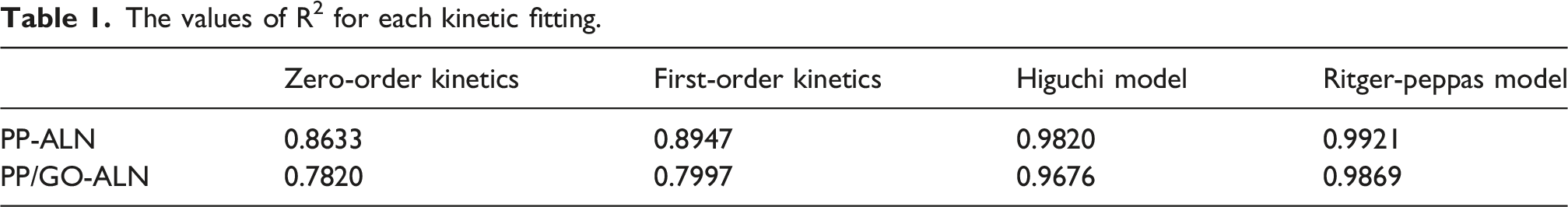
To gain deeper insights into the drug release mechanism from the MNs, the data from the in vitro release and ex vivo permeation experiments were fitted to four commonly used drug release kinetic models: zero-order kinetics, first-order kinetics, Higuchi model, and Ritger-Peppas model.27,28 The coefficient of determination (R2) served as the primary evaluation metric, with the model exhibiting an R2 closest to 1 considered the most suitable for describing the release behavior. Furthermore, the release exponent n in the Ritger-Peppas model was analyzed: if n < 0.45, the release mechanism conforms to Fickian diffusion; if n > 0.89, the release is predominantly governed by matrix erosion; and if 0.45 < n < 0.89, it indicates non-Fickian diffusion, signifying that drug release results from the synergistic effects of diffusion and erosion.
Antibacterial test
Preparation of LB solid medium
Peptone, yeast extract, sodium chloride, agar powder, and distilled water were used to prepare the LB solid medium. The specific preparation steps are as follows: The above mixed solution was placed in a pressure steam sterilizer and sterilized at 121°C for 30 min. The autoclave was cooled to approximately 50°C, and then plates were poured in a clean bench. After the culture medium completely solidifies, the petri dishes were sealed and inverted for storage to prevent surface contamination caused by condensate dripping.
Antibacterial performance assessment method
In this study, Staphylococcus aureus (S. aureus) and Escherichia coli (E. coli) were selected as model strains to evaluate the antibacterial performance of the fabricated MNs. Bacterial cultivation and colony counting were conducted using LB liquid medium and LB solid medium, respectively. 29 S. aureus and E. coli were activated and cultured in a shaking incubator at 37°C for 24 h.
S. aureus and E. coli were activated and cultured in a 37°C shaking incubator for 12 h. Each MN was immersed in a mixed solution containing 100 μL of live bacterial suspension and 900 μL of sterile PBS solution (pH = 7.4). After culturing at 37°C for 12 h, 100 μL of the mixed bacterial solution was withdrawn and 900 μL of sterile PBS solution was added (designated as 10-1 dilution solution). Subsequently, 100 μL of the 10-1 dilution solution was mixed with 900 μL of sterile PBS solution (designated as 10-2 dilution solution) until diluted to 10-6. 50 μL of the final dilution solution (i.e., 10-6 dilution solution) was spread on the surface of LB solid medium and incubated at 37°C in a constant temperature incubator for 12 h.
The PP MNs group was used as the blank control group, while the other groups (including MNs with different added components) were the experimental groups. Based on the colony count results, the antibacterial rates of each group were calculated using the following formula:
The data on colony counts and antibacterial rates were statistically analyzed using Origin and GraphPad software to compare the antibacterial performance differences among the four types of MNs, and to evaluate the effects of ALN and GO on the antibacterial performance of the PP MNs.
Antibacterial rate = (Blank group - Experimental group)/Blank group.
Aseptic operation and waste disposal
The whole operation was carried out in a sterile ultra-clean table. The instrument reagents were sterilized by pressure steam, and the waste containing bacteria was sterilized by 75% ethanol and treated as medical waste.
In vitro cell studies
Cell culture
M3T3-E1 cells were cultured in medium containing 10% fetal bovine serum and 1% streptomycin and penicillin solution, and kept in an incubator at 37°C and 5% CO2. Cells in logarithmic growth phase were cultured in well plates, and they were randomly divided into MNs experimental group, negative control group and blank control group.
Extract and differentiation medium preparation
As described in previous literature, the extract solution and differentiation medium were prepared. 30
The weight of MNs was precisely weighed, and the MNs were immersed in complete medium (containing 10% Fetal Bovine Serum and 1% Penicillin-Streptomycin solution), and then the MNs were placed in an incubator at 37°C and 5% CO2 for 24 h to obtain the extract with different MNs concentrations of 0.02 g/mL.
Differentiation medium was prepared by adding 10 mmoL/L β-glycerophosphate disodium salt hydrate and 50 μg/mL L-ascorbic acid to complete medium. Differentiation medium was used to extract the MNs as described above.
CCK-8 assay
In order to explore the effects of different components on the physiological and functional status of MC3T3-E1 cells, CCK-8 kit was used to detect the proliferation of osteoblasts. 31 MC3T3-E1 cells were seeded in 96-well plates at a density of 2 × 10³ cells/well, and the culture medium was 100 μL per well. After 1 d of cultivation, the culture medium was replaced with the extract solution. After another 1 d of cultivation, 10 μL of CCK-8 reagent was added to each well, and the samples were incubated in the dark at 37°C for 2 h. The multifunctional microplate detector was used to detect the absorbance (OD) value of each well at a wavelength of 450 nm, and the data were recorded and the cell proliferation rate was calculated. Cell proliferation rate = (OD value of experimental group - OD value of blank group)/(OD value of control group - OD value of blank group) × 100%.
Alkaline phosphatase (ALP) activity detection
The intracellular alkaline phosphatase staining and activity assay were performed on MC3T3-E1 cells. The cells were seeded in 6-well plates at a density of 2 × 10⁵ cells per well. After 24 h of culture, the old culture medium was replaced with differentiation medium. The medium was changed every other day. After 25 d of culture, the culture medium was discarded, and the wells were washed twice with PBS. Then, 200 μL of RIPA lysis buffer was added to each well, and the cells were scraped off with a cell scraper. The cells were homogenized and the lysate was collected in a 1.5 mL centrifuge tube. The tube was centrifuged at 12,000 rpm for 10 min at 4°C. The staining procedure was carried out according to the Biorad ALP Alkaline Phosphatase Assay Kit. The 96-well plate was shaken, incubated at 37°C for 5∼10 min, and then 100 μL of reaction termination solution was added to terminate the reaction. The absorbance was measured at 405 nm (400∼415 nm) and the alkaline phosphatase activity in the samples was calculated based on the enzyme activity definition. The yellow color produced by the staining remained stable within a few hours.
Mineralization assay (Alizarin Red S staining)
The Alizarin Red S staining method was used to evaluate the effects of four different MN samples on the mineralization nodule formation ability of MC3T3-E1 osteoblasts. The osteoblasts were inoculated into 24-well plates, with 500 μL per well (1 × 10⁴ cells). After the cells adhered to the plate, the old culture medium was replaced with the differentiation medium. The induction was stopped on the 6th day. The extract solution was discarded, and the cells were washed twice with PBS buffer solution and then fixed with 4% paraformaldehyde at room temperature for 10 min. Then, 0.5 mL of alizarin red S staining solution (2%, pH = 8.3, stained at 37°C for 30 min) was added to each well for staining. Finally, the residual alizarin red S staining solution was quickly washed with distilled water, and an appropriate amount of absolute ethanol was added to prevent cell drying. The cells were observed and photographed under a microscope.
Statistical analysis
The data were statistically analyzed and plotted using GraphPad 10.6.0 software. The t-test was used for comparison between the two groups, and one-way ANOVA was used for comparison among multiple groups. P value less than 0.05 was considered statistically significant.
Results and discussion
Morphology of MNs
Using PDMS molds, MNs with different compositions were fabricated. Figure 2(a) shows the frontal and side views of drug-free MNs. The results indicate that MNs prepared with 5% PVA–8% PVP and 8% PVA–8% PVP exhibited good performance during demolding. Among them, the MNs fabricated with 8% PVA–8% PVP exhibited optimal morphology, sharp tips, and minimal defects. Higher PVA concentration (12%) led to increased viscosity, bubble retention, and curling. Higher PVA concentration (12%) led to increased viscosity, bubble retention. Although the number of needles was close to the mold design, peripheral needle tips showed breakage, and edge regions displayed curling and unevenness. Overall, as the PVA concentration increased, the needle tips tended to become sharper and straighter. However, when the PVA concentration was too high, the MNs showed a tendency to curl inward. This may be attributed to the increased viscosity of the solution due to high PVA content, which hindered bubble removal. Simultaneously, the higher proportion of PVA molecules reduced the relative content of water molecules, making the MNs more prone to curling during drying due to water loss. Considering both mechanical properties and skin insertion performance, 8% PVA–8% PVP was selected as the matrix filling solution for subsequent experiments. The morphological characteristics (a, b) and optical images (c) of the MNs. (The above MNs are all square-shaped with dimensions of 1 cm × 1 cm).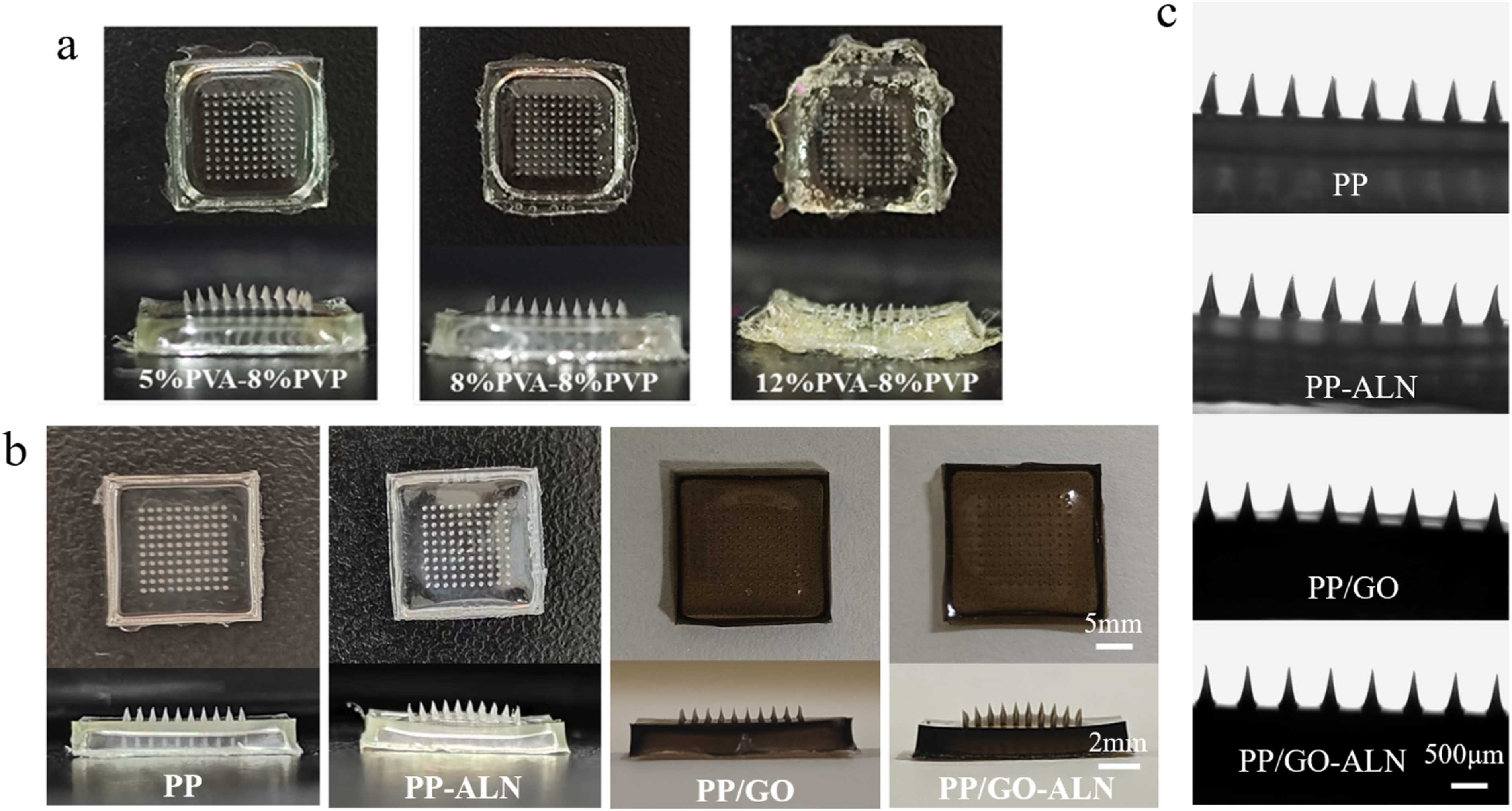
On this basis, GO dispersion was added to 8% PVA–8% PVP to prepare PP-GO MNs. Addition of GO (PP/GO MNs) resulted in uniform black coloration without altering needle morphology (Figure 2(b)). Drug loading with ALN did not significantly affect MN structure (Figure 2(c)). As a small phosphorus-containing molecule, ALN possesses polar groups (—PO43-, —NH2) that may form hydrogen bonds or electrostatic interactions with hydroxyl (—OH) or carbonyl (C = O) groups in PVA/PVP.
Mechanical properties
Figure 3 shows the puncture performance test results of MNs with four different compositions. As can be seen from the puncture rate and the images of each layer of parafilm after puncture, PP MNs and PP-ALN MNs could mostly penetrate the second layer of parafilm, while only a small number of needle tips penetrated the third layer, with puncture rates of merely 48.33% and 51.33%, respectively. In contrast, GO-loaded PP/GO MNs and PP/GO-ALN MNs could completely penetrate the second layer of parafilm, and most of them could penetrate three layers of parafilm, achieving puncture rates of 88.33% and 66%. Literature has shown that the skin stratum corneum thickens with aging, and the relatively thick stratum corneum of the upper limb can reach 30 μm, which is far smaller than the thickness of three layers of parafilm (approximately 400 μm). This indicates that PP/GO-ALN MNs possess excellent puncture capability, which can meet the requirements of transdermal drug delivery. In conclusion, MNs containing GO are superior in mechanical strength to GO-free formulations, and they can still maintain good puncture performance and structural integrity after loading 1% ALN, facilitating drug penetration through the skin. Characterization of MNs penetration into Parafilm®M: (a) penetration efficiency statistics, (b) macroscopic photograph of the penetrated region. (n=3).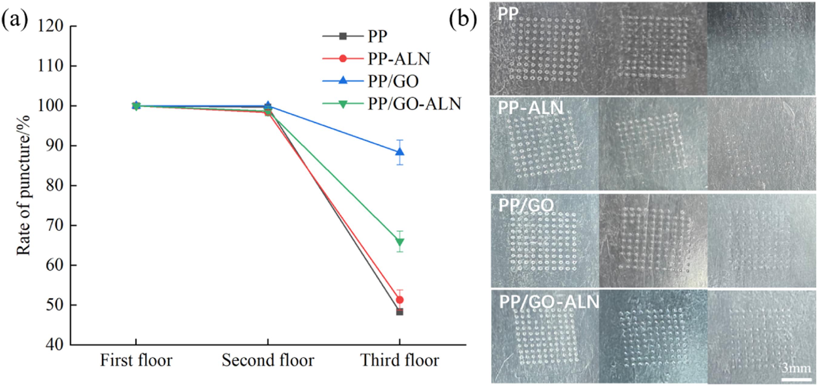
Swelling ratio
Figure 4 shows the swelling profiles of the four MNs (PP, PP-ALN, PP/GO, and PP/GO-ALN). Experimental observations show that all groups exhibited significant swelling and partial dissolution at the MNs, indicating their solubility. Swelling kinetics results indicate that all four formulations reached swelling equilibrium at approximately 1260 s (21 min), with equilibrium swelling ratios in the following order: PP/GO-ALN MNs (299%) > PP/GO MNs (287%) > PP MNs (265%) > PP-ALN MNs (236%). This order indicates that the two groups containing GO (PP/GO and PP/GO-ALN) exhibited higher expansion rates, suggesting that the presence of GO is associated with increased expansion. The expansion equilibrium time for all four MNs was approximately 1260 s, indicating that the matrix composition had no significant effect on the equilibrium time under the experimental conditions, and the system exhibited relatively consistent expansion rates. (a) The image of the swelling rate after 21 min of swelling in PBS and (b) the schematic diagram of the MNs after swelling. (n = 3).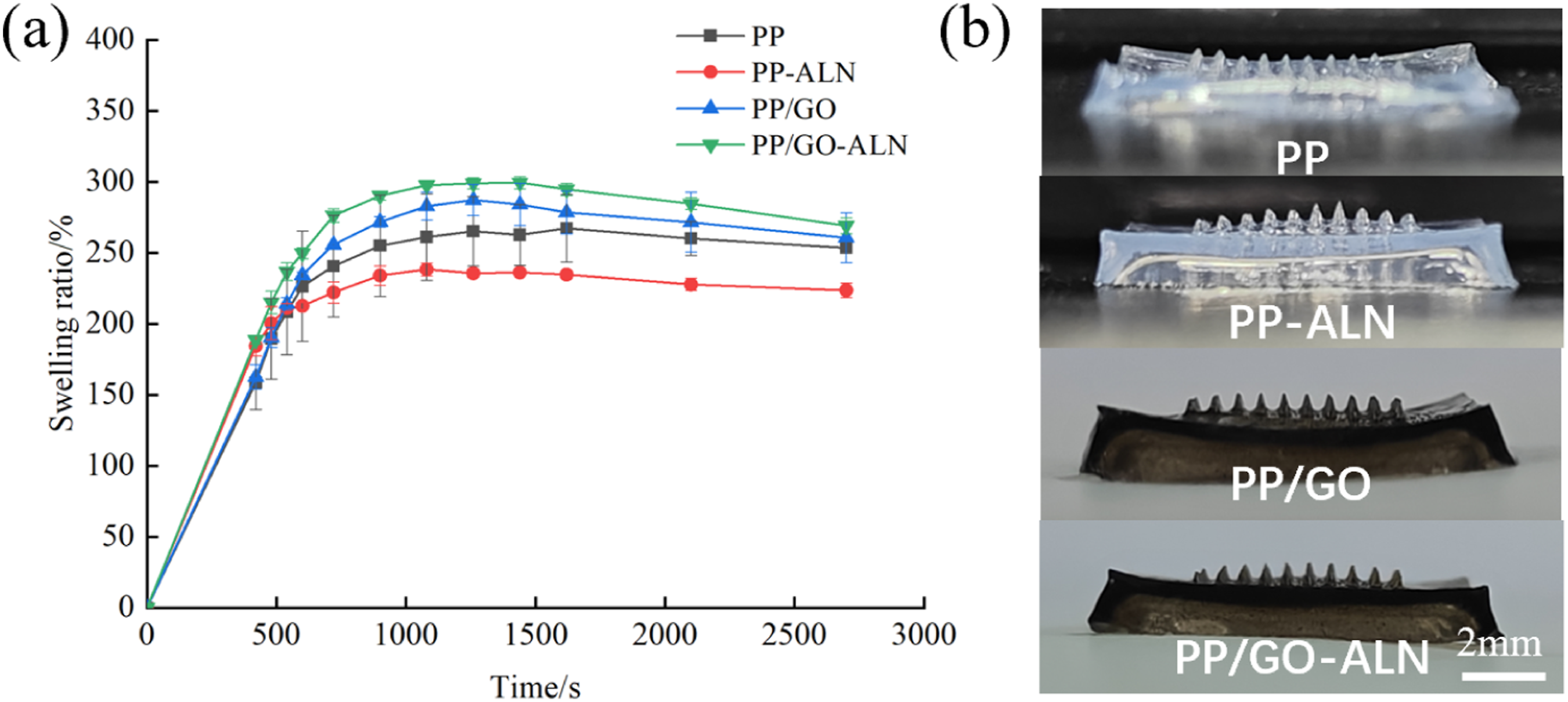
In addition, all samples exhibited a slight decrease in expansion rate after 1620 s (27 min), a phenomenon consistent with the partial dissolution observed at the tip of the MNs. This suggests that the expansion process of the soluble MNs was accompanied by gradual dissolution of the material; in the later stages, the rate of mass loss due to dissolution may exceed the water absorption rate, leading to a decrease in the apparent expansion rate.
While the underlying mechanisms cannot be directly confirmed by the present swelling experiments alone, we hypothesize, based on previous literature, that the enhanced swelling behavior may be attributed to the following factors. GO may enhance the system’s swelling capacity due to its hydrophilic functional groups and nanochannel effects. 32 In addition, the three-dimensional assembly of graphene-based nanosheets provides high porosity as water reservoirs which supply water to intersheet spacings. 33 Based on this, a tentative explanation is proposed: the addition of GO in this system could potentially enhance the hydrophilicity and swelling properties of the MNs system; meanwhile, a possible synergistic effect between GO and ALN might further modulate these properties.
In vitro release and transdermal permeation studies
The in vitro drug release experiment (Figure 5(a)) showed that in the dialysis bag model without a skin barrier, the cumulative drug release amounts of PP-ALN MNs and PP/GO-ALN MNs within 24 h were 1038.90 μg and 955.07 μg respectively, with no statistical difference between the two. This result indicates that under the condition of only using PBS as the release medium, the ALN molecules (with molecular weights much lower than the 8000∼14,000 Da retention molecular weight of the dialysis bag) in both types of MNs can freely diffuse, and their release behaviors are basically similar. (a) The cumulative release amount within the dialysis bag and (b) the ex vivo cumulative permeation amount through rat skin. (n = 3).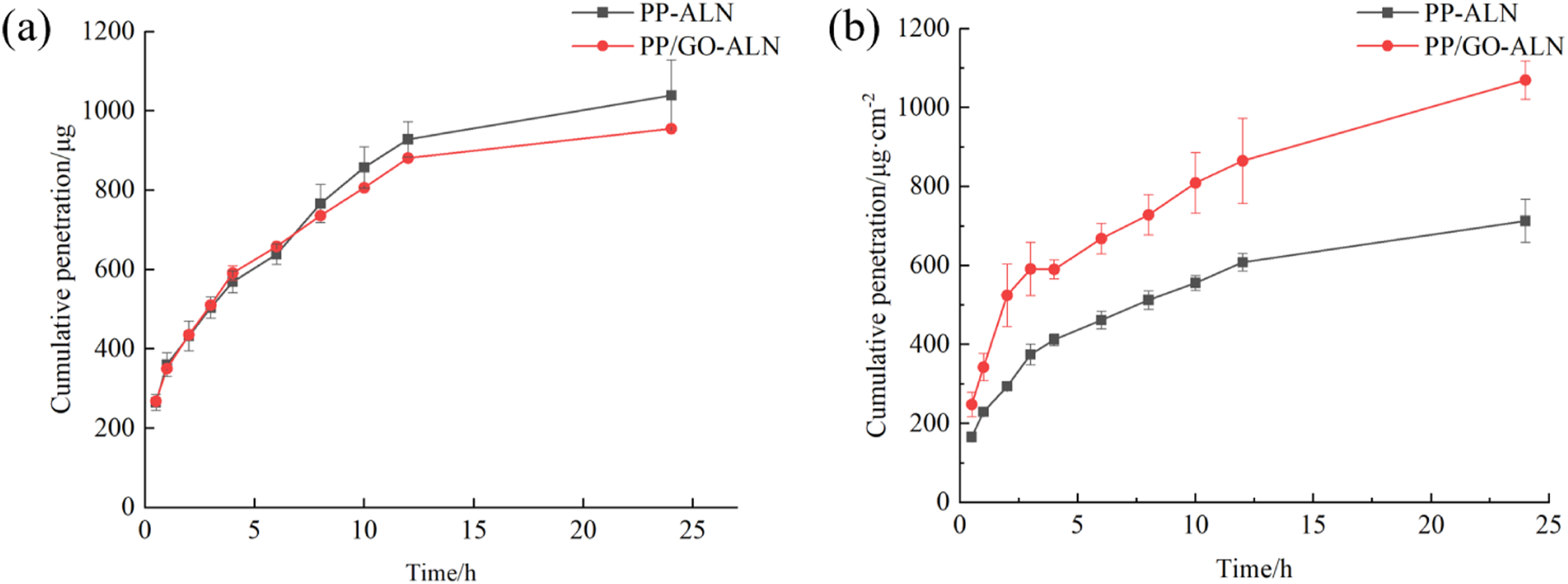
The in vitro transdermal experiment (Figure 5(b)) demonstrated that in the rat skin model, the 24 h cumulative transdermal permeation amount of the PP/GO-ALN MNs (1069.53 μg/cm2) was significantly higher than that of the PP-ALN MNs (712.89 μg/cm2, *p < 0.05). This result indicates that the introduction of GO is associated with a higher transdermal permeation amount. While the direct mechanisms underlying this enhancement remain to be further validated, we hypothesize that the improved transdermal performance may arise from two potential contributions of GO, based on the literature and the present results: (1) GO may enhance the mechanical strength of the PVA-PVP matrix, improving the insertion depth and structural integrity of the microneedles, thereby creating more efficient microchannels for drug delivery 34 ; and (2) the lamellar structure of GO may promote the dispersion of ALN within the matrix, reducing drug aggregation and facilitating its release and permeation. 35 These synergistic effects may contribute to the enhanced transdermal drug delivery efficiency observed for PP/GO-ALN-MNs.
The values of R2 for each kinetic fitting.
Antibacterial activity
It is known that GO is a highly effective antibacterial nanomaterial.
36
To investigate the regulatory effects of GO and ALN on the antibacterial properties of MNs, this study systematically evaluated the antibacterial activity of four types of MNs—PP (control), PP-ALN, PP/GO, and PP/GO-ALN—using the plate counting method, with S. aureus and E. coli as model strains. Figure 6 showed that the colony counts for S. aureus and E. coli in the blank control group (PP) were 528 ± 29 CFU and 219 ± 39 CFU. In the PP-ALN group, the colony counts were 383 ± 8 CFU for S. aureus and 250 ± 52 CFU for E. coli, showing no significant difference compared to the control group, indicating that ALN alone failed to confer antibacterial functionality to the MNs. In contrast, the two groups containing GO demonstrated significant antibacterial effects: in the PP/GO group, the colony counts for S. aureus and E. coli decreased to 317 ± 18 CFU and 79 ± 5 CFU, respectively, representing reductions of approximately 40% and 64% compared to the control group; in the PP/GO-ALN group, the counts decreased to 251 ± 28 CFU for S. aureus and 84 ± 15 CFU for E. coli, with reductions of 52% and 62%, respectively. Both groups exhibited higher inhibition rates against E. coli than against S. aureus, reflecting the stronger antibacterial activity of GO against Gram-negative bacteria, which is associated with multiple mechanisms. The inhibition rates against E. coli were higher than those against S. aureus in both groups, indicating that GO exhibits stronger antibacterial activity against Gram-negative bacteria. Although the underlying mechanisms may involve multiple factors, they remain unclear; it is speculated that this may be due to the disruption of the lipopolysaccharide structure on the outer membrane of E. coli, the induction of oxidative stress, and bacterial encapsulation. It is worth noting that for the PP group and the PP-ALN group, both the PP/GO group and the PP/GO-ALN group exhibited good antibacterial capabilities, indicating that the addition of GO enhances the inhibitory effect on bacteria, which also confirms the antibacterial properties of GO. In summary, GO is the key component for enhancing the antimicrobial performance of MNs. Both PP/GO and PP/GO-ALN MNs demonstrated good antimicrobial performance, providing experimental evidence for their application in synergistically inhibiting pathogenic microorganisms during transdermal drug delivery. (a) Counting of colonies of S. aureus and E. coli, (b) Images of S. aureus and E. coli colonies on the agar plate. (n = 3, *p < 0.05,**p < 0.01).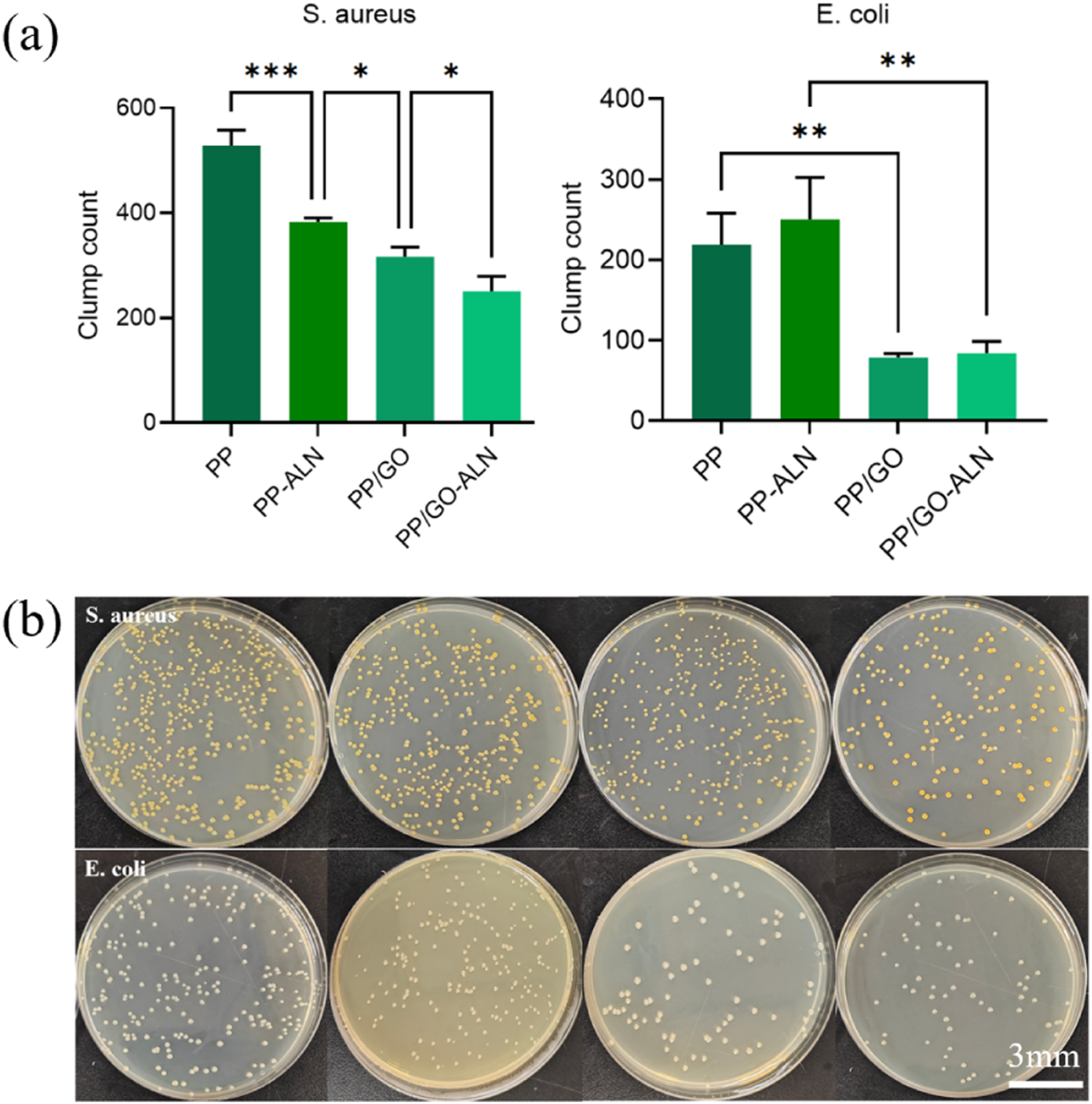
MC3T3-E1 cell level experiments in vitro
CCK-8 assay
To systematically evaluate the biocompatibility and biological functionality of MNs materials, this study employed the CCK-8 assay to examine the effects of extracts from different MNs formulations on osteoblast viability. Cells were cultured in extracts from four types of MNs—PP, PP-ALN, PP/GO, and PP/GO-ALN—for 24 h, and the results are presented in Figure 7. The data showed that the relative cell viabilities for the PP, PP-ALN, and PP/GO groups were 97.88% ± 2.35%, 96.64% ± 1.35%, and 99.23% ± 4.39%, respectively, indicating no significant cytotoxic or proliferative effects under the experimental conditions. In contrast, the PP/GO-ALN group demonstrated a significant increase in cell viability to 107.42% ± 2.74%, which was statistically higher than all other groups (p < 0.05). These findings indicate that a clear pro-proliferative effect on osteoblasts was only observed when GO and ALN were co-loaded into the MNs, whereas neither ALN nor GO alone produced a similar effect. This phenomenon suggests a synergistic interaction between GO and ALN, the mechanism of which may be related to GO acting as a nanocarrier to facilitate the sustained release of ALN, enhance its bioavailability, or improve drug delivery efficiency within the cellular microenvironment. The cell proliferation rate of MC3T3-E1 in the extract solution. (n = 3, *p < 0.05,**p < 0.01).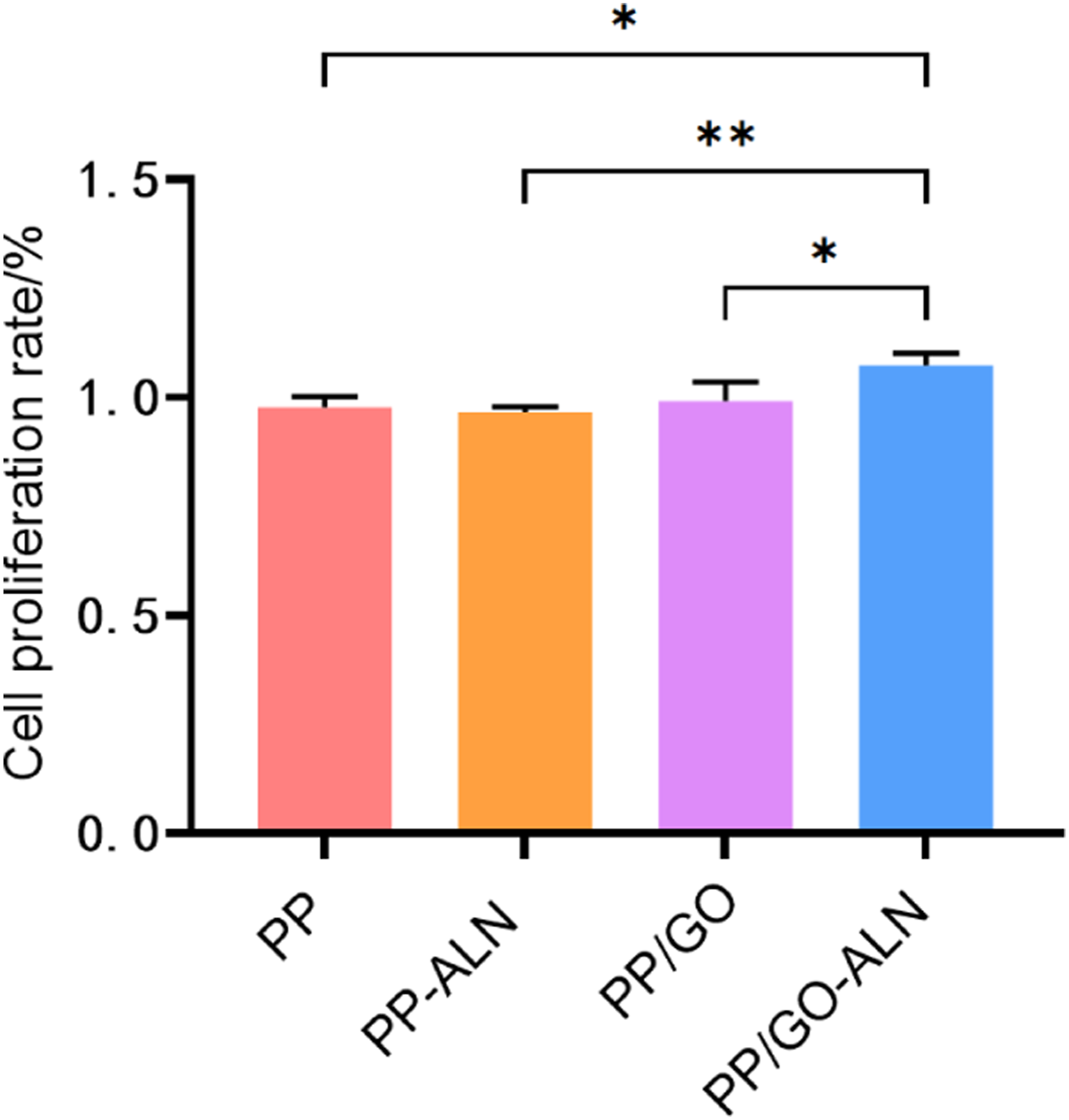
ALP activity detection
In MC3T3-E1 mouse osteoblasts, ALP activity is a key indicator of the early stage of osteogenic differentiation, mainly reflecting the initiation degree of osteoblasts’ transformation into mature osteoblasts. It is an early characteristic of cells possessing osteogenic phenotype and mineralization potential, and its activity level can directly indicate the activation state of osteogenic differentiation in the early stage. As shown in Figure 8(b), the ALP activity in the bone cell lysates of the PP group and PP/GO group was 28.97 and 29.89 DEA U/L respectively, while that in the PP-ALN group and PP/GO-ALN group was 34.02 and 33.73 DEA U/L respectively, which was 17.43% and 12.85% higher than that of the PP group and PP/GO group (p < 0.05). This indicates that osteoblasts have ALP activity, and the incorporation of ALN promotes the generation of ALP, suggesting that MC3T3-E1 cells cultured in the differentiation medium loaded with ALN have better osteogenic differentiation activity. (a) ALP standard curve, (b) After 25 d of induction differentiation, the changes in ALP activity of osteoblasts in different MNs differentiation culture media were observed. (n = 3; *p < 0.05; **p < 0.01).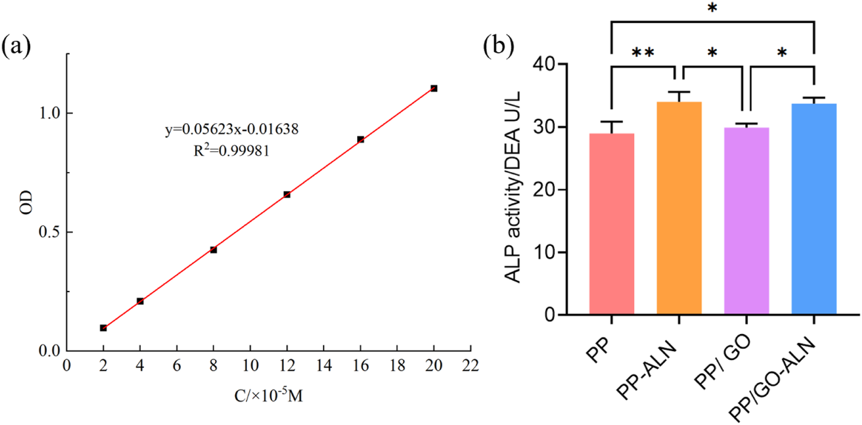
Mineralization assay
Alizarin Red S staining was used to qualitatively assess late-stage mineralization, which reflects the formation of calcium nodules by osteoblasts. As shown in Figure 9, the number of mineralized nodules was slightly higher in the PP and PP/GO groups than in the blank control, while a clear increase in nodule formation was observed in the PP-ALN and PP/GO-ALN groups compared to the unloaded groups. These qualitative observations are consistent with the ALP activity results, suggesting that ALN loading may promote osteogenic differentiation. It should be noted that these results are qualitative, and further quantitative analysis (e.g., extraction and colorimetric measurement of the stain) would provide more robust evidence of mineralization levels, which we plan to address in future work. Alizarin red S staining was used to identify the ability of osteogenic mineralization nodules to form in different MNs differentiation culture media. (n = 3).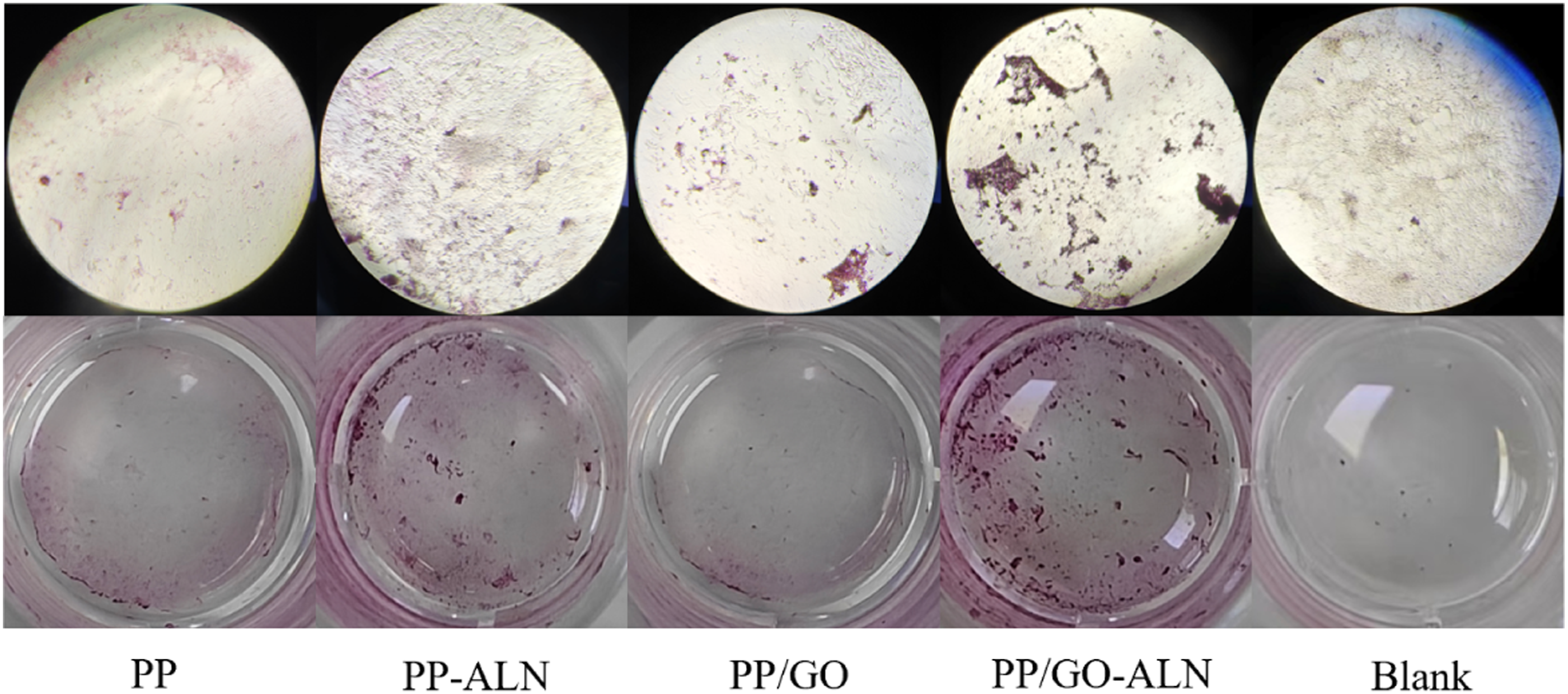
Conclusions
In this study, a GO-enhanced dissolvable MN system was successfully developed for transdermal ALN delivery. The incorporation of GO significantly improved the mechanical strength (penetration efficiency: 66∼88%), welling capacity (PP/GO-ALN swelling ratio: 299%), and antibacterial activity. In an ex vivo skin model, the cumulative transdermal permeation of PP/GO-ALN MNs over 24 h (1069.53 μg/cm2) was significantly higher than that of the PP-ALN group (712.89 μg/cm2). Furthermore, cell experiments demonstrated that PP/GO-ALN synergistically promoted osteoblast proliferation (cell viability: 107.42%, p < 0.05), promoted osteoblast differentiation (ALP activity) and mineralization nodule formation. These findings highlight the potential of this composite MN system as an effective, noninvasive platform for osteoporosis treatment.
Footnotes
Author contributions
Data curation Qianqian Sun and Xiaowen Li; Funding acquisition, Litao Wang and Mei Lv; Investigation, Guofa Zhang; Methodology, Bing Li; Project administration, Yanlian Niu; Supervision, Litao Wang; Writing – original draft, Qianqian Sun; Writing – review & editing, Mei Lv.
Funding
The authors disclosed receipt of the following financial support for the research, authorship, and/or publication of this article: This work was supported by the Natural Science Foundation of Shandong Province ZR2023MH018; Jining Medical College high-level scientific research project cultivation plan, JYGC2022 KJ007.
Declaration of conflicting interests
The authors declared no potential conflicts of interest with respect to the research, authorship, and/or publication of this article.
Data Availability Statement
The data presented in this study are available on request.