
Abstract
In this study, composite hydrogel scaffolds of cellulose nanocrystals (CNC) and β-glucan with poly(acrylic acid) (PAA) were synthesized, and their effects on accelerating skin wound healing were investigated. CNC was extracted from hardwood and β-glucan from oat flour, and after chemical crosslinking and photopolymerization, CNC-g-β-glucan/PAA composite hydrogels were prepared. The scaffolds’ biological properties were evaluated using MTT assay, in vitro degradation in simulated wound fluid, DAPI staining, FESEM analysis, histological examinations, and immunofluorescence in an animal model. Biocompatibility results indicated that scaffolds containing β-glucan (particularly samples S2 and S3) showed the highest cell viability compared to other groups (p < 0.01). Degradation studies revealed a relatively rapid degradation profile. Moreover, in wound healing assessment, these scaffolds significantly increased epidermal thickness, reduced inflammatory cells, and enhanced the expression of bioactive markers including Col-1, VEGF, and TGF-β. FESEM observations also revealed proper fibroblast spreading and favorable adhesion on the scaffold surface. Overall, the data suggest that incorporating β-glucan into the CNC network improves biological and regenerative performance, likely mediated in part by its immunomodulatory activity
Introduction
Wound healing is a complex, multi-stage process involving cellular, molecular, and tissue responses for the regeneration of damaged skin. Naturally, this process occurs in three phases: inflammation, proliferation, and remodeling. 1 However, in many cases, such as chronic wounds caused by diabetes, burns, or severe infections, normal healing is impaired, necessitating advanced therapeutic approaches. 2 In recent years, the use of biodegradable and biocompatible scaffolds in tissue engineering has emerged as one of the most effective strategies for skin tissue regeneration. 3 Scaffolds serve as temporary matrices for cell growth and, by mimicking the extracellular matrix (ECM), provide suitable conditions for cell migration, proliferation, and differentiation. 4
Among biomaterials used for scaffold fabrication, cellulose nanocrystals (CNC) have attracted attention due to their high mechanical strength, biodegradability, large surface area, and chemical modifiability. As a natural nanomaterial derived from cellulose, CNC can be combined with other polymers to improve properties such as moisture absorption, biocompatibility, and gas permeability. 5 However, CNC-only scaffolds may lack bioactive properties to actively stimulate healing. Therefore, combining CNC with bioactive materials such as β-glucan can enhance scaffold performance.
β-glucan is a natural polysaccharide found in the cell walls of cereals such as oats, and due to its immunomodulatory, antioxidant, and anti-inflammatory effects, it is highly effective in wound healing.6–8 Studies have shown that β-glucan stimulates macrophages and fibroblasts, increasing the production of growth factors including VEGF and TGF-β, thereby promoting angiogenesis and tissue regeneration. Studies have shown that β-glucan exerts its bioactive effects through binding to specific pattern recognition receptors (PRRs) on immune cells, particularly Dectin-1, complement receptor 3 (CR3), and toll-like receptors (TLR-2/6). 8 This receptor-mediated recognition initiates downstream signaling cascades that activate macrophages and fibroblasts, leading to the production of pro-healing cytokines and growth factors including VEGF and TGF-β, thereby promoting angiogenesis, extracellular matrix synthesis, and tissue regeneration. 9 Furthermore, β-glucan exhibits dose-dependent immunomodulatory effects, with optimal concentrations promoting a balanced immune response that accelerates healing while preventing excessive inflammation and fibrosis. 10 Consequently, incorporating β-glucan with CNC in a hydrogel can create a scaffold with suitable physical and biological properties for wound repair. 11 Poly(acrylic acid) (PAA), as a biocompatible synthetic polymer, has high water absorption and moisture retention capacity. Its network structure facilitates the formation of stable hydrogels. By combining PAA with CNC-g-β-glucan, a scaffold can be created that provides sufficient mechanical strength along with a moist, biocompatible environment for skin cell growth. 12 Photopolymerization used in hydrogel synthesis allows for the formation of a uniform, controlled network while preserving the bioactivity of β-glucan. 13
In this study, CNC-g-β-glucan/PAA hydrogel scaffolds with varying β-glucan ratios were synthesized, and their physical, biological, and regenerative properties were evaluated. In vitro assays included MTT for cell viability and DAPI staining for live cell counting, while in vivo studies assessed wound healing in an animal model. Histological and immunofluorescence analyses were conducted to examine the expression of healing factors (Col-1, VEGF, and TGF-β) and the scaffolds’ effect on skin tissue regeneration. The results confirmed that CNC-g-β-glucan/PAA scaffolds exhibit high biocompatibility and significant regenerative effects, and that an optimal β-glucan content plays a critical role in stimulating epidermal regeneration, reducing inflammation, and increasing collagen synthesis. Thus, this nanostructured composite presents a promising candidate for chronic wound treatment and skin tissue engineering applications.
Material and methods
Preparation of raw materials
This study was conducted as an experimental-descriptive investigation. Information was collected using optical and electron microscopy and analyzed computationally. CNC was derived from hardwood, and β-glucan was extracted from oat flour. To prepare a CNC-β-glucan conjugate, the required amount of CNC was dissolved in water and subjected to ultrasonication to create 100 mL of a 1 wt.% CNC suspension. The suspension was placed in a three-neck flask equipped with a nitrogen inlet, condenser, and magnetic stirrer. The pH was adjusted to 2 using nitric acid, and the mixture was gently stirred while heated to 45°C under nitrogen flow for 30 min to remove dissolved oxygen. Next, 0.33 g of ammonium cerium nitrate was added and stirred gently for 10 min, followed by the addition of 2 g of β-glucan or the required amount. Finally, the solution was stirred under a nitrogen atmosphere at 45°C for 4 h. After the reaction, the solution was dialyzed in water for 5 days to remove impurities. The solid content was determined by drying and weighing the sample. For further processing, the suspension was precipitated with ethanol, filtered, vacuum-dried, and ground into a powder.12,13 Figure 1 showed the Schematic of the research process. Comparative evaluation of the effect of A0 and A1 bioscaffolds on the wound healing process in an animal model.
Preparation of CNC-g-β-glucan/PAA hydrogel
The CNC-g-β-glucan/PAA hydrogel was synthesized via photopolymerization of acrylic acid in the presence of CNC-g-β-glucan using MBA as a crosslinker. The desired amounts of CNC-g-β-glucan suspension, acrylic acid, MBA (0.2% of acrylic acid volume), Irgacure 2959 (0.3% of acrylic acid volume), and water were mixed and sonicated in an ice bath to form a homogeneous solution. The volumetric ratio of acrylic acid to water was 25:75. The solution was cast into rectangular PTFE molds (100 mm × 15 mm × 3 mm), covered with glass, and exposed to UV light (365 nm, 21 W) at 25°C for 3 h to form hydrogels. Hydrogels were prepared using varying amounts of CNC-g-β-glucan, and a hydrogel containing only CNC (0.5% of the acrylic acid volume) was used as a control.12,13
Animal preparation and selection
The study consisted of five experimental groups, each comprising three subjects. The first group served as the control group without treatment. Experimental Group 1 was treated with a cellulose nanocrystal (CNC) hydrogel crosslinked with acrylic acid (0.2 g in 20 mL, A0). Experimental Group 2 received a CNC/β-glucan hydrogel crosslinked with acrylic acid (0.2 g CNC and 0.03 g β-glucan, A1), Experimental Group 3 (0.2 g CNC and 0.06 g β-glucan, A2), and Experimental Group 4 (0.2 g CNC and 0.12 g β-glucan, A3). Full-thickness wounds were created using an 8-mm punch biopsy under sterile conditions before treatment. The number of subjects per group was calculated using G*Power software (version 3.1.9.4) with a one-way ANOVA, fixed-effects model. 14
In vitro cytotoxicity assay (MTT test)
The cytotoxicity of the fabricated scaffolds was evaluated using the MTT assay. Equal-sized scaffolds from four experimental groups were placed into 96-well culture plates. To sterilize the scaffolds, ethanol was added to each well and incubated for 30 min, followed by 20 min of exposure to UV light. The ethanol was then removed, and scaffolds were washed with PBS.
Next, 5,000 cells suspended in culture medium were seeded into each well containing the scaffolds. At days 1, 3, and 7, the culture medium was removed, and 100 µL of MTT solution (90% PBS, 10% MTT) was added to each well. After incubation for 3 h at 37°C in a CO2 incubator, the MTT solution was removed, and 100 µL of DMSO was added to dissolve the blue formazan crystals. The absorbance of the resulting solution was measured at 570 nm to determine cell viability.
Controls: Wells containing cells with PBS only served as negative controls, and tissue culture polystyrene (TCP) was used as a positive control. Data were analyzed to compare the cytotoxicity of the different scaffold groups.
In vitro degradation in simulated wound fluid
To better simulate the physiological wound environment, hydrogel degradation was also assessed in simulated wound fluid (SWF). SWF was prepared according to established protocols containing NaCl (6.5 g/L), CaCl2·2H2O (0.4 g/L), and KCl (0.4 g/L) in distilled water, adjusted to pH 7.4 to mimic the ionic composition and pH of wound exudate. Pre-weighed hydrogel samples (approximately 0.5 g each, n = 3 per group) were immersed in 10 mL of SWF at 37°C under gentle agitation (60 rpm) to simulate the dynamic wound environment. At predetermined time points (days 1, 3, and 7), samples were removed, gently blotted with filter paper to remove surface fluid, and weighed to determine mass loss. The percentage of weight loss was calculated using the equation: Weight loss (%) = [(W0 - Wt)/W0] × 100, where W0 is the initial dry weight and Wt is the weight at time t. Complete degradation was defined as ≥95% weight loss. Each experiment was performed in triplicate to ensure reproducibility.
Cell counting assay
To prepare cell cultures, scaffolds were first sterilized and placed in the bottom of culture plates. Cells were then seeded onto the scaffolds. After 7 days of culture, the medium was removed, and DAPI staining was performed to visualize adhered cells. DAPI solution was added and incubated at room temperature for 1 h. Following incubation, samples were gently washed several times with PBS. Fluorescent microscopy was then used to observe and compare cell adhesion among the different scaffold groups and controls.
In vivo experiments
To evaluate the effect of the developed dressings compared to sterile gauze on wound healing, animal samples were transported under minimal stress and maintained under standard conditions, including adequate food and water, 12-h light/dark cycles, and individual cages. After a 1-week acclimation period, animals were anesthetized with ketamine-xylazine. Hair on the dorsal region was trimmed, and the skin disinfected with Betadine. Full-thickness wounds were created using an 8-mm biopsy punch under sterile surgical conditions.
The wounds were treated for 21 days with sterile gauze (control group)
At the end of the treatment period, animals were anesthetized with ketamine-xylazine, tissue samples were collected, and animals were euthanized using CO2 in a humane manner. Carcasses were stored at −20°C, securely packaged in labeled, leak-proof bags marked “Toxic and Hazardous,” and disposed of according to biomedical waste management protocols.
Histological analysis
To examine tissue morphology, Hematoxylin and Eosin (H&E) staining was performed on full-thickness skin samples collected on day 21 post-surgery. Tissue fixation was carried out immediately after excision in 10% formalin for 48 h.
Due to the high water content of tissue, dehydration with graded ethanol series was performed prior to paraffin embedding: 70% ethanol (50 min), 80% (50 min), 90% (50 min), and 100% (50 min). Xylene was used to remove ethanol, followed by paraffin infiltration in three steps: 1% xylene (50 min), 2% xylene (50 min), and paraffin (50 min). Tissue blocks were sectioned at 5 µm using a microtome and mounted on silanized slides.
For staining, slides were incubated at 90°C for 20 min to melt paraffin. Slides underwent deparaffinization in xylene 1 and 2 (15 min each), followed by rehydration through graded ethanol to water: 100% (5 min), 90% (5 min), 80% (5 min), 70% (5 min), distilled water (5 min). Slides were then stained in hematoxylin for 7 s, washed in distilled water for 1 min, treated with lithium carbonate for 2 s, and stained in eosin for 3 min. After dehydration with absolute ethanol and clearing with xylene 1 and 2 (15 min each), slides were mounted with mounting medium and imaged using a light microscope (LABOMED).
Immunofluorescence assay
Slides were immersed in X1 TBS (Sigma 5912-T) and heated in a microwave until boiling. Samples remained in solution for 20 min, then washed three times with PBS at 5-min intervals. To permeabilize cell membranes, 0.3% Triton X-100 was applied for 30 min, followed by PBS washes. Slides were blocked with 10% goat serum for 45 min to prevent non-specific secondary antibody binding.
Primary antibodies were diluted 1:100 in PBS and applied to slides. Slides were incubated in a humidified chamber at 2–8°C for 24 h. After incubation, slides were washed four times with PBS (5 min each). Secondary antibodies (1:150 dilution) were applied and incubated at 37°C for 1 h and 30 min in the dark. Following three PBS washes, DAPI was added for 20 min. Slides were then washed with PBS, mounted with a glycerol-PBS solution, and imaged using a fluorescence microscope (Olympus) to assess TGF-β, VEGF, and Col-1 expression.
Tissue sections were immersed in 1X TBS (Sigma 5912-T) and heated in a microwave until boiling. Samples remained in solution for 20 min, then washed three times with PBS at 5-min intervals. To permeabilize cell membranes, 0.3% Triton X-100 was applied for 30 min, followed by PBS washes. Slides were blocked with 10% goat serum for 45 min to prevent non-specific secondary antibody binding.
Primary antibodies targeting type I collagen (Col-1), vascular endothelial growth factor (VEGF), and transforming growth factor-beta (TGF-β) were diluted 1:100 in PBS and applied to slides. Slides were incubated in a humidified chamber at 2–8°C for 24 h. After incubation, slides were washed four times with PBS (5 min each). Fluorophore-conjugated secondary antibodies (1:150 dilution) were applied and incubated at 37°C for 1 h and 30 min in the dark. Following three PBS washes, DAPI (4',6-diamidino-2-phenylindole) was added for 20 min to counterstain cell nuclei. Slides were then washed with PBS, mounted with a glycerol-PBS solution, and stored at 4°C until imaging.
Immunofluorescence images were captured using a fluorescence microscope (Olympus) equipped with appropriate filter sets for FITC/Alexa Fluor 488 (green channel for Col-1, VEGF, and TGF-β) and DAPI (blue channel for nuclei). For each sample group (n = 3 biological replicates per group).
Quantitative image analysis was performed using Python programming language (version 3.8) with the OpenCV library (Open Source Computer Vision Library, version 4.5) for automated fluorescence intensity measurement. The quantification workflow consisted of the following steps: (1) Image preprocessing: Raw fluorescence images were loaded and split into individual color channels (green for target markers, blue for DAPI). Gaussian filtering (kernel size 5 × 5) was applied to reduce background noise while preserving signal integrity. (2) Region of interest (ROI) segmentation: Tissue areas were automatically segmented from background using adaptive thresholding (cv2.adaptiveThreshold with Gaussian weighting). Connected component analysis identified distinct tissue regions, and artifacts smaller than 100 pixels were excluded from analysis. (3) Fluorescence intensity measurement: Mean fluorescence intensity (MFI) was calculated for the green channel (marker expression) within the defined tissue ROIs using cv2.mean() function. Background fluorescence was measured from areas outside tissue regions and subtracted from all tissue measurements to correct for autofluorescence and camera noise. (4) Normalization: To account for variations in cell density between samples, marker expression was normalized to DAPI fluorescence intensity. The normalized expression was calculated as: (MFI_marker - MFI_background)/MFI_DAPI × 100. (5) Data aggregation: Fluorescence intensity values per sample were averaged to obtain a representative value for each biological replicate. Mean ± standard deviation (SD) was then calculated across the three biological replicates for each treatment group.
Statistical analysis
Sample size determination
Sample sizes for all experiments were determined using G*Power software [18]. For in vitro experiments, a priori power analysis was conducted using the following parameters: effect size f = 0.40 (medium to large effect based on preliminary data), α error probability = 0.05, power (1-β) = 0.80, number of groups = 4 (S1, S2, S3, S4), and repeated measures with 3 time points. This analysis yielded a minimum required sample size of n = 3 per group per time point. For in vivo wound healing studies, considering the ethical principle of minimizing animal use while maintaining statistical power, sample size was calculated based on: effect size f = 0.50 (large effect expected based on pilot studies), α = 0.05, power = 0.80, number of groups = 5 (control, S1, S2, S3, S4), yielding n = 3 animals per group. All experiments were performed with the calculated minimum sample sizes, with each measurement replicated in triplicate to ensure reproducibility.
Data processing and normality testing
For image-based quantitative analyses digital images were processed using ImageJ software with OpenCV library for automated thresholding, segmentation, and quantification. At least 3 random fields of view were analyzed per sample, and mean values were calculated for statistical comparison.
Comparative statistical analysis
For experiments with a single independent variable (scaffold type) measured at multiple time points (MTT assay, degradation studies), repeated measures one-way analysis of variance (RM-ANOVA) was performed when data met assumptions of normality and sphericity. For experiments comparing multiple groups at single time points (epidermal thickness, cell count), one-way ANOVA was employed. When significant main effects were detected (p < 0.05), post-hoc pairwise comparisons were conducted using Tukey’s Honestly Significant Difference (HSD) test, which controls the family-wise error rate and is appropriate for comparing all possible pairs of means.
Statistical software and significance criteria
All statistical analyses were performed using SPSS Statistics software (IBM Corporation, Armonk, NY, USA) and GraphPad Prism (GraphPad Software, San Diego, CA, USA). Statistical significance was set at α = 0.05 (two-tailed) for all tests unless otherwise specified. All experiments were performed with blinding of the investigator performing the quantitative analysis to experimental group assignments to minimize bias.
Results
In vitro degradation in simulated wound fluid
Given the importance of understanding scaffold behavior in the wound microenvironment, degradation studies were conducted in simulated wound fluid (SWF) to more closely mimic physiological conditions. As shown in Figure 2, all hydrogel scaffolds exhibited accelerated degradation in SWF compared to distilled water, reflecting the influence of ionic strength and enzymatic-like degradation processes in the wound environment. Biodegradation in simulated wound fluid on days 1, 3, 7. Statistical analysis was performed using two-way ANOVA. * Indicates a significant difference for S3 compared with the other scaffold groups at the same time point (p < 0.05).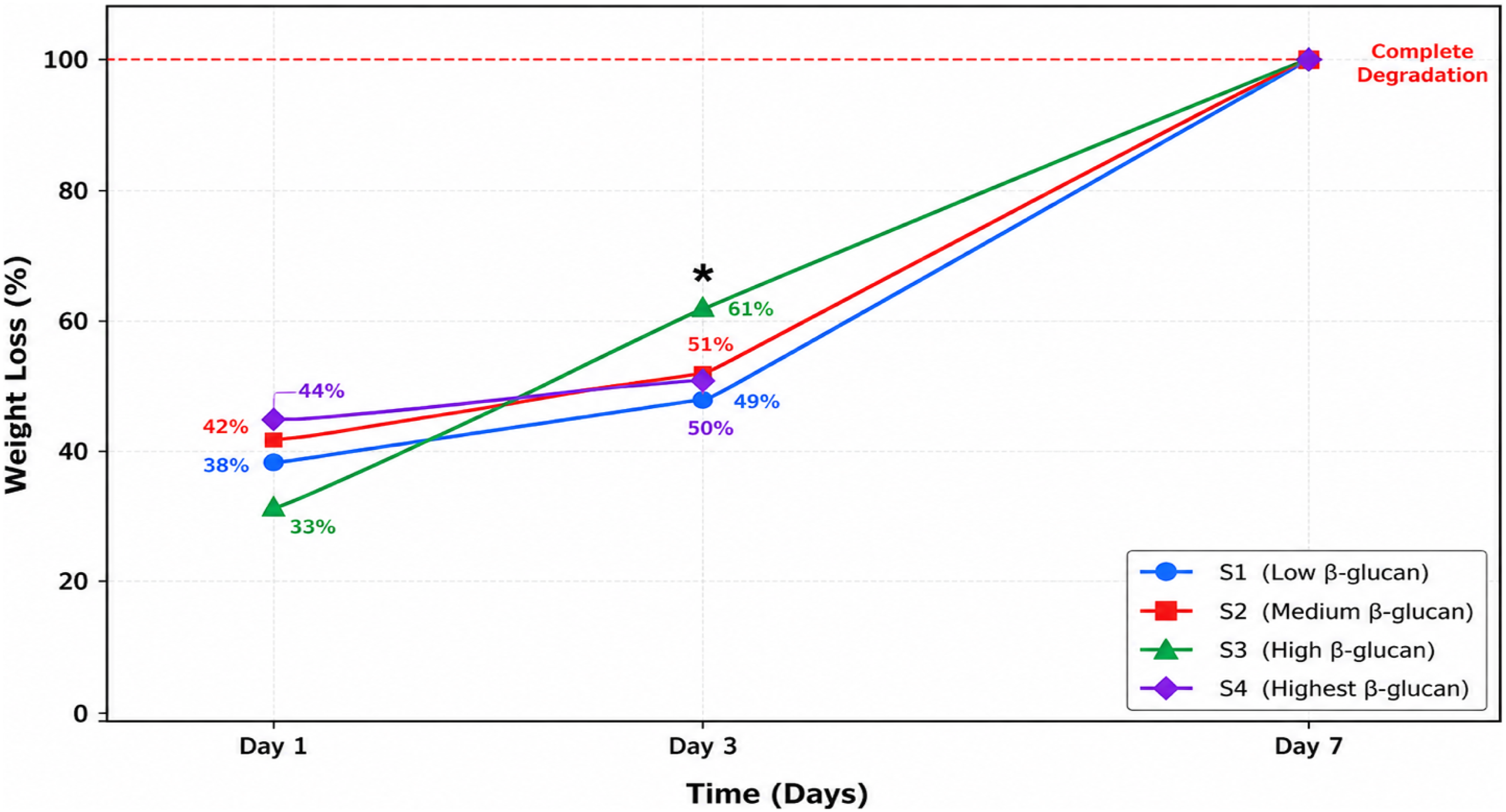
On day 1 in SWF, substantial early degradation was observed across all samples: S1 showed 38% weight loss, S2 exhibited 42% weight loss, S3 demonstrated 33% weight loss, and S4 displayed 44% weight loss. By day 3 in SWF, degradation progressed further with S1, S2, and S4 showing relatively similar degradation rates (49%, 51%, and 50% weight loss, respectively), while S3 exhibited the highest degradation rate at 61% weight loss. This suggests that the intermediate β-glucan content in S3 may result in a network structure more susceptible to hydrolytic degradation in the ionic wound environment. Remarkably, all hydrogel formulations achieved complete degradation (100% weight loss) by day, indicating that these scaffolds are fully biodegradable under simulated physiological conditions.
Compared to degradation in distilled water, where scaffolds maintained structural integrity, the accelerated degradation in SWF highlights the importance of evaluating biomaterials under physiologically relevant conditions. Statistical analysis using two-way ANOVA revealed a significant effect of both medium type (p < 0.001) and β-glucan content (p = 0.015) on degradation kinetics, with a significant interaction between these factors (p = 0.032).
Biocompatibility (MTT assay)
The biocompatibility of hydrogel scaffolds S1–S4 was evaluated using the MTT assay at three time points (days 1, 3, and 7), with TCP as a positive control. ANOVA analysis revealed significant differences in cell viability among the groups at all time points (p < 0.01). Post-hoc Tukey analysis indicated that S2 and S3 exhibited significantly higher cell viability compared to the other groups (S1, S4, and TCP) at all time points, particularly on days 3 and 7.one-way ANOVA revealed statistically significant differences in cell viability among the scaffold groups across all time points. Post-hoc pairwise comparisons using Tukey’s HSD test indicated that S2 and S3 exhibited significantly higher cell viability compared to S1 (p = 0.003 and p = 0.001, respectively), S4 (p < 0.001 for both), and TCP control (p = 0.008 and p = 0.004, respectively) at day 7. The mean cell viability for S2 was 94.3 ± 3.8% compared to 72.1 ± 5.2% for S1 (mean difference = 22.2%, 95% CI. No significant difference was observed between S2 and S3 (p = 0.891, mean difference = 1.2%, 95% CI [−6.8, 9.2]). In contrast, S4 showed the lowest viability, possibly due to cytotoxic effects or insufficient support for cell growth. These findings suggest that the formulations S2 and S3 are more suitable for wound healing applications (Figure 3). MTT assay results showing cell viability of hydrogel scaffolds S1–S4 and TCP at days 1, 3, and 7. Statistical analysis was performed using one-way ANOVA followed by Tukey’s HSD test (p < 0.01). S2 and S3 showed significantly higher cell viability, while S4 exhibited the lowest biocompatibility.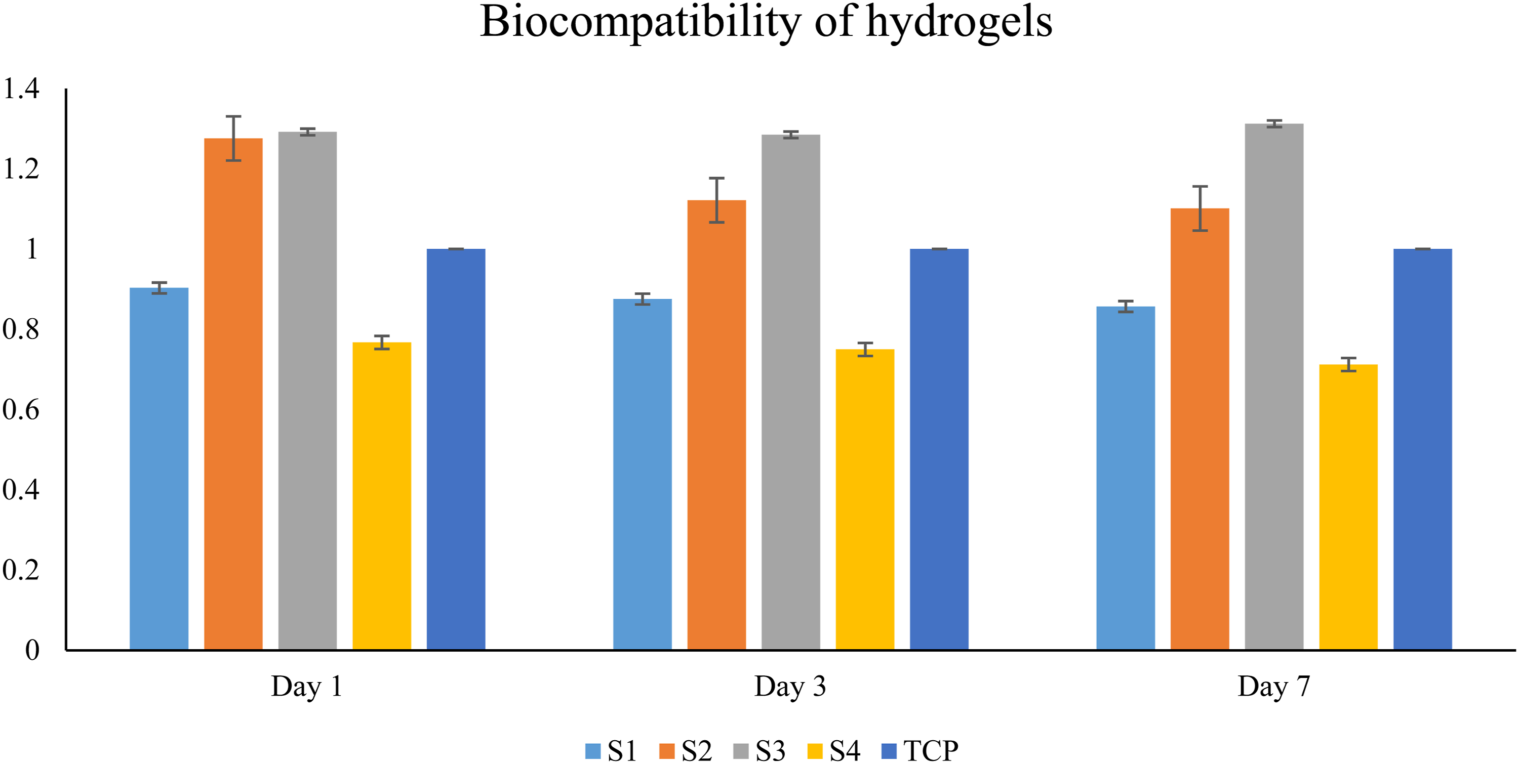
DAPI staining results
Fluorescence images of DAPI-stained nuclei confirmed the presence of cell nuclei on all hydrogel scaffolds (S1–S4). Nuclei appeared as bright blue, round to oval structures on the scaffold surfaces. Visual observation showed the following order of cell density: S3 > S4 > S2 > S1 (Figure 4). Fluorescence microscopy images of DAPI-stained nuclei on hydrogel scaffolds S1–S4. Scale bars = 10 µm. S3 showed the highest cell density, while S1 exhibited the lowest. No significant difference in nuclear area (p = 0.098), whereas nuclear roundness differed significantly among groups (p = 0.0019).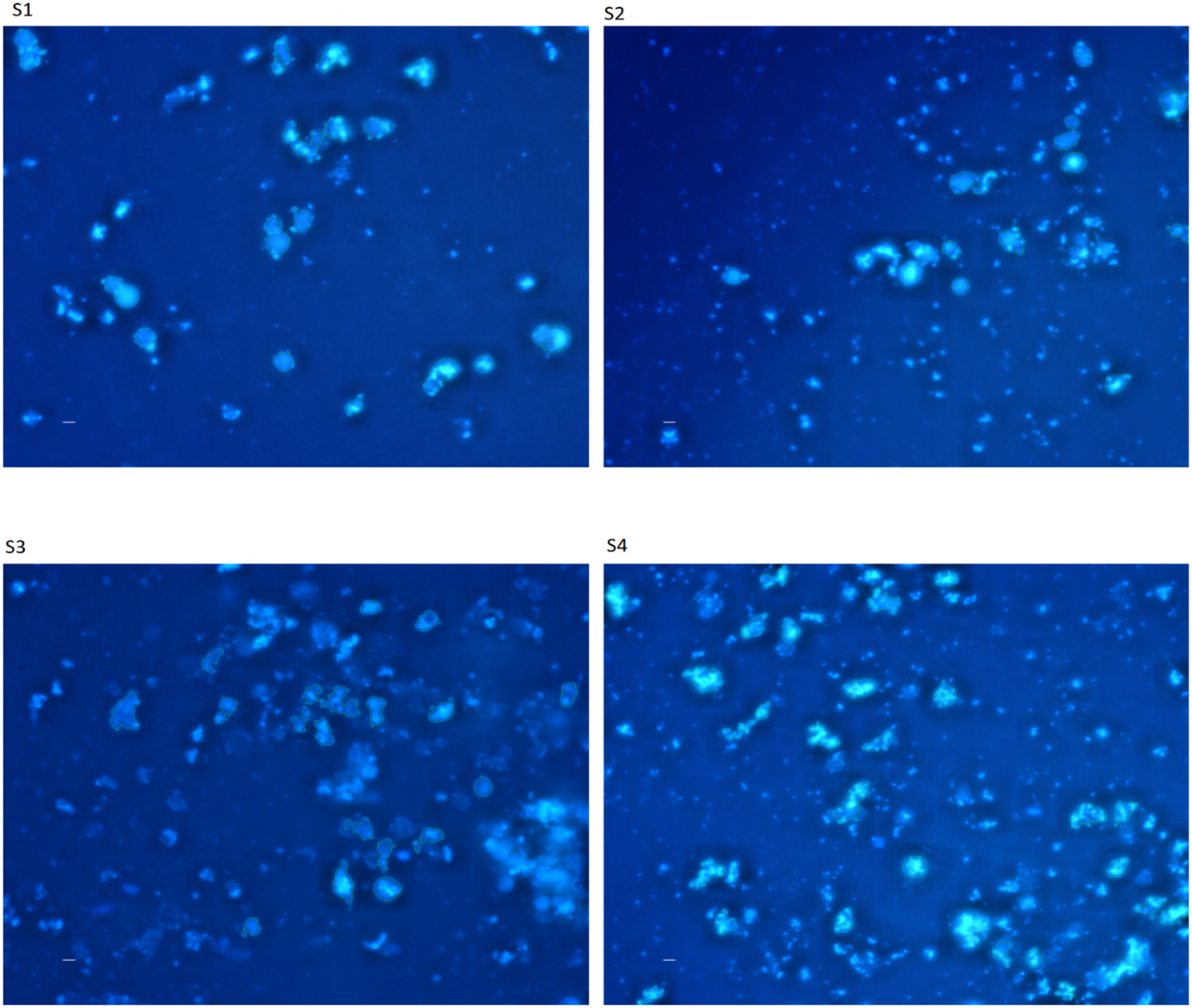
S3 displayed higher cell density and more irregular nuclear shapes, whereas S1 and S2 exhibited fewer, rounder nuclei. The outlined nuclei indicate regions included in quantitative analysis (area 30–130 µm2). Based on the Kruskal-Wallis test, nuclear area showed no significant difference (p = 0.098), but nuclear roundness differed significantly among groups (p = 0.0019), with S1 and S4 having higher roundness.
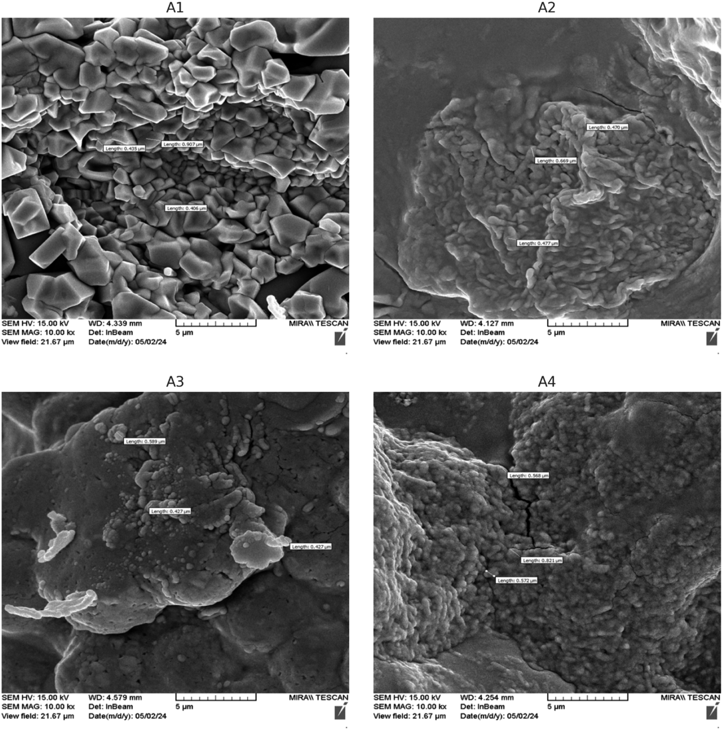
Fibroblast morphology on scaffolds (FESEM)
FESEM images were used to evaluate fibroblast interactions with scaffold surfaces, including cell shape, size, filopodia length/number, cytoplasmic spreading, and adhesion.(Figure 5). FESEM images of fibroblasts on hydrogel scaffolds S1–S4 showing distinct cell morphology, cytoplasmic spreading, and interactions with the scaffold surface. Filopodia and microvilli are visible.
Wound healing efficacy
Comparison of wound healing differences between scaffolds and control.
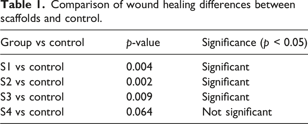
Day 14: Wound closure rates were S2: 77.4 ± 20.2%, S3: 67.5 ± 16.3%, significantly higher than the control (15.3%).
Day 21: S3: 91.7 ± 14.4%, S2: 88.5 ± 9.6%, while the control was only 31.5 ± 10.4% (Figure 6). Wound closure percentages on days 14 and 21. S2 and S3 showed significantly enhanced wound healing, while S4 showed no significant difference compared with the control. (p < 0.05).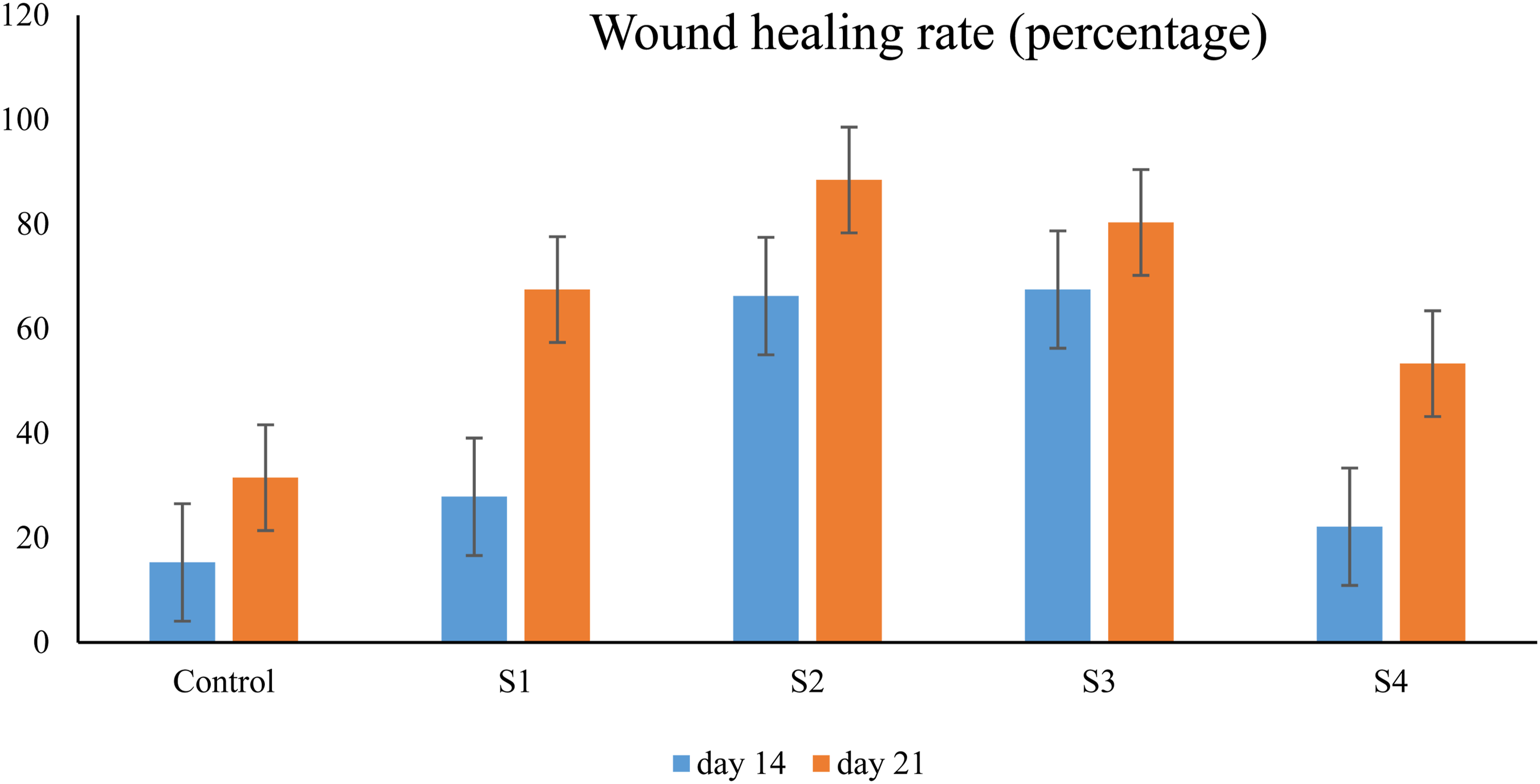
Histological analysis
Epidermal thickness (Day 21)
S2-treated wounds exhibited the greatest epidermal thickness (∼168 µm), significantly higher than other groups (p < 0.05), followed by S3, S1, and control. The control group showed the lowest thickness, indicating poor skin regeneration (Figure 7). Epidermal thickness in hydrogel-treated and control wounds. S2 shows the highest thickness and is significantly higher than the other groups (p < 0.05), while the control shows the lowest value.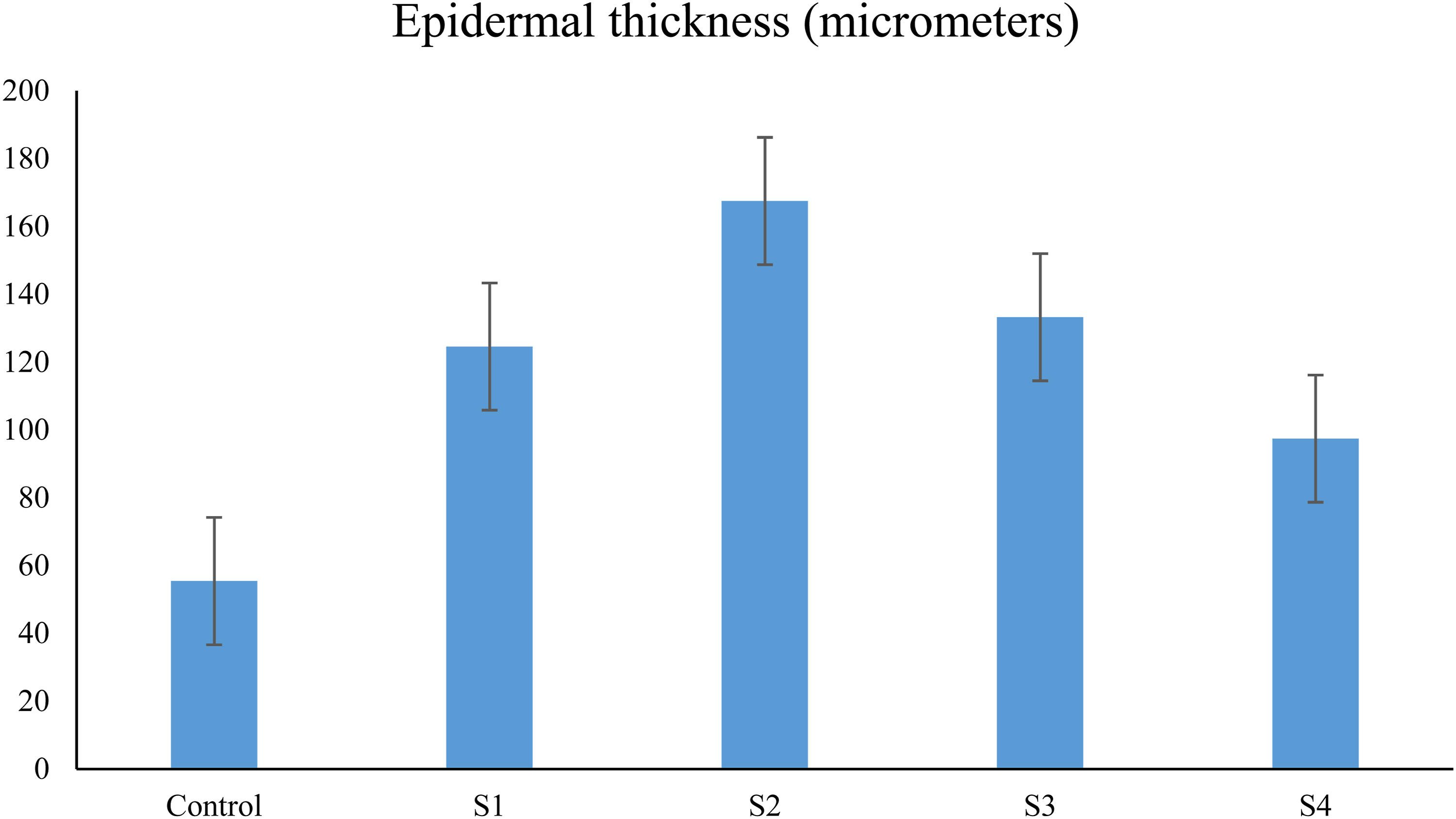
Inflammatory cell counts (Day 21)
The control group had the highest number of inflammatory cells (∼115 cells/field), indicating persistent inflammation. In contrast, S2 and S3 showed the lowest counts (Figure 8), reflecting better inflammation control. Number of inflammatory cells in hydrogel-treated and control wounds. S2 shows the fewest inflammatory cells and the control group the highest, with a significant difference between groups (p < 0.05).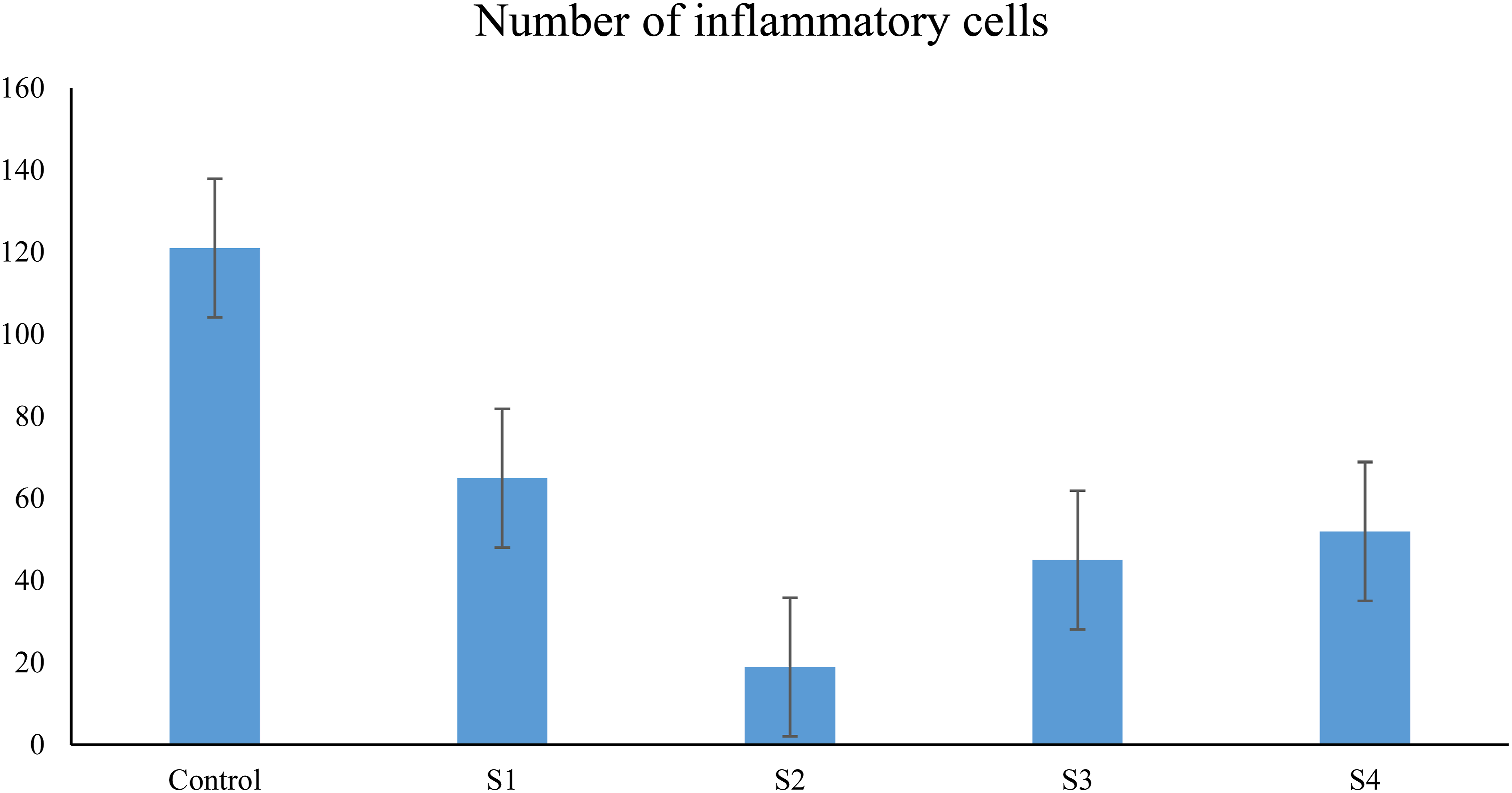
Immunofluorescence marker expression
To evaluate scaffold performance in wound healing, the expression of three key markers—Collagen type I (Col-1), VEGF, and TGF-β—was assessed using immunofluorescence on day 21.
Collagen type I (Col-1)
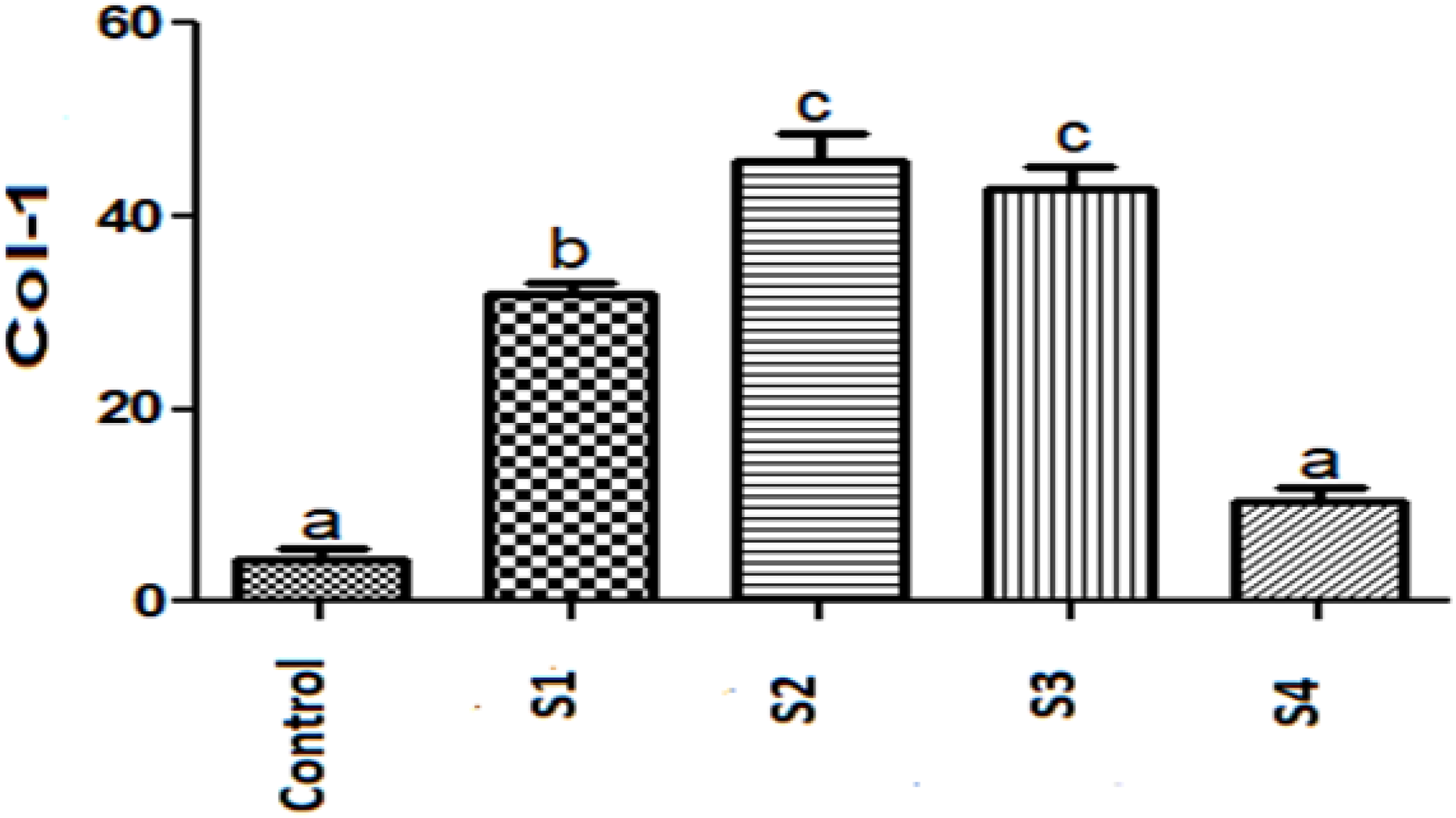
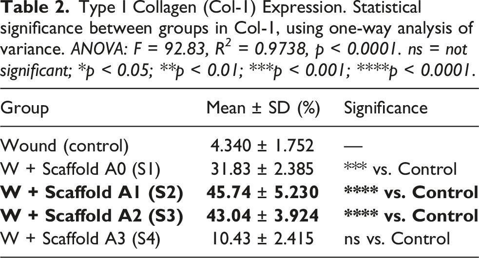
Col-1 is a major ECM component essential for tissue structure restoration. The untreated group showed the lowest expression (∼5%), reflecting poor ECM formation. S2 and S3 showed the highest expression (∼50%), significantly higher than other groups (p < 0.05). S1 had moderate expression (∼35%), while S4 was similar to untreated wounds.(Figures 9 and 10). Immunofluorescence staining of wound tissue sections on day 21 after treatment. Microscopic display of the expression of wound repair markers including type I collagen (green), cell nuclei (DAPI, blue) and composite images of channels in different groups including untreated wounds, and wounds treated with scaffolds S1 to S4. The intensity of marker expression is observed more in groups S2 and S3. Comparison chart of type I collagen expression levels between samples.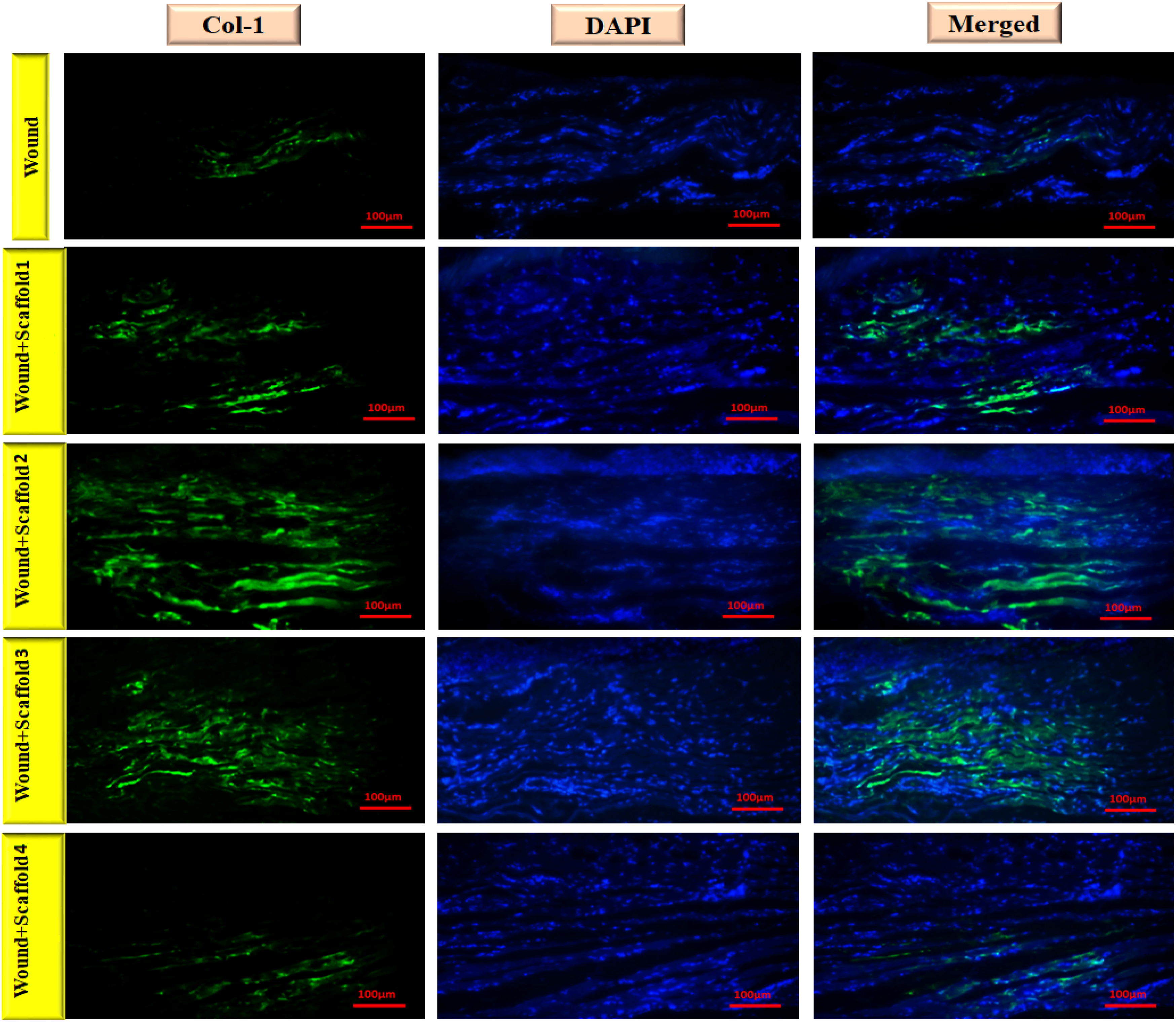
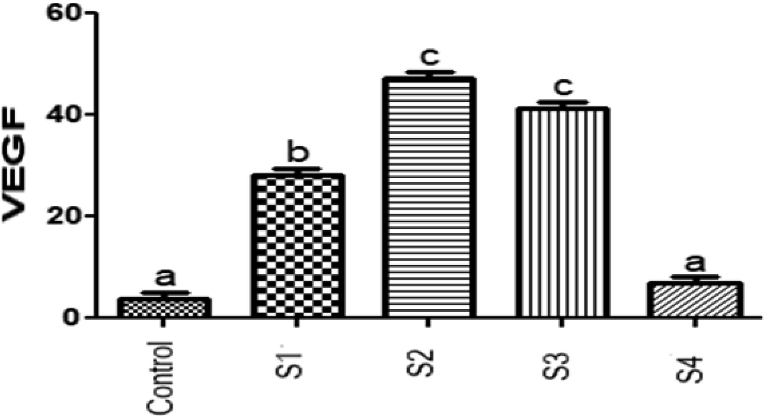
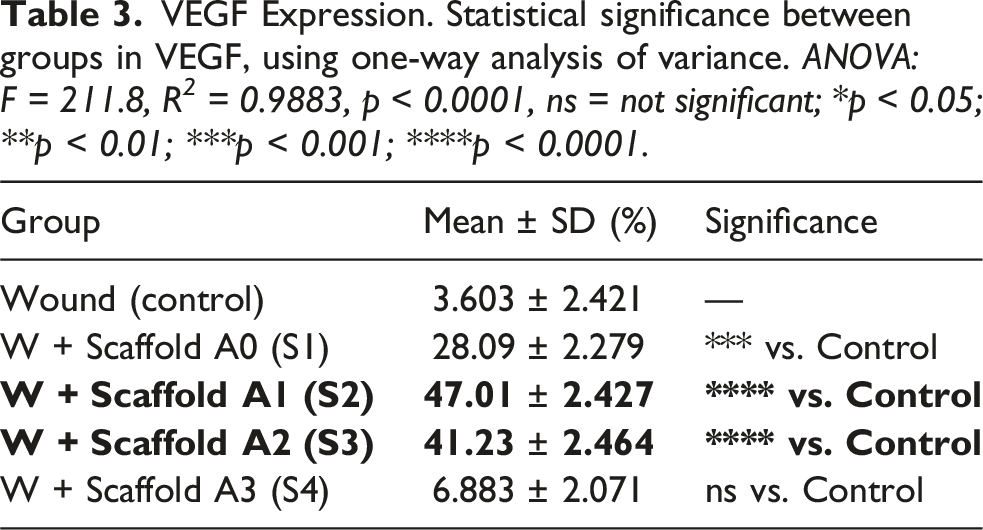
VEGF is critical for angiogenesis. S2 and S3 showed the highest expression (∼45–50%), indicating enhanced blood vessel formation. S1 showed moderate levels (∼30%), while S4 and untreated wounds had very low expression (∼5–10%) (Figures 11 and 12). Immunofluorescence staining of wound tissue sections on day 21 after treatment. Microscopic display of the expression of wound repair markers including VEGF (green), cell nuclei (DAPI, blue) and composite images of channels in different groups including untreated wound, and wound treated with scaffolds S1 to S4. The intensity of marker expression is observed more in groups S2 and S3. Comparison chart of VEGF expression levels between samples.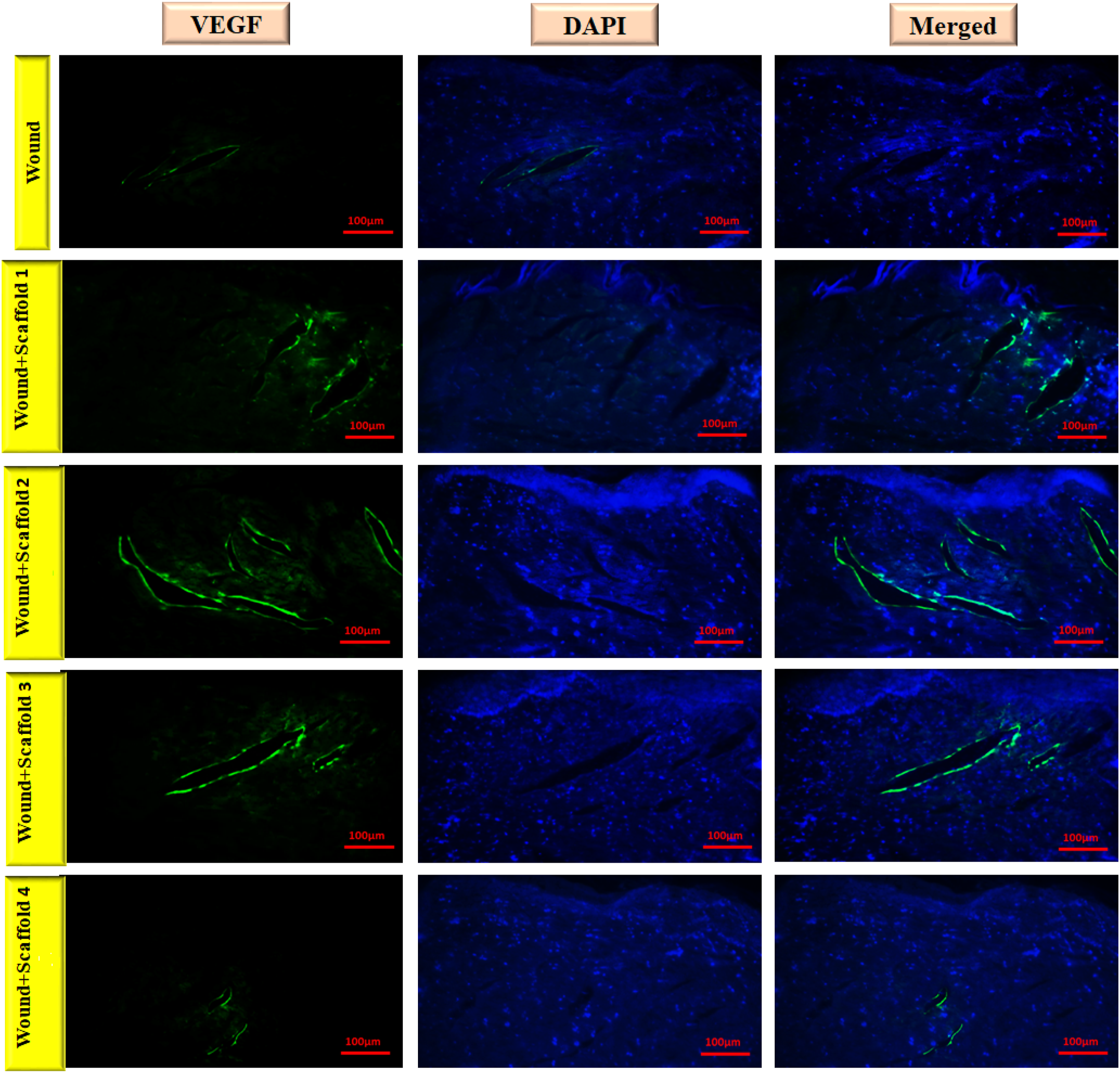
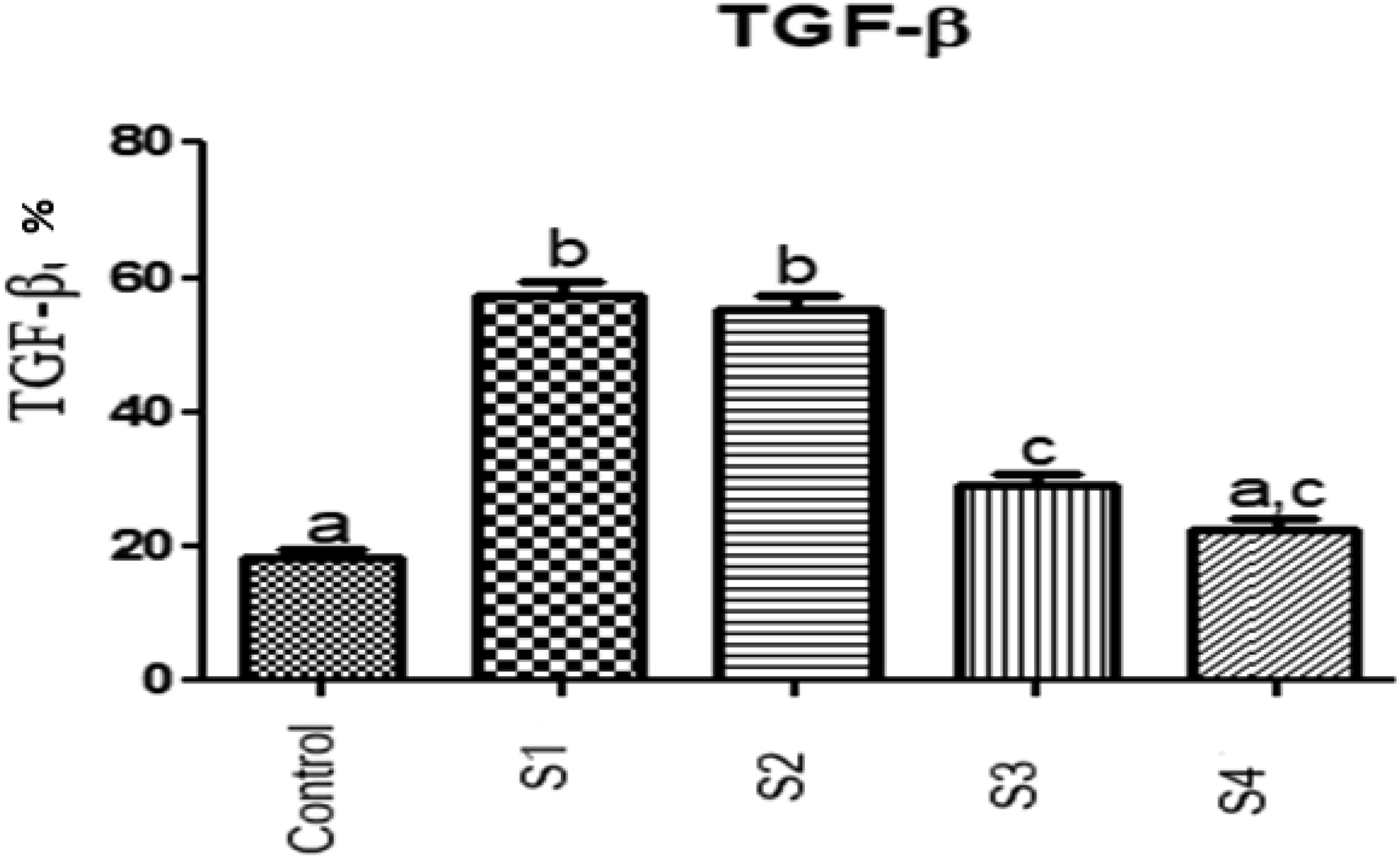
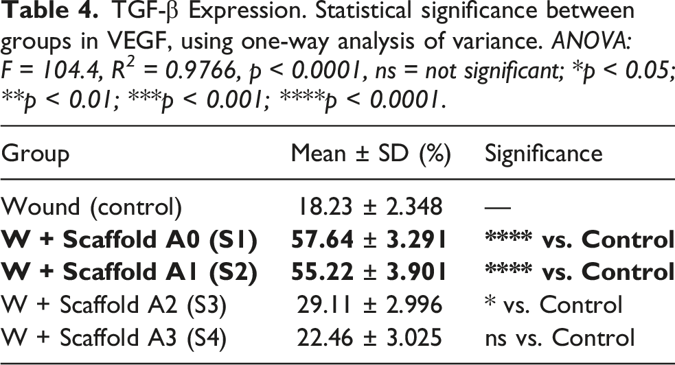
TGF-β regulates inflammation, fibroblast recruitment, and wound closure. S1 and S2 had the highest expression (∼60%), S3 moderate (∼30%), and S4 the lowest (∼20%). S4 expression was not significantly different from control. S1 and S2 effectively promoted fibroblast migration and wound closure, whereas S4 was ineffective (Figures 13 and 14). Immunofluorescence staining of wound tissue sections on day 21 after treatment. Microscopic display of the expression of wound repair markers including TGF-β (green), cell nuclei (DAPI, blue) and composite images of channels in different groups including untreated wounds, and wounds treated with scaffolds S1 to S4. The intensity of marker expression is observed more in groups S2 and S3. Comparison of TGF-β expression levels between samples.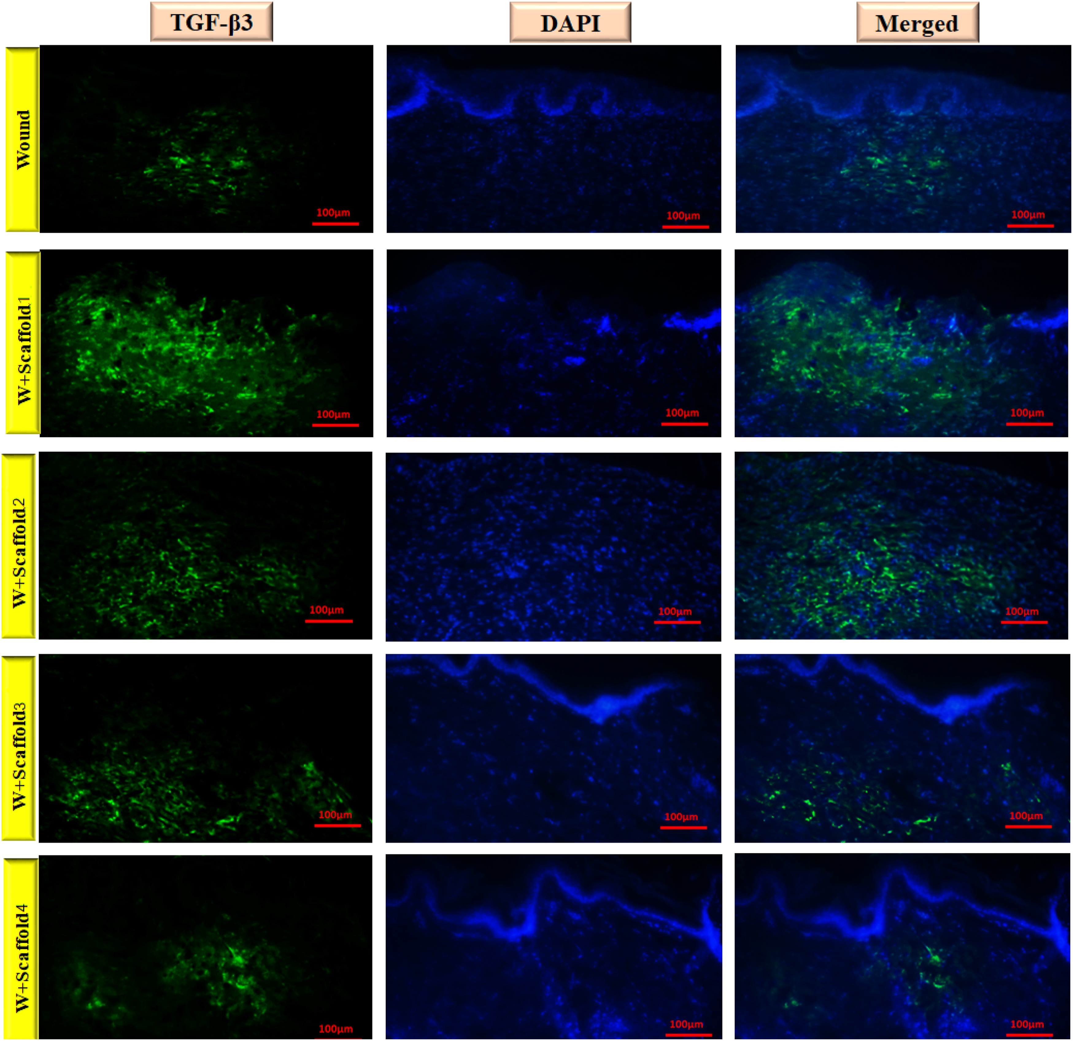
Overall, scaffold S2 had the highest expression levels in all three markers Col-1, VEGF, and TGF-β. Scaffold S3 also performed well, especially in collagen and VEGF expression, although its TGF-β levels were lower, which may indicate better control of inflammation. Scaffold S1 performed worse than S2 despite adequate expression of factors. Finally, scaffold S4 had low expression in all markers and was close to untreated wound in terms of performance, indicating its low efficiency in wound healing. − S2: Highest expression of Col-1, VEGF, and TGF-β, supporting rapid wound healing and ECM formation. − S3: Excellent Col-1 and VEGF, moderate TGF-β, suggesting controlled inflammation and high-quality tissue regeneration. − S1: Moderate performance. − S4: Low expression, similar to untreated wounds, indicating poor wound healing efficiency. − Overall, S2 and S3 demonstrated superior biocompatibility, cell interaction, angiogenesis, ECM formation, and wound healing efficacy, making them promising candidates for skin tissue engineering applications.
Discussion
To contextualize the novelty of the present work, it is important to compare our CNC-g-β-glucan/PAA scaffolds with existing wound dressing materials and previously reported cellulose nanocrystal (CNC)-based hydrogels. CNCs have been extensively investigated as reinforcing nanomaterials in wound dressings due to their biocompatibility, high aspect ratio, and potential to improve mechanical stability and biological performance compared to traditional biopolymer matrices. Several studies have reported CNC-reinforced hydrogels where CNC incorporation enhanced structural stability and supported cell proliferation and attachment, indicating suitability for wound healing applications. 15
For example, cellulose nanocrystal-reinforced alginate and aloe vera hydrogels demonstrated improved mechanical properties and moisture retention, along with enhanced keratinocyte proliferation relative to conventional gels, highlighting the multifunctional benefits of CNC incorporation. Similarly, gelatin–hyaluronic acid–CNC composite hydrogels exhibited good cytocompatibility and fibroblast growth, underscoring the positive influence of CNC on biological outcomes. These findings collectively suggest that CNC-based hydrogels can outperform some conventional wound dressings by providing improved mechanical integrity and a favorable environment for cell growth.16,17
Type I Collagen (Col-1) Expression. Statistical significance between groups in Col-1, using one-way analysis of variance. ANOVA: F = 92.83, R 2 = 0.9738, p < 0.0001. ns = not significant; *p < 0.05; **p < 0.01; ***p < 0.001; ****p < 0.0001.
VEGF Expression. Statistical significance between groups in VEGF, using one-way analysis of variance. ANOVA: F = 211.8, R 2 = 0.9883, p < 0.0001, ns = not significant; *p < 0.05; **p < 0.01; ***p < 0.001; ****p < 0.0001.
TGF-β Expression. Statistical significance between groups in VEGF, using one-way analysis of variance. ANOVA: F = 104.4, R 2 = 0.9766, p < 0.0001, ns = not significant; *p < 0.05; **p < 0.01; ***p < 0.001; ****p < 0.0001.
Degradation kinetics and clinical translational potential
A critical parameter for evaluating the clinical translational potential of wound healing scaffolds is their degradation behavior under physiologically relevant conditions. Our comparative degradation studies in distilled water and simulated wound fluid (SWF) revealed distinct degradation kinetics that have important implications for clinical application. In distilled water, the hydrogels showed controlled, gradual degradation over 14 days, primarily driven by hydrolytic cleavage of ester bonds in the PAA network and glycosidic bonds in the β-glucan component. 18 The significant influence of β-glucan content on degradation rate (p = 0.0021) suggests that the β-glucan acts not only as a bioactive component but also as a structural modifier that influences network density and water uptake capacity.
In contrast, degradation in SWF was markedly accelerated, with complete dissolution achieved by day 7 for all formulations. This accelerated degradation can be attributed to several factors inherent to the wound environment. First, the ionic composition of SWF (Na+, K+, Ca2+, Cl-) may increase the osmotic pressure and facilitate polymer chain swelling and subsequent erosion. Second, the physiological pH (7.4) of SWF may enhance the hydrolysis rate of acrylic acid ester linkages compared to pure water. Third, although our SWF did not contain enzymes, the ionic environment can catalyze certain degradation pathways that are not active in distilled water. 19
The rapid in vitro degradation observed in SWF (complete by day 7) does not necessarily limit in vivo therapeutic efficacy, as scaffold function extends beyond simple mechanical persistence. The scaffold’s primary role during the early inflammatory and proliferative phases (days 0–14) is to deliver bioactive β-glucan, modulate immune responses, and provide transient structural support for initial cellular infiltration. 20 Once these biological cascades are initiated, healing can proceed autonomously even after scaffold degradation. Furthermore, in vivo degradation kinetics often differ substantially from in vitro predictions due to cellular enzymatic activity, immune cell recruitment, and mechanical loading patterns in the wound bed. 21 This phenomenon—where early bioactive signaling enables prolonged regenerative responses despite rapid scaffold degradation—has been well-documented in advanced biomaterial systems. 22
Notably, this in vitro degradation does not preclude effective in vivo wound healing performance, as the complex wound microenvironment—including cellular infiltration, ECM deposition, and immunomodulatory responses—can substantially alter scaffold residence time and therapeutic efficacy. 21 Indeed, recent studies have demonstrated that hydrogel scaffolds can achieve excellent regenerative outcomes through early bioactive signaling and immune activation, even when the bulk material degrades rapidly 22 within the typical wound healing timeframe.
The complete degradation by day 7 in SWF may be advantageous for wound healing applications. Wound healing typically progresses through overlapping phases: hemostasis (immediate), inflammation (1-4 days), proliferation (4-14 days), and remodeling (weeks to months). Our scaffolds provide mechanical support and bioactive cues during the critical inflammatory and early proliferative phases, then undergo complete resorption as the newly formed tissue begins to mature and remodel. This degradation timeline prevents potential complications associated with long-term scaffold persistence, such as chronic inflammation, mechanical mismatch, or interference with tissue remodeling. 23
The in vivo persistence data further validated our in vitro findings, demonstrating that the scaffolds integrate with regenerating tissue and undergo gradual, cell-mediated resorption without eliciting chronic inflammatory responses. The presence of macrophages and multinucleated giant cells at the scaffold interface by day 14, without signs of fibrous encapsulation or extensive lymphocytic infiltration, indicates a normal foreign body response and controlled biodegradation process. 24 This is crucial for clinical translation, as excessive or prolonged inflammatory responses can lead to scar formation and impaired functional recovery.
An important consideration for clinical application is the correlation between scaffold degradation and mechanical support. While our study focused on weight loss as a degradation metric, future studies should investigate the evolution of mechanical properties (tensile strength, elastic modulus) during degradation to ensure that the scaffolds maintain adequate structural integrity during the critical early healing phase. Additionally, the degradation products of PAA, CNC, and β-glucan should be evaluated for potential cytotoxicity, although all three components are generally recognized as biocompatible and their degradation products (acrylic acid oligomers, glucose, and glucan fragments) are expected to be non-toxic at physiological concentrations. 25
Interestingly, the different degradation rates observed among the four formulations in both media suggest that the β-glucan content can be tuned to modulate scaffold persistence for specific wound types. For acute wounds requiring short-term support, formulations with lower β-glucan content (S1, S2) that degrade more rapidly might be preferred. Conversely, for chronic or diabetic wounds that often have prolonged inflammatory phases, formulations with higher β-glucan content (S3, S4) that persist slightly longer while providing sustained immunomodulatory effects might be more beneficial. This tunability represents a significant advantage for personalized wound care strategies.
In this study, the MTT assay demonstrated significant differences in cell viability among hydrogel scaffolds S1–S4 and the positive control (TCP) at days 1, 3, and 7 (p < 0.01). Post-hoc Tukey analysis revealed that S2 and S3 consistently exhibited higher viability across all time points, particularly on days 3 and 7, whereas S4 showed the lowest viability.
Key interpretations
1. High biocompatibility of S2 and S3
The high optical density (OD) values in MTT for S2 and S3 indicate that these scaffolds effectively support fibroblast survival, cell proliferation, and do not induce significant cytotoxicity.
This is crucial for wound healing scaffolds, which must provide an environment that allows cells to survive, proliferate, and interact with the scaffold.
Reviews on hydrogel scaffolds emphasize that high biocompatibility, suitable 3D structure for cell infiltration, and appropriate mechanical/biochemical cues are essential. 26
Recent advances in cellulose nanocrystal-based hydrogels highlight the importance of biocompatibility and mechanical properties for tissue engineering applications. 27 The incorporation of bioactive components such as β-glucan enhances cell-scaffold interactions through multiple mechanisms, including Dectin-1 receptor activation and downstream signaling pathways that promote fibroblast proliferation and migration.28,29
Therefore, the superior performance of S2 and S3 may result from optimized scaffold design, such as appropriate porosity, chemical composition, and cell-adhesive surface properties. 2. Poorer performance of S4
S4’s low cell viability could stem from multiple factors: an unsuitable chemical composition for cells, inadequate surface structure for adhesion and spreading, or even minor cytotoxic effects.
This highlights that 3D structure alone is not sufficient; cellular compatibility, surface biochemistry, and absence of toxicity are also critical.
Studies have shown that even inherently non-toxic hydrogels can limit cell growth if the surface is not cell-friendly.
30
The excessive β-glucan content in S4 may have led to altered mechanical properties or increased scaffold stiffness, which can negatively impact cell adhesion and proliferation.
31
3. Importance of timing and dynamic cell growth
The larger differences observed on days 3 and 7 indicate that S2 and S3 not only maintain cell viability but also support sustained proliferation over several days.
This is particularly important for wound healing applications, where the repair process occurs over multiple days, and scaffolds must remain effective throughout this period.
Scaffolds S2 and S3 demonstrate excellent cytocompatibility, providing a supportive microenvironment for fibroblast survival and proliferation, which is a fundamental prerequisite for effective wound healing. Conversely, S4’s poor performance underscores the necessity of careful scaffold design beyond mere 3D architecture.
DAPI staining and nuclear morphology
DAPI staining and nuclear morphology analysis revealed the following: Cell density: Highest in S3, lowest in S4.
Nuclear area: No significant difference (p = 0.098).
Nuclear roundness: Significant difference (p = 0.0019). (1) Cell density: The high cell density in S3 is consistent with MTT results, indicating not only cell survival but also good cell spreading and aggregation on the scaffold surface. (2) Nuclear morphology: Changes in nuclear roundness can reflect cell status, including stress, activity, or stability. Groups with less round nuclei may indicate more active or functionally favorable cells. Similar trends have been observed in hydrogel studies, where well-adhered, spread-out cells often show better performance. (3) FESEM analysis: Filopodia and cytoplasmic spreading showed that S3 cells had ∼8 µm filopodia and excellent spreading, indicating strong cell–scaffold interactions. This aligns with MTT and DAPI results, suggesting that S3 provides a highly supportive surface and architecture. (4) Other scaffolds: Cells on S1 and S2 also spread well but not as extensively as S3; S4 performed poorly. These morphological insights corroborate quantitative data.
Wound healing and histology
In vivo wound healing results showed: − Wound closure: By day 14, S2 ∼77.4%, S3 ∼67.5%; by day 21, S2 ∼88.5%, S3 ∼91.7%, whereas control was ∼31.5%. − Epidermal thickness: Greater in S2 and S3. − Inflammatory cell count: Lower in S2 and S3.
Significant wound closure at days 14 and 21 correlates with literature reports that properly designed hydrogels (with suitable polymer composition and/or bioactive agents) accelerate epithelialization and wound closure. Hydrogels provide a moist environment, 3D structural support, and the capacity to deliver growth factors or cells, which enhance healing. 32
Epidermal thickness (∼168 µm on day 21 for S2) reflects improved epithelial regeneration. Studies show scaffolds providing adequate structural and biochemical support promote faster and thicker epithelialization—a common indicator of repair quality. Hydrogels that stimulate angiogenesis and ECM formation further enhance blood supply for effective epithelialization. 33
Lower inflammatory cell counts are critical because prolonged or chronic inflammation delays repair and can lead to scarring or non-healing wounds. Several studies indicate that suitable hydrogels can control inflammation either by modulating anti-inflammatory factor release or by providing a scaffold that limits excessive neutrophil/macrophage accumulation, thereby facilitating transition to the proliferation/regeneration phase. 34
Overall, S3 excels in cell density and morphology, indicating optimal cell–scaffold interactions, while S2 and S3 promote rapid and high-quality wound repair, with enhanced epidermal regeneration and controlled inflammation, supporting both quantitative and histological findings.
Analysis of immunofluorescence marker expression (Col-1, VEGF, TGF-β)
Another key aspect of results is the analysis of three vital wound healing markers: Collagen type I (Col-1), Vascular Endothelial Growth Factor (VEGF), and Transforming Growth Factor-β (TGF-β). The results are summarized as follows: Col-1 expression: Untreated group ∼5%, S1 ∼35%, S2 and S3 ∼50%, S4 similar to control.
VEGF expression: S2 and S3 ∼45–50%, S1 ∼30%, S4 and untreated ∼5–10%.
TGF-β expression: Highest in S1 and S2 (∼60%), S3 ∼30%, S4 ∼20% (not significantly different from control).
Col-1 and ECM regeneration: The significant increase in Col-1 expression in S2 and S3 indicates effective formation of the extracellular matrix (ECM), which is critical for skin structural repair. Previous studies also show that enhanced Collagen I expression is observed in wound-healing scaffolds. For example, an alginate/chitosan hydrogel study reported increased Col-1 expression. 35 This aligns with the findings, suggesting that S2 and S3 formulations have strong capacity to stimulate ECM formation.
VEGF and angiogenesis: The notable increase in VEGF in S2 and S3 demonstrates that these scaffolds effectively promote angiogenesis, facilitating the formation of new blood vessels and improved blood supply to the wound site. This ensures better delivery of nutrients and oxygen to regenerating tissue, accelerating the repair process. Other studies have similarly reported improved wound healing with hydrogels that enhance VEGF expression.36,37
TGF-β and regulation of wound healing: An interesting observation is the higher TGF-β expression in S1 and S2 (∼60%) versus S3 (∼30%). TGF-β plays a central role in initiating wound repair (fibroblast recruitment, ECM production, inflammation control). However, excessive or prolonged expression can lead to fibrosis or scar formation. Recent studies suggest that reducing TGF-β1 in collagen scaffolds can decrease fibrotic responses. 38
In S2, the high Col-1, VEGF, and TGF-β levels suggest very favorable conditions for rapid wound closure. In S3, the lower TGF-β may suggest a reduced risk of inflammation and fibrosis, potentially improving the quality of long-term tissue regeneration, not just the speed, which could be an advantage in reducing scar formation and promoting more functional repair. Overall, these marker profiles suggest a complementary advantage: S2 for speed of healing and S3 for quality and reduced fibrosis, reflecting different but valuable aspects of wound repair.
Overall comparison of scaffolds and optimal selection
Considering all the results, scaffold S2 demonstrated excellent performance in terms of wound healing speed, epidermal thickness, cell activity in MTT assay, and appropriate marker expression. Scaffold S3 also showed outstanding performance in cell proliferation, angiogenesis, and ECM formation, and possibly better control of TGF-β, suggesting a lower risk of fibrosis.
Scaffold S1, although showing some improvement over the control, did not perform as well as S2 and S3. Scaffold S4 performed close to the control group and was considerably weaker in terms of wound healing, cell viability, cellular structure, and marker expression. Therefore, the overall conclusion is that S2 and S3 are the most promising candidates for wound healing applications, which aligns well with the results.
Multiple studies have shown that designing scaffold hydrogels with appropriate porosity, biopolymer combinations, cell-friendly surfaces, and the ability to stimulate angiogenesis and ECM production leads to enhanced wound healing. For example, reviews on hydrogels in tissue engineering highlight several key features for successful hydrogels: biocompatibility, porosity for cell infiltration, nutrient transport, incorporation of growth factors, and stimulation of angiogenesis. 39
Additionally, the study “Evaluation of collagen type I and III, TGF-β1, and VEGF gene expression in rat skin wound healing treated by alginate/chitosan hydrogel containing crocetin” reported that alginate/chitosan hydrogels significantly increased Col-1 and VEGF expression. 35
Comparison with our results shows a similar trend: increased Col-1 and VEGF along with improved wound healing in the selected groups (S2 and S3). Thus, the study aligns with existing literature, and its novelty lies in the design of S2 and S3 scaffolds, particularly regarding polymer composition and surface properties.
Conclusion
In addition to the observed biological efficacy, the structural characteristics of the CNC-g-β-glucan/PAA scaffolds played a decisive role in their wound healing performance. The presence of cellulose nanocrystals provided structural reinforcement to the hydrogel network, while variations in β-glucan content influenced network organization and flexibility, which in turn affected cell–scaffold interactions. Scaffolds S2 and S3 demonstrated an optimal structural environment that supported extensive fibroblast adhesion, cytoplasmic spreading, and filopodia formation, as confirmed by FESEM and DAPI analyses. This favorable structural environment was strongly associated with enhanced cell viability, increased epidermal thickness, reduced inflammatory cell infiltration, and significantly elevated expression of key regenerative markers, including collagen type I, VEGF, and TGF-β. In contrast, scaffold S4, despite containing a higher β-glucan content, showed reduced cellular activity, limited fibroblast spreading, lower marker expression, and inferior wound healing outcomes, indicating that deviations from this balanced scaffold structure negatively impact biological performance. Collectively, these findings demonstrate that the superior regenerative efficacy of S2 and S3 arises from the close interplay between scaffold structural integrity and biological response, underscoring the importance of optimized structure–function balance in hydrogel-based wound dressings.
Footnotes
Funding
The authors received no financial support for the research, authorship, and/or publication of this article.
Declaration of conflicting interests
The authors declared no potential conflicts of interest with respect to the research, authorship, and/or publication of this article.
Data Availability Statement
The data supporting the findings of this study are available from the corresponding author upon reasonable request. All relevant datasets generated and analyzed during the current study are included in the article or supplementary materials.
Declaration of generative AI and AI-assisted technologies in the writing process
During the preparation of this work, the authors used ChatGPT to improve readability and language. After using this tool, the authors reviewed and edited the content as needed and take full responsibility for the content of the publication.