
Abstract
The development of biomaterial scaffolds that combine structural support with biological activation remains critical for bone defect repair. In this study, gelatin-based cryogels (GBCs) that incorporate β-tricalcium phosphate (β-TCP) and hyaluronic acid (HA) were fabricated and optimized to balance porosity and mechanical stability. Increasing the HA concentration increased the water content, swelling ratio, and porosity while reducing the compressive stiffness. The cryogels that contained 0.3% HA exhibited interconnected macropores (200–300 μm after swelling), ∼30% porosity, and a compressive modulus of 3.67 MPa and were selected for biological evaluation. Human umbilical cord-derived mesenchymal stem cell (hUC-MSC)-derived exosomes were incorporated to increase osteogenic bioactivity. Compared with the control treatment, exosome treatment significantly increased MG-63 proliferation, alkaline phosphatase activity, and mineral deposition (p < 0.05). Notably, compared with exosomes alone, exosome-seeded cryogels synergistically increased osteogenic differentiation (p < 0.001). In a rabbit femoral defect model, compared with cryogels without exosomes, exosome-functionalized cryogels promoted denser bone matrix formation and neovascularization. These findings demonstrate that GBCs seeded with exosomes provide a structurally permissive and biologically active microenvironment that improves bone regeneration. This cell-free strategy represents a promising platform for translational bone repair applications.
Introduction
Bone defects commonly occur following fractures, trauma, skeletal tumor resection, and degenerative bone diseases. With the increasing prevalence of osteoporosis and age-related musculoskeletal disorders in the aging population, the clinical demand for effective bone regeneration strategies continues to increase.1,2 Current treatment options for critical-sized bone defects include autologous bone grafts, allogenic bone grafts, and artificial bone substitutes. Autologous bone grafts remain the gold standard because of their osteogenic, osteoinductive, and osteoconductive properties and are commonly harvested from the iliac crest or tibial plateau. 3 However, autologous grafting is limited by donor site morbidity, postoperative pain, and restricted graft availability. 4 Although allogenic grafts provide an alternative treatment option, concerns regarding immune reactions, disease transmission, and infection remain unresolved. Consequently, biomaterial-based scaffolds have attracted increasing attention as alternatives for bone defect repair. An ideal scaffold should provide not only mechanical support but also a biologically favorable microenvironment that promotes cellular infiltration, angiogenesis, osteogenesis, and tissue remodeling.
Cryogels are a particular type of hydrogels fabricated under subzero conditions through cryopolymerization, during which ice crystals act as porogens to generate highly interconnected macroporous structures. 5 These scaffolds possess several advantageous characteristics, including high porosity, rapid swelling ability, osmotic stability, and mechanical resilience, which make them attractive for tissue engineering applications. The interconnected porous architecture of cryogels facilitates nutrient diffusion, cell infiltration, and extracellular matrix (ECM) deposition, thereby providing a favorable microenvironment for tissue regeneration. Accordingly, cryogel-based scaffolds have emerged as promising candidates for bone tissue engineering applications.
Among biomaterials used for regenerative scaffolds, gelatin has been extensively investigated because of its excellent biocompatibility, biodegradability, and cell adhesive properties. Gelatin, which is a denatured protein devoid of lipids and cholesterol, contains a diverse array of amino acids is derived from collagen hydrolysis and contains abundant Arg–Gly–Asp (RGD) sequences that promote cellular adhesion and proliferation. 6 Furthermore, gelatin is a Food and Drug Administration (FDA)-approved biomaterial with favorable hydrophilic and thermoresponsive properties; gelatin can form a thermally reversible gel with water, with a gel state at low temperature (<25°C) and a solution state at high temperature (>35°C). 7 Previous studies have demonstrated that gelatin-based biomaterials can enhance osteoblast adhesion, proliferation, and alkaline phosphatase activity, thereby supporting osteogenic differentiation and bone formation.8–11 In addition to gelatin, hyaluronic acid (HA), a naturally occurring polysaccharide component of the ECM, has gained increasing attention in tissue engineering because of its remarkable water-retention capability and biological regulatory functions. 12 HA contributes to hydration maintenance, cell infiltration, extracellular matrix remodeling, and tissue repair.12–14 Moreover, HA-containing biomaterials can improve scaffold swelling behavior and create a hydrated microenvironment favorable for cellular infiltration and proliferation.
To further improve osteoconductive properties, calcium phosphate-based ceramics are frequently incorporated into scaffolds for bone regeneration. Tricalcium phosphate (TCP), particularly β-tricalcium phosphate (β-TCP), closely resembles the inorganic mineral composition of native bone and exhibits excellent biocompatibility and biodegradability.15,16 β-TCP can release calcium and phosphate ions during degradation, thereby supporting osteoblast activity and matrix mineralization. Furthermore, β-TCP-containing biomaterials have been shown to enhance osteogenic differentiation and bone tissue formation. 16 Although gelatin, HA, and β-TCP each possess favorable biological characteristics, balancing hydration capacity, porosity, and mechanical stability within a single scaffold system remains challenging. Excessive swelling and high porosity may compromise scaffold stiffness, whereas overly rigid scaffolds may impair cellular infiltration and tissue remodeling. Therefore, optimizing the structural and biological interactions among these components is critical for successful scaffold design in bone regeneration.
In addition to scaffold architecture, recent advances in regenerative medicine have highlighted the therapeutic potential of mesenchymal stem cell-derived exosomes as cell-free biological mediators. Exosomes are extracellular vesicles secreted by various cell types and contain bioactive proteins, lipids, and regulatory nucleic acids that can modulate intercellular signaling and tissue repair. 17 Recent studies have demonstrated that stem cell-derived exosomes can promote osteogenesis and angiogenesis, both of which are essential for effective bone regeneration and fracture healing.17,18 Compared with stem cell transplantation, exosome-based therapies may reduce immunogenicity, tumorigenic risk, and regulatory concerns while preserving regenerative bioactivity. Incorporating exosomes into porous biomaterial scaffolds may therefore provide sustained local bioactivity while simultaneously supporting structural regeneration.
Bone regeneration requires a coordinated combination of osteoconductive architecture, mechanical support, and biological activation. In this study, we developed gelatin-based cryogels (GBCs) incorporating HA and β-TCP to establish a structurally permissive and ECM-mimetic scaffold for bone regeneration. Human umbilical cord-derived mesenchymal stem cell (hUC-MSC)-derived exosomes were subsequently incorporated to further enhance osteogenic bioactivity. The novelty of this study lies in the integration of optimized cryogel architecture with exosome-mediated biological stimulation to simultaneously improve porosity, hydration behavior, osteogenic differentiation, and early bone regeneration. The physicochemical properties, osteogenic activity, and regenerative potential of the exosome-seeded cryogels were evaluated through in vitro experiments and a rabbit femoral defect model.
Methods
Synthesis of gelatin-based cryogels (GBCs)
Cryogels were prepared with HA (MW: 1,000,000 Da; Kinkasei Chemilab, Taichung City, Taiwan) and gelatin from type B bovine skin (225 g Bloom, MW: 50,000 Da; Sigma-Aldrich, St Louis, MO, USA), β-tricalcium phosphate (β-TCP) (Sigma-Aldrich, St Louis, MO, USA), and the gelling agent 1-(3-dimethylaminopropyl)-3-ethylcarbodiimide hydrochloride (EDC) (98+%; Thermo Fisher Scientific, Waltham, MA, USA). HA, at varying concentrations (0 g (0% w/v), 0.03 g (0.3% w/v), 0.05 g (0.5% w/v), or 0.1 g (1% w/v)), was initially dissolved in 9 cm3 of deionized distilled water (ddH2O) and heated to 70°C. Subsequently, 4 g of gelatin (40% w/v) was incorporated into the HA solution, followed by the addition of 6 g of β-TCP (60% w/v). To facilitate cross-linking, 0.005 g of EDC (0.05% w/v) was dissolved in 1 cm3 ddH2O, and the obtained solution was added to the mixture while stirring, which resulted in a final volume of 10 cm3. Afterward, the solution was transferred into two 5 cm3 cylindrical molds with a diameter of 10 mm and sealed with Parafilm®. The molds were concealed in plastic wrap before being oscillated in an ultrasonic bath for 30 s. Afterward, the sealed molds were submerged in anhydrous ethanol inside a centrifuge tube and placed at a subzero temperature of −20°C for 16 h for the cryopolymerization process to occur. Cryogelation is performed under subzero conditions, where ice crystals serve as porogens to define the macroporous architecture of the scaffold. The use of an ethanol bath at −20°C provides a more homogeneous heat transfer environment compared with direct air cooling, which helps minimize temperature gradients and promotes more uniform ice crystal formation, thereby contributing to a consistent pore structure. Afterward, the prepared material was sectioned into cylindrical discs with a thickness of approximately 3 mm. A series of dehydration steps followed, which involved immersion in ethanol solutions of increasing concentration (25%, 50%, 75%, and 99.5%) for 3 min each. This graded ethanol dehydration was used to gradually replace water within the cryogel network. Direct freeze-drying of water-containing hydrogels may lead to structural collapse due to capillary forces generated during solvent removal. Gradual solvent exchange with ethanol reduces surface tension effects and helps preserve the integrity of the porous architecture. Next, the discs were fully saturated with anhydrous ethanol and frozen at −20°C for 12 h. The cryogels were then removed and placed on a Petri dish at room temperature for 2 h to allow the ethanol to evaporate, followed by another freezing cycle at −20°C for 12 h. Ethanol was removed prior to lyophilization because it is not suitable for direct freeze-drying. The additional refreezing step allows the remaining solvent to be uniformly frozen before sublimation, thereby facilitating effective lyophilization and helping maintain the pore structure formed during cryogelation. The final step in the processing involved lyophilization for 24 h, which resulted in the formation of gelatin-based cryogels.
Analysis of the characteristics of the GBCs
Water content
The water content quantifies the ability of a cryogel to retain water molecules within its structure, which determines the ability of the cryogel to swell, as cellular metabolism relies heavily on water molecules. The dry mass (Md) of a sample was first weighed. Afterward, the material was completely submerged in ddH2O for 1 h and then removed. Excess water was blotted away using Kimwipes®, and the wet mass (Mw) was measured. The water content was then calculated using the following formula:

Swelling ratio
The swelling ratio is a measure of the ability of a cryogel to absorb water and expand in volume, which indicates its efficacy in filling defects or voids. Similarly, a sample was weighed to determine its dry mass (Md). It was then fully immersed in ddH2O and removed at each predetermined time point, which included 5 min, 10 min, 15 min, 30 min, 1 h, 2 h, 3 h, 6 h, 12 h, 24 h, 36 h, 48 h, and 72 h. For each duration, excess water was once again absorbed with Kimwipes®, and the wet mass (Mw) of the material was recorded. The swelling ratio was calculated using the following formula:

Porosity measurement
The porous structure plays a crucial role in supporting cell survival, proliferation, and infiltration, thereby facilitating extracellular matrix (ECM) formation within the cryogel scaffold. The dry mass (Md) and volume (V) of a sample were first measured. The volume of each sample was determined by measuring its dimensions (length, width, and height) using a digital caliper and calculating the volume accordingly. The material was subsequently completely submerged in ethanol at a concentration of 99.5% for impregnation. The wet mass (Mw) was measured after the excess surface liquid was wicked away using Kimwipes®. The porosity was calculated with (0.7893) as the density of ethanol using the following formula:

The ethanol displacement method estimates the pore volume of the cryogels.
Scanning electron microscopy (SEM) and pore size quantification
After freeze-drying, the surfaces of the cryogels were coated with a layer of gold film by a vacuum sputter coater to increase the conductivity. The pore size, density, and surface cross-sectional structure of the cryogels were observed by SEM (Zeiss Auriga FIB-SEM) at 10 kV at various magnifications. For swollen samples, cryogels were first immersed in deionized water (ddH2O) for 24 h, followed by freeze-drying prior to SEM observation. Accordingly, the SEM images represent the pore morphology after swelling and freeze-drying. In addition, the crystalline characteristics of the cryogels were analyzed by X-ray diffraction (XRD), and the results are provided in the Supporting Information (Figure S1).
Mechanical testing
A universal testing machine (Tinius Olsen, Horsham, PA, USA) was used to perform a uniaxial compression test on the swollen hydrogels at 6 h (with a diameter of 10 mm and a depth of 3 mm) with a loading rate of 0.5 mm/min. The compressive strength and the change in the thickness of the cryogels were recorded. After testing, the data were converted to stress (MPa) and strain (%). Young’s modulus was determined from the slope of the initial linear elastic region (0–10% strain) using linear regression.
Cell viability testing
Cell viability and cytotoxicity were determined using an MTT (3-(4,5-dimethylthiazol-2-yl)-2,5-diphenyl tetrazolium bromide) assay. L929 mouse fibroblasts (1 × 10⁵) obtained from the Bioresource Collection and Research Center (BCRC), Hsinchu, Taiwan, were seeded onto the cryogels with various concentrations of HA in a 24-well plate. Cells seeded on cryogel scaffolds without HA incorporation (GBC) at the same initial density were used as the control group. The cells were cultured in high-glucose Dulbecco’s modified Eagle’s medium (DMEM, HyClone), supplemented with 10% fetal bovine serum (Cellgro), and supplemented with 1% penicillin‒streptomycin (15140122, Gibco) at 37°C with 5% CO2. After 48 h of incubation, the plates were washed with phosphate-buffered saline (PBS), and the cells were incubated with serum-free medium supplemented with MTT (0.5 mg/mL) for 3 h before centrifugation at 12,000 rpm for 5 min. After the supernatants were discarded, the formazan products were dissolved in 250 μL dimethyl sulfoxide (DMSO). The samples were measured at 570 nm using a microplate reader (SPECTROstar Nano, BMG Labtech, Ortenberg, Germany) to determine the light absorbance value. Cell viability was normalized to the control group and expressed as a percentage.
Characterization of human umbilical cord-derived mesenchymal stem cell (hUC-MSC)-derived exosomes
Human umbilical cord mesenchymal stem cell (hUC-MSC)-derived exosomes were obtained from Yia Jia Biotechnology Co., Ltd (New Taipei City, Taiwan). To assess exosome surface marker expression, exosomes were bound to 4 μm aldehyde/sulfate latex beads (Thermo Fisher Scientific, Waltham, MA, USA) for 1 h at a concentration of 4% (w/v). The bead-to-exosome ratio was maintained constant during adsorption. The reaction was terminated by addition of 100 mM glycine, followed by washing with PBS. The exosome–bead complexes were then incubated with mouse monoclonal anti-CD63 antibodies at room temperature for 1 h, followed by incubation with fluorescein isothiocyanate (FITC)-conjugated secondary antibodies for an additional 1 h. After final PBS washing, samples were resuspended in PBS and analyzed using an ImageStream®X Mark II flow cytometer (Amnis, Vaduz, Liechtenstein). Particle size distribution and concentration were analyzed using nanoparticle tracking analysis (NanoSight LM10-HS, Malvern Panalytical, Malvern, UK) at a 1:1000 dilution in PBS. Morphological characterization was performed using transmission electron microscopy (JEM-1400, JEOL, Tokyo, Japan). Exosome samples were loaded onto glow-discharged carbon-coated copper TEM grids. Excess liquid was removed, and samples were negatively stained prior to imaging.
MG-63 cell proliferation after treatment with various concentrations of exosomes
This experiment was designed to evaluate the effect of exosome incorporation on the proliferation of MG-63 osteoblast-like cells. The human preosteoblastic MG-63 cell line was obtained from the Bioresource Collection and Research Center (BCRC NO. 60279; Hsinchu, Taiwan). MG-63 cells were cultured in a medium that consisted of Dulbecco’s modified Eagle’s medium (Invitrogen) supplemented with 10% fetal bovine serum (Invitrogen), 50 µg/mL ascorbic acid, 10 mM ß-glycerophosphate, 1 mM sodium pyruvate, 100 U/mL penicillin and 100 µg/mL streptomycin and incubated at 37°C in a humidified atmosphere of 5% CO2. MG-63 cells were seeded at an initial density of 1 × 10⁵ cells per well. Cells treated without exosome addition were used as the control group and were cultured under identical conditions. Thus, the only variable between groups was the concentration of exosomes. The MG-63 cells were treated with various concentrations of exosomes for 24, 48, 72, or 168 h. Afterward, the cells were incubated with a serum-free medium that contained MTT (0.5 mg/mL) for 3 h prior to centrifugation at 12,000 rpm for 5 min. This step was performed to facilitate the collection of formazan crystals and ensure complete removal of the supernatant prior to DMSO dissolution, thereby improving measurement accuracy and reproducibility. After the supernatants were discarded, the formazan products were dissolved in 250 μL of DMSO by vortexing. The samples were measured at 570 nm using a microplate reader (SPECTROstar Nano, BMG Labtech, Offenburg, Germany). Cell proliferation was normalized to the control group and expressed as a percentage, with the control group defined as 100%.
Alkaline phosphatase staining and activity quantification
Alkaline phosphatase (ALP) activity is an important early marker of osteoblast differentiation. ALP activity was measured by an ALP Colorimetric Assay Kit (BioVision, Milpitas, CA, USA) on a microplate reader at 405 nm. Briefly, the cryogels were washed with PBS three times. Next, the MG-63 cells on the cryogels were lysed with 1% Triton X-100 (Sigma) for 15 min, and the supernatant was collected by centrifugation (14,000 × g, 5 min). Then, 50 μl of p-nitrophenyl phosphate solution was added to each well and incubated for 20–30 min in the dark at room temperature until a clear yellow color developed. The reaction was subsequently stopped by the addition of 20 μL of stop solution. Optical density was then measured at 405 nm using a microplate reader (SPECTROstar Nano, BMG Labtech, Offenburg, Germany) at 24, 48, 72, and 168 h in each group.
Alizarin Red S staining
Alizarin Red S staining was used to observe the mineralization of MG-63 cells on the cryogels. First, the MG-63 cells were seeded on the cryogel alone, exosomes alone, or exosome-seeded cryogel and cultured for 24, 48, 72, or 168 h. Then, the samples were washed with PBS three times and fixed in formaldehyde for 15 min. Subsequently, the cryogels were washed with PBS three times and stained with ARS solution (2%, pH 4.2) for 15 min. The mineralization of the MG-63 cells was observed under a light microscope. The intensity of the Alizarin Red S staining in each sample was measured using ImageJ software (Version 1.53 t, National Institutes of Health, United States of America).
Calcein AM staining
An EZViableTM Calcein AM Cell Viability Assay Kit (BioVision Research) was used following the manufacturer’s instructions to verify the viability of the MG-63 cells that migrated into the scaffold. In summary, MG-63 cells were treated with either the cryogel alone, the exosomes alone, or the exosome-seeded cryogel and then cultured for various durations (24, 48, 72, and 168 h). The cultures were stained with 1 mM dye and examined under a fluorescence microscope, with the excitation wavelength set to 488 nm and the emission wavelength set to 515 nm. The fluorescence intensity of the Calcein AM staining in each sample was quantified using ImageJ software (Version 1.53 t; National Institutes of Health, United States).
In vivo animal study in rabbits
Six healthy 12-week-old male New Zealand rabbits (3.0 ± 0.2 kg) without previous surgical procedures were included in the animal study. After anesthesia with isoflurane, bone defects measuring 3 mm in diameter were surgically created in the distal femur of the posterior limb using a drill. The cryogels were subsequently implanted into the bone defects (Figure 1). After the bone defects were created, all the experimental limbs were divided randomly into three groups: group A (n = 2), the control group without any treatment; group B (n = 2), the group treated with cryogels without exosomes; and group C (n = 2), the group treated with cryogels seeded with exosomes. The exosome-loaded cryogels were prepared by incubating the cryogels with hUC-MSC-derived exosome suspensions at a concentration of 106 particles/mL for 24 h at 4°C prior to implantation. The rabbits were housed individually and randomly assigned to the experimental groups. The sample size was determined in accordance with the 3 Rs principles of animal research (Replacement, Reduction, and Refinement) to minimize the number of animals used in this preliminary study, and all animals were included in the final analysis. All animals had free access to food and water throughout the study period. Postoperatively, the animals received Neomycin ointment “N.Y.” (5 mg/g; Genuine Chemical Pharmaceutical, Taiwan) once daily for infection prophylaxis and Buprenorphine (0.03 mg/kg; Bayer, Switzerland) twice daily for analgesia for 1 week. Animal welfare was monitored daily based on surgical wound condition, food and water intake, body weight, and movement behavior. a: A 3 mm-diameter defect was created on the distal femur (see the white arrow). b: The cryogel was implanted into the defect (see the yellow arrow).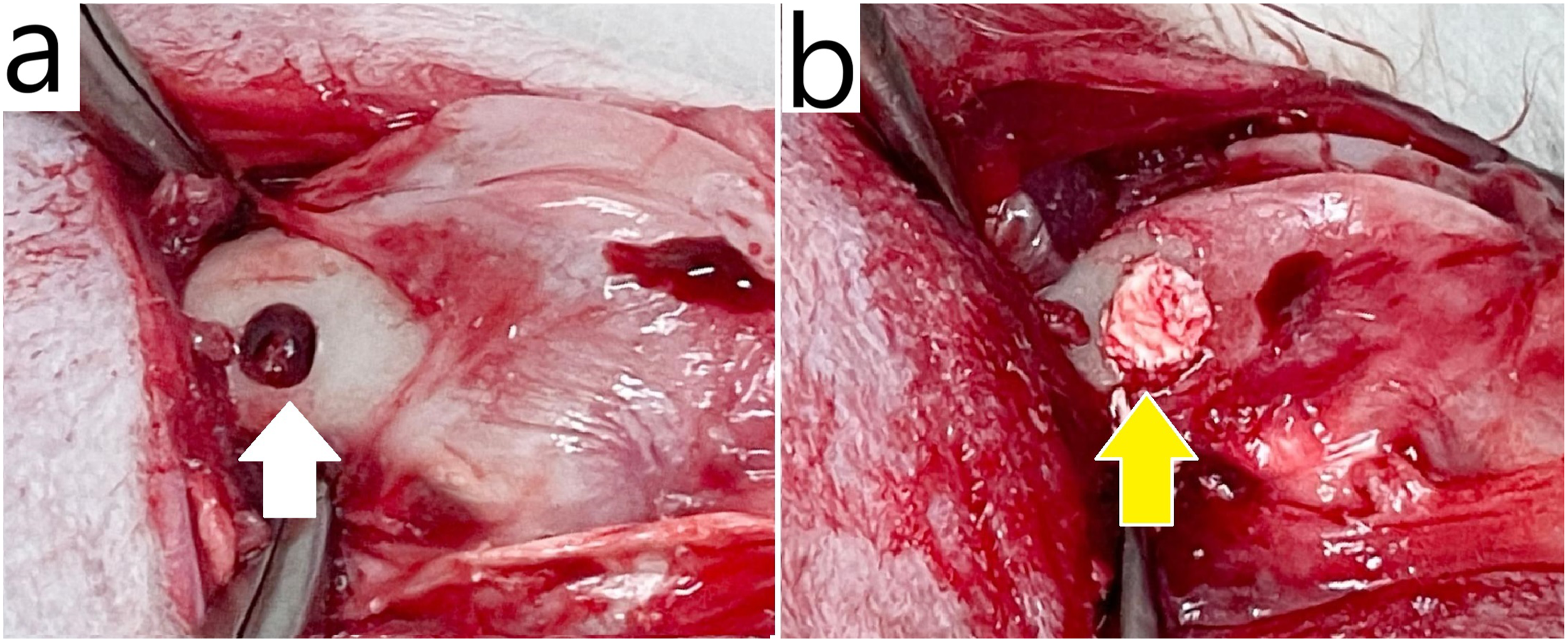
All the rabbits were sacrificed 4 weeks after surgery via cardiac injection of a saturated potassium chloride (KCl) solution. Following euthanasia, the distal femurs were extracted, immersed in 4% formaldehyde, and washed three times with phosphate-buffered saline (PBS). The specimens were then immersed in a decalcifying solution (Decalcifier I; Leica Biosystems, Wetzlar, Germany) until the femur was sufficiently softened for sectioning. After decalcification, the tissue was washed three times with PBS and dehydrated through an ethanol gradient for 20 min at each step, followed by paraffin embedding. Hematoxylin and eosin (H&E) staining and Masson’s trichrome staining were performed for histological analysis. All the procedures adhered to the ethical guidelines of the Institute of Animal Care and Use Committee of I-Shou University (IACUC ISU-113012).
Statistical analysis
All the quantitative data are presented as the mean ± the standard deviation (SD). The sample size (n) for each experiment is indicated in the corresponding legend. The normality of the data distribution was assessed using the Shapiro‒Wilk test prior to parametric analysis. For comparisons among multiple groups, one-way analysis of variance (ANOVA) was performed, followed by Tukey’s post hoc multiple comparison test. For time-dependent experiments that involved multiple groups, two-way ANOVA was used to assess the interaction between treatment and time, followed by Tukey’s post hoc test when appropriate. For comparisons between two groups, unpaired two-tailed Student’s t-tests were applied. Statistical significance was defined as p < 0.05. Exact p-values are reported where applicable. Statistical analyses were conducted using GraphPad Prism 8.0 (GraphPad Software, San Diego, CA, USA).
Results
Water content
To determine the optimum HA concentration of the GBCs, the water contents of the cryogels with various HA concentrations (0%, 0.3%, 0.5%, and 1%) were determined. In this study, after the GBCs were impregnated with ddH2O for 1 h, as the concentration of HA increased, the water content within the material also increased significantly (39.76% ± 1.59% in 0% HA, 45.48% ± 2.05% in 0.3% HA, 47.79% ± 2.13% in 0.5% HA, and 51.26% ± 1.55% in 1.0% HA) (Figure 2). Even the cryogels with only 0.3% HA still presented a water content of nearly 50%. Water contents of GBCs with varying HA concentrations (data are presented as the mean ± SD; n = 10; *p < 0.05 and **p < 0.01 compared with 0%).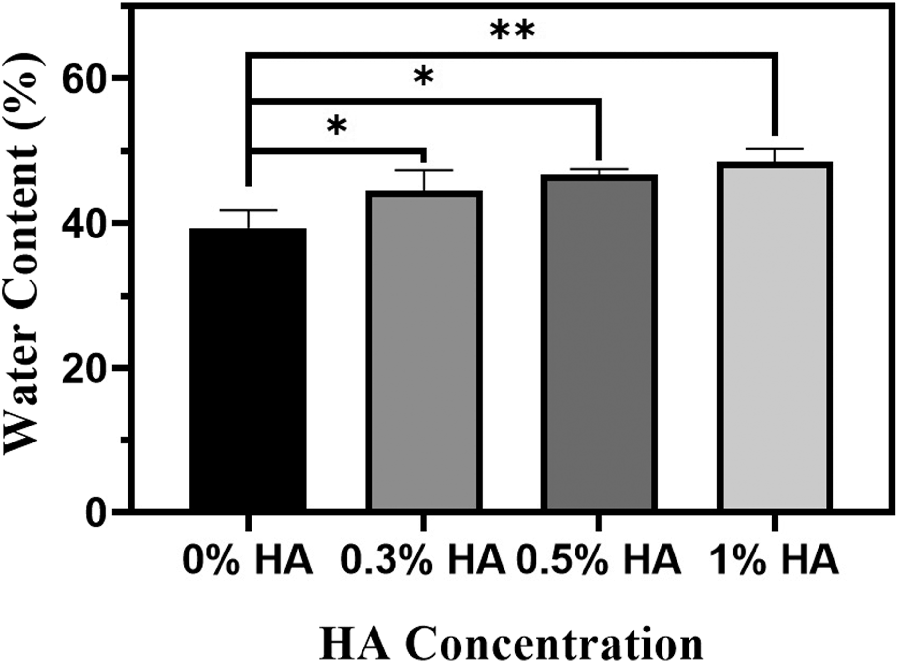
Swelling ratio
The water content affects the swelling ratio. We evaluated the swelling ratios of cryogels with different HA concentrations, and the swelling ratios increased gradually over time. The cryogels with higher HA concentrations exhibited higher swelling ratios over time (Figure 3). After 48 h, a swelling ratio of up to 300% was observed for the 0.3% HA cryogels (321.52 ± 9.69%), whereas the swelling ratio of the 1% HA cryogels (584.57 ± 30.50%) increased to nearly 600%, which was a significant increase compared to their initial weight (p < 0.0001). Swelling ratios of GBCs with varying HA concentrations at various time points (data are presented as the mean ± SD; n = 10).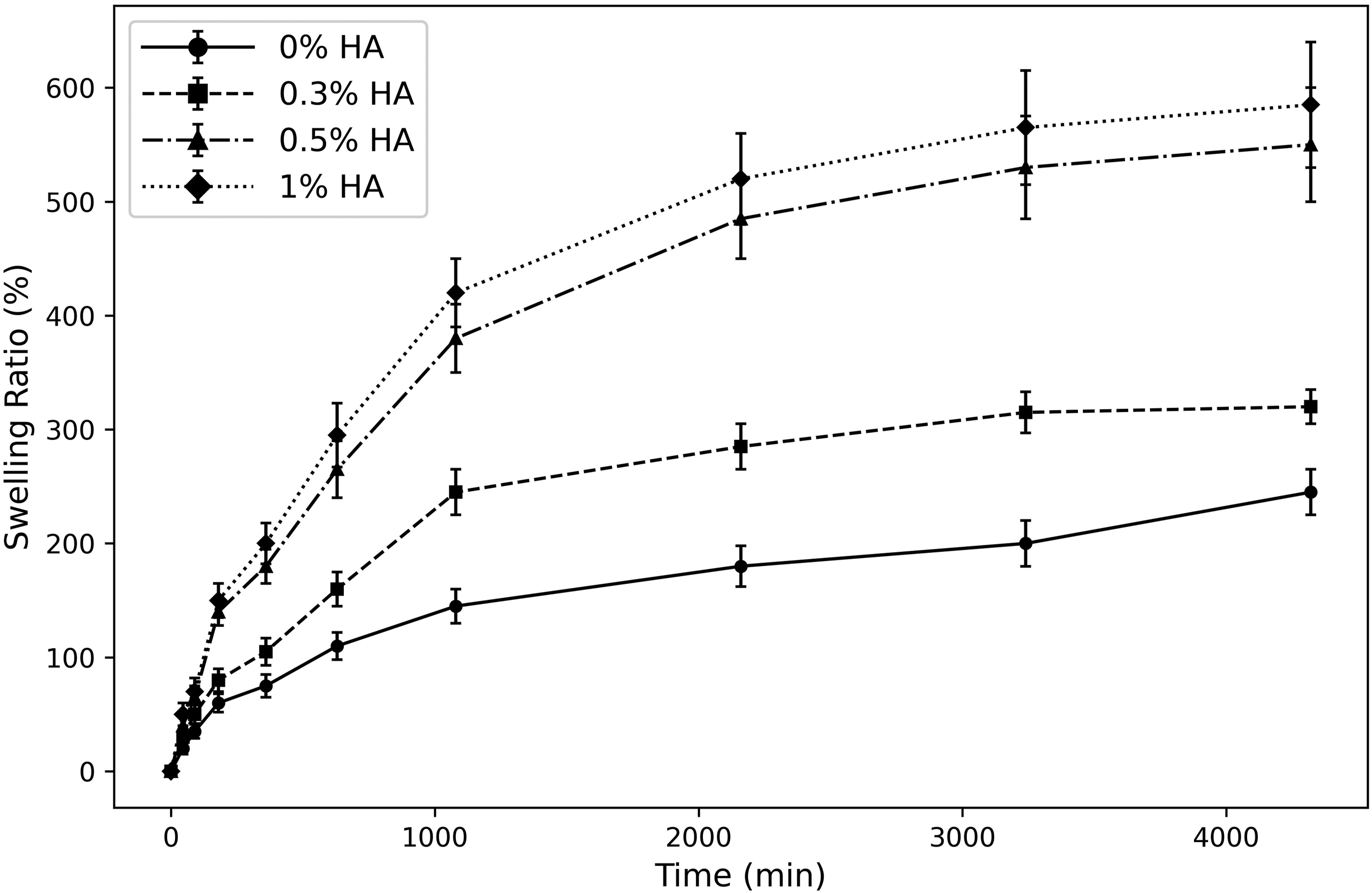
Porosity
In our study, cryogels that contained higher HA concentrations led to higher porosity before and after swelling for 24 h (Figure 4). Before swelling, the porosity ratios (%) were 1.86 ± 0.05 for 0% HA, 2.58 ± 0.39 for 0.3% HA, 4.31 ± 0.88 for 0.5% HA, and 4.85 ± 0.85 for 1.0% HA. After swelling, the porosity ratios (%) were 20.98 ± 4.12 for 0% HA, 29.87 ± 0.85 for 0.3% HA, 36.7232 ± 2.08 for 0.5% HA, and 48.34 ± 4.21 for 1.0% HA. After swelling for 24 h, the ethanol-accessible pore volume of all cryogels increased markedly compared with that before swelling. Because ethanol displacement reflects accessible pore volume rather than intrinsic three-dimensional structural porosity, this increase is likely attributable to enhanced ethanol penetration into the hydrated cryogel network after swelling. Compared with the cryogel without HA, the three cryogels with HA exhibited ethanol-accessible pore volumes of approximately 30% to 50%, which represented significant increases. Porosities of GBCs with varying HA concentrations. a: Before swelling and b: after swelling. A higher concentration of HA resulted in greater porosity. (The data are presented as the mean ± SD; n = 10; **p < 0.01 and ***p < 0.001 compared with 0%).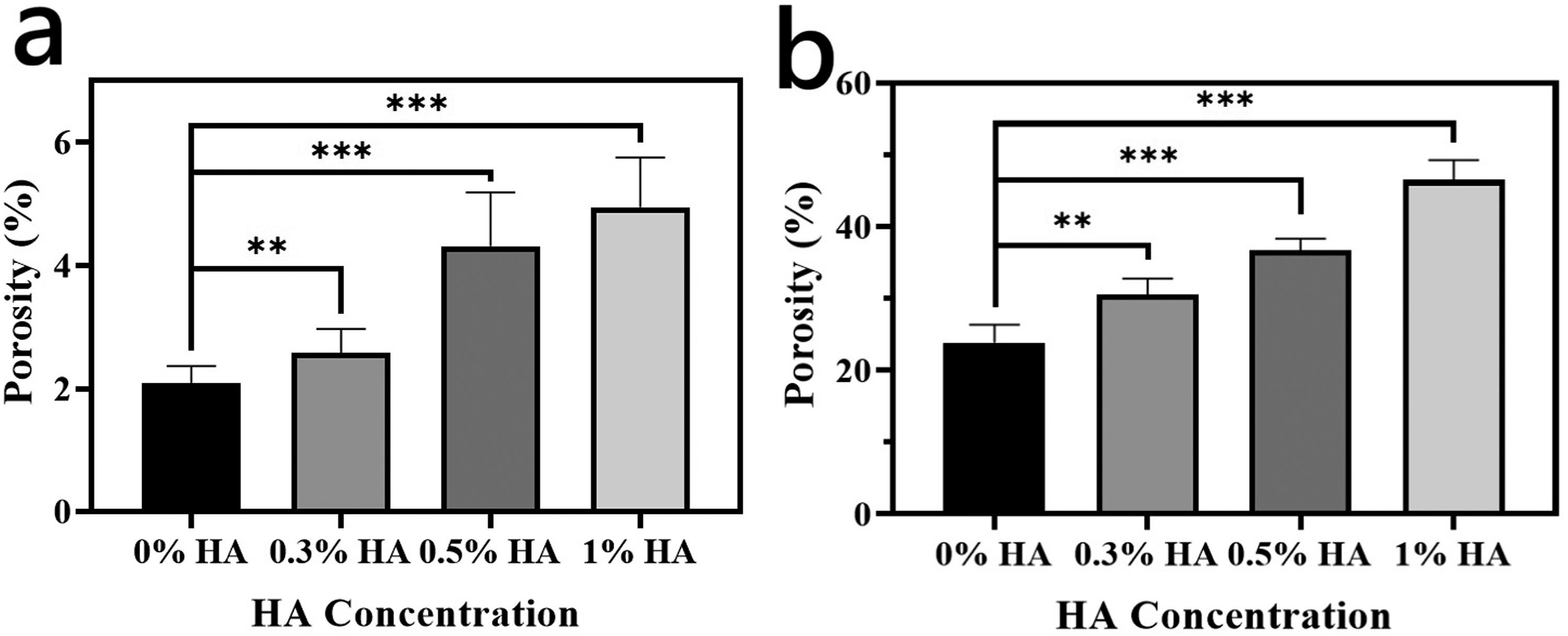
SEM analysis
Under a scanning electron microscope, the material surface before swelling exhibited a consistent distribution of rough particles. These particles were indicative of β-TCP dispersed within the gelatin matrix; notably, all four groups displayed uniform particle distributions, which confirmed the homogeneous incorporation of β-TCP within the composite material (Figure 5(a)–(d)). The crystalline characteristics of the scaffold were further confirmed by XRD analysis (Figure S1, Supporting Information), which indicated the presence of β-TCP-related peaks. After impregnation in ddH2O for 24 h, each group demonstrated a distinct and evenly distributed pore structure (Figure 5(e)–(h)). SEM images were used to illustrate pore morphology and structural characteristics rather than to directly confirm three-dimensional pore interconnectivity. Additional SEM micrographs of the outer surface and inner core structures are provided in the Supporting Information (Figure S2), where pore wall fenestrations were observed, suggesting potential pathways for fluid transport within the scaffold. Furthermore, swelling behavior analysis after rehydration and lyophilization (Figure S3, Supporting Information) demonstrated rapid liquid uptake, which is consistent with a permeable porous structure that may facilitate fluid infiltration throughout the scaffold. This observation suggests that the water molecules were uniformly absorbed throughout the cryogel, thereby facilitating swelling. As the concentration of HA increased, the pore size increased: in the 0% HA group, a few pores increased in size to approximately 100 to 200 μm, but most pores were smaller than 100 μm; in the 0.3% HA group, most pores increased to between 150 and 250 μm; in the 0.5% HA group, the pore sizes increased to a range of 250 to 350 μm, and larger cavities were observed; and in the 1.0% HA group, the largest pore sizes were observed, which increased to more than 300 μm. After the exosomes were seeded in the 0.3% HA group, the pore sizes increased from 200 to 300 μm (Figure 5(i)). Hence, the incorporation of exosomes into the cryogel did not affect the inherent pore size of the material. SEM micrographs of GBCs with various HA concentrations. (a–d) Cryogels with 0% HA, 0.3% HA, 0.5% HA, and 1% HA, respectively, before swelling. (e–h) Cryogels with 0.3%, 0.5%, and 1% HA, respectively, after swelling for 24 h. (i) Cryogel seeded with exosomes with 0.3% HA after swelling for 24 h.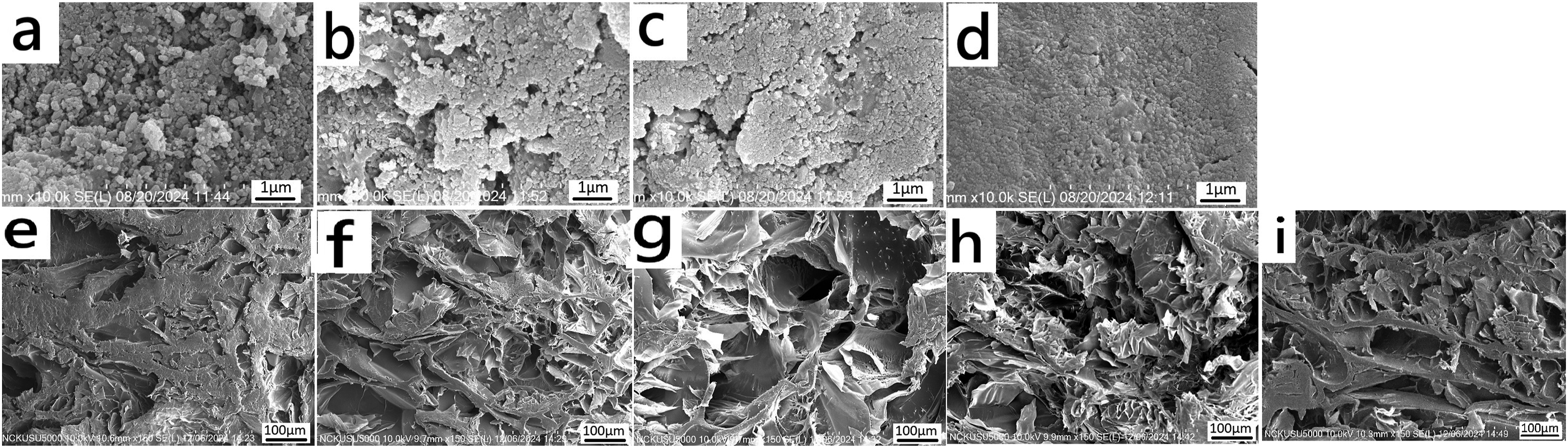
Mechanical test
The GBCs were designed to promote bone regeneration in bone defects, which requires adequate mechanical strength to tolerate the stress that occurs during movement. As shown in Figure 6, the stress–strain curves of all groups exhibited an initial linear region within 0–10% strain. The Young’s modulus was calculated from the slope of this initial linear region (0–10% strain). Increasing the HA concentration resulted in a lower Young’s modulus. The calculated Young’s modulus values were 13.61 MPa for the 0% HA group, 3.67 MPa for the 0.3% HA group, 1.50 MPa for the 0.5% HA group, and 1.36 MPa for the 1% HA group. A clear decrease in stiffness was observed with increasing HA concentration, indicating that incorporation of HA reduced the mechanical stiffness of the cryogel scaffolds. Compressive stress–strain curves of GBCs with various HA concentrations after swelling for 6 h. Young’s modulus was calculated from the initial linear region (0–10% strain) of each curve (n = 5).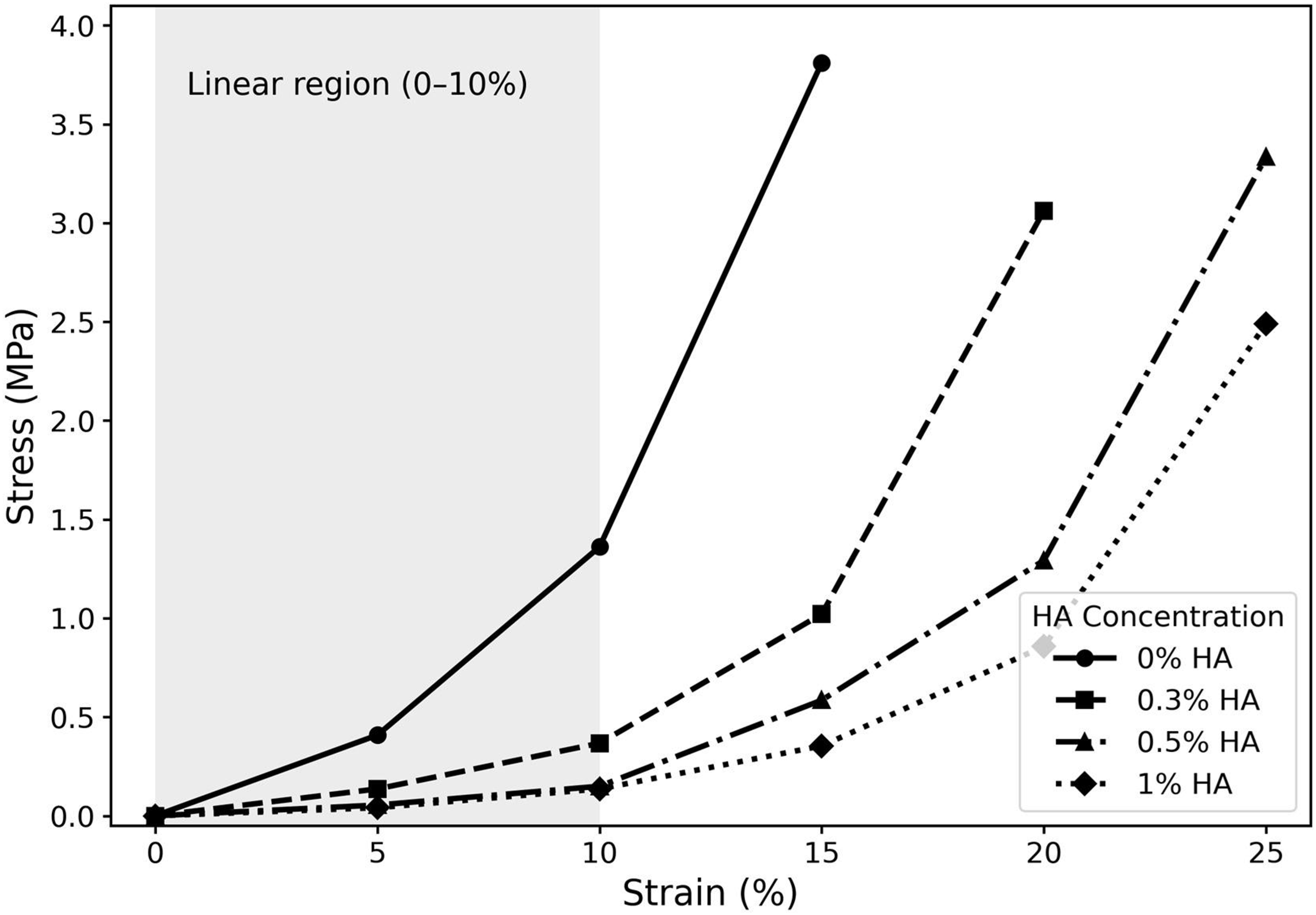
Cell viability
The results of the MTT assay with L929 cells after implantation with cryogels with different concentrations of HA indicate that after 48 h of incubation, cell growth in all the cryogel groups was not significantly different (0% HA: 0.243 ± 0.008, p = 0.632; 0.3% HA: 0.248 ± 0.031, p = 0.248; 0.5% HA: 0.258 ± 0.03, p = 0.361; and 1% HA: 0.254 ± 0.034, p = 0.511) from that in the control group (0.239 ± 0.013) (Figure 7). These findings indicated that the materials are noncytotoxic and biocompatible. MTT assay of GBCs with varying HA concentrations compared with the control (data are presented as the mean ± SD; n = 5).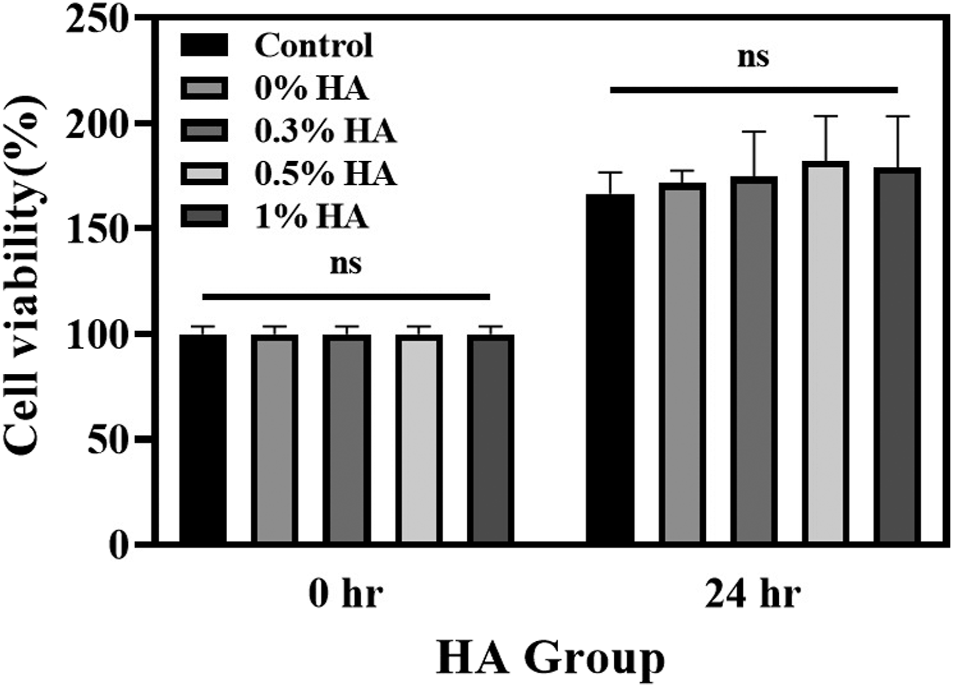
Analysis of human umbilical cord-derived mesenchymal stem cell-derived exosomes
NTA of the hUC-MSC-derived exosomes revealed a heterogeneous population of particles with sizes that ranged from 50 to 500 nm, and the majority of the population was 50–200 nm in size (Figure 8(a)). TEM imaging was performed to evaluate the morphological characteristics of the particles (Figure 8(b)). Taken together, these complementary techniques provide information on particle size distribution in suspension (NTA) and morphology under dehydrated conditions (TEM), supporting the presence of exosome-like vesicular structures suitable for subsequent biological experiments. Additional characterization of exosome incorporation into the cryogel scaffold is provided in the Supporting Information. The exosome encapsulation efficiency was approximately 40% (Figure S4), indicating effective incorporation of exosomes within the porous cryogel matrix. Furthermore, the release profile demonstrated sustained exosome release behavior, with an initial burst release followed by gradual release over 72 h (Figure S5). (a): Relative concentration, measured in particles/mL, of hUC-MSC-derived exosomes of varying sizes. (b): TEM micrograph of hUC-MSC-derived exosomes.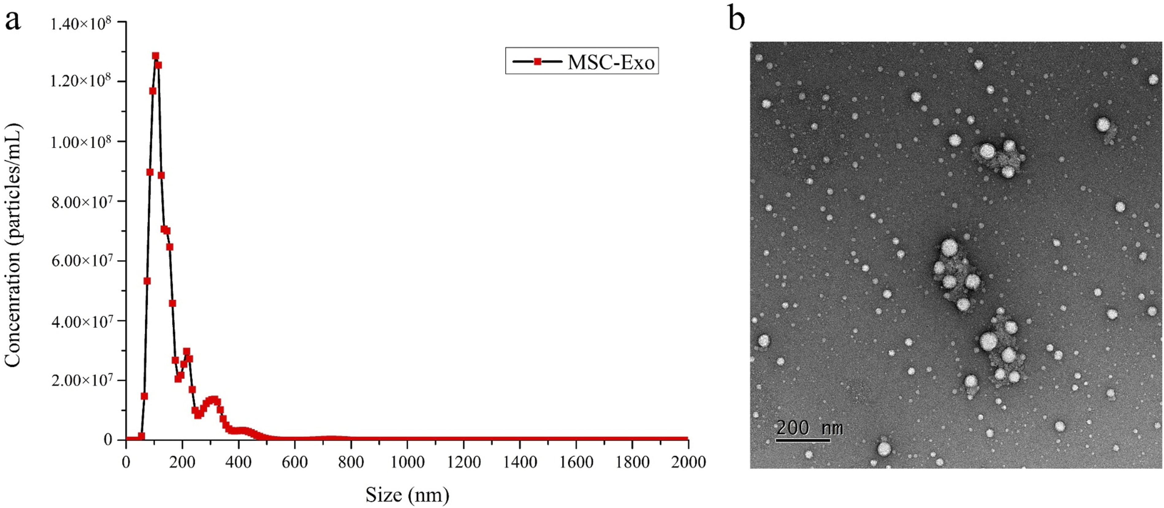
Cell proliferation after treatment with various concentrations of exosomes
The MG-63 cells were cultured with various concentrations of hUC-MSC-derived exosomes (103, 104, 105, 106, and 107 particles/mL). MTT assays were used to test the cell toxicity and viability after the cells were seeded with exosomes compared with those of the untreated control cells. After 168 h, compared with control MG-63 cells, MG-63 cells treated with various concentrations of hUC-MSC-derived exosomes exhibited significantly greater viability (0.958 ± 0.042). Specifically, compared with the control MG-63 cells, the MG-63 cells cultured with an exosome concentration of 106 particles/mL had significantly greater cell viability (1.388 ± 0.023) (p < 0.001) (Figure 9). However, compared with 106 particles/mL, the concentration of 107 particles/mL resulted in lower cell viability (1.122 ± 0.052) (p < 0.001), which indicated that excessively high concentrations may be detrimental. Therefore, 106 particles/mL of exosomes was selected as the optimal condition for subsequent experiments. MTT assay of MG-63 cell cultures with various concentrations of hUC-MSC-derived exosomes compared with the control condition (data are presented as the mean ± SD; n = 5; **p < 0.01 and ***p < 0.001.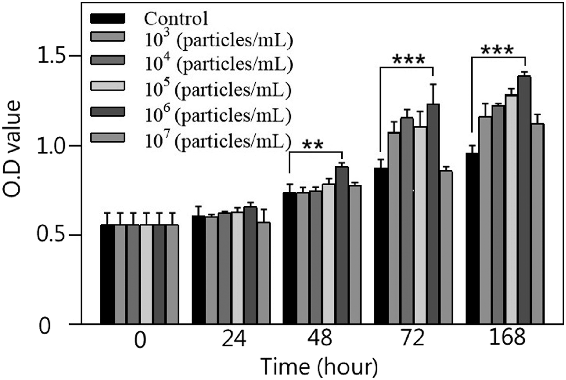
Considering the potential of the cryogels as bone defect filler materials, adequate compressive strength is essential. HA (0.3%) demonstrates a good swelling ratio and porosity, and its compressive strength is higher than those of 0.5% and 1% HA, thus providing the best supportive stability. Therefore, 0.3% HA cryogel combined with 106 particles/mL exosomes was selected to prepare GBCs as the material for subsequent in vivo experiments.
Before the in vivo experiments were conducted, the cell viability of MG-63 cells in the presence of 0.3% HA cryogel, 106 particles/mL hUC-MSC-derived exosomes, and cryogel seeded with 106 particles/mL hUC-MSC-derived exosomes for 24 h at 4°C was evaluated using the MTT assay (Figure 10). Both the exosome group and the cryogel-seeded exosome group exhibited significantly greater cell viability at 24, 48, 72, and 168 h than the control group did (p < 0.05). At 168 h, the cell viability increased by approximately 57% in the exosome group (0.703 ± 0.006, p < 0.001) and 55% in the exosome-seeded cryogel group (0.692 ± 0.027, p < 0.001) compared with that in the control group (0.447 ± 0.029). No significant difference was observed between the cryogel group and the control group. These results indicated that the standalone use of the cryogel did not promote MG-63 cell growth, although it was not cytotoxic. MTT assay results for MG-63 cell cultures after cryogel implantation, exosome addition, and exosome-seeded cryogel implantation compared with the control condition (data are presented as the mean ± SD; n = 5; *p < 0.05, **p < 0.01, and ***p < 0.001).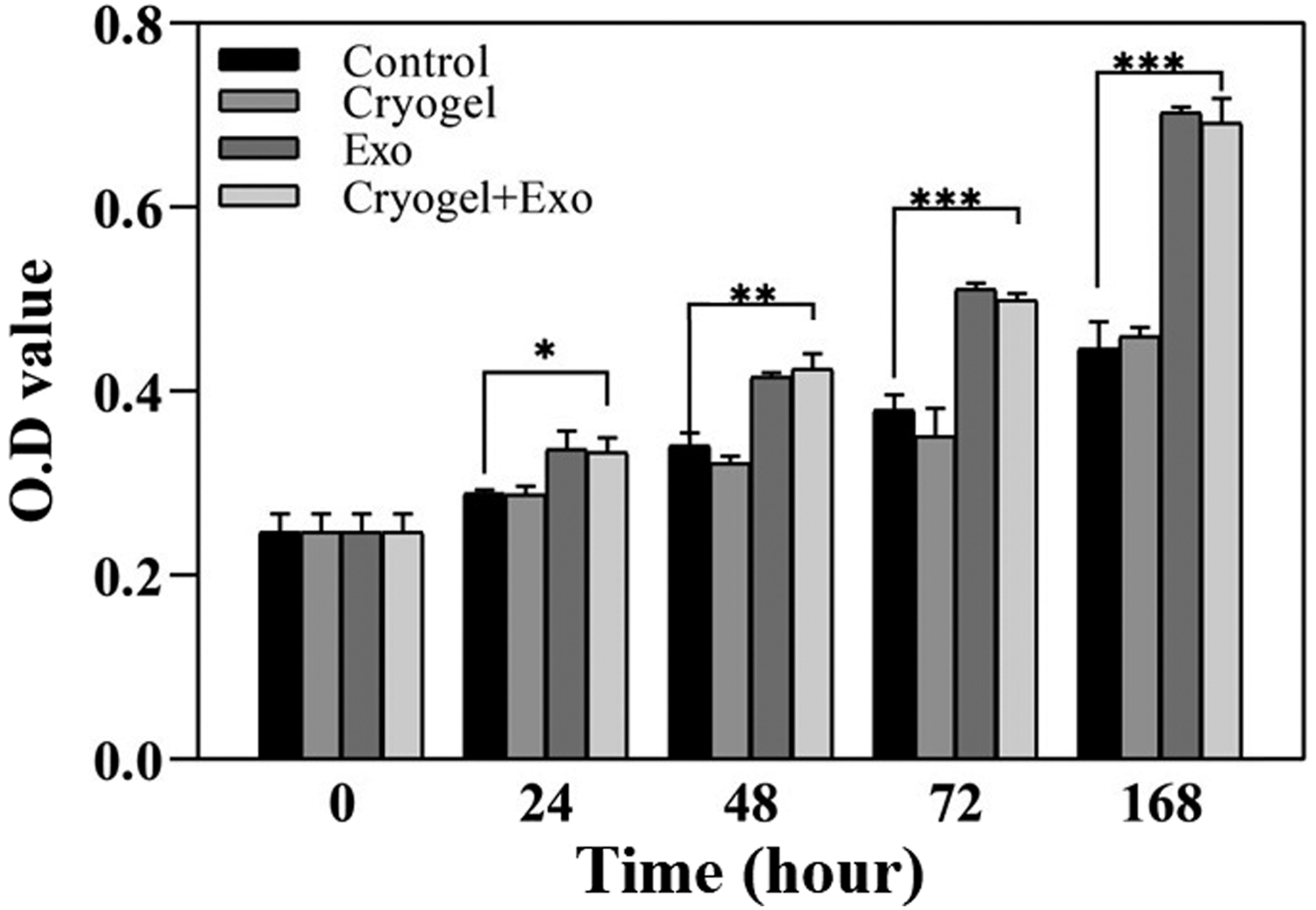
ALP activity
ALP activity was tested in MG-63 cells treated with cryogel alone, 106 particles/mL hUC-MSC-derived exosomes alone, and cryogel seeded with hUC-MSC-derived exosomes to evaluate osteoblast differentiation. Compared with the control group, both the exosome group and the cryogel-seeded exosome group exhibited higher ALP activity at 24, 48, 72, and 168 h (p < 0.001) (Figure 11). ALP activity significantly increased in the cryogel group at 24 and 48 h compared with that in the control group (18.34 ± 1.96 vs 6.64 ± 2.66, p < 0.001; 28.58 ± 3.57 vs 16.41 ± 2.61, p < 0.01); however, this effect was no longer observed after 72 h (33.48 ± 0.88 vs 28.34 ± 2.94, p = 0.015). ALP activity significantly increased in both the exosome group and the cryogel-seeded exosome group, even after 72 h. At 168 h, one-way ANOVA demonstrated a significant difference among the four groups (p < 0.001). Tukey post hoc analysis revealed no significant difference between the control and cryogel alone groups (42.73 ± 1.23 vs 42.68 ± 1.19, p > 0.05). However, these parameters were significantly greater in the exosome group and the cryogel-seeded exosome group than in both the control group and the cryogel-alone group (p < 0.001). Moreover, the ALP activity of cryogels seeded with exosomes was significantly greater than that of exosomes alone (64.38 ± 1.52 vs 53.01 ± 1.10, p < 0.001). The exosome-seeded cryogel group consistently exhibited the highest ALP activity. This could suggest an increased effect between the cryogel scaffold and incorporated exosomes because the cryogel provides the initial environment for bone growth, but a longer growth effect requires exosomes to continuously promote bone growth activity. ALP activity of MG-63 cell cultures after cryogel implantation, exosome addition, and exosome seeded cryogel implantation compared with that of the control condition (data are presented as the mean ± SD; n = 5; **p < 0.01 and ***p < 0.001).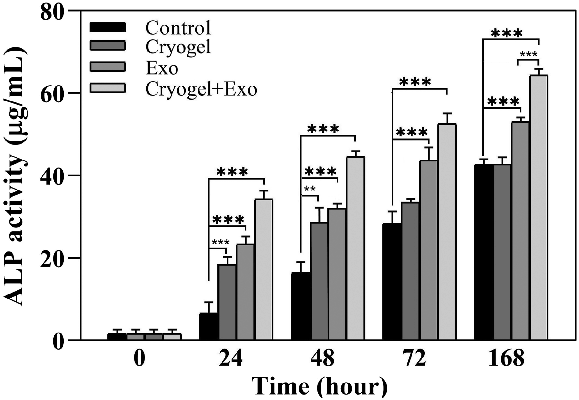
The fluorescence images of Calcein AM-stained samples after cell culture for 168 h revealed that the fluorescence intensity increased over time, thus indicating cell proliferation, under all the conditions (Figure 12(a)). All the semiquantitative data were compared among groups using ANOVA followed by Tukey’s post hoc test for multiple comparisons. At 168 h, compared with the control group (429.73 ± 12.52, p < 0.001), the cryogel group (506.38 ± 18.63), exosome group (515.48 ± 17.87), and exosome-seeded cryogel group (693.27 ± 18.19) presented significantly greater intensity values (429.73 ± 12.52, p < 0.001). The exosome-seeded cryogel group demonstrated a marked increase, which was approximately 61% greater than that of the control group and significantly greater than those of the cryogel group and the exosome groups (p < 0.001) (Figure 12(b)). a: Calcein AM staining of seven-day-old MG-63 cell cultures after cryogel implantation, exosome addition, and exosome-seeded cryogel implantation compared with the control condition. b: Fluorescence intensity that was measured using ImageJ to evaluate cell viability (data are presented as the mean ± SD; n = 5; *p < 0.05, **p < 0.01, and ***p < 0.001).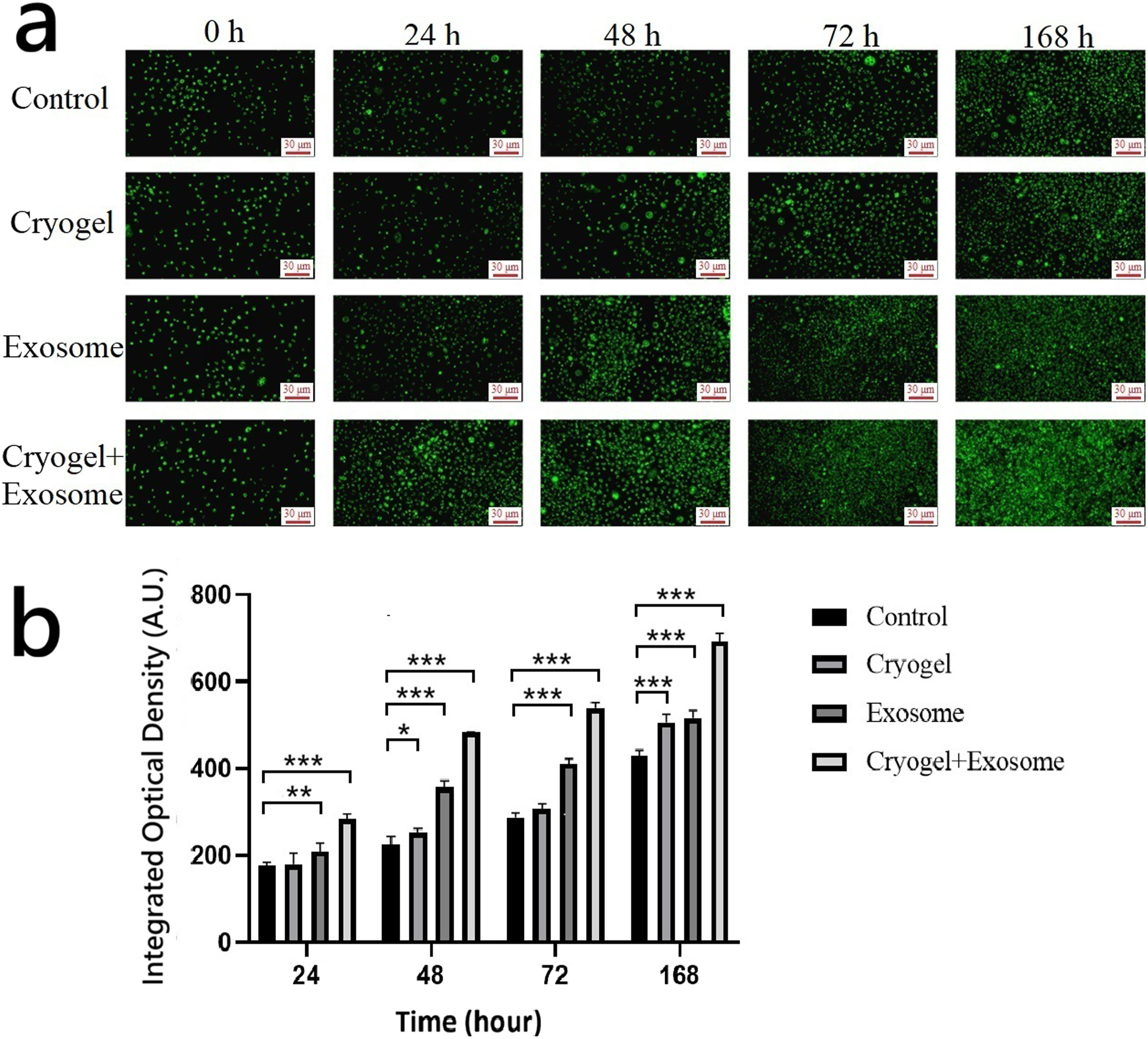
Alizarin Red S staining
Alizarin Red S staining was used to observe the mineralization of the MG-63 cells in the cryogel, exosome, and exosome-seeded cryogel groups (Figure 13). The staining intensity was measured using ImageJ and analyzed with ANOVA followed by Tukey’s post hoc test for multiple comparisons. The results indicated increased calcium deposition as reflected by ARS staining intensity, in the cryogel group at 24h compared with that in the control group (39.56 ± 2.68 vs 30.77 ± 1.53, p < 0.001), and no difference was detected between the cryogel group and the control group at 48, 72 or 168 h. However, compared to control group (48.54 ± 2.20, 50.80 ± 1.99), the significant increase in intensity persisted to 72 and 168 h in the exosome group (54.25 ± 3.39, 94.94 ± 5.67) and exosome-seeded cryogel group (62.10 ± 3.39, 115.29 ± 9.14). Moreover, the intensity of the exosome-seeded cryogel group was significantly greater than that of the exosome group at 72 and 168 h (p < 0.05 and p < 0.01, respectively). These results were consistent with the ALP activity results, suggesting enhanced early osteogenic activity in the presence of exosomes and cryogels. Exosome incorporation appeared to promote early-stage osteogenic responses, and the combination of cryogel with exosomes further enhanced this effect. It should be noted that, under the relatively short culture duration used in this study, ARS staining primarily reflects early-stage calcium deposition associated with osteogenic differentiation rather than fully developed mineralized matrix formation. a: Alizarin Red S staining of 168-h-old MG-63 cell cultures after cryogel implantation, exosome addition, and exosome-seeded cryogel implantation compared with the control condition. b: Integrated optical density analysis of Alizarin Red S staining was performed using ImageJ (data are presented as the mean ± SD; n = 5; *p < 0.05, **p < 0.01, and ***p < 0.001).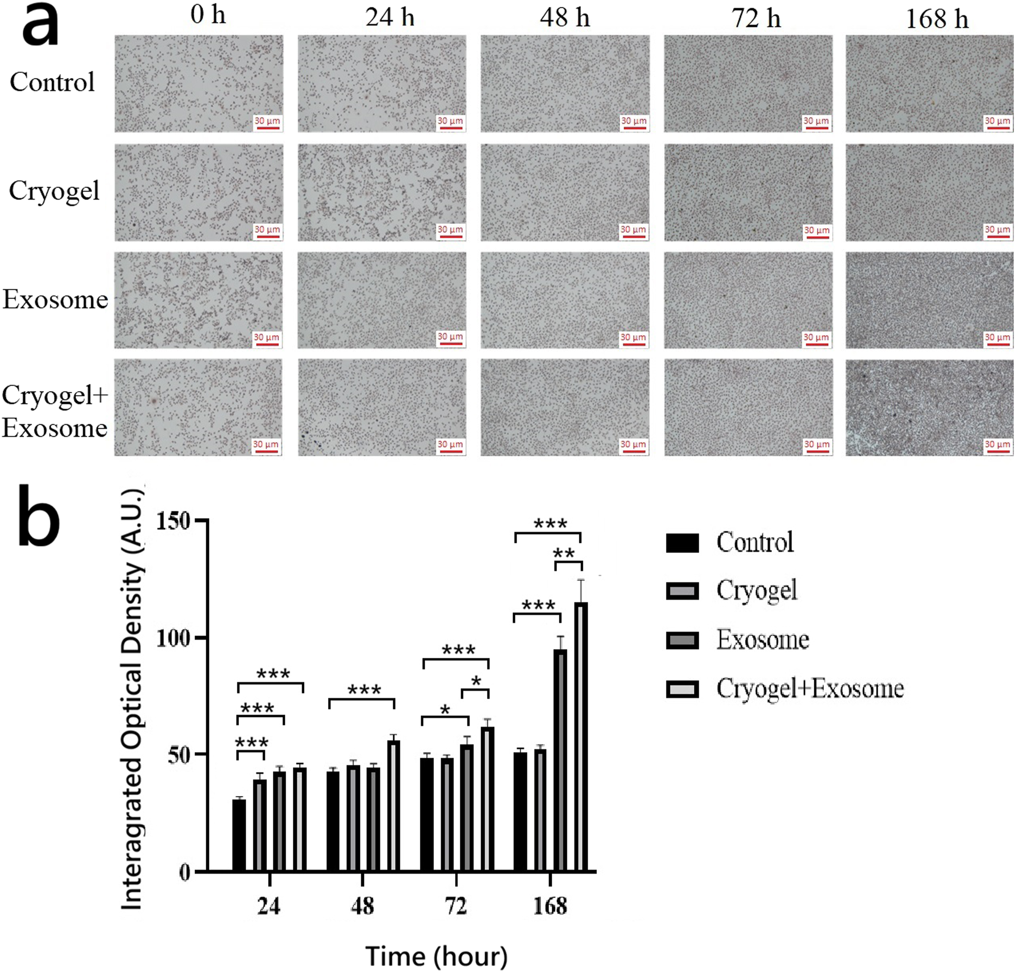
Repair of bone defects in vivo: Animal study
The rabbits were sacrificed after 4 weeks, and the defects were evaluated both grossly and histologically. In the control group, the defect was still clearly visible, with some tissue formation grossly observed within the defect. In both the cryogel group and exosome-seeded cryogel group, new tissue formation was demonstrated, which fully covered the defects (Figure 14). Gross evaluation of cortical defects 4 weeks after the operation in the rabbit model. (a) Control group defect (yellow circle), (b) cryogel group defect (yellow circle), and (c) exosome-seeded cryogel group defect (yellow circle).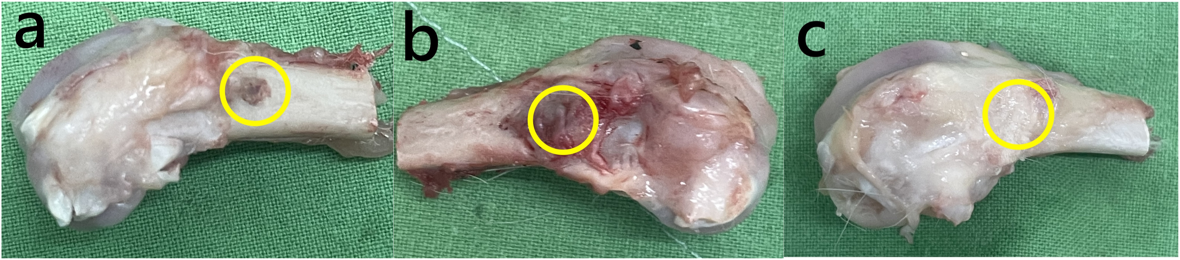
Histology
All the specimens were subjected to H&E staining. In the control group, no bone matrix was observed within the defect; only fibrous tissue filled the defect (Figure 15(a)). In the cryogel group, the defects were filled with loose bone matrix (Figure 15(b)). In exosome-seeded cryogel group, the defects were filled with dense bone matrix, and angiogenesis and neovascularization were evident (Figure 15(c)). Histological images with H&E staining that show recovery of the bone defects after 4 weeks. Low-magnification images of defects in the control (a), cryogel implantation (b), and exosome-seeded cryogel implantation (c) groups. The black boxes in (a)–(c) indicate the defect regions. High-magnification images of the control (d), cryogel implantation (e), and exosome-seeded cryogel implantation (f) groups. Fibrous tissue within the bone defect in the control group (black hollow arrow), loose bone matrix within the defect in the cryogel group (blue circle), and dense bone matrix (solid blue arrow) and neovascularization (solid black arrow) in the exosome-seeded cryogel group.
Masson’s trichrome staining was used to differentiate between mineralized bone and surrounding connective tissue. In the control group, the newly formed tissue within the defect was identified as fibrous tissue without mineralized bone matrix (Figure 16(a)). In contrast, both the cryogel group and the exosome-seeded cryogel group showed the formation of mineralized bone matrix within the defects (Figure 16(b) and (c)). However, denser mineralization was observed in the exosome-seeded cryogel group, whereas a looser matrix was observed in the cryogel alone group. Histological images with Masson’s trichrome staining that show recovery of the bone defects after 4 weeks. Low-magnification images of defects in the control (a), cryogel implantation (b), and exosome seeded cryogel implantation (c) groups. The black boxes in (a)–(c) indicate the defect regions. High magnification images of the control (d), cryogel implantation (e), and exosome-seeded cryogel implantation (f) groups. Denser mineralization was observed in the exosome-seeded cryogel group (yellow rectangle), and a looser matrix was observed in the cryogel alone group (black circle).
Discussion
The structural and biochemical properties of biomaterial scaffolds are fundamental for their function in bone tissue engineering. In this study, cryogels were designed using gelatin, β-TCP, and varying concentrations of HA to create a composite material that mimics the ECM microenvironment while offering sufficient mechanical and biological support. The material leverages the complementary benefits of its components: gelatin provides biocompatible and cell-adhesive structures (e.g., RGD peptides), β-TCP offers osteoconductive mineral content, and HA contributes to the capacity for hydration and cell signaling. The cryogels fabricated at subzero temperatures exhibited an interconnected macroporous structure because of ice crystal templating, which promoted cellular infiltration and mass transport. These traits are consistent with previous findings on cryogels in which ice acts as a porogen to produce large, interconnected pores that are beneficial for tissue regeneration. 5
Our experimental results demonstrated that increasing the HA concentration led to higher water contents and swelling ratios in the cryogel network. Notably, the 1.0% HA cryogel reached a water content of 51.26% and a swelling ratio near 600% after 24 h. These findings align with previous studies that have described the remarkable hygroscopicity of HA and its role in maintaining hydration in ECM analogs. 12 Improved swelling properties are desirable for materials to form irregular pores, thereby increasing their effective integration with host tissue. Importantly, the porosity analysis in our study revealed that the addition of more HA resulted in higher porosity, and even 0.3% HA resulted in a porosity of more than 30% after swelling, which was attributable to expansion of the hydrophilic network during gelation and lyophilization. The role of high porosity in supporting osteoblast infiltration, proliferation, and nutrient diffusion has been reported previously. 19 The integration of β-TCP particles, which was confirmed by SEM, provided a bioactive mineral phase that supported osteointegration. The structural resemblance of β-TCP to native bone minerals and its ionic dissolution profile contribute calcium and phosphate ions to the microenvironment, thereby supporting matrix mineralization and cellular differentiation. 20
Although high porosity and water retention are advantageous for biological interactions, they often compromise the mechanical integrity of scaffolds. In our study, as the HA concentration increased, the compressive stiffness of the cryogels decreased. Young’s modulus decreased from 13.61 MPa in the 0% HA group to just 1.36 MPa in the 1.0% HA group, which was an 88% reduction in stiffness. This trend was consistent with previous studies that have indicated that excessive swelling capacity and large pore sizes reduce the density of the polymer network, thereby leading to a loss in load-bearing capacity. 21 In the context of bone regeneration, mechanical properties must be precisely adjusted. A scaffold that is too rigid may cause stress shielding and hinder bone remodeling, whereas an overly soft scaffold may collapse and fail to support defect bridging. A suitable Young’s modulus of the scaffold to native tissue helps minimize the mechanical mismatch that could otherwise impair cellular behavior. Studies on mechanobiology have also demonstrated that material stiffness modulates osteoblast differentiation via focal adhesion signaling pathways. 22 Another study also suggested that for non-load-bearing sites or early-stage bone repair, a compressive modulus of 5–20 MPa is acceptable for providing temporary mechanical support while promoting host cell invasion. 23 Our 0.3% HA cryogels had a Young’s modulus of 3.67 MPa, which was closer to the optimal range than 0.5% and 1% HA cryogels, thus providing better biomechanical stability and sufficient flexibility to match the physiological condition of the host bone and presenting a mechanically favorable environment for osteogenesis. Collectively, these physicochemical improvements created a scaffold that could allow hydration, support cell attachment, and facilitate osteogenic signaling. Furthermore, as the degradability of the natural and mineral components used—gelatin, hyaluronic acid, and β-TCP—is well-documented in literature and inherently susceptible to enzymatic hydrolysis, this study prioritized the optimization of these structural properties and the synergistic biological functions elicited by exosome incorporation. Porosity and pore size are crucial structural features for bone scaffolds, as they directly affect cell infiltration, nutrient diffusion, vascular ingrowth, and overall osteoconduction. Optimized pore architecture contributes to improved osteogenic activity and graft–host integration in scaffold-based bone repair strategies. Previous studies have shown that pore architecture and size significantly influence cell infiltration and scaffold osteoconduction in bone tissue engineering, with larger, interconnected pores improving cellular invasion and matrix deposition. 24 In our study, SEM analysis demonstrated that increasing the concentration of HA led to increased porosity and larger average pore sizes, which ranged from <100 μm in the 0% HA group to >300 μm in the 1.0% HA group. Previous studies in scaffold-based bone tissue engineering have suggested that interconnected macropores in the range of approximately 150–400 μm are favorable for bone ingrowth, cell infiltration and angiogenesis, which are essential for sustained bone remodeling.19,25,26 In our study, we selected cryogels with 0.3% HA seeded with 106 particles/mL exosomes as the most appropriate conditions for our experiments, which exhibited pore sizes between 200 and 300 μm. In our in vivo study, the cryogel group seeded with exosomes exhibited extensive neovascularization and bone regeneration. Moreover, postimplantation swelling is a defining advantage of cryogel scaffolds, as it can aid in defect filling and stabilization. It increases the effective pore volume and enables a sponge-like conformation that better conforms to irregular defect geometries. This pore adaptation contributes to more effective osteoconduction and bone matrix deposition.
Cortical bone porosity has been reported to range from approximately 2–5% in young adults to 10–30% or higher in elderly individuals, which reflects accumulated remodeling events.27,28 Porosity increases during the remodeling stage because of bone resorption by osteoclasts. Bone remodeling is a dynamic process in which osteoclasts resorb aged bone tissue, which results in the generation of temporary resorption cavities and local intracortical pores that are subsequently refilled by osteoblasts, thereby resulting in the formation of secondary osteons. In the present study, our GBCs with exosomes (0.3% HA) exhibited a porosity of approximately 30% after swelling, which mimicked the transient microenvironment of increased porosity generated during the resorptive phase of remodeling. Specifically, macropores may facilitate vascular infiltration and osteogenic cell infiltration. Consistent with this structural profile, our in vitro results demonstrated increased MG-63 proliferation, elevated ALP activity, and increased mineral deposition in the exosome-seeded cryogel group. The porous architecture of cryogels may increase local exosome retention, thereby contributing to sustained bioactivity. Furthermore, in vivo implantation in a rabbit femoral defect model revealed improved bone matrix formation and neovascularization compared with those of the controls, which suggests that the porosity of the scaffold contributes to effective host cell infiltration and bone regeneration during the early remodeling phase.
Exosomes have emerged as potent mediators of tissue regeneration because of their ability to transfer regulatory RNAs, proteins, and lipids to recipient cells. In our study, compared with control cells, MG-63 cells cultured with exosomes at 106 particles/mL exhibited significant increases in cell viability and proliferation over 168 h, with a 57% increase, thus indicating a strong bioactive response to exosome stimulation. These results are consistent with those of previous studies in which MSC-derived exosomes promoted osteoblast proliferation, cellular activity, and matrix synthesis. Exosomal miRNAs such as miR-196a, miR-21, and miR-29b have been implicated in osteogenic differentiation pathways, including the Wnt/β-catenin and BMP/Smad signaling cascades.29,30 Moreover, in our present study, ALP activity assays revealed that both exosome-treated cells and exosome-seeded cryogel scaffolds significantly increased enzymatic activity, particularly at 72 and 168 h, which indicated that osteoblastic activity increased. Compared with exosomes alone, exosome-seeded cryogels further increased ALP expression, which suggested an enhanced effect between the scaffold architecture and the exosomal content. Mineralization potential was confirmed by Alizarin Red S staining, with the exosome group and exosome-seeded cryogel group showing markedly increased calcium deposition. The observed increases in ALP activity and mineral deposition indicates that exosome incorporation may support both early differentiation and subsequent matrix maturation processes during osteogenesis, which is consistent with previous reports. 31 It should be noted that the in vitro experiments in this study were conducted over a relatively short duration (up to 168 h) to evaluate early-stage osteogenic responses, including cell proliferation, ALP activity, and early extracellular matrix calcium deposition associated with osteogenic differentiation. Longer-term mineralization studies may be required to fully assess late-stage osteogenic maturation.
In our in vivo study, gross observation revealed defects that were not repaired in the control group, whereas both the cryogel group and exosome-seeded cryogel group showed soft tissue coverage within the defect, which indicated tissue proliferation. However, histological staining with H&E and Masson’s trichrome revealed a distinct qualitative difference: cryogel alone resulted in a loose bone matrix with fibrous interposition, whereas the exosome-seeded cryogels exhibited superior healing with dense bone matrix formation and neovascularization. The presence of vascularized structures may play a key role in accelerating matrix deposition and remodeling, given that angiogenesis is essential for supplying oxygen, nutrients, and progenitor cells to the regeneration site.32,33 Our findings reinforce the premise that exosome incorporation transforms the cryogel into a bioactive scaffold that can stimulate robust osteogenic activity, even without the introduction of live cells or the regulatory complexities of stem cell therapy. This cell-free approach is promising for clinical translation in bone defect management.
Although the results of this study are promising, some limitations still are acknowledged. First, the in vivo assessment was limited to a relatively short duration (4 weeks). Although this time frame is sufficient for assessing early bone regeneration and matrix deposition, it does not provide insight into long-term remodeling, scaffold resorption, or functional load-bearing capacity. Our study is a preliminary study. Although quantitative micro-CT was not performed in this preliminary study, the consistent histological findings across specimens support improved osteogenic remodeling in the exosome-functionalized group. The presence of organized mineralized matrix and vascular structures suggests accelerated early-stage bone regeneration. The present work focuses on early regenerative outcomes at 4 weeks, which is a time point commonly used to evaluate initial bone matrix deposition and angiogenic activity. Future studies that incorporate longer follow-up and volumetric imaging analyses, such as quantitative micro-CT, to evaluate volumetric bone fill and architectural integration over time may be necessary. Second, the dose‒response relationship of exosome concentration warrants further exploration, as our data indicated reduced viability at excessively high doses (107 particles/mL), which was potentially because of vesicle aggregation or cellular stress. Standardizing exosome isolation, quantification, and storage is another critical step for clinical applications. Despite these limitations, our preliminary study confirmed the potential efficacy of gelatin-based cryogels seeded with hUC-MSC-derived exosomes in promoting bone regeneration.
Conclusions
We developed a gelatin-based cryogel that incorporates β-TCP and hyaluronic acid with adequate physicochemical and mechanical properties for bone tissue engineering applications. Among the tested formulations, 0.3% HA GBCs achieved a favorable balance between porosity (30% after swelling) and compressive modulus (8.53 MPa) and provided sufficient structural stability while maintaining an interconnected macroporous architecture conducive to cellular infiltration and mass transport.
The incorporation of hUC-MSC-derived exosomes further improved the biological performance of the scaffold by promoting osteogenic cell proliferation, ALP activity, and matrix mineralization in vitro. In a rabbit bone defect model, compared with cryogels alone, exosome-seeded cryogels resulted in superior bone matrix formation and neovascularization, which suggested that the scaffold architecture and exosome-mediated bioactivity were improved.
Collectively, these findings indicate that hUC-MSC-derived exosome-seeded gelatin cryogels provide a structurally permissive and biologically active microenvironment that supports bone regeneration. This cell-free strategy represents a promising platform for future translational development in bone defect repair.
Supplemental material
Supplemental Material - Gelatin-based cryogels seeded with exosomes enhance osteogenic activity and bone regeneration in a rabbit femoral defect model
Supplemental Material for Gelatin-based cryogels seeded with exosomes enhance osteogenic activity and bone regeneration in a rabbit femoral defect model by Joseph Yang, Daniel Yang, Jhe-Lun Hu, Yong-Ji Chen, Shwu-Jen Chang and Shan-Wei Yang in Journal of Biomaterials Applications
Footnotes
Acknowledgments
The authors gratefully acknowledge the use of SEM, TEM, and NTA equipment belonging to the Instrument Center of National Cheng Kung University (NCKU), supported by the National Science and Technology Council.
Ethical considerations
Animal experiments conducted in this study were approved by the Institutional Animal Care and Use Committee (IACUC) of I-Shou University, Taiwan (Approval No. ISU-IACUC-113002).
Author contributions
Shwu-Jen Chang and Shan-Wei Yang contributed equally to this work.
All authors have read and agreed to the published version of the manuscript
Funding
The authors disclosed receipt of the following financial support for the research, authorship, and/or publication of this article: This research was funded by the National Science and Technology Council, grant number MOST113-2221-E-214-003-MY3.
Declaration of conflicting interests
The authors declared no potential conflicts of interest with respect to the research, authorship, and/or publication of this article.
Data Availability Statement
The raw data supporting the conclusions of this article will be made available by the authors on request.
Supplemental material
Supplemental material for this article is available online.
References
Supplementary Material
Please find the following supplemental material available below.
For Open Access articles published under a Creative Commons License, all supplemental material carries the same license as the article it is associated with.
For non-Open Access articles published, all supplemental material carries a non-exclusive license, and permission requests for re-use of supplemental material or any part of supplemental material shall be sent directly to the copyright owner as specified in the copyright notice associated with the article.