






Abstract
Background:
The genus Streptococcus is 1 of the dominant bacterial groups in human milk, but the taxonomic identification of some species remains difficult.
Objective:
The objective of this study was to investigate the discriminatory ability of different methods to identify streptococcal species in order to perform an assessment of the streptococcal diversity of human milk microbiota as accurately as possible.
Methods:
The identification of 105 streptococcal strains from human milk was performed by 16S rRNA, tuf, and sodA gene sequencing, phylogenetic analysis, and Matrix Assisted Laser Desorption Ionization-Time of Flight (MALDI-TOF) mass spectrometry.
Results:
Streptococcus salivarius, Streptococcus mitis, and Streptococcus parasanguinis were the streptococcal dominant species in the human milk microbiota. Sequencing of housekeeping genes allowed the classification of 96.2% (16S rRNA), 84.8% (sodA), and 88.6% (tuf) of the isolates. Phylogenetic analysis showed 3 main streptococcal clusters corresponding with the mitis (73 isolates), salivarius (29), mutans (1)-pyogenic (2) groups, but many of the mitis group isolates (36) could not be assigned to any species. The application of the MALDI-TOF Bruker Biotyper system resulted in the identification of 56 isolates (53.33%) at the species level, but it could not discriminate between S pneumoniae and S mitis isolates, in contrast to the Vitek-MS system.
Conclusion:
There was a good agreement among the different methods assessed in this study to identify those isolates of the salivarius, mutans, and pyogenic groups, whereas unambiguous discrimination could not be achieved concerning some species of the mitis group (S mitis, S pneumoniae, S pseudopneumoniae, S oralis).
Well Established
Recent studies have shown that human milk is a source of bacteria for the infant gut. The assessment of this bacterial diversity has revealed the frequent presence of streptococci, but the identification of some streptococcal species is still problematic.
Newly Expressed
This is the first study focused on the assessment of the streptococcal diversity in human milk. We have investigated the discriminatory ability of several methods such as sequencing of housekeeping genes, phylogenetic analysis, and Matrix Assisted Laser Desorption Ionization-Time of Flight mass spectrometry. There was a good agreement among these approaches to identify those isolates of the salivarius, mutans, and pyogenic groups, but the identification of some species of the mitis group still remains a challenge.
Background
Human milk represents a continuous supply of commensal, mutualistic, and/or potentially probiotic bacteria to the infant gut. 1 Studies about the bacterial diversity of human milk have revealed that the streptococci are among the dominant groups in this biological fluid.2-5
At present, the genus Streptococcus comprises up to 111 species, including major pathogens, such as Streptococcus pyogenes, Streptococcus agalactiae, or Streptococcus pneumoniae, but also some commensal members of the human microbiota. 6 Despite the spectacular advances in bacterial identification methods over the past decades, the identification of some streptococcal species is still problematic, causing considerable confusion for both clinical microbiologists and taxonomists.
This is particularly true for the so-called viridans group streptococci (VGS), a pseudotaxonomic term that comprises a very heterogeneous group of organisms. 7 Traditionally, VGS have been poorly classified and characterized; in fact, their taxonomy has been very controversial since, for many years, there was not a standardized naming scheme or typing system for these microorganisms.8,9 DNA-based methods have greatly contributed to improved VGS taxonomy and, as a result, phylogenetic analysis of 16S rRNA gene sequences allowed their classification into 5 major phylogenetic groups: anginosus, mitis, mutans, bovis, and salivarius. 10 However, the 16S rRNA nucleotide sequences of some VGS and, particularly, of some mitis group members (S mitis, S oralis, S pseudopneumoniae, S pneumoniae) share a very high percentage of identity (≥ 99%), and the use of this gene is often unable to discriminate among them.11,12 Alternate identification procedures, based on other conserved housekeeping genes such as dnaJ, gyrB, recA, recN, sodA, rpoB, or tuf13-18 or in the application of the Matrix Assisted Laser Desorption Ionization-Time of Flight (MALDI-TOF) mass spectrometry, 19 have been proposed to improve the discrimination among streptococcal species.
To date, most of the streptococcal species identified in human milk belong to the salivarius (S salivarius, S vestibularis) or the mitis (S mitis, S oralis, S parasanguinis, S infantis, S peroris, S australis, S lactarius) groups.3,20-24 In this context, and taking into account the present difficulties for a correct taxonomic assignment of some streptococcal species, the objective of this study was to compare different procedures currently available for such purposes in order to perform an assessment of the streptococcal diversity of human milk microbiota as accurately as possible.
Methods
Bacterial Isolates
A total of 105 streptococcal strains isolated from milk samples were analyzed in this study. Milk samples were provided by 67 healthy breastfeeding women with no clinical symptoms of mastitis during current lactation. These samples were collected in a sterile tube by manual expression using sterile gloves. Previously, nipple and mammary areola had been cleaned with soap and sterile water and the first drops of milk were discarded. All volunteers gave written informed consent to the protocol approved by the Ethical Committee of Hospital Clinico (Madrid, Spain).
The isolates were originally selected from Columbia Nalidixic Acid agar (CNA; BioMérieux, Marcy l’Etoile, France) plates and were initially observed by optical microscopy to determine morphology and Gram staining, tested for catalase activity, and stored at −80ºC in glycerol (30%, v/v). All the streptococcal isolates were submitted to RAPD genotyping as previously described by Ruiz-Barba et al. 25 Multiple bacterial isolates from a single sample were included only if they showed different genotype profiles after RAPD genotyping. Reference strains included in the study are shown in Table 1.
Identification Results for the Reference Strains according to the Sequence Analysis of 16S rRNA, sodA, and tuf Genes, and Matrix Assisted Laser Desorption Ionization-Time of Flight (MALDI-TOF).

Abbreviations: ATCC, American Type Culture Collection; CECT, Spanish Type Culture Collection; DSM, Leibniz-Institut DSMZ German Collection of Microorganisms and Cell Cultures; T, type strain.
Identification Based on the Sequencing of Housekeeping Genes (16S rRNA, tuf, and sodA)
PCR amplification of fragments of the 16S rRNA (450 bp) and tuf (560 bp) genes was carried out using the primer sets plb16 (5’-AGAGTTTGATCCTGGCTCAG-3’) and mlb16 (5’-GGCTGCTGGCACGTAGTTAG-3’),
The sodA gene fragment was amplified by using the primer pair d1 (5’-CCITAYICITAYGAYGCIYTIGARCC-3’) and d2 (5’-ARRTARTAIGCRTGYTCCCAIACRTC-3’). 16 The PCR cycling conditions included an initial denaturation at 95°C for 3 minutes, followed by 35 cycles at 95°C for 30 seconds, 37°C for 90 seconds, and 72°C for 90 seconds, followed by a final extension at 72°C for 10 minutes.
A few bacterial colonies removed from pure cultures on CNA agar plates and resuspended in 50 μL of sterile deionized water were used as a source of DNA template for the PCRs. The total volume of all the PCR mixtures was 25 μL, containing 5 μL MyTaq Red Mix (Bioline, London, UK), 0.15 μL MyTaq polymerase (0.75U), 13.85 μL sterile PCR-grade water, 0.5 μL of each pair of primer (0.2 μM), and 5 μL of template DNA. The amplicons were purified using the Nucleo Spin Gel and PCR Clean-up (Macherey-Nagel, Düren, Germany) and sequenced at the Genomics Unit of the Universidad Complutense de Madrid, Spain. The resulting sequences were compared with those available in the NCBI by using the BLAST algorithm, and the identity of the isolates was determined on the basis of the following scores: ≥ 98% for species level identification and 95% to 97% for genus level; the results were considered as “no identification” when values < 95% were achieved.
Phylogenetic Analysis
Concatenated sequences of 16S rRNA, sodA, and tuf gene fragments from each strain were aligned and subjected to phylogenetic analysis by the neighbor-joining algorithm using MEGA version 5. 28 The robustness of the nodes was evaluated by bootstrapping (1000 replicates).
For reference purposes, sequencing of 16S rRNA, sodA, and tuf genes and phylogenetic analysis of the type strains were carried out. In addition, sequences obtained from the genomes of the following reference strains were also included: S cristatus ATCC 51100T (GenBank: AY584476, DQ232543.1, and AF276261.2); S oligofermentans CIP 108229T (GenBank: AY099095.1, DQ232554.1, and EU156926.1); and S pseudopneumoniae CCUG 49455T (GenBank: EU156785.1, HM560723.1, and EU156956.1).
Unequivocal clustering with a single type strain of a particular species in the phylogenetic tree was taken as a criterion indicating the identification at the species level.
Identifications by MALDI-TOF Mass Spectrometry
Identification of the bacterial isolates was performed by using the MicroFlex LT system (Bruker Daltonics, Bremen, Germany). Briefly, bacterial colonies were suspended in 300 μL of sterile water and vortexed; 900 μL of absolute ethanol was subsequently added to the bacterial suspension, vortexed, and centrifuged at 13 000 rpm for 2 minutes. After discarding the supernatant, residual ethanol was removed by a second centrifugation and drying in a vacuum centrifuge. The pellet was resuspended in 50 μL of 70% formic acid and mixed with 50 μL of acetonitrile by pipetting. The suspension was centrifuged at ≥ 13 000 rpm for 2 minutes, and 1 μL of the supernatant was spotted onto the steel MALDI target plate, allowing drying at room temperature before the addition of 1 μL of matrix. The MALDI Biotyper system was calibrated with the Bruker bacterial test standard (Escherichia coli lysate). Spectra were analyzed using MALDI Biotyper automation control and Bruker Biotyper 2.0 software. Identification criteria used were those recommended by the manufacturer: a score of ≥ 2 indicated species level identification, 1.70 to 1.99 genus level identification, and < 1.70 was interpreted as no identification.
A subset of streptococcal isolates was also identified by MALDI-TOF using a Vitek-MS instrument (BioMérieux) in the facilities of Probisearch (Tres Cantos, Spain). Briefly, a portion of a bacterial colony (~1 µL) was spotted onto a MALDI sample plate, overlaid with 1 µL of a saturated solution of α-cyano-4-hydroxycinnamic acid in acetonitrile (28%), and then allowed to dry at room temperature. For each isolate, a mean spectrum was constructed with at least 50 m/z spectra profiles and used for the identification by comparison with the spectra contained in the Myla database (BioMérieux). Identification was defined as a 99% to 100% match to the species-specific m/z values in the database.
Results
In this study, the taxonomic identification of 105 streptococcal strains isolated from human milk samples has been performed by different methods (Supplementary Table, available online).
Identification Based on the Sequencing of Housekeeping Genes
Amplification of the 3 housekeeping genes from all the streptococcal strains generated a single amplicon of the expected size for each gene. Sequence analysis of 16S rRNA, sodA, and tuf resulted in the identification at the species level of 96.19%, 84.76%, and 89.52% of the bacterial isolates, respectively. A total of 15 different streptococcal species have been identified from the 105 isolates (Table 2).
Comparison of the Identification Results Obtained by 16S rRNA, sodA, and tuf Gene Sequencing of 105 Streptococcal Strains Isolated from Human Milk.
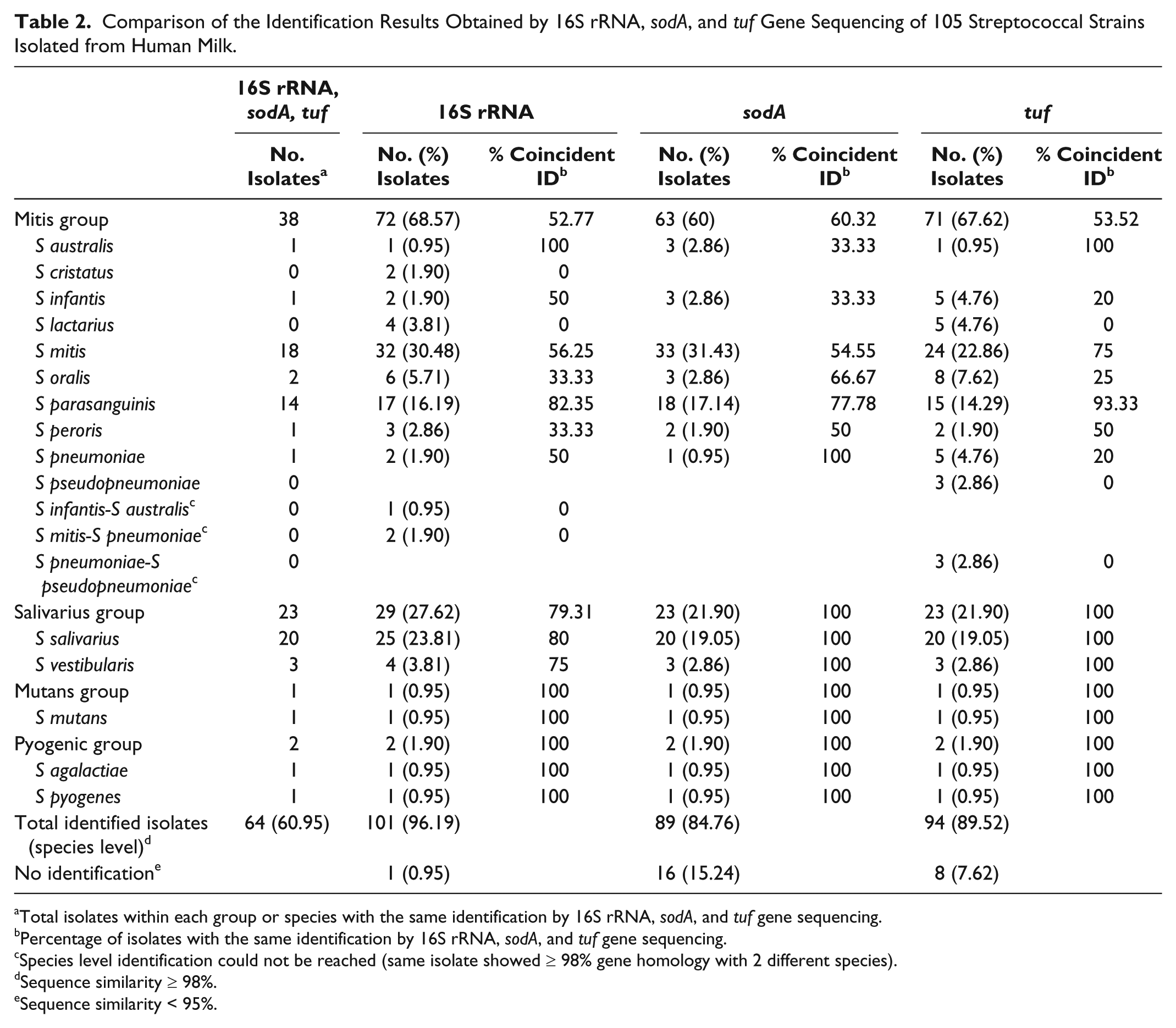
Total isolates within each group or species with the same identification by 16S rRNA, sodA, and tuf gene sequencing.
Percentage of isolates with the same identification by 16S rRNA, sodA, and tuf gene sequencing.
Species level identification could not be reached (same isolate showed ≥ 98% gene homology with 2 different species).
Sequence similarity ≥ 98%.
Sequence similarity < 95%.
Globally, 60% to 69% of the bacterial isolates were identified as belonging to the mitis group and 22% to 28% to the salivarius group by the sequencing of these housekeeping genes. Presence of other streptococci, such as those of the mutans (0.95% of the isolates) or pyogenic (1.90%) groups, was scarce among the human milk strains isolated in this study (Table 2).
The use of the 3 genes led to a coincident identification at the species level in 60.95% of the bacterial strains, mainly corresponding to isolates identified as S salivarius (20 isolates, 19.05%), S mitis (18 isolates, 17.14%), and S parasanguinis (14 isolates, 13.33%). On the other hand, a disagreement between the results provided by 2 genes and that obtained with the remaining gene was observed for 27 isolates (25.71%). Most of the disagreements involved isolates identified by 1 or another method as S mitis, S pneumoniae, S pseudopneumoniae, or S oralis. For the remaining 14 isolates (13.33%), there was either a disagreement among the 3 genes or an impossibility to get a species identification (Supplementary Table).
The data show that tuf gene sequencing resulted in a higher percentage of isolates of S parasanguinis and S mitis (93.33% and 75%) properly identified (ie, the same identification was achieved with the 3 genes) when compared to the 16S rRNA (82.35% and 56.25%) and sodA (77.78% and 54.55%) approaches. Finally, 20 isolates were identified as S salivarius by the 3 genes; this represented 100% of the isolates identified as belonging to such species by using the sodA and tuf genes but only 80% of those ascribed to this species using the 16S rRNA gene (25 isolates) (Table 2).
All the reference strains were correctly identified by the 3 genes, except for S lactarius CECT 7613, for which no identification at the species level could be attained by sodA gene sequencing (Table 1).
Identification Based on Phylogenetic Analysis
Figure 1 shows a neighbor-joining radial tree based on 122 concatenated sequences (1364 bp) from 105 human milk isolates and 17 type strains that included all the species commonly present in human milk. The tree showed 3 main clusters that were well resolved, with ≥ 98% bootstrap confidence values for the nodes, corresponding to the mitis, salivarius, and mutans-pyogenic groups. A more detailed dendrogram, indicating the position of the milk isolates and bootstrap values exceeding 60%, is shown in Figure 2.

Phylogenetic Tree Based on the Concatenated Sequences of 16S rRNA, sodA, and tuf Genes of 122 Streptococcal Isolates (105 Human Milk Isolates and 17 Type Strains).

Dendrogram Obtained with 122 Streptococcal Isolates (105 Human Milk Isolates and 17 Type Strains) after a Phylogenetic Analysis Based on the Concatenated Sequences of 16S rRNA, sodA, and tuf Genes.
The mitis cluster contained 73 milk isolates and included all the type strains belonging to species of this streptococcal group; however, many of the milk isolates (36) could not be assigned to any species within the mitis group since, globally, there was not a clear separation among them (Figures 1 and 2). This was particularly relevant for those strains identified as S mitis, S pneumoniae, or S pseudopneumoniae by sequence analysis of at least 1 gene, since they grouped together, including the reference type strains, without forming defined groups (Figure 1). The only exceptions were those isolates identified as S lactarius or S parasanguinis, including the respective reference strains, since they clustered in a well-resolved branch; in fact, the node separating the S lactarius-S parasanguinis branch from that including other species of the mitis group had a bootstrap value of 100% (Figures 1 and 2). Subsequently, 2 sub-branches separating the S lactarius isolates from S parasanguinis ones (bootstrap values of ≥ 80%) were also observed (Figures 1 and 2).
The salivarius cluster included 29 milk isolates and the type strains of the species of this streptococcal group. The milk isolates were distributed, forming 3 different and well-defined subgroups, as assessed by the bootstrap values (Figures 1 and 2); 2 of the subgroups were correlated with the type strains of S salivarius and S vestibularis, whereas the third one did not include any of the type strains. This third group included all the isolates that had been identified as S salivarius or S vestibularis by 16S rRNA sequencing but not by sodA and tuf sequencing (BLAST identity score < 95%). Finally, the mutans and pyogenic groups clustered separately, containing 1 and 2 milk isolates, respectively.
Identifications by MALDI-TOF Mass Spectrometry
The 105 milk isolates and reference strains were submitted to MALDI-TOF identification using the Bruker Biotyper system. Application of this technique resulted in a reliable identification (score value ≥ 2) of 56 isolates (53.33%) at the species level (Table 3). Among the isolates with such a score value, 33.33% (35 isolates) belong to the mitis group, and they were identified as S oralis (1), S parasanguinis (14), S peroris (3), and S pneumoniae (17). The isolates with a score value ≥ 2 included in the salivarius group (18 isolates, 17.14%) were classified as S salivarius (14) and S vestibularis (4). The remaining isolates identified at species level corresponded to S mutans (1), S agalactiae (1), or S pyogenes (1).
Identification Results Obtained by MALDI-TOF Bruker Biotyper of 105 Streptococcal Strains Isolated from Human Milk
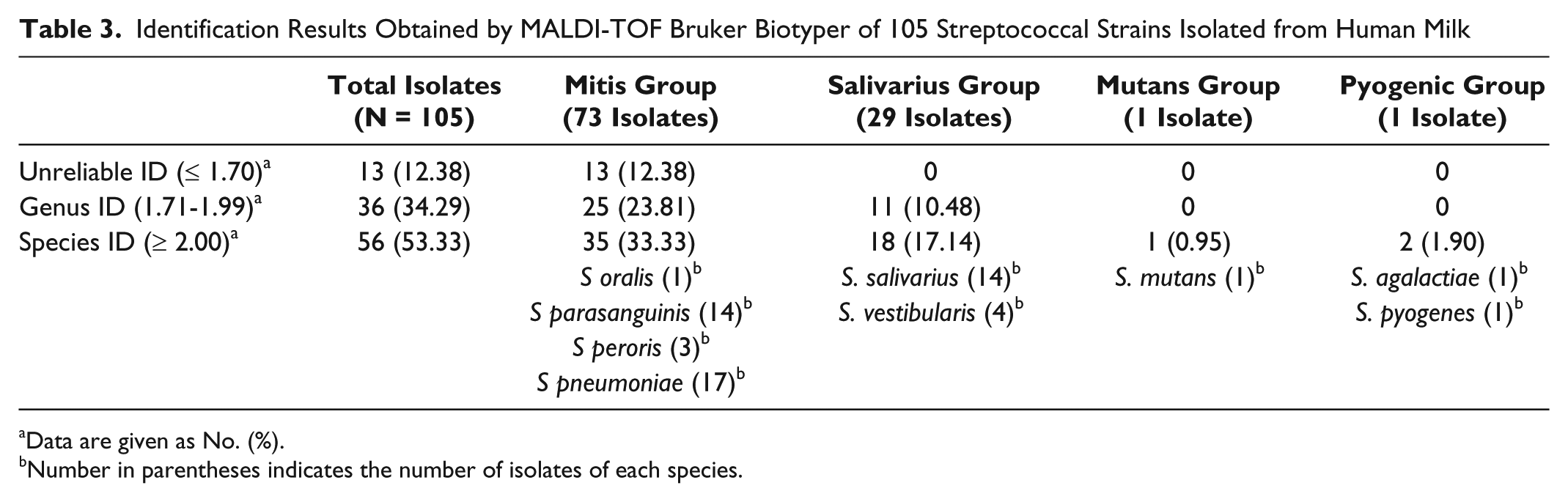
Data are given as No. (%).
Number in parentheses indicates the number of isolates of each species.
Identification was possible only at the genus level for 34.29% of the isolates, whereas 12.38% could not be identified since score values were < 1.7 or no peaks were found by the system. The reference strains were correctly identified by the Bruker Biotyper system, with the exception of S lactarius CECT 7613
The high percentage of identifications only at the genus level as well as nonreliable identifications in the mitis group, together with the fact that a high number of the milk isolates were identified as S pneumoniae but none as S mitis, led us to perform a MALDI-TOF identification of a selection of 34 streptococcal strains along with the reference strains by using different equipment with a different database (Vitek-MS system). The selected strains were those isolates that had been identified as S pneumoniae by the Bruker Biotyper system. Most of the isolates identified as S pneumoniae by the Bruker Biotyper were identified as S mitis/S oralis by the Vitek-MS system (Table 4). Therefore, although the Vitek instrument was unable to discriminate between S mitis and S oralis, it showed a better agreement with the results obtained by sequencing of the housekeeping genes. In relation to the reference strains, the Vitek-MS system also misidentified S lactarius CECT 7613
Comparison of the Identification Results Obtained by Matrix Assisted Laser Desorption Ionization-Time of Flight (MALDI-TOF) Bruker Biotyper and MALDI-TOF VITEK MS of 34 Streptococcal Strains Isolated from Human Milk.

Discussion
The application of both culture techniques and culture-independent molecular techniques for the assessment of bacterial diversity of human milk has revealed the frequent presence of streptococci and streptococcal DNA in this biological fluid.5,20,27,29,30 In fact, Streptococcus seems to be among the core genera of the human milk microbiome. 4
Human milk streptococci mainly belong to the viridans group and, more specifically, to the mitis and salivarius groups. Difficulties in the correct classification and laboratorial identification of species within such groups have been extensively reported.7,9,31,32 The use of conventional phenotypic and biochemical tests often fails to accurately classify VGS to species level. 31 Previous reports have demonstrated the low sensitivity of standardized phenotypic identification methods, such as the API or the VITEK 2 systems, for speciation of VGS,32,33 limitations that could be associated with the relatively low number of biochemical traits that can be analyzed in comparison with the high number of VGS species and the absence of updated databases incorporating the re-evaluations faced by streptococcal taxonomy in the last years.
DNA-based methods represent an alternate approach for discriminating among VGS species. Various genes have been proposed for streptococci species identification and, among them, the 16S rRNA, tuf, and sodA genes were selected in this study. Partial sequencing of the first 2 genes has been previously used for the detection and identification of human milk streptococci.2,27 In such studies, most isolates were identified as S mitis, S parasanguinis, and S salivarius, in agreement with this and previous works.3,5,20,22
In this study, a relatively high percentage of isolates could not be identified at the species level. The fact that some Streptococcus species share a high degree of DNA sequence identity complicates the identification by using DNA-based techniques. Thus, although 16S rRNA sequencing has been widely accepted for bacterial identification, it is not adequate to discriminate among some streptococcal species, such as S mitis, S oralis, S pneumoniae, and S pseudopneumoniae.11,12 It has been shown that interspecies recombination occurs at a high frequency among members of VGS, invalidating identification procedures based on sequence analysis of a single gene.34-36
Alternatively, the simultaneous sequence analysis of various housekeeping genes has been proposed for a more precise identification of streptococci. 10 In our study, analysis of concatenated sequences of the 3 genes resulted in a defined cluster within the salivarius group, composed of bacterial isolates that could not be identified by the single-gene analysis of tuf or sodA sequences. Similarly, this approach allowed the clustering of all the isolates identified as S lactarius by 16S rRNA and/or tuf sequencing together with the reference strain S lactarius CECT7613. Such isolates could not be identified as S lactarius by sodA sequencing. Therefore, analysis of concatenated sequences of the 3 genes showed a higher level of discrimination among these species than single gene sequencing. Trees based on the concatenated sequences of some housekeeping genes can identify sequence clusters that can be assigned as species clusters by the location on the tree of the type strains of each of the viridans or mitis group species. The resulting tree can show whether a strain falls within 1 of the known viridans species clusters or within an unassigned sequence cluster (a potential new species), or whether it is a unique divergent genotype. 10
In contrast, this approach could not discriminate among the rest of the species of the mitis group, probably because of their high sequence identity in the analyzed genes. In addition, the number of new species of the mitis group has increased rapidly in the last years, and the fact that there could be a high number of new streptococcal species waiting for discovery may be responsible for the apparent disagreement between procedures targeting different housekeeping genes.
Recently, MALDI-TOF has emerged as a useful, rapid, and cost-effective method for the identification of clinically relevant microorganisms, including streptococci.19,37,38 However, difficulties for a proper distinction between S mitis, S oralis, and S pneumoniae were observed in this work. A low discrimination power among VGS species has been reported in previous works.39,40 The close relationships among 16S rRNA sequences of some species of the mitis group may explain the difficulty in differentiating them by MALDI-TOF, since this method is based on the analysis of the ribosomal protein spectra. In fact, the spectral profiles of some mitis group streptococci species are very similar, and recent studies have searched for characteristic MALDI-TOF peaks to distinguish these species.41,42
It is interesting that a high percentage of S mitis isolates were misidentified as S pneumoniae using the Bruker Biotyper system, whereas they were identified as S mitis/S oralis by the VITEK MS system as it has been previously reported.43-45 Differences in the identification algorithms may make VITEK MS more sensitive than Bruker Biotyper in the detection of specific peaks and may be related to its better ability to distinguish between S pneumoniae and other mitis group streptococci. 46 However, the VITEK MS system was not able to differentiate between S mitis and S oralis. Implementation of further improvements in the current MALDI-TOF systems, including better and wider databases and the recognition of new differential peaks of the spectral profile, is needed to improve mass spectrometry-based identification of mitis group streptococci.
At present, the limits between the relevant pathogen S pneumoniae and other mitis group species, especially its closest relative S mitis, are unclear not only taxonomically but also regarding their pathogenic potential.36,47,48 However, accurate species-level identification is still a relevant issue, and therefore, clear differentiation of pneumococci from other viridans streptococci remains a challenge.
There was a good agreement among the different techniques in relation to the identification of isolates belonging to S pyogenes, S mutans, and S agalactiae, pathogens involved in invasive infections in humans. 49 However, these pathogens are rarely present in human milk and can be easily identified by the methods routinely used in most clinical laboratories.
Conclusion
The methods assessed in this study have identified S salivarius, S mitis, and S parasanguinis as the streptococcal dominant species in the human milk microbiota. These techniques have shown a good ability to identify isolates belonging to S parasanguinis and other streptococcal species within the salivarius, mutans, and pyogenic groups; however, our methodology failed in the identification of closely related species belonging to the mitis group, particularly S mitis, S pneumoniae, S pseudopneumoniae, and S oralis. These results show that the identification of some streptococcal species remains extremely difficult in spite of the spectacular advances in bacterial identification techniques. Future studies are in progress to improve our tools for the identification and characterization of the streptococci present in human milk. This work includes the study of streptococci’s virulence factors, resistance to antibiotics, and potential involvement in mastitis.
Footnotes
Acknowledgements
The authors thank Tomás García for his technical assistance with the Matrix Assisted Laser Desorption Ionization-Time of Flight analyses.
Declaration of Conflicting Interests
The authors declared no potential conflicts of interest with respect to the research, authorship, and/or publication of this article.
Funding
The authors disclosed receipt of the following financial support for the research, authorship, and/or publication of this article: This work was supported by AGL2013-41980-P project from the Ministerio de Economía y Competitividad (Spain).

References
Supplementary Material
Please find the following supplemental material available below.
For Open Access articles published under a Creative Commons License, all supplemental material carries the same license as the article it is associated with.
For non-Open Access articles published, all supplemental material carries a non-exclusive license, and permission requests for re-use of supplemental material or any part of supplemental material shall be sent directly to the copyright owner as specified in the copyright notice associated with the article.
