
Abstract
Objective
To evaluate the diagnostic usefulness of effluent endotoxin by Limulus amoebocyte lysate (LAL) assay in gram-negative peritonitis patients on continuous ambulatory peritoneal dialysis (CAPD) therapy.
Design
Prospective study with patients serving as their own controls. Standard microbiologic work up and endotoxin analysis of effluents (night dwell) were done during the pre- and posttreatment phases.
Setting
Specimens from three teaching hospitals were processed and tested at a common center. Patients were left for treatment at their respective centers without intervention.
Patients
32 clinical peritonitis and 40 infection-free CAPD patients were studied.
Results
75% (n = 24) of cultures were positive: 41.6% (n = 10) gram-negative and 58.4% (n = 14) gram-positive bacteria. Effluents of pre- and posttreated gram-negative cultures had endotoxin levels of 1.53 ± 0.169 and 0.214 ± 0.085 endotoxin units (EU)/mL, respectively (p < 0.0001); pre- and posttreated gram-positive levels of 0.102 ± 0.06 and 0.122 ± 0.052 EU/mL, respectively (p > 0.05); pre- and posttreated culture-negative peritonitis levels of 0.110 ± 0.025 and 0.087 ± 0.031 EU/mL, respectively (p > 0.05); peritonitis-free effluents contained 0.117 ± 0.079 EU/mL. The diagnostic specificity and the sensitivity of LAL assay were 100% and 98.2% respectively.
Conclusion
Where initial microbiological work-up cannot demonstrate a pathogen, effluent endotoxin determined by quantitative LAL assay is a useful marker for diagnosis and management, within safe time limits, of gram-negative peritonitis in CAPD patients.
With the current microbiological techniques available, 3% – 30% of CAPD-related peritonitis remain culture negative (1,5). Techniques to concentrate bacteria, mechanical liberation of sequestrated bacteria from leukocytes before inoculation, culturing of large volumes of effluents, and the use of specialized culture media (6-8) are reported to reduce the rate of negative culture results. However, the length of time required for a definitive microbiological report has not been shortened.
Despite attractive earlier results (9-14), the Limulus amoebocyte lysate (LAL) assay has not been accepted in the diagnosis and management of peritonitis in CAPD. With this conflict still unsettled, we aimed at retesting the method's reliability and determining the “cutoff” value of endotoxin level that could guide a clinician for the initial management of CAPD-related peritonitis patients.
Patients and Method
Patients
The study population was comprised of 32 clinical peritonitis patients and a peritonitis-free group (n = 40). Average age was 52 years, range 19 – 78 years, for male and female patients on a standard CAPD program for at least 6 months.
Peritonitis was diagnosed when there was one or a combination of (1) generalized abdominal pain, (2) cloudy effluent, or (3) effluent white blood cell (WBC) count of more than 100/mm3. Exclusion criteria included (1) recent (1 week) major gastrointestinal or extragastrointestinal trauma or operation, (2) recent insertion of peritoneal catheter, (3) recent major gastrointestinal invasive diagnostic work up, (4) current major clinical infection other than CAPD peritonitis, or (5) being on antibiotic therapy or within a week of its discontinuation.
Materials
Reagent
The quantitative chromogenic LAL assay kit, Coatest Endotoxin, a color-producing substrate S-2423, and a control standard endotoxin solution (E. coli 0111:B4) manufactured by Charles River Endosafe, Charleston, SC, U.S.A., were supplied by Chromogenix AB, Mölndal, Sweden. Pyrogen-free glassware and pipettes were provided with the kit.
Study Design
This was a prospective study consisting of two phases, with some patients serving as their own controls. In the first phase, patients suspected of having peritonitis had their peritoneal fluid, preferably night-dwell, drained. Within a maximum of 1 hour, bags of effluent were transferred to a study center for immediate processing.
The fluid was examined for WBC count on Thoma glass, then a 10-mL sample was drawn and stored in an apyrogen glass container at –20°C. Microbiologists performed routine Gram stain and cultures. Patients were treated at their respective centers until a cure was effected, proven by clinical recovery and negative cultures.
In the second phase, similar amounts of effluent samples from cured and 40 clinically- and microbiologically-proven peritonitis-free CAPD patients were collected and stored, as described above, for future endotoxin measurements.
Cultures
A 60-mL aliquot of each sample was centrifuged at room temperature in a sterile glass test tube at 3000g for 20 minutes. The sediment was vigorously agitated with a vortex mixer before inoculation on standard agar plates, then incubated at 37°C for 14 days. Culture results were retrospectively correlated to endotoxin levels.
Endotoxin
The LAL and standard endotoxin solutions were reconstituted according to the manufacturer's instructions. After thawing stored effluents and bringing all samples to room temperature, 50 μL of each of the test and standard samples was immediately pipetted into wells of an apyrogen microplate and incubated at 37°C for 5 minutes. 50 μL of reconstituted LAL solution was added into each well, mixed, and incubated at 37°C for another 12 minutes, after which 100 μL of substrate buffered solution was added and the plate briefly agitated before re-incubating for an additional 8 minutes. Finally, 100 μL of 20% acetic acid was added into each well to stop the reaction. Plates were read on Metertech Σ-960 ELISA auto analyzer (Metertech; Nankang, Taipei, Taiwan) at 405 nm. Absorbencies of standards were plotted on a curve and concentrations of endotoxin in the sample solutions were extracted from the standard curve.
Statistics
Values are given as mean ± standard deviation (SD). Average levels of endotoxin concentrations were compared using a one-way variant analysis (ANOVA), a p value of less than 0.05 was considered statistically significant. Significance tests were performed by using Tukey–Kramer multiple comparison test.
Results
Cultures
Of 32 clinical peritonitis episodes, 75% (n = 24) were culture-positive, of which 42% (n = 10) had gram-negative and 58% (n = 14) had gram-positive bacteria; 25% (n = 8) were sterile throughout 14 days of incubation. Within the first 48 hours, 60% (n = 6) of gram-negative and 86% (n = 12) of gram-positive cultures were positive, while 40% (n = 4) of gram-negative and 14% (n = 2) of gram-positive cultures were positive after 72 hours of follow-up.
Endotoxin (Figure 1)
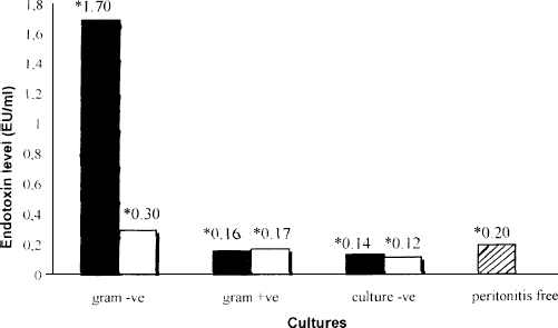
Maximum endotoxin values (EU/ mL) obtained from effluents of peritonitis-free and pre- (black bars) and post- (white bars) treated CAPD-related peritonitis patients. Gram (-ve) = gram-negative; Gram (+ve) = gram-positive; Culture (-ve) = culture-negative.
Effluents of pre- and posttreated gram-negative cultures had endotoxin levels of 1.53 ± 0.169 endotoxin units (EU)/mL and 0.214 ± 0.085 EU/mL, respectively (p < 0.0001); pre- and posttreated gram-positive cultures, 0.102 ± 0.06 and 0.122 ± 0.052 EU/mL, respectively (p > 0.05); pre- and posttreated culture-negative, 0.110 ± 0.025 and 0.087 ± 0.031 EU/mL, respectively (p > 0.05). The endotoxin level in the peritonitis-free group was 0.117 ± 0.079 EU/mL.
Diagnostic Accuracy and Reliability of Lal Assay
A receiver operating characteristic (ROC) plot described the alteration in the diagnostic sensitivity and specificity that occurred when the hypothetical “cutoff” value for endotoxin was changed. In our study, the cutoff limit of 0.38 EU/mL corresponded to a sensitivity of 100% while maintaining a specificity of 98.2%.
Discussion
The present rate of gram-negative peritonitis and our continued current inability to discover the pathogen in a timely manner has, in most cases, continued to endanger the peritoneum, the dialyzing organ in peritoneal dialysis.
Advances in bacteria processing before inoculation (6-8) have not shortened the length of time for a definitive microbiological report, leading to costly empirical management that at times could be dangerous. Given that 40% of our positive routine culture results are available only beyond 72 hours of inoculation, and the fact that culture-negative peritonitis accounts for 25% of our results, it is strongly suggested we find a rapid alternative diagnostic technique for CAPD peritonitis. It is unfortunate that, in the current antibiotic era, we record an unacceptably high (3%) mortality rate, and an equal rate of transfer to hemodialysis, due to gram-negative bacteria.
The early diagnostic usefulness of the LAL assay was demonstrated by Ghandi et al. (15) in the late 1970s. Despite attractive later studies (9-14), we continued to view the LAL assay with a doubting eye.
We utilized the ELISA technique and performed our study with a quantitative chromogenic method that was able to detect endotoxin levels as low as 0.118 EU/mL. All ten samples of effluent with gram-negative growth contained endotoxin concentrations as high as 1.7 EU/mL; this level dropped sharply after treatment to below 0.3 EU/mL. Levels in gram-positive and in all sterile samples were below 0.2 EU mL and never changed significantly after treatment, indicating the high sensitivity and specificity of the LAL assay.
Freezing, thawing, and mechanical shear caused by centrifugation or by the chemical reactants used for analysis may liberate endotoxin from intact bacteria. As our samples were frozen and analyzed in constant and standard environments, it was assumed in our study that any increase in endotoxin during storage and processing would be by a constant factor in all the samples containing gram-negative bacteria, unless they were allowed to stand unacceptably longer. To avoid a possible reporting of exaggerated levels, bacteria filtration before storage is recommended.
Additionally, it should be noted that the presence of fungal β-glucans in the media might interfere with the Limulus test (16,17), especially when qualitative rather than quantitative technique is used. Therefore, high LAL activity in a patient who fails to recover within acceptable limits of treatment should raise concern about the presence of a fungal pathogen.
The ROC plot described a sensitivity of 100% and a specificity of 98.2% that was reached at endotoxin levels greater than or equal to 0.38 EU/mL.
Ultimately, we report excellent usefulness of endotoxin and the LAL assay in the management of gram-negative peritonitis in CAPD patients. The test, which is rapid and easy to perform, can be relied upon when initial microbiological techniques fail to demonstrate a pathogen. However, results of a larger study are needed before any particular cutoff value can be accepted as a guide to management.