
Abstract
Background
Glucose is still used as an osmotic solute in peritoneal dialysis fluids, despite evidence of its local (peritoneal) and systemic toxicities. However a constant search is underway for a new, more biocompatible osmotic solute for peritoneal dialysis fluids.
Objective
The present study evaluated N-acetylglucosamine (NAG) in a concentration of 220 mmol/L as an alternative to glucose for the osmotic solute in peritoneal dialysis fluid, during chronic peritoneal dialysis in rats.
Methods
For 8 weeks, male Wistar rats were infused with glucose-based or NAG-based dialysis fluid. Intraperitoneal inflammation and peritoneal permeability and morphology were evaluated in all rats during the study.
Results
Repeated intraperitoneal infusion of the NAG-based dialysis fluid resulted in a weaker intra-abdominal inflammatory reaction as compared with the reaction in rats infused with glucose-based dialysis solution. At the end of the study, the concentration of hyaluronan in the peritoneal interstitium obtained from NAG-treated rats was higher than that found in the interstitium taken from animals exposed to dialysis fluid containing glucose. Also, peritoneal permeability to total protein was lower in NAG-treated rats.
Conclusion
As an alternative to glucose, NAG used for the osmotic solute in peritoneal dialysis solution decreases the intraperitoneal inflammatory reaction induced by the process of peritoneal dialysis and, indirectly (owing to the increased hyaluronan content in the peritoneal interstitium), diminishes peritoneal permeability to protein.
Keywords
Materials and Methods
The experiment was performed on healthy male Wistar rats with peritoneal catheters implanted according to the method used in our lab (5). To begin, all animals underwent a 4-hour dwell study performed with 30 mL Dianeal 4.25% (Baxter Healthcare SA, Castlebar, Ireland). After the 4-hour dwell, 5 mL of the dialysate was drained. At the same time, under ether anesthesia, a blood sample was drawn from the tail vein in all animals. Concentrations of urea, creatinine, and total protein were measured in dialysate and serum.
The animals were divided into two groups (6 rats in each group), and during the next 8 weeks were dialyzed with either glucose-based or NAG-based dialysis fluid. The electrolyte composition of the fluids was as follows: Na, 132 mmol/L; Ca, 1.75 mmol/L; Mg, 0.75 mmol/L; Cl, 102 mmol/L; and lactate, 35 mmol/L. Glucose or NAG was used as the osmotic solute in a concentration of 220 mmol/L. The osmolality of the fluids was 480 mOsm/kg H2O (glucose) and 481 mOsm/kg H2O (NAG). Fluids were sterilized by filtration and their pH was 7.05. The solutions were supplemented with antibiotics: cefuroxime 50 mg/L and gentamicin 5 mg/L.
Animals were infused with 20 mL of solution twice daily; the instilled fluid was allowed to gradually absorb from the peritoneal cavity. At the end of every week, after a 4-hour dwell, dialysate was drained for evaluation of the dialysate inflammatory parameters. A cell count was done in a Neubauer chamber, and the differential was evaluated in cell smears after Giemsa staining of the cytospun cell pellets. Cytokine levels [tumor necrosis factor alpha (TNFα) and monocyte chemoattractant protein-1 (MCP-1)] in dialysate samples were measured using ELISA kits (Biosource Europe SA, Nivelles, Belgium).
At the end of the 8-week experiment, peritoneal permeability was assessed in all rats during a 4-hour dwell performed with 30 mL Dianeal 3.86% (Baxter Healthcare SA). The dialysate was gravity drained. Glucose concentration was measured in the dialysate and the absorption rate was calculated. The peritoneal permeability coefficient (K: cm x min–1) was calculated for urea, creatinine, and total protein.
At the end of the study, a biopsy of the visceral peritoneum covering the liver was taken for evaluation by light microscopy. Additionally, a piece of the mesentery was removed for measurement of the hyaluronan concentration in the tissue as previously described (4). Hyaluronan content in the digested tissue was measured by ELISA (Chugai Pharmaceuticals Inc., Tokyo, Japan).
Results are presented as mean ± standard deviation. Statistical analysis was performed by ANOVA or Mann–Whitney test, as appropriate. A p value less than 0.05 was considered significant.
Results
Over the course of the experiment, a gradual decline in the dialysate cell count was observed in the NAG-treated rats as compared with the glucose-treated animals (Table 1). At the end of week 8, the percentage of macrophages in the NAG-based dialysate was higher than the percentage in the glucose-based dialysate (59.8% ± 4.0% vs 47.0% ± 3.0% respectively; p < 0.05), but the percentage of neutrophils was lower in the NAG group as compared with the glucose group (16.3% ± 2.0% vs 29.0% ± 3.8% respectively; p < 0.05).
Dialysate Cell Count, Monocyte Chemoattractant Protein-1 (MCP-1), and Tumor Necrosis Factor Alpha (TNFα) Levels in 4-Hour Dialysate Samples Obtained at Weekly Intervals from Rats Infused with Glucose (GLU)–Based or N-Acetylglucosamine (NAG)–Based Dialysis Fluid
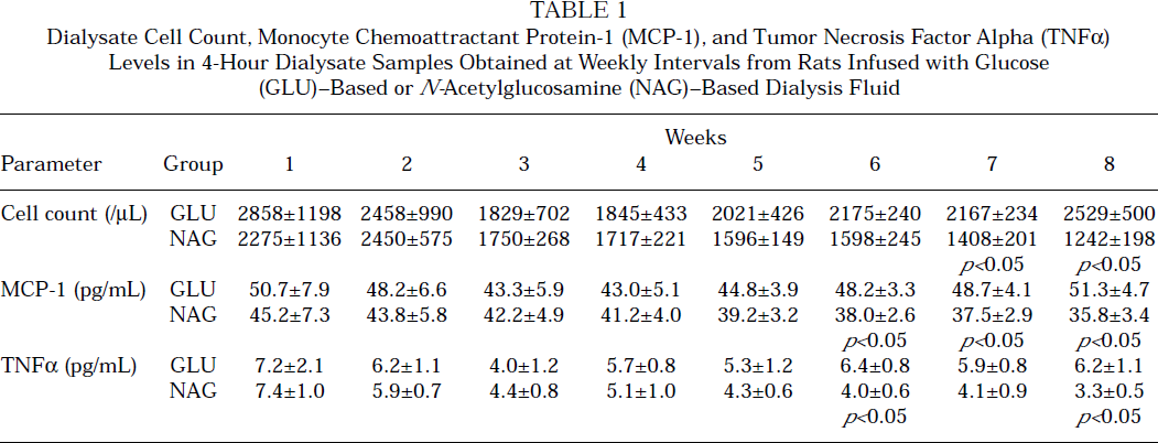
In dialysate samples obtained from NAG-treated rats, we observed a decline in MCP-1 and TNFα concentrations with time. No such effect was seen in animals exposed to glucose-based dialysis fluid (Table 1).
After the 8 weeks of the study, the quantity of hyaluronan in the mesentery was higher in NAG-treated animals than in rats exposed to glucose (0.232 ± 0.035 μg/mg wet tissue vs 0.153 ± 0.015 μg/mg wet tissue, p < 0.05). Under light microscopy, we observed a thickening of the submesothelial interstitium in the visceral peritoneum in all animals, but no significant differences were seen between the groups (NAG: 36.9 ± 23.6 μm; glucose: 55.3 ± 29.3 μm).
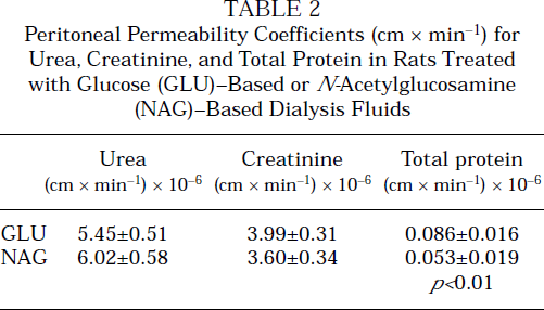
The peritoneal equilibration test (PET) revealed that the volume of drained dialysate tended to be higher in NAG-treated rats than in glucose-treated animals (34.6 ± 4.1 mL vs 31.7 ± 1.7 mL). In both groups of animals, a similar amount of glucose was absorbed during the dwell (86.7% ± 5.1% and 88.3% ± 3.1% respectively). Peritoneal permeability coefficients for urea and creatinine were comparable in both groups of rats, but peritoneal permeability to total protein was lower in NAG-treated animals (Table 2).
Peritoneal Permeability Coefficients (cm x min–1) for Urea, Creatinine, and Total Protein in Rats Treated with Glucose (GLU)–Based or N-Acetylglucosamine (NAG)–Based Dialysis Fluids
Discussion
N-Acetylglucosamine has been proposed as an alternative to glucose as the osmotic agent in peritoneal dialysis fluids (6). Owing to its molecular size (221 Da), N-acetylglucosamine induces a similar transperitoneal water flow during acute, short dwells in rats. During longer dwells, dialysate volume was higher in the presence of NAG.
The results of the present study show that NAG may be used as an alternative for glucose in peritoneal dialysis fluids. Omentectomy and surgical implantation of a peritoneal catheter cause intra-peritoneal irritation as reflected by an increased dialysate cell count and higher cytokine levels. During the subsequent days of experimental infusion with NAG-based dialysis fluid, a gradual decrease in the intraperitoneal inflammatory reaction was seen. No such effect was observed in animals exposed to glucose solution (Table 1).
The anti-inflammatory action of amino-sugars is well documented in various tissues. Shikhman et al (7) showed that NAG prevents interleukin-1β (IL-1β)– mediated activation of human chondrocytes in an in vitro culture. Supplementation of NAG in children with chronic inflammatory bowel disease resulted in an improvement in clinical status and partial remission of the morphologic changes in the intestinal wall (8).
In in vitro experiments, exposure to NAG was observed to stimulate hyaluronan synthesis by peritoneal mesothelial cells and fibroblasts (3). In the present in vivo study, an increased hyaluronan content in the peritoneal mesentery was seen in rats treated with NAG-based dialysis fluid. Hyaluronan given as an additive in dialysis fluid causes a reduction in transperitoneal protein loss during a dialysis exchange (4). We suggest that the increased hyaluronan content in the peritoneal interstitium might explain why, in the present study, peritoneal permeability to total protein was reduced in NAG-treated rats. In that respect, Flessner demonstrated that increasing the hyaluronan concentration in the abdominal wall muscles retards movement of water and protein across the peritoneum (9).
On the other hand, Carlsson et al (10) concluded from experiments on rats treated with hyaluronidase that hyaluronan in physiologic concentrations in the peritoneal membrane does not limit fluid and protein movement across the peritoneum. However, we cannot exclude the possibility that decreased intraperitoneal inflammation in NAG-treated rats also contributed to the lower permeability of their peritonea to protein.
We think that the findings presented in this paper support our previous observations, suggesting potential usefulness for NAG as an alternative to glucose in peritoneal dialysis.