





Abstract
Objective
3-Deoxyglucosone (3-DG) and acetaldehyde were found to be the major reactive carbonyl compounds in conventional heat-sterilized peritoneal dialysis fluids (PDFs). The aim of this study was to identify factors in the production of PDFs promoting or inhibiting the formation of acetaldehyde and 3-DG.
Design
Single-chamber bag PDFs with different buffer systems and pH values were analyzed for acetaldehyde. 3-Deoxyglucosone was determined in double-chamber bag PDFs with different pH values, in commercially available samples, and in double-chamber products stored under defined conditions.
Results
Acetaldehyde was found in the presence of lactate and malate, whereas in 2-hydroxybutanoate-buffered solution propionaldehyde was detected instead. Between pH 5.0 and 6.0 the acetaldehyde content in lactate-buffered solutions increased strongly. The concentration of 3-DG in the chamber containing glucose in double-chamber bags increased between pH 3.0 and 5.0 by a factor of 6. 3-Deoxyglucosone concentrations in commercially available products vary greatly, reflecting the different pH values of these products. A time- and temperature-dependent reaction leads to a reduction in 3-DG and an increase in 5-hydroxymethyl-furan-2-carbaldehyde during storage.
Conclusion
Acetaldehyde is produced by a reaction that requires both lactate and glucose. Thus, its formation can be prevented by a separation of the reaction partners, glucose and lactate, in a double-chamber bag. In double-chamber bags, pH greatly influences the formation of 3-DG. Minimal formation is observed in the region of pH 3.0. This finding should be taken into account for the development of new double-chamber bag PDFs.
Keywords
Commercially available single-chamber bag peritoneal dialysis fluids (PDFs) contain electrolytes and glucose as osmotic agents. They are buffered with sodium lactate in a weakly acidic pH range. This composition is far from ideal and implies several risks (1). These PD solutions have proved cytotoxic in in vitro studies (2-7), and the irritant effect of inflowing dialysis solution has been attributed to glucose degradation products (GDPs) formed during heat sterilization and storage (8,9). Several reactive carbonyl compounds (RCC) have been identified in PD solutions, in particular, 5-hydroxymethyl-furan-2-carbaldehyde (HMF), formaldehyde, acetaldehyde, glyoxal, and methylglyoxal (10). The quantities found in these investigations were below 1 ppm for formaldehyde and glyoxal, approximately 1 ppm for methylglyoxal, and more than 10 ppm for acetaldehyde. For some of these products a cytotoxic potential could be confirmed (11,12). Recently, 3-deoxyglucosone (3-DG) was detected in single- and double-chamber PDFs in concentrations higher than those of any other RCC so far determined (13,14). Moreover, 3-DG is of particular importance because of its high reactivity and its potential to induce the formation of advanced glycation end-products (AGEs) in vivo (15-17).
The chemical reactions leading to GDPs are summarized in Figure 1: HMF and methylglyoxal are formed via the 3-deoxy degradation pathway; enolization and dehydration of glucose yields 3-DG, a key intermediate in glucose degradation. Cyclization and further dehydration lead to HMF, whereas methylglyoxal is formed via a retroaldol reaction (18,19). In contrast to these elucidated mechanisms, formation of acetaldehyde from glucose is not so easy to understand. One of the objectives of this study was, therefore, to reveal how acetaldehyde is formed in PDFs and which parameters influence its formation.

Formation of certain aldehydes by degradation of glucose via enolization (a), dehydration (b), cyclization (c), and C–C-cleavage (d).
In the development of a double-chamber PD system, the pH value of the compartment containing glucose can be selected from a range between 2 and 6. Therefore, we investigated how pH value influences the formation of 3-DG in double-chamber bag PDFs and in which concentration range 3-DG is present in different commercially available new double-chamber bag PDFs. Finally, we analyzed 3-DG and HMF in samples that were stored under defined conditions.
Methods
Solutions
Single-Chamber Bags
All chemicals used were at least p.a. quality; water was demineralized and bidistilled. Solutions were based on a standard PD formulation containing 99 mmol/L sodium chloride, 35 mmol/L sodium lactate, 1.75 mmol/L calcium chloride, 0.5 mmol/L magnesium chloride, and 15 g/L glucose. Prior to heat sterilization, the pH was adjusted to 5.8 using hydrochloric acid. Solutions prepared according to this specification but lacking glucose or lactate served as controls.
Variation of the Buffer System
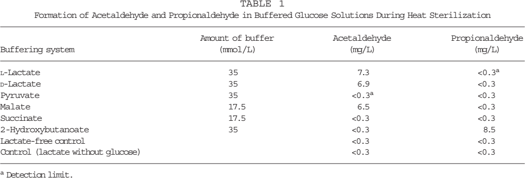
Peritoneal dialysis fluids of the analogous composition were produced where sodium lactate was replaced by sodium salts of several organic acids (Table 1). Malic acid and succinic acid were used, at a concentration of 17.5 mmolL, because they represent two base equivalents compared to lactate. Aliquots of these solutions (5 mL) were filled into glass vials and placed into an oil bath for 30 minutes at 121°C for sterilizing.
Formation of Acetaldehyde and Propionaldehyde in Buffered Glucose Solutions During Heat Sterilization
Detection limit.
Variation of pH Value
The pH was adjusted to values of 5.0 – 6.0 in steps of 0.1. The solutions were filled into flexible non-PVC plastic containers and sealed. Steam sterilization was performed to an Fo value of approximately 15.
All samples were produced in duplicate. After sterilization, the bags/vials were opened and each was examined twice for acetaldehyde and propionaldehyde by gas chromatography. The results are given as mean of the four measurements. The standard deviation was below 3% in all experiments and is not shown.
Double-Chamber Bags with Different pH Values in the Glucose Compartment
The glucose compartment (1 L) contained 193 mmol/L sodium chloride, 3.50 mmol/L calcium chloride, 1.0 mmol/L magnesium chloride, and 45 g/L glucose. The pH was adjusted to values between 2.5 and 5.0 in steps of 0.5 using hydrochloric acid. The lactate compartment (1 L) contained 70 mmol/L sodium lactate and 5 mmol/L sodium bicarbonate. The solutions were filled into flexible non-PVC plastic containers and sealed. Steam sterilization was performed to an Fo value of approximately 15. Afterwards, the peel seam between the compartments was ruptured and the mixed solutions were analyzed for 3-DG.
Commercially Available Double-Chamber Bags
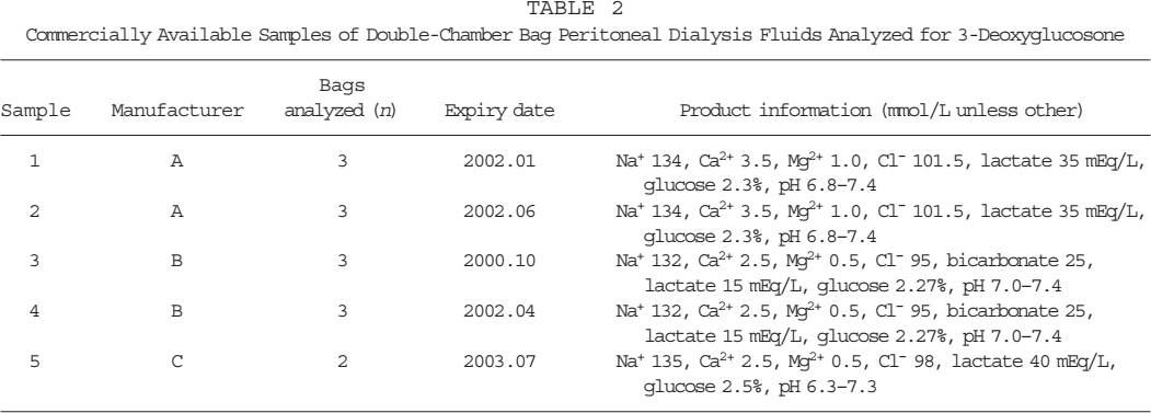
Peritoneal dialysate bags were bought from a pharmacy or supplied directly from the manufacturer. Table 2 gives a survey of the samples. 3-Deoxyglucosone was determined after mixing. All samples were analyzed in October to December 2000 before the expiry date of the respective solutions.
Commercially Available Samples of Double-Chamber Bag Peritoneal Dialysis Fluids Analyzed for 3-Deoxyglucosone
Influence of Storage Conditions on 3-DG and HMF Concentrations
Commercial double-chamber bags containing bicarbonate (Fresenius Medical Care, Bad Homburg, Germany) with 134 mmol/L sodium, 1.75 mmol/L calcium, 0.5 mmol/L magnesium, 104.5 mmol/L chloride, 34 mmol/L bicarbonate, and 1.5% or 4.25% glucose (the glucose compartment has pH 2.8 and contains 3.0% and 8.5% glucose respectively) were stored under different climatic conditions [25°C and 60% relative humidity (RH); 30°C and 35% RH; 40°C and 75% RH] for 6 months. 3-Deoxyglucosone and HMF were determined at 0 and 6 months in 3 samples of each solution.
Gas Chromatography Analysis
The gas chromatography system for quantitative determination consisted of a Hewlett Packard (HP, Waldbronn, Germany) gas chromatograph 5890 with a split injector and a flame ionization detector and an HP headspace sampler, model 19395A. The gas chromatography system for the identification of propionaldehyde consisted of an HP gas chromatograph, model 5890 II, with a split injector and a mass selective detector (HP MSD), model 5971A. The conditions were as follows: injector temperature 250°C; oven temperature 50°C; detector temperature 300°C; carrier gas, helium; pressure 1.0 bar; flow 7.0 mL/minute; split 18 mL/min; column HP-FFAP (modified polyethylene glycol), internal diameter 0.32 mm, length 50 m, film thickness 0.52 μ; headspace conditions: equilibration for 30 minutes, bath temperature 60°C, transfer line 90°C.
High Performance Liquid Chromatography (Hplc) Analysis of 3-Dg
The HPLC analyses were carried out with a diode-array detector system (Pump PU-1580, Degaser 980-50, ternary gradient unit LG-980-02S, diode array detector MD-1510 from JASCO, Gross-Umstadt, Germany; quantification at 237 nm/316 nm) using an RP-18 column (LC-18-DB 25 cm × 4.6 mm, Supelco, Deisenhofen, Germany). For elution, a binary gradient was used with 0% – 70% solvent A from 0 to 18 minutes and 70% solvent B from 18.1 to 25 minutes (solvent A, ammonium formate buffer 5 mmol/L, pH 7.4; solvent B, methanol); 8 mL PDF was mixed with 1 mL o-phenylenediamine solution (0.02 mol/L in methanol) and incubated at room temperature overnight. Retention time of the 3-DG-quinoxaline derivative with the HPLC method described above was 17.5 minutes. All samples were assessed in duplicate. Derivatization of synthesized 3-DG (20) yielded the standard curve for calculation of concentrations.
Determination of HMF
HMF was derivatized with o-toluidine in barbituric acid/acetic acid solution as described in Ref. (21). The derivatization and the following photometric measurement at 550 nm were performed with an ELAN analyzer (Eppendorf, Hamburg, Germany).
Results
Analysis of Acetaldehyde in Pdfs Dependent on the Buffer Solution
Table 1 shows the amounts of acetaldehyde and propionaldehyde in differently buffered solutions. Acetaldehyde was only detected in glucose solutions containing lactate and malate, and not in solutions containing pyruvate, succinate, and 2-hydroxybutanoate. In the latter, propionaldehyde was found instead. Identity of propionaldehyde was confirmed by gas chromatography/mass spectrometry. Neither of the aldehydes was detected in the solutions containing only glucose or lactate.
Formation of Acetaldehyde in Single-Chamber Pdfs Dependent on pH Value
Figure 2 illustrates the influence of different pH values on the formation of acetaldehyde in lactate-buffered glucose solution. The formation of acetaldehyde is pH dependent: by increasing the pH from 5.0 to 6.0, the amount of acetaldehyde quadruples from 1.5 to 5.8 ppm.

The formation of acetaldehyde in lactate-buffered glucose solutions is pH dependent. Data are mean values of four measurements; standard deviation is below 3%.

The formation of 3-deoxyglucosone in acidic lactate-free glucose solution is pH dependent. Data are mean values of four measurements; standard deviation is below 3%.
Formation of 3-Dg in Double-Chamber Bag Pdfs Dependent on pH Value
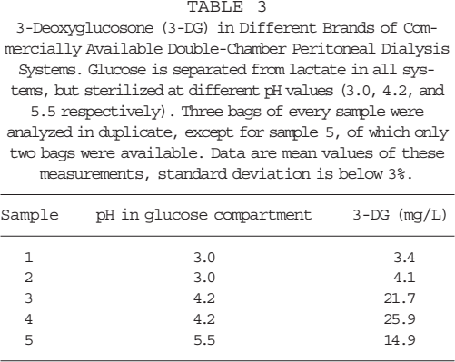
3-Deoxyglucosone concentration in solutions sterilized at pH 2.5, 3.0, and 3.5 is comparatively low (3.1 ppm); whereas it rapidly increases when the pH increases to 4.0 (8.9 ppm), 4.5 (14.7 ppm), and 5.0 (20.9 ppm) (Figure 3). The 3-DG concentrations measured in different samples of commercially available double-chamber PDFs are shown in Table 3. The concentration of 3-DG ranged from 3.4 to 25.9 ppm, which represents a difference between the lowest and the highest concentration as high as 660%.
3-Deoxyglucosone (3-DG) in Different Brands of Commercially Available Double-Chamber Peritoneal Dialysis Systems. Glucose is separated from lactate in all systems, but sterilized at different pH values (3.0, 4.2, and 5.5 respectively). Three bags of every sample were analyzed in duplicate, except for sample 5, of which only two bags were available. Data are mean values of these measurements, standard deviation is below 3%.
Changes in 3-DG and HMF Concentrations during Storage of Double-Chamber Bag Pdfs
Double-chamber bag PDFs were stored after heat sterilization under defined conditions. After 6 months, changes in the concentrations of 3-DG and HMF were measured. The amount of 3-DG decreased after sterilization (between 21% and 68%, Figure 4), whereas between 50% and 187% more HMF was found (Figure 5). The decrease in 3-DG and the increase in HMF concentrations are stronger at higher storage temperature. The sum of 3-DG and HMF on a molar basis remains unchanged.

3-Deoxyglucosone decreases during storage under defined conditions: 4.25% glucose stored at 25°C (open squares); 4.25% glucose at 30°C (open triangles); 4.25% glucose at 40°C (open circles); 1.5% glucose at 25°C (closed squares); 1.5% glucose at 30°C (closed triangles); 1.5% glucose at 40°C (closed circles). Data are mean values of six measurements; standard deviation is below 3%.

5-Hydroxymethyl-furan-2-carbaldehyde (HMF) increases during storage under defined conditions: 4.25% glucose stored at 25°C (open squares); 4.25% glucose at 30°C (open triangles); 4.25% glucose at 40°C (open circles); 1.5% glucose at 25°C (closed squares); 1.5% glucose at 30°C (closed triangles); 1.5% glucose at 40°C (closed circles). Data are mean values of six measurements; standard deviation is below 3%.
Discussion
Acetaldehyde has been referred to as a GDP (10,12). According to the results obtained in this study, the formation of acetaldehyde is not dependent only on the presence of glucose, but also on the presence of lactate. In control experiments, acetaldehyde (detection limit 0.3 mg/L) could not be detected in lactate-free glucose solution or in glucose-free lactate solution. Furthermore, acetaldehyde was not found in solutions in which lactate was replaced by pyruvate or succinate. These findings strongly indicate that the precursor of acetaldehyde is lactate. Lactate could be oxidatively decarboxylated in a reaction that is mediated by glucose. This hypothesis was confirmed by the fact that propionaldehyde, which was identified by gas chromatography/mass spectrometry analysis, was detected instead of acetaldehyde when lactate was replaced by 2-hydroxybutanoate. Oxidative decarboxylation of 2-hydroxybutanoate leads to the formation of propionaldehyde. According to the same mechanism, malate is oxidized to malonsemialdehyde. The latter compound is not stable and decarboxylates to acetaldehyde. As a result, acetaldehyde was detected in samples that were buffered with malate. All reactions are summarized in Figure 6. Acetaldehyde has previously been identified as a degradation product of glucose (22), but the reaction was carried out in concentrated sulfuric acid at a strongly acidic pH, which is in contrast to the almost neutral diluted PD solution. Therefore, this report is not in contradiction to our findings. Thus, we conclude that acetaldehyde, which is detected as a major RCC in single-chamber bag PDFs, is formed from lactate, during heat sterilization, by a mechanism that is mediated by glucose. Consequently, separation of glucose and lactate during heat sterilization by the use of double-chamber bags should inhibit the formation of acetaldehyde. Indeed, in all samples of double-chamber bag PDFs that were investigated, acetaldehyde concentration was below detection limit.

Formation of aldehydes from α-hydroxycarbonic acids by oxidative decarboxylation (a) and decarboxylation (b).
Commercially available PD solutions are usually buffered at a pH between 5.0 and 5.8 because the formation of GDPs is highly favored at neutral pH, but is reduced in weakly acidic solution. In the pH range between 5 and 6, the formation of acetaldehyde is strongly pH dependent (Figure 2), whereas it is rather low at pH 5; concentrations of acetaldehyde are four times as high at pH 6.0. For that reason, the pH of single-chamber bag PD solutions is adjusted to weak acidic values, although a neutral pH is more physiological.
In double-chamber bags, glucose is dissolved in a rather acidic milieu, whereas the electrolytes are buffered at higher pH. Therefore, in double-chamber systems a neutral physiological pH can be achieved after mixing the compartments. In the double-chamber system, the pH milieu of the glucose compartment can be selected in the range between pH 2 and 6. Technological aspects have to be taken into consideration, for example, if sodium bicarbonate is used to neutralize the glucose compartment, pH can be adjusted to comparatively low values (pH 2.0 – 3.0). It has been shown that the low pH value in the glucose compartment considerably lowers glucose degradation. Until now, the only GDPs that could be found in double-chamber bags were 3-DG and HMF. The concentration of 3-DG, however, was reduced by as much as 80% in certain types of double-chamber bags compared to a single-chamber bag PDF (14). In this study, we measured the formation of 3-DG in double-chamber bag PDFs in relation to the pH value in the glucose compartment. The analysis showed a considerable increase in 3-DG concentration between pH 3.5 and 5.0, whereas changes between pH 2.5 and 3.5 were minimal (Figure 3). These results suggest that sterilizing the glucose in the latter pH range is favorable to reducing the formation of 3-DG.
The differences found in these laboratory-made solutions are reflected by differences in commercially available double-chamber bag PDFs, where the glucose solutions were sterilized at pH values between 3.0 and 5.5. Solutions where glucose is sterilized at pH 4.2 show a sixfold increase in 3-DG concentration compared to fluids with pH 3.0 in the glucose compartment (samples 1 – 4 in Table 3). Therefore, we conclude that our results are relevant for industrially produced double-chamber bags.
It seems, however, that pH is not the only determinant for 3-DG formation because sample 5, with a pH of 5.5, contained less 3-DG than samples 3 and 4. Moreover, the latter two showed higher 3-DG levels than one would expect from the data in Figure 3. The parameters of sterilization might equally contribute to glucose degradation.
As we found less 3-DG in old lots compared to new lots of PDFs of the same composition, we included samples in our study that were stored under defined conditions for 6 months. In 4.25% glucose solutions, 3-DG decreased at temperatures of 25°C and 40°C, by 21% and 61% respectively after a storage period of 6 months. As 3-DG is known as a precursor of HMF (19), we analyzed HMF as well. During storage, HMF increased by 52% and 178% respectively (Figures 4 and 5). The sum of both GDPs on a molar basis is nearly constant. We therefore conclude that, under storage conditions, only minimal amounts of 3-DG are formed de novo, but considerable amounts of 3-DG are transformed to HMF. This transformation is enhanced by higher storage temperatures.
Until now, there have been no biological function data or clinical outcome studies supporting the hypothesis that double-chamber solutions with a low 3-DG level are superior to those with higher concentrations of this GDP. On the other hand, 3-DG reacts rapidly with proteins and is a well-known precursor of AGEs. So the principle of precaution might recommend a reduction of 3-DG to the lowest technically possible level. Still, the possible clinical implications of higher 3-DG and HMF levels should be investigated in in vivo studies.