






Abstract
Objectives
Glucose and other bioincompatible factors of conventional peritoneal dialysis solutions may damage the peritoneal membrane. The aim of our study was to investigate whether replacement of glucose with icodextrin (ID) or amino acids (AA) affects inflammatory parameters or cancer antigen 125 (CA125).
Design
Either ID or AA was used, in random order, in one daily exchange during an 8-week period. After the first study period, the patients entered a washout period and then switched to the other study solution for an 8-week period. C-reactive protein (CRP) was measured in serum, and CA125, tumor necrosis factor alpha (TNF-α), interleukin-6 (IL-6), soluble intercellular adhesion molecule-1 (sICAM-1), and hyaluronan (HA) were measured in the overnight dwell dialysates at the beginning and end of the study periods.
Setting
A university hospital.
Patients
22 patients with duration on peritoneal dialysis of 1.5 – 6.3 months.
Main Outcome Measures
Levels of serum CRP and dialysate CA125, IL-6, HA, and sICAM-1 during use of ID and AA were compared to levels during use of glucose-only-based solutions.
Results
CRP increased significantly during use of ID. CA125 increased significantly during 8 weeks’ use of AA, from 22.8 (5.4 – 89.0) to 42.9 (7.1 – 92.9) kU/L (p = 0.007). IL-6 increased during 8 weeks’ use of AA, from 22.0 (9.0 – 108.0) to 36.5 (14.0 – 93.0) ng/L (p = 0.002) and ID, from 25.5 (8.0 – 82.0) to 40.0 (12.0 – 118.0) ng/L (p = 0.008). TNF-α also increased significantly during use of ID, but showed no significant changes during use of AA.
Conclusions
The use of glucose-free solutions, especially AA, may lead to preservation of mesothelial cell mass and host defense. However, activation of systemic and peritoneal inflammation may appear during the use of ID and to a lesser extent during use of AA.
Peritoneal dialysis (PD) is an established treatment modality for end-stage renal disorders. However, its long-term use is often limited by diminishing dialysis and ultrafiltration capacity of the peritoneal membrane. Recurrent episodes of peritonitis, as well as glucose, its degradation products (GDP), and other bioincompatible components of the dialysis solutions, may damage the peritoneal membrane. Two alternative osmotic agents have been developed to avoid the harmful effects of glucose. Amino acid (AA)-based solution (Nutrineal; Baxter Healthcare, Deerfield, Illinois, USA) has a more physiological pH than conventional glucose-based solutions and it does not contain glucose; thus, formation of GDP and advanced glycation end products (AGE) can be avoided. It has also been associated with positive effects on lipid profile and modest nutritional benefits (1-5). Icodextrin (ID; Extraneal, Baxter) is a glucose polymer that allows improved and sustained ultrafiltration (6). It is iso-osmolar and acts as a colloid osmotic agent, but has a low pH. It has been shown that use of ID causes less production of GDP, Amadori albumin, and AGE (7-9). Icodextrin has also been associated with positive effects on glucose and lipid metabolism (10,11).
Both mesothelial cells and fibroblasts are capable of producing various cytokines and growth factors, which are important to host defense but may also reflect inflammatory processes in the peritoneum. Interleukin-6 (IL-6) and tumor necrosis factor alpha (TNF-α) are mediators of inflammation (12,13). Together with other inflammatory transmitters, IL-6 stimulates the production of adhesion molecules such as intercellular adhesion molecule-1 (ICAM-1) (14); ICAM-1, in turn, stimulates hyaluronan (HA), which forms a critical component of extracellular matrices (15). Furthermore, cancer antigen 125 (CA125) is an ovarian tumor marker that serves as an indicator of mesothelial cell mass (16).
The aim of the present study was to evaluate the influence of ID and AA on systemic and peritoneal inflammation and on preservation of the peritoneum compared with glucose-based solutions.
Materials and Methods
Subjects
Twenty-two patients with a PD duration of 1.5 – 6.3 months participated in the crossover study, which was approved by the Committee of Research Ethics at the Helsinki University Hospital and conducted between January 2001 and January 2003. All patients were on continuous ambulatory PD. Patients with type I diabetes, malignancy, or systemic inflammation were excluded. Eight patients used cholesterol-lowering medication and the doses remained constant throughout the study. Also, the doses of other drugs, except phosphate binders, were kept similar during the study. Clinical characteristics of the patients are shown in Table 1. The study was interrupted in 4 patients during the follow-up: 2 patients received a cadaveric kidney transplant, 1 patient developed a prolonged infection at the beginning of the ID study period, and 1 patient was excluded because of symptoms of acute coronary syndrome during the washout period. Eighteen patients completed both study periods and 20 patients completed either study period (ID or AA).
Clinical Characteristics of the Patients
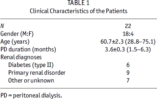
PD = peritoneal dialysis.
Methods
The patients used one daily exchange of ID (overnight dwell) or AA (one daytime dwell) dialysis solution, in a random order, during an 8-week study period (Figure 1). After completing the first study period, the patients entered a washout period of 8 weeks’ duration (using glucose-only-based dialysis solutions) and were then switched to the other study solution (Figure 1). Twelve patients started with ID and 10 patients started with AA. Conventional glucose-containing lactate-based solutions were used for the other three exchanges. The glucose concentrations of the solutions varied according to individual need for dialysis and ultrafiltration.
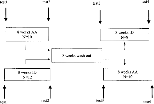
Study design (AA = amino acid solution; ID = icodextrin).
Blood samples for measurements of serum CRP were drawn and samples from overnight dwell dialysate were obtained at the beginning and end of the study periods. Concentrations of CA125, IL-6, soluble ICAM-1 (sICAM-1), TNF-α, and HA were measured from the dialysate. All samples were stored at –20°C until analyzed. A Personal Dialysis Capacity test (PDC; Gambro, Lund, Sweden) was performed before and after the study (17).
Assays
The assays that were used showed no detectable cross-reactivity with any other cytokines or adhesion molecules. Dialysate concentrations of sICAM-1 were analyzed by high sensitivity ELISA [overall intra-assay coefficient of variation (CV) of 5.6% and overall interassay CV of 7.8%] (Bender Med Systems, Vienna, Austria); IL-6 (intra-assay CV 4.0% – 6.1%, interassay CV 9.3% – 14.1%) and TNF-α (intra-assay CV 5.1% – 8.2%, interassay CV 5.7% – 9.4%) were analyzed by radioimmunoassay (12,13). CA125 was quantitated with an immunoenzymatic assay (Immuno1; Bayer, Tarrytown, New York, USA), with a detection limit of 0.9 kU/L; in the concentration range 15 – 500 kU/L, interassay CV is <4% and intra-assay CV <3%. Hyaluronan was determined by ELISA (Corgenix, Denver, Colorado, USA), with intra- and interassay CVs of 4.2% – 4.7% and 5.7% – 6.2% respectively. Sensitive CRP in serum was measured by radioimmunoassay (RIA), with a detection range from 0.01 to 10 mg/L and intra- and interassay CVs of 4.0% and 6.5% respectively (mean of normal ± SD: 1.1 ± 0.3 mg/L) (18). The levels frequently exceeded 10 mg/L, demanding repeated dilutions of the samples, which affects the precision of the analyses. Therefore, if the levels of sensitive CRP exceeded 10 mg/L, the samples were remeasured by Hitachi 911 analyzer using reagents from Roche, and in these cases this value is reported.
Statistical Analyses
Values are given as mean ± SEM or median with range in text and median with quartiles and range in figures (CRP and soluble factors in dialysate). Wilcoxon signed rank test and Spearman rank correlation test (StatsDirect statistical software, Cheshire, UK) were used for statistical analyses. A p value of <0.05 was considered statistically significant. The level of significance was adjusted according to the number of comparisons (Bonferroni).
Results
Both ID and AA were well tolerated. Neither episodes of sterile peritonitis nor skin rashes were seen during ID. Some patients complained of inflow pain when ID was started. The total amount of glucose used in the dialysis solutions (glucose load) decreased during both ID (before ID 154.6 ± 4.8 g/day vs during use of ID 107.4 ± 4.7 g/day, p < 0.0001) and AA (before AA 157.3 ± 6.6 g/day vs during use of AA 125.5 ± 6.7 g/day, p < 0.0001). The glucose load during the use of ID was less than during the use of AA (p = 0.03). Mean ultrafiltration rates increased during use of ID (from 1250 ± 167 to 1625 ± 195 mL/24 hours, p = 0.01) but did not change during use of AA (1375 ± 152 vs 1350 ± 134 mL/24 hours).
Body mass index (BMI) did not change significantly during use of either study solution (25.1 ± 0.5 kg/m2 before and after 8 weeks’ use of ID; 25.3 ± 0.4 kg/m2 before AA and 25.5 ± 0.4 kg/m2 after 8 weeks’ use of AA; p = NS). CRP values are shown in Figure 2(a). The medians of sensitive CRP values (18) were above the normal range in all measurements. CRP increased during the use of ID [from 2.3 (0.7 – 9.0) to 5.0 (0.8 – 16.0) mg/L,p = 0.01], whereas no significant changes during the use of AA [4.3 (0.7 – 13.0) vs 4.2 (0.8 – 27.0) mg/L,p = NS] were seen. The CRP values correlated with BMI in all measurements (r = 0.5 – 0.6, p = 0.009 – 0.05).
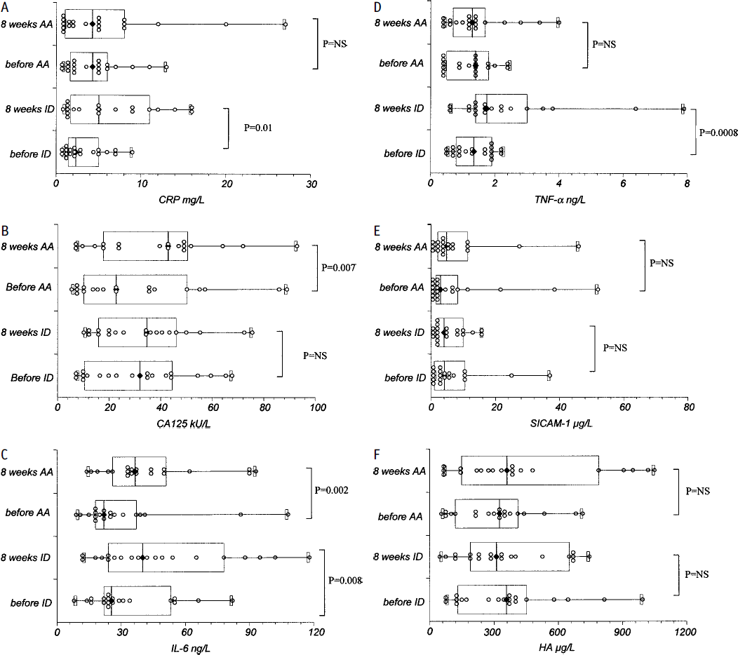
C-reactive protein (CRP) in serum (A) and cancer antigen 125 (CA125) (B), interleukin (IL)-6 (C), tumor necrosis factor alpha (TNF-α) (D), soluble intercellular adhesion molecule-1 (SICAM-1) (E), and hyaluronan (HA) (F) in dialysate, before and after 8 weeks’ use of icodextrin (ID) and amino acid (AA) solutions. Values are expressed as minimum–(lower quartile–median–upper quartile)–maximum, and as patients’ individual values.
Measurements of the soluble factors in dialysate before and after 8 weeks’ use of ID and AA are shown in Figures 2(b) – 2(f). CA125 increased significantly during the use of AA [from 22.8 (5.4 – 89.0) to 42.9 (7.1 – 92.9) kU/L,p = 0.007]; IL-6 increased significantly during the use of both ID [from 25.5 (8.0 – 82.0) to 40.0 (12.0 – 118.0) ng/L,p = 0.008] and AA [from 22.0 (9.0 – 108.0) to 36.5 (14.0 – 93.0) ng/L,p = 0.002]; and TNF-α increased during the use of ID [from 1.4 (0.5 – 2.3) to 1.8 (0.6 – 7.9) ng/L,p = 0.0008], but no significant changes during the use of AA were seen.
When adjusting the level of significance according to the number of comparisons, the changes in the soluble factors remained significant: CA125 before versus during the use of AA, p = 0.035; IL-6 before versus during the use of ID, p = 0.039; IL-6 before versus during the use of AA, p = 0.01; and TNF-α before versus during use of ID, p = 0.004. Soluble ICAM-1 and HA did not change significantly during either study solution. The levels of the soluble factors measured during use of glucose-only-based solutions (i.e., before ID and before AA) did not show any significant differences compared with each other (Figure 2).
The patients underwent clinical examination in connection with the test measurements. There were no exit-site infections or other episodes of infection or inflammation during the study. However, 4 patients had an episode of bacterial peritonitis during the study: two of them appeared shortly after beginning the use of ID and the other two shortly after beginning the use of AA. This means that at least 2 weeks after completion of the treatment of peritonitis had passed before the next measurements were performed. Excluding the patients with peritonitis during follow-up did not affect the changes in levels of CRP or the soluble factors in dialysate (separate data not shown).
Characteristics of the peritoneal membrane did not change during the study according to the PDC test measurements [area: 16 397 ± 1043 (78%) vs 17481 ± 1366 (83%) cm/1.73 m2,p = NS; absorption 2.05 ± 0.2 (171%) vs 2.04 ± 0.2 (170%) mL/min/1.73 m2,p = NS; plasma loss 0.1 ± 0.001 (121%) vs 0.1 ± 0.001 (121%) mL/min/1.73 m2,p = NS]. Also, PD-delivered creatinine clearance remained stable (4.5 ± 0.2 vs 4.3 ± 0.2 mL/min/1.73 m2, p = NS). Residual renal function declined slightly (4.1 ± 0.6 vs 2.9 ± 0.7 mL/min/1.73 m2, p = 0.03).
Discussion
It has previously been shown that glucose-free dialysis solutions have positive effects on glucose and lipid metabolism in PD patients (1-5,10,11). In vitro studies suggest enhanced biocompatibility of ID and AA, but in vivo evidence is still incomplete. In the present study, we showed an increase in CA125 during the use of each study solution (not significant during use of ID), indicating a positive effect on mesothelial cells. However, both CRP in serum and inflammatory markers in dialysate increased during the use of ID solution, and less extensively during the use of AA solution.
C-reactive protein is a marker of inflammation and is considered a negative prognostic factor, predicting enhanced risk of cardiovascular events in both the common population (19) and PD patients (20,21). In the present study, half the patients had elevated CRP values in all four measurements. The levels did not change significantly during the use of AA solution, but did increase during the use of ID. Although the study was partly conducted during the period when some ID batches contained a higher level of peptidoglycans, no signs or symptoms of sterile peritonitis were observed during the study. The elevation of CRP during the use of ID solution was unexpected because ID has been regarded as more biocompatible than glucose-based solutions. We are not aware of any reports of CRP values during use of ID except during episodes of sterile peritonitis. Since CRP increased in most patients using ID and, further, no clinical signs of sterile peritonitis were observed, it is unlikely that the increase would be related to sterile peritonitis. The study was planned before the problems with ID and episodes of sterile peritonitis came out. At that point, no one could really imagine the importance of repeated CRP measurements during the use of ID solutions. When the study was initiated, the malnutrition–inflammation–atherosclerosis syndrome (22) was also not as well known as it is today. Even though it has been shown that a single time point CRP has significant and predictive prognostic value in PD patients (20), the importance of following CRP values continuously in uremic patients came up after the present study was planned and performed. Thus, more information in this area is urgently needed. We showed a correlation between CRP levels and BMI, which could indicate a connection between inflammation and obesity. There are also other recent reports that assume features of low-grade inflammation may be associated with obesity (23,24).
Cancer antigen 125 is considered a marker of mesothelial cell mass (16). We found a significant increase in CA125 in dialysate during the use of AA solution, but the slight increase noticed during the use of ID solution was not significant. CA125 concentrations also increased in all 4 patients with peritonitis during the follow-up. All episodes of peritonitis were clinically mild. They were problem free and cured with antibiotics and did not seem to damage the peritoneal membrane. In a previous study, we showed that CA125 increased in patients who used ID solution in one daily exchange, but decreased slightly in patients who used glucose-only-containing solutions during 1 year of follow-up (unpublished observations). Furthermore, Cappelli et al. showed an increase in CA125 concentration and mesothelial cell mass during the use of a GDP-free solution by using the three-compartment bag system (25). Production of GDP can also be avoided or reduced by using non-glucose-containing solutions. The increase in CA125 during the use AA solution, and to some extent also during ID, in the present study may indicate preservation of mesothelial cell mass during the use of AA and ID solutions.
We showed an increase in dialysate IL-6 concentrations during the use of both ID and AA solutions and an increase in TNF-α concentrations during the use of ID solution. Neither the levels nor the changes in the soluble factors differed in patients with and without peritonitis during the follow-up. A few questions arise on the basis of these findings: Are the measured factors markers of enhanced capacity of host defense, or markers of irritation and inflammation in the peritoneum? Because the normal levels of the measured factors in dialysate are poorly defined, interpretation becomes even more complex.
There are several reports of in vitro studies assessing the biocompatibility of ID. Ha et al. showed less production of vascular endothelial growth factor and procollagen III N-terminal peptide secretion by mesothelial cells during ID, pointing out the enhanced biocompatibility of ID (26). Formation of Amadori albumin, GDP, and AGE was reduced during ID in several studies (7-9). Bajo et al. showed greater ex vivo proliferation of mesothelial cells taken from ID effluent than from glucose effluent (27). However, some studies have shown equal cell culture cytotoxicity with ID as with conventional glucose-based solutions (28-30). On the other hand, Parikova et al. found more signs of subclinical inflammation during the use of ID than during the use of glucose-based solutions (31). Furthermore, Gotloib et al. found mesothelial dysplastic changes and lipid peroxidation induced by ID (32).
The biocompatibility of AA-containing dialysis solution has been studied by several investigators. Brulez et al. found better preservation of macrophage function during the use of AA solution than during the use of glucose-based solution (33). Chan et al. compared the effects on mesothelial cells of dialysate obtained from 4-hour dwells with AA-based and glucose-based dialysis solutions (34). Ultrastructure and viability of mesothelial cells were better preserved and cell proliferation less reduced during AA (34), but IL-6 secretion by cultured mesothelial cells increased during AA (34). In an experimental in vivo study, mesothelial damage and vascular changes could be avoided in rabbits when AAs were used instead of glucose as osmotic agent in dialysis solutions (35). The enhanced biocompatibility of AA-based dialysis solutions is likely based on both the reduction of glucose load, leading to less formation of GDP and AGE, and the more physiological pH of AA.
We assume that the increase in IL-6 and TNF-α during ID and AA shown in the present study is a sign of less cellular inhibition and better preservation of cytokine release than during glucose-free solutions. This is supported by findings of better mesothelial preservation, especially during AA. Reducing the glucose load probably plays the major role in these favorable changes. Nevertheless, it cannot totally be denied that elevated inflammatory mediators in dialysate induced by the alternative osmotic agents may be indicators of silent inflammation and increase the risk of vascular and fibrotic changes of the peritoneal membrane in the long term. Larger long-term studies are needed to confirm the results.
Parikova et al. found elevated HA concentrations in dialysate during the use of ID solution (31). Unlike that study, we could not show any changes in HA concentrations during the use of either solution. Also, sICAM concentrations remained stable during the use of both glucose-free solutions. ICAM-1 mediates interaction of cells with extracellular matrix and other cells, and binds and stimulates fibrinogen and HA (36). It binds to HA in a dose-dependent way (14). Hyaluronan forms a critical component of extracellular matrices (15). It is present in tissues undergoing remodeling and has an important role in wound repair, adhesion and locomotion of cells, and water homeostasis (15). Changes in ICAM-1 and HA levels may take more time, and the negative findings in the current study may be due to the relatively short duration of follow-up. Again, larger studies with longer follow-up would be needed to further confirm the results.
Only relatively new patients with PD duration of 6 months or less were included in the study. Half the patients started with ID and the other half with AA in order to minimize the effect of time during the study. The crossover design of the study enabled the patients to serve as their own controls. There were no significant differences in measurements during the use of glucose-only-based solutions at the start of the first and the second study periods. Patients showed no signs or symptoms indicating inadequate dialysis or ultrafiltration during the study and PD-delivered dialysis doses and characteristics of the peritoneal membrane remained stable. Because the measured soluble factors are either of large molecular size or bound to carrier proteins in circulation, we assume that they are produced mainly locally in the peritoneal membrane (37) and, therefore, we do not expect that declining residual renal function would have any significant impact on the results. Thus, we do not assume that PD duration or changes in membrane characteristics or dialysis efficacy had any essential influence on the results.
We chose to analyze the soluble factors from overnight dwells in order to have stable conditions, considering the time of day and dwell duration. This means that the analyses were performed in ID solution during the ID period and in glucose solutions during the AA and washout period. We assume that the 10-hour dwell times allow stabilized excretion of the soluble factors and we are not aware that the osmotic agent per se would affect the analyses. However, acute changes due to measuring the factors from ID solution cannot be totally excluded. Furthermore, it is likely that peritoneal host defense and immune systems were stable during the 8-week study periods. The levels measured after 8 weeks’ use of ID and AA thus represent the chronic situation. However, it is possible that a longer study duration would have allowed, for example, changes in sICAM-1 and HA levels to appear.
The findings of the present study point to better preservation of mesothelial cell mass during the use of AA than during the use of glucose-based solutions. However, there are some hints of systemic and peritoneal inflammation during the use of ID and, to some extent, during AA. Enhanced dialysate inflammatory markers may be an advantage to host defense, but a negative influence on long-term preservation of the peritoneal membrane cannot be excluded. Thus, advanced in vivo long-term studies are urgently required to illuminate this matter. The vision for more biocompatibility of PD treatment in the future also includes the use of bicarbonate instead of lactate as buffer, and combinations of several non glucose-containing dialysis solutions.
Conclusions
The findings of the current study suggest that the use of one daily exchange of a glucose-free dialysis solution, especially amino acid-based solution, leads to better preservation of mesothelial cell mass. However, markers of systemic and peritoneal inflammation increased during the use of icodextrin and to a lesser extent during the use of amino acid-based solution. Increased dialysate inflammatory marker levels may signal better-preserved host defense capacity of the peritoneum. Because of the short follow-up time of the present study, silent inflammatory processes, which may be detrimental to the peritoneal membrane in the long term, cannot be totally excluded.
Footnotes
Acknowledgments
This work was supported by grants from the Finnish Kidney Foundation and Finska Läkaresällskapet.
The authors thank the nurses from the PD ward for assistance.