
Abstract
Hemoperitoneum is seen in patients receiving peritoneal dialysis (PD) because the PD catheter provides a window to the peritoneum. Gynecological associated phenomena account for the majority of cases. Intra-abdominal pathology of solid organs such as the kidney, liver, and spleen as well as the gastrointestinal tract is recognized. Unique to PD patients, hemoperitoneum may be associated with the catheter itself, uremic bleeding, or peritonitis. A successful PD program requires nephrologists, PD nurses, and patients assess and manage hemoperitoneum in a systematic fashion. This paper reviews hemoperitoneum in adult PD patients.
Approximately 15 months after starting PD, she noticed during an exchange that her PD effluent was bloody. Immediately at the end of the exchange she called the PD nurse. The nurse determined that this patient had never had this happen previously. The patient was not experiencing any abdominal pain or tenderness. She was not taking aspirin or anticoagulant. She was on day 2 of her menstrual period. The nurse told the patient to perform a rapid exchange. The patient was to call back with a description of the next effluent. The patient was told to report to the clinic as soon as possible and to bring in both bags.
The patient's vital signs were blood pressure 120/ 80 mmHg, pulse 80 beats per minute, temperature 98.6°F, and respiratory rate 12 per minute. She was in no acute distress. Lungs were clear to percussion and auscultation and heart sounds were normal and regular. The abdomen was soft and without rebound tenderness. The exit site was clean and without drainage. She had no pedal edema. Bag 1 was grossly bloody; bag 2 was also bloody. The fluid from bag 1 was sent for cell count and differential, Gram stain, and culture and sensitivity.
The nurse performed several rapid exchanges while the patient was in the clinic; each effluent became less bloody. The nurse explained to the patient that this was probably related to her menstrual period and that hemoperitoneum is not an uncommon occurrence. The nurse reassured the patient and sent her home.
The next day, the patient called, stating that the effluent, which cleared the day before, was bloody again. The physician ordered a computerized axial tomographic (CT) scan of the abdomen and pelvis to determine whether abdominal lesions, such as renal cysts, were playing a role in this hemoperitoneum. The patient had bilateral multiple renal cysts as well as cysts in the liver. There was no blood in any of the cysts. The PD catheter was seen; the tip was located in the infero-posterior portion of the abdomen. There were no other abnormalities seen in the abdomen or pelvis. The etiology of hemoperitoneum was presumed to be from retrograde menstrual flow. Peritoneal effluent again cleared after several exchanges, without further incidence during that menstrual cycle. Over the next 3 years, she had two similar episodes, both presumed related to retrograde menstrual flow.
Hemoperitoneum
The PD catheter provides a window to the peritoneum. Only grossly bloody peritoneal dialysate brings the patient to medical attention. As little as 2 mL of blood will render a 1 L bag of peritoneal dialysate visibly blood tinged (1). Hemoperitoneum is not an uncommon event. All nurses and doctors, especially those providing PD, as well as patients, should be aware of hemoperitoneum. Thus, this paper reviews hemoperitoneum in adult PD patients. Table 1 summarizes possible etiologies for hemoperitoneum. A discussion of the major etiological categories, evaluation, and management of hemoperitoneum follows.
Etiologies for Bloody Peritoneal Dialysate (Hemoperitoneum)
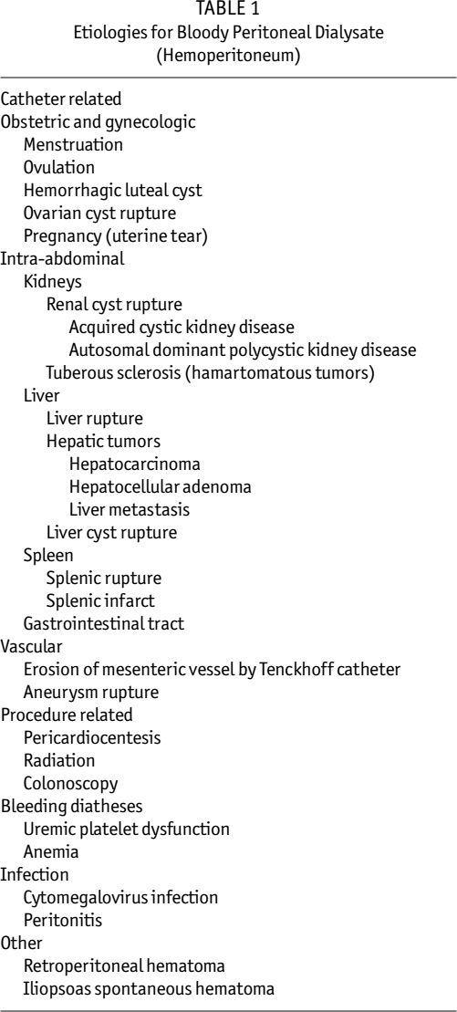
Catheter Related
Hemoperitoneum can develop shortly after peritoneal catheter insertion. Bleeding diathesis due to platelet dysfunction is common in uremic patients (2,3). Hemostasis can be established at the time of surgery to prevent bleeding. Patients with known coagulation disorders, such as hemophilia A and von Willebrand disease, have had peritoneal catheters implanted successfully with coagulation factor coverage (4,5). Garcia Falcon et al. reviewed the incidence of and risk factors for complications after permanent PD catheter implantation (5). In a total of 192 peritoneal catheters implanted in 137 patients over 10 years, they identified 53 complications in 50 catheters implanted in 42 patients. Hemoperitoneum accounted for 3.6% compared to higher rates of complications, such as catheter malposition or omental entrapment (13%) and dialysate leak (8.9%), and lower complication rates such as peritonitis (1%), surgical wound infection (0.5%), and chylous ascites (0.5%). Bernardini showed hemoperitoneum to be among the common mechanical problems hindering adequate function, which also included obstruction, dialysate leaks, hernia, and pain (6).
Vascular or visceral damage can occur during catheter placement. Laparoscopic-assisted placement of PD catheters in selected patients with previous abdominal surgery is very successful due to direct visualization and thus minimizes complications. Previous abdominal surgery may include appendectomy, ovarian resection, hysterectomy, cesarean section, open cholecystectomy, segmental resection of the small intestine, and truncal vagotomy with pyloroplasty. Temporary hemoperitoneum was described postoperatively in 3 of 20 patients operated on between 1999 and 2001 using laparoscopic-assisted technique (7). Of the 3 patients having hemoperitoneum, all had mild coagulopathy and 2 had simultaneous laparoscopic adhesiolysis.
The Tenckhoff catheter itself may cause hemoperitoneum. In one case report, the Tenckhoff catheter tip eroded a small bowel mesenteric vessel (8). In another case report, massive hemoperitoneum was due to splenic injury caused by a dislocated Tenckhoff catheter, with the tip located in the left hypochondrium (9).
Gynecologic Related
Gynecologic events, either physiologic or pathophysiologic, are by far the most common cause of hemoperitoneum in adult female PD patients. Menstruation-associated bleeding accounts for 33% of all causes of hemoperitoneum, with percentages ranging from 8% to 82% depending on whether the denominator is women in general, women of reproductive age, women of child-bearing age, or premenopausal women (10-12). Over half of menstruating females on PD will experience hemoperitoneum at least once during their time on PD (13). In a survey conducted by Holley et al. in female patients below the age of 55 years, it was found that 52% experienced hemoperitoneum of varying frequency, as shown in Table 2 (13).
Hemoperitoneum in Women on Peritoneal Dialysis: Patient Demographics and Survey Results (n = 24)
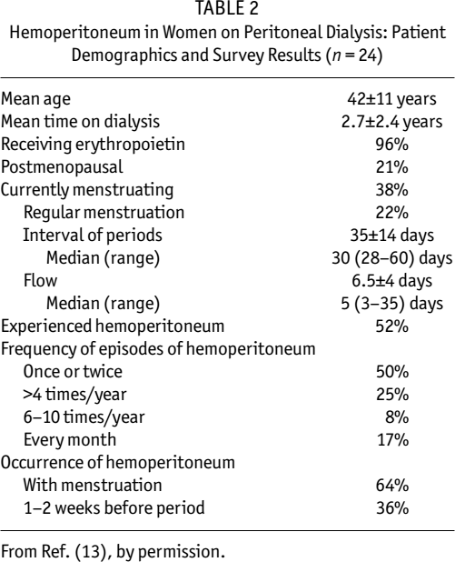
From Ref. (13), by permission.
In the reproductive age group, hemoperitoneum from physiologic events of the menstrual cycle (either menstruation, albeit retrograde flow, or ovulation causing mid-cycle bleeding) outnumbers other events (10-15). Documented hemoperitoneum from ovarian etiologies including ruptured ovarian cyst (16), luteal cysts (12,17), and follicular cysts (12) have been reported. Endometriosis, although looked for, has not been documented (11).
In general, a menstrual history is used initially to determine whether hemoperitoneum in a female is due to ovulation or menstruation. For the most part, hemoperitoneum resolves after a few rapid exchanges with warm or cold solution (10,18).
For repeated or persistent hemoperitoneum, other diagnostic procedures, including ultrasound (12,16), laparoscopy (19), or laparotomy (16), may be needed to identify the etiology of the gynecologic disorder causing hemoperitoneum. Bleeding diagnosed by angiography has never been reported.
Treatment for recurrent gynecological events include tubal ligation to prevent retrograde blood flow (12), or suppression of ovulation using oral anovulant therapy (12,16).
Obstetric Related
Hemoperitoneum has been reported in a female with end-stage renal disease receiving PD during pregnancy. After persistent hemoperitoneum during the second trimester of pregnancy, lacerations of the uterus were identified by laparoscopic technique (19). Laparotomy was performed to stop the bleeding using sutures, cauterization, or direct pressure with Surgicel [Ethicon (Johnson & Johnson), Piscataway, New Jersey, USA] on the sites. There was no etiology for the lesions. Lew proposed that the Tenckhoff catheter tip might have irritated the uterus during a contraction when the peritoneal cavity was free of peritoneal fluid.
In another case, amniocentesis performed due to advanced maternal age in a woman at 18 weeks of pregnancy resulted in blood-tinged dialysate drainage and preterm uterine contraction (20). Administration of tocolysis stopped the contractions and the PD fluid cleared with subsequent exchanges. However, 9 days post procedure, the patient developed bloody PD fluid. Exploratory laparotomy revealed serosal tearing on the uterine fundus, with a well-located Tenckhoff catheter. The tear was 1 cm in length and bleeding actively. There was an 800 mL blood clot in the peritoneal cavity. Primary suture failed to approximate the wound and hemostasis was ineffective. Serosal tearing of the pregnant uterus led to an inevitable surgical intervention. Chou et al. postulated several possible mechanisms for the uterine tear (20): the tip of the catheter might have caused the wound, the lesion might have been induced by topical pressure of the tocometer, or, alternatively, spontaneous serosal tearing may have been due to the enlarging uterus.
Both cases reported lacerations or tears on the uterine fundus, which are not found in normal pregnancy. In addition to these two case reports, the differential diagnosis for hemoperitoneum during pregnancy includes placental abruption, with retrograde blood flow into the peritoneum, and bleeding associated with disseminated intravascular coagulopathy or HELLP syndrome (hemolysis, elevated liver enzymes, and low platelet count) (21).
Intra-Abdominal Organs
Intra-abdominal pathology of solid organs such as the kidney, liver, and spleen, as well as the gastrointestinal tract, is a potential source of bleeding causing hemoperitoneum.
Kidneys: Renal cyst rupture from either acquired cystic kidney disease (22) or polycystic kidney disease (23,24) can cause hemoperitoneum. Acquired cystic kidney disease develops in patients with longstanding end-stage renal disease. Patients with polycystic kidney disease have large cysts with stretched blood vessels, putting them at higher risk of rupturing. In addition, the large kidneys are more susceptible to trauma. It is not clear why a retroperitoneal organ such as the kidney would result in hemoperitoneum. A possible explanation is that adhesions form between the cyst's wall and the peritoneum, favored by their anatomical proximity and inflammation secondary to intracystic hemorrhage. These adjoining structures could then rupture as a result of rising intracystic pressure (22).
In a patient with tuberous sclerosis, intratumoral bleeding of the renal angiomyolipoma (hamartomatous tumors) was identified on ultrasound and CT as the etiology for hemoperitoneum (25).
Liver: Peng and Yang described 3 PD patients with hepatic tumor causing hemoperitoneum (26). Two of these patients were treated with transcatheter arterial embolization, with subsequent spontaneous hepatic tumor rupture. The third patient developed tumor necrosis after transcatheter arterial embolization. This caused the surrounding vessels to bleed since the PD fluid prevented a blood clot from sealing the small vessels, which could not be embolized completely. Bleeding stopped when PD was discontinued but restarted when continuous ambulatory peritoneal dialysis (CAPD) was restarted.
A ruptured subcapsular hepatic hematoma along the posterior surface of the right lobe of the liver, originating from hepatic metastases from renal cell carcinoma found in a previously removed kidney with acquired renal cystic disease, resulted in hemoperitoneum (27).
Dozio et al. reported a case of a 70-year-old anti-hepatitis C virus-positive woman with bloody peritoneal effluent after 48 months of CAPD treatment (28). Abdominal CT scan revealed three hepatic lesions with signs of hepatic neoplasms. Selective hepatic arteriography confirmed the diagnosis. Chemoembolization of the three lesions resulted in resolution of hemoperitoneum.
Hepatic cysts, frequent complications of polycystic kidney disease, may rupture and cause hemoperitoneum. Magnetic resonance imaging (MRI) and CT may be used to make the diagnosis. Treatment includes cauterization of the cysts (23).
Laparotomy was needed to control the bleeding from a large hematoma originating from a large soft-feeling tumor that was later identified as hepatocellular adenoma secondary to possible oral contraceptive use (29). Ultrasound showed an enlarged and inhomogeneous left liver lobe, whereas CT showed a large solitary cystic tumor with a solid central mass.
Spontaneous liver rupture was noted in a patient with amyloidosis and hepatitis C. Hemoperitoneum was caused by a laceration of the liver that was diagnosed with arteriography of the celiac tripod. Bleeding was controlled by embolization with titanium microcoils and Gelfoam (Pharmacia & Upjohn, Kalamazoo, Michigan, USA) particles (30).
Spleen: Splenic rupture is the most common splenic injury reported to cause hemoperitoneum. Contributing underlying conditions include splenic infarction (31), chronic myelogenous leukemia (32), and amyloid deposition (33,34).
An atraumatic splenic infarction due to hemorrhage was diagnosed by laparotomy when a patient presented with left shoulder pain, hypotension, and hemoperitoneum (31). No underlying splenic pathology was noted.
Emergency splenectomy performed on a PD patient presenting with hemoperitoneum after a fall revealed a splenic capsule rupture with amyloid deposits in the spleen (33). CT scan revealed an enlarged spleen with intra-splenic hematomas and splenic capsule rupture.
Massive hemoperitoneum after colonoscopy was reported as a complication of the procedure (35). In fact, the patient was bleeding from the spleen, which was avulsed from its diaphragmatic attachments. This occurred during a difficult colonoscopy with many attempts to pass the colonoscope around the splenic flexure. Strong pulling forces on the splenocolic ligament tore the diaphragmatic splenic ligament and a small laceration of the splenic capsule led to an expanding subcapsular hematoma, while there was continuity of the splenocolic ligament.
In general, the diagnosis of splenic involvement is made with CT. When the spleen is the cause of hemoperitoneum, surgical intervention (e.g., splenectomy) is needed to control bleeding.
Hemoperitoneum from splenomegaly associated with acute cytomegalovirus (CMV) antigenemia has been reported (36). Peritoneal fluid CMV DNA was also positive. Splenectomy was not required since the hemoperitoneum resolved spontaneously 10 days after presentation.
Gastrointestinal Tract: It is amazing that there are not more gastrointestinal complications associated with PD, given the large surface area and length of the intestines. Although hemoperitoneum has been reported with abdominal processes such as diverticulosis, diverticulitis, acute appendicitis, acute cholecystitis, and acute pancreatitis, there are no reported cases involving PD patients. The Table in Ref. (36) entitled Reported Causes of Hemoperitoneum in CAPD Patients included these abdominal processes, among others; however, no references were cited.
Colon perforation presenting as hemoperitoneum in a patient with dialysis-related amyloidosis was reported by Min et al. (37). Colon perforation generally presents with fecal material in the peritoneum.
Vascular Disorders
Hemorrhage from vascular complications may cause hemoperitoneum. Miller et al. reported erosion of a mesenteric vessel by a Tenckhoff catheter (8). In another case report, a true aneurysm of the omental artery led to hemoperitoneum and shock in a CAPD patient (38). A novel approach to treating blood vessel abnormalities is using percutaneous embolization using Gianturco coils (Cook, Bloomington, Indiana, USA), as in the case of splenic artery pseudoaneurysm (39). Embolization using a percutaneous transcatheter approach obfuscates a laparotomy. Complications, such as hernia, adhesions, and the inability to use the peritoneum for PD for several days while the wounds are healing, can be avoided by using percutaneous embolization rather than laparotomy.
Procedure Related
Hemoperitoneum may occur after procedures involving the abdomen and thorax. Since the Tenckhoff catheter offers a “window” to the peritoneum, more cases of hemoperitoneum post procedure are reported in the CAPD population than in any other group.
Laparoscopic cholecystectomy is a frequent and preferred method of removing the gall bladder (40). Hemoperitoneum is common immediately post surgery and during the first post-procedure exchange (40,41). Most surgeons would like a 1 – 2 day dialysate-free peritoneum. Low volume exchanges are performed for several days to decrease wound complications. Thereafter, PD is resumed with minimal complications.
Radiation to the peritoneum results in acute and delayed complications. Hassell et al. reported a case of radiation therapy for carcinoma of the bladder and subsequent radiation-induced peritoneal injury resulting in catheter-related trauma and hemorrhage (42).
Bender reported a procedure of the thorax, namely pericardiocentesis, that resulted in hemoperitoneum (43). Pericardiocentesis was performed under fluoroscopic guidance using a subxiphoid approach to drain 400 mL of serosanguineous fluid in a patient with a moderate to large pericardial effusion with evidence of right atrial and ventricular collapse. A pericardial drain remained in place for 24 hours for an additional 100 mL drainage. When intermitted PD was initiated, the peritoneal dialysate was blood tinged, with 275 red blood cells (RBCs) U/L; 24 hours later the peritoneal dialysate was clear, with 4 RBCs U/L. The authors concluded that, during pericardiocentesis, the needle went through the peritoneum, causing minor bleeding (43).
Massive hemoperitoneum was reported as a complication of colonoscopy but it turned out to be splenic in origin (35). However, Nace et al. reported hemoperitoneum after colonoscopy (1).
Bleeding Diatheses
End-stage renal disease patients have an increased bleeding tendency due to uremic platelet dysfunction, as expressed by prolonged bleeding time (44). Prothrombin time, partial thromboplastin time, and platelet count are generally normal. Low hemoglobin or hematocrit also increases the risk of bleeding after a procedure. In the presence of an insufficient number of RBCs, thrombin cannot trap RBCs to form a clot. Bleeding potential may be minimized with either blood transfusion (45) or administration of erythropoietic-stimulating proteins (46) to raise hemoglobin to a desirable level before an elective procedure.
Infectious Complications
Peritonitis generally presents with cloudy peritoneal dialysate effluent due to white blood cells and clinically with abdominal pain. When cell count and differential are performed on peritoneal fluid, RBCs are frequently seen due to inflammatory changes of the peritoneal membrane (47). However, gross hemoperitoneum is rarely seen.
Sclerosing peritonitis is a rare but serious complication of PD. Sclerosing peritonitis is characterized by small-bowel obstruction due to encapsulation, dense adhesions, or mural fibrosis. Although small-bowel obstruction is the most frequent presenting clinical finding (92%), some patients presented with hemoperitoneum (8%) in a survey of Australian PD patients from 1980 to 1994 (48).
Other Causes of Hemoperitoneum
An iliopsoas muscle hematoma that infiltrated the anterior abdominal wall and superior part of the hip resulted in hemoperitoneum after an episode of coughing (49). Manipulation of a double-lumen femoral catheter for hemodialysis led to retroperitoneal bleeding and hemoperitoneum (50).
There are cases of hemoperitoneum without an etiology or an association to any specific intra-abdominal pathology. These cases tend to clear spontaneously. Tse et al. believe the source of bleeding in most cases of so-called idiopathic episodes of hemoperitoneum is likely a minor Tenckhoff catheter-related tear in omental venules (15). They recommend empirical treatment of bed rest and administration of intraperitoneal heparin to prevent blockage of the Tenckhoff catheter.
Assessment and Management of Pd-Related Hemoperitoneum
In the evaluation of PD-related hemoperitoneum, bleeding into the peritoneal dialysate can be classified as benign, minor, or significant (10). Benign bleeding includes retrograde menstruation, ovulation, and post colonoscopy. Minor intraperitoneal bleeding with significant accompanying pathology includes polycystic kidney disease with intra-cyst bleeding or cholecystitis. Significant bleeding includes ruptured ovarian cyst, post-pelvic irradiation, adhesions after peritonitis, ruptured spleen after colonoscopy, ruptured spleen after catheter dislodgment, idiopathic thrombocytopenia purpura, and calcific peritonitis. Causes of hemoperitoneum can be classified into three groups based on severity of the underlying pathology as well as whether intervention is needed. This classification process helps in diagnosis and treatment.
An outline of the assessment of and action plan for hemoperitoneum from a nursing perspective is provided by Maaz (51).
When PD hemoperitoneum presents, several rapid PD exchanges are performed to determine if bleeding is persistent or is an acute event. In the absence of a recent procedure or concurrent acute illness, hemoperitoneum is associated with a gynecologic event (33% of hemoperitoneum cases are menstruation related) or a capillary rupture. Rapid exchanges with room temperature dialysate (cold dialysate) are associated with vasoconstriction and help control PD-related hemoperitoneum (18). For the most part, the procedure clears PD-related hemoperitoneum.
Coagulopathy should be corrected to halt or retard further bleeding. Administration of fresh frozen plasma or cryoprecipitate is effective in treating uremic coagulopathy (52). Intravenous desmopressin increases the release of factor VIII–von Willebrand factor multimers from endothelial storage sites (53). Blood transfusion not only increases the hematocrit but also reduces bleeding time when the hematocrit is above 25% – 30% (45). The reduction in bleeding time is due to enhanced platelet aggregation and increased platelet adhesion to endothelial cells (45). In addition, conjugated estrogen has been used to improve uremic bleeding (54,55).
Addition of heparin 500 U/L PD fluid is recommended to prevent catheter malfunction due to a clot obstructing the flow of dialysate.
In the presence of a recent procedure or concurrent acute illness, correlation or association with the event and hemoperitoneum should be investigated. With persistent hemoperitoneum, diagnostic procedures may include imaging (e.g., ultrasound, CT, MRI) or angiography, as well as direct visualization via laparoscopy or laparotomy.
Depending on the etiology of gynecological bleeding, hormonal or surgical intervention may be used to control bleeding. Bleeding from an intra-abdominal organ requires surgical intervention to control hemorrhage. Laparoscopic techniques are replacing laparotomy for obvious reasons. However, there are more-recent reports on using intravascular techniques such as angiography to diagnose the source of bleeding, and intravascular embolization techniques to stop the bleeding.
Complications of Hemoperitoneum
With minor bleeding, complications are minimal. However, with severe bleeding, blood may clot, causing dialysate flow obstruction. Addition of heparin, 500 U/L PD fluid, may prevent this complication.
Long-term follow-up of individuals with hemoperitoneum while on PD, whether one episode or recurrent, does not show compromise in membrane characteristics (56) or ultrafiltration (15), or predisposition of the peritoneum to peritonitis (15). Hemoperitoneum after catheter placement can result in catheter failure (60%) and adhesion formation, especially if the abdomen is dry. When the abdomen was irrigated until the PD fluid was clear, followed by low volume (500 – 1000 mL) short dwells, there was no catheter failure (57).
Summary
Hemoperitoneum is not an uncommon event in patients receiving PD. Bleeding may range from mild to severe. The etiology of the bleeding may be benign or pathological. Assessment of the etiology may range from a simple history with a few rapid exchanges to an invasive procedure such as laparotomy or laparoscopy. The use of imaging techniques to identify the source of bleeding is currently advocated due to advances in this field over the past decade. Management depends heavily on the etiology of the bleeding. Despite the alarming presence of blood in the peritoneum, there is generally minimal damage to the peritoneum.
The approach to hemoperitoneum in a patient receiving peritoneal dialysis is different from that in the general population. A knowledge base of hemoperitoneum by the patient as well as the nursing and medical staff provides for a systematic approach to diagnosis and management.