
Abstract
Animal models of peritoneal dialysis fluid (PDF) exposure are key tools in the study of mechanisms involved in alterations of the peritoneal membrane and in the design of therapies. We recently developed a mouse model of chronic peritoneal exposure to high glucose dialysate. Herein, we make a sequential analysis of the effects of glucose-based PDF on mouse peritoneal membrane and on mesothelium. We demonstrate that chronic exposure to PDF induces thickness and fibrosis of the peritoneal membrane in a time-dependent manner. We also show that mesothelial cells progressively detach and lose cytokeratin expression. In addition, we demonstrate that some mesothelial cells invade the submesothelial space, where they appear as cytokeratin- and alpha-smooth muscle actin-positive cells. These findings demonstrate that epithelial-to-mesenchymal transition (EMT) of mesothelial cells takes place in mouse peritoneum exposed to PDF, validating this model for the study of effects of drugs on the EMT process as a therapy for peritoneal deterioration.
We have demonstrated that effluent-derived MCs cultured ex vivo show progressive loss of their epithelial phenotype and acquire a fibroblast-like phenotype on extension of PD. In addition, these cells acquire the expression of alpha-smooth muscle actin (αSMA), characteristic of a myofibroblast nature (2). Immunohistochemical analysis of peritoneal biopsies from PD patients demonstrated the presence of mesothelial markers in stromal αSMA-positive myofibroblasts, suggesting that these cells could originate from the local conversion of MCs via epithelial-to-mesenchymal transition (EMT) (3). There is emerging evidence supporting the idea that the EMT of MCs plays an important role in the onset of PM deterioration (1). The production of extracellular matrix components and large amounts of vascular endothelial growth factor by transdifferentiated MCs (4) may induce vasodilation, hyperpermeability, high solute transport, rapid disappearance of glucose osmotic gradient, poor ultrafiltration capacity, and, later on, angiogenesis (5). Upon in vitro stimulation with inflammatory cytokines, omentum-derived MCs also suffer functional and morphological alterations similar to those observed in vivo (2).
Animal models contribute to our understanding of the mechanisms involved in the response to PD. We recently developed a mouse model of PD in which exposure to classic glucose-based peritoneal dialysis fluids (PDF) induced fibrosis and angiogenesis of PM in 5 weeks of treatment (unpublished observation). These morphological alterations were accompanied by ultrafiltration failure of mouse PM. In that work we addressed the effect of chronic exposure to PDF on mouse PM in the context of EMT of MCs. Our data demonstrated that this animal model will provide a valuable tool to unravel the molecular basis of PM deterioration and to study the anti-EMT effects of drugs as therapy for peritoneal deterioration.
Material and Methods
Experimental Animals, Surgery, and PDF Exposure
This study was performed in nonuremic C57BL/6 female mice aged 12 – 14 weeks (Harlan Interfauna Iberica, Barcelona, Spain). The mice were housed under standard conditions and with free access to food and water. The experimental protocol was approved by the Animal Ethics Review Committee of the Unidad de Cirurgía Experimental of Hospital La Paz.
A customized vascular access port (mouse port; Access Technologies, Strategic Applications, Libertyville, Illinois, USA) was implanted in the mice. Catheters containing ten holes were located within 1 cm from the tip to help the fluid exit and to prevent obstructions. Animals were anesthetized intraperitoneally with ketamine (100 mg/kg) and xylazine (10 mg/kg). Afterward, the end of catheter was introduced into the peritoneal cavity by the right flank of the animal and the port was displaced at the subcutaneous space of the mouse's back. During the first week post surgery, 0.2 mL saline was instilled in order to prevent catheter trapping. Thereafter, during the experimental procedure, 1.5 mL standard PDF with lactate and 4.25% glucose (Fresenius Medical Care, Bad Homburg, Germany) was instilled. A second group, age matched and not manipulated, was used as control.
During the experiment, the health of the mice was checked daily by the veterinarian. Mice were weighed every week and those presenting more than 10% weight loss or any sign of port infection or abnormal activity were excluded from the experiment. Following sacrifice, peritoneum was carefully examined for any sign of bleeding or infection.
Antibodies and Histological Analyses
For this study we used mouse anti-pan-cytokeratin and mouse anti-αSMA antibodies (Sigma–Aldrich, Saint Louis, Missouri, USA). In order to avoid unspecific staining with secondary antibodies, these antibodies were previously conjugated with specific Zenon fab (Invitrogen, Paisley, UK) according to the manufacture's instructions.
We collected tissue from parietal peritoneum, fixed it in neutral-buffered formalin, and embedded it in paraffin, which we cut into 5-μm sections. The sections were stained with Masson's trichrome for fibrosis detection and submesothelial layer thickness was determined microscopically (Leica CTR6000 with an LAS-AF6000; Leica Microsystems, Wetzlar, Germany). For the immunofluorescence analysis of MCs, samples were cut into 3-μm sections and, after treating with xylol to remove the paraffin, samples were heated for 15 minutes. After that, samples were stained with anti-pan-cytokeratin and anti-αSMA Zenon conjugate. Nuclei were stained with DAPI. Micrography was performed using a fluorescence microscope (Leica CTR6000 with an LAS-AF6000).
Statistical Analyses
Statistical analysis of data was performed using GraphPad Prism 4 software (GraphPad, La Jolla, California, USA). Comparison among groups was made using the Mann–Whitney test. A p value of less than 0.05 was considered statistically significant.
Results
Exposure to PDF Induces Peritoneal Fibrosis
We have previously demonstrated that chronic exposure to conventional glucose-based PDF induces fibrosis in mouse PM. In order to better understand the mechanism of peritoneal damage in this model, we analyzed progression of peritoneal thickening against time of peritoneal exposure to dialysis solution.
Thickness of PM increased with time of exposure to dialysis solution [Figure 1(a)]. Quantitative analysis demonstrated that this increment was significant at the second week of treatment [Figure 1(b)].
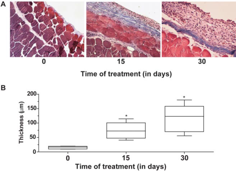
Chronic exposure to peritoneal dialysis fluid results in increased deposition of extracellular matrix (A) and thickness (B) of the peritoneal membrane in a time-dependent manner. Comparisons between times of observations were performed using the nonparametric Mann–Whitney test.
Mesothelial Cells are Detached during Pdf Treatment and Lose Cytokeratin Expression
To analyze the effect of dialysis solution exposure on mice MCs, we stained these cells with anti-cytokeratin and myofibroblasts with anti-αSMA antibodies. Before starting dialysis solution instillation, the PM showed a continuous layer of cytokeratin-positive MCs, properly positioned. Two weeks after PDF instillation, the mesothelium showed evidence of MC loss. At day 30, fill cells of PM were positive for cytokeratin. In addition, we observed dull cytokeratin-positive cells embedded in the submesothelial compact zone. This suggests that MCs are not only denudating but are also migrating to the submesothelial space (Figure 2).
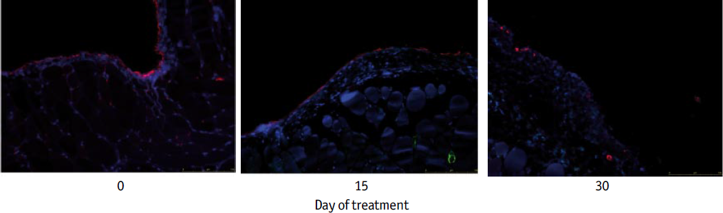
Immunofluorescence detection of mesothelial cell markers (green: αSMA; red: cytokeratin; blue: DAPI) at different times in a chronic mouse model of peritoneal dialysis fluid exposure demonstrates a time-dependent loss of cytokeratin by mesothelial cells and submesothelial location of mesothelial cells.
Mesothelial Cells Undergo Emt in Response to Chronic Exposure to PDF
We have previously described the presence of cytokeratin- and αSMA-positive cells embedded in the submesothelial space in peritoneal biopsies from PD patients, suggesting that these cells stem from the conversion of MCs into myofibroblasts (3). In order to determine the presence of transdifferentiated MCs in the submesothelial space of mice exposed to dialysis solution, we performed an analysis at high magnification. In the control group, we observed a simple line of MCs expressing cytokeratin. The only αSMA-positive cells were observed in smooth muscle around the blood vessels located deep between muscles (Figure 3, left panels). In PM of mice exposed to dialysis solution, we observed loss of cytokeratin-positive cells in the mesothelium. The presence of cells expressing both cytokeratin and αSMA in the submesothelial space (Figure 3, white arrows, upper and lower right panels) indicates that at least some of the myofibroblasts involved in peritoneal damage originated from MC conversion in the peritoneum of mice exposed to PDF (Figure 3, right panels).

Mesothelial cells at the submesothelial space express alpha-smooth muscle actin (αSMA). Images of peritoneal biopsies of mice at day 0 (control; left panels) and after 30 days of treatment with peritoneal dialysis fluid (PDF; right panels) were analyzed at x40 (upper panels) and x63 (lower panels). The presence of cells expressing both cytokeratin and αSMA at the submesothelial space in PDF-treated mice are shown (white arrows).
Discussion
Compromise of PM due to exposure to dialysis solution is a serious event in PD because the treatment is based on the functional integrity of the PM (6). In order to better develop efficient therapies to prevent or treat compromised PM, it is necessary to know the intimate mechanisms involved in its deterioration. The study of human peritoneal biopsies permits only a snapshot of a long and complex process that culminates with peritoneal deterioration. Therefore, the development of animal models of PD is important in improving knowledge of the mechanisms responsible for peritoneal deterioration and in testing potential counteractions.
We recently described a mouse model of chronic exposure to dialysis fluid in which 5 weeks of treatment caused morphological and functional changes similar to those observed in patients (unpublished observations). Exposure to standard high glucose dialysis solution induced thickening of PM due to the deposition of extracellular matrix. In addition, the treatment also induced angiogenesis that culminated with a lesion similar to that in humans and associated with loss of dialytic capacity of the PM.
One of the important events involved in peritoneal deterioration is myofibroblast conversion of MCs (3). Evidence suggests that these cells play an important role in the deposition of extracellular matrix and the angiogenesis that compromise the PM of PD patients (4). Here, we addressed the effect of chronic exposure to a conventional high glucose dialysis solution on the progression of PM damage. The results demonstrate that peritoneal thickness increases progressively with time of treatment (Figure 1). We also observed that mesothelium denudates while treatment is maintained (Figure 2). In addition, we observed the presence of cytokeratin- and αSMA-positive cells at the submesothelial space, suggesting that EMT of MCs also takes place in our model of chronic exposure to conventional dialysis solution.
The results presented in this work suggest that EMT of MCs also contributes to the progression of peritoneal deterioration in a mouse model. We do not yet know how important this process is in the onset of peritoneal deterioration or the initial mechanisms responsible for MC conversion. However, the mouse model may contribute to answering these questions through the utilization of genetically modified mice.
Footnotes
Acknowledgments
This work was supported by grants SAF2007-61201 and PET2006-0256 (from Ministerio de Educación y Ciencia) to M. López-Cabrera and FIS PI06/0098 and RETICS 06/0016 (from Fondo Investigaciones Sanitarias) to R. Selgas. This work was also partially supported by Fresenius Medical Care and Gambro Europe. L.S. Aroeira was supported by RETICS 06/0016. G.T. González-Mateo received financial support from Gambro Europe.
We thank Javier Benito de la Víbora (DVM) and Carlota Largo Aramburu (DVM, PhD) for their assistance with care of the mice.