
Abstract

The Addition of a NO Inhibitor to a More Biocompatible Peritoneal Dialysis Solution in a Long-Term Rat Model with Kidney Failure
Background: Long-term peritoneal dialysis (PD) with a conventional glucose-based and lactate-buffered PD fluid can lead to morphological and functional alterations of the peritoneal membrane. A recent study showed that a mixture of osmotic agents and a bicarbonate/lactate buffer (glycerol 1.4%/amino acids 0.5%/dextrose 1%; GLAD) resulted reduced peritoneal vessel density but faster small solute transport rates. It was hypothesized that the latter was due to the influence of vasoactive substances, such as NO. Purpose: To investigate whether high transfer rates of small solutes, induced by exposure to GLAD could be influenced by L-NMMA (Nω– methyl-L-arginine acetate, L-NMMA), an inhibitor of NO, in a long-term rat model with chronic kidney failure. Methods: All rats underwent a peritoneal catheter implantation and 70% nephrectomy (Nx). Thereafter a division in 3 groups was made: GLAD L-NMMA (n=9), GLAD (n=7), and Buffer (control n=8). All rats were infused daily (6 mL/100 g BW) for 16 weeks post Nx with the appropriate solution. All rats underwent a standard peritoneal permeability analysis and were sacrificed afterwards. Results: Residual renal clearance was the same in all groups, allowing comparison of them. Data (Table) are expressed in mean±SD and *different from Buffer (p<0.05).
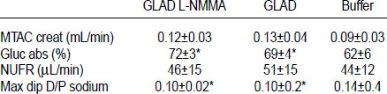
Conclusion: Inhibition of NO did not influence small solute transport. This is in line with our previous findings in human exposed to an amino acid-based dialysis solution.
The Inhibition of Peritoneal Fibrosis with the Novel Angiogenesis Inhibitor Sunitinib
Background: Encapsulated peritoneal sclerosis (EPS) is characterized by neoangiogenesis and fibrosis. Increased inflammation is the leading cause of EPS. In turn, neoangiogenesis is both consequence and contributor of inflammation. The effects of sunitinib, a multi-targeted receptor tyrosine kinase inhibitor, have been postulated in various anti-angiogenesis, anti-inflammatory, and antifibrotic processes both in vitro and in vivo. This novel angiogenesis inhibitor, sunitinib maleate (SUT), was investigated in our rat EPS model. Methods: 40 nonuremic Wistar albino rats were divided into 4 groups as follow: 2 mL isotonic saline intraperitoneally (IP) daily, 3 weeks (wk) (Control group); daily IP 2 mL/200 g injection of chlorhexidine gluconate (0.1%) and ethanol (15%) dissolved in saline, 3 wk (CG group); CG + additional 3 wk without any treatment, total 6 wk (Resting group); CG + additional 3 wk 1 mg/kg daily SUT in drinking water, total 6 wk (SUT group). At the end of the study, 1-hour PET was performed. Functional parameters and morphological changes of peritoneum with dialysate cytokine levels were examined. Results: The results are given in the Table (mean±SEM). Discussion: SUT renewed ultrafiltration (UF) failure, D1/D0 glucose levels, and dialysate protein loss. Peritoneal thickness, WBC count, and inflammation of peritoneum were also decreased with SUT treatment. SUT significantly improved overexpression of dialysate transforming growth factor (TGF)-β1, monocyte chemoattractant protein (MCP-1), and vascular endothelial growth factor (VEGF) levels compared to resting. Conclusion: SUT may preserve membrane viability even at lower dosages in long-term peritoneal dialysis patients. Morphological Changes with Dialysate Cytokine Profile
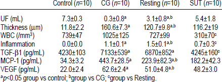
p<0.05 group vs control;
group vs CG;
group vs Resting.
Bozkurt D.,1 Timur O.,3 Hur E.,1 Bicak S.,3 Taskin H.,3 Sarsik B.,2 Akcicek F.,1
Uric Acid Transperitoneal Transport: Impact of p-Cresol and Sodium Hyaluronate in Vitro
P-Cresol is a one of the uremic toxins. Its high concentration in blood is observed in peritoneal dialysis patients with end-stage kidney disease. Cytoprotective molecule such sodium hyaluronate may protect the peritoneum, which are modified by renal replacement therapy. The object of study was uric acid transport across the isolated parietal peritoneum from anterior abdominal wall of white New Zealand rabbits, placed inside the modified Ussing-type chamber. Values of the transfer, directed from the mesothelial (M) to the interstitial (I) side of membrane (M→I) were calculated using mathematical model of mass transport and expressed as coefficient of diffusive permeability P (in centimeters per second). Three separate series of the experiments were done. In the first we examined uric acid transfer in the control series (for 120 minutes), in the next, coefficients of diffusive permeability before (15–60 minutes) and after p-cresol (0.005 g/dL) or sodium hyaluronate (0.04 g/dL) applications (75–120 minutes). The dynamics of transperitoneal transport of uric acid was stable. The values of P ± standard error of the mean were 0.842±0.128 (×0.0001; cm/s). Hyaluronan did not change transfer of uric acid. p-Cresol application on the mesothelial side of membrane decreases by 10% transport of uric acid. The results may be clinically important.
A Damaged Endothelial Glycocalyx in Peritoneal Dialysis Patients
Background and Objectives: Endothelial dysfunction and accelerated vascular disease are common features in patients with chronic renal failure (CRF). The glycocalyx is a negatively charged mesh lining the inner wall of blood vessels. It is a main regulator of vascular homeostasis. No data are available on the state of endothelial glycocalyx in CRF. In the present study we investigated whether the microvascular glycocalyx is damaged in patients with end-stage renal disease (ESRD) treated with peritoneal dialysis (PD), compared to healthy controls.
Methods: Investigations were carried out in 8 patients with ESRD undergoing PD (male/female 7/1; median age 37.7 (18.25–53.3) years; median time on dialysis 45.7 (7.4–145.6) months, and 12 healthy age- and sex-matched controls with normal kidney function. Exclusion criteria: diabetes mellitus, use of antioxidants, use of statins 6 weeks prior to the measurement, use of angiotensin converting enzyme inhibitors or angiotensin II receptor blockers on the day of the measurements. The status of endothelial glycocalyx in individual blood vessels was assessed by measuring the dynamic range of red blood cell (RBC) width in the sublingual microvasculature, using sidestream dark-field (SDF) imaging with a MicroScan video microscope.
Results: Compared to healthy controls, PD patients appeared to have a 0.13±0.6 μm (p=0.03) increase in the dynamic range of RBC width indicating the loss of glycocalyx barrier properties. This correlated with total time on renal replacement therapy (r=0.81, p=0.02).
Conclusions: Patients with ESRD on PD have a loss of glycocalyx barrier properties compared to age- and sex-matched healthy controls. Impaired glycocalyx barrier properties may be an early indicator of pathogenic activation of vascular endothelium as a marker of increased cardiovascular risk.