






Abstract
Neurological toxicity is a relatively rare adverse reaction reported in elderly patients treated with cephalosporins. We present a case of ceftazidime-induced encephalopathy in the context of acute kidney injury in an 80-year-old female treated for a Pseudomonas aeruginosa prosthetic joint infection. During the course of treatment, the patient developed sudden confusion and disorientation. The patient’s mental state progressively worsened, eventually leading to intubation and admission to the intensive care unit. As imaging and laboratory analyses revealed no alternative causes explaining the patient’s symptoms, ceftazidime was stopped under the suspicion of drug-induced neurotoxicity. Shortly after ceftazidime discontinuation, the patient’s condition drastically improved and returned to baseline within 5 days. This case reveals the potential severity of cephalosporin-induced neurotoxicity in elderly patients and highlights the importance of quickly detecting such adverse events in order to prevent dire outcomes.
Introduction
Cephalosporins are widely prescribed antibiotics with a generally favourable safety profile. However, an accumulating body of evidence indicates that cephalosporins can cause significant neurological toxicity, particularly in patients with renal dysfunction, the elderly and patients with pre-existing neurological disease.1-3 Indeed, several cases of cephalosporin-induced neurotoxicity have been reported in the literature, highlighting a rare clinical feature of this antibiotic class. 2 These reports suggest that the risk of neurotoxicity is particularly significant with third-generation and fourth-generation cephalosporins, more specifically for ceftazidime and cefepime.1,4,5 Although numerous reports have already been published on cefepime-induced neurotoxicity, fewer cases have been reported on the neurotoxic effects of ceftazidime.5,6
Ceftazidime is a broad-spectrum bactericidal antibiotic with coverage against Gram-positive and Gram-negative bacteria. Although it exhibits less activity against Staphylococcus species as compared to first and second generation cephalosporins, it is often used for its activity against Pseudomonas aeruginosa. 7 Specifically, the molecule targets penicillin-binding proteins of the bacterial cell and inhibits the biosynthesis of the peptidoglycan cell wall, causing cell lysis. 7 According to the manufacturer’s monograph, central nervous system adverse events have been reported in less than 1% of patients treated with ceftazidime. Neurological sequelae, including tremor, myoclonia, convulsions, encephalopathy and coma, have been reported in patients with renal dysfunction and unadjusted dosages. 8 As the potential causative factors for neurotoxicity in hospitalized patients are diverse, beta-lactam associated neurological adverse effects may be difficult to diagnose and are often overlooked. We present a case of ceftazidime-induced neurotoxicity in an elderly woman with acute renal dysfunction.
Case Report
An 80-year-old woman presented to the hospital with a 2-week history of progressive hip pain and purulent drainage. The patient was known to have a history of osteoarthritis of the hip, for which she underwent a left total hip arthroplasty approximately 2 years prior. This procedure was complicated by a periprosthetic fracture of the left hip 2 weeks after, which required a revision hip arthroplasty. Other past medical history included type II diabetes, hypertension, dyslipidemia, chronic kidney disease, stroke, mild aortic stenosis, abdominal aortic aneurysm, obesity, obstructive sleep apnea and gastroesophageal reflux disease.
On presentation, physical examination of the hip revealed an erythematous and hypertrophic zone with a deep opening, discharging odorless purulent fluid. Laboratory analysis showed a significant increase in inflammatory markers with a white blood cell count of 8.8 x 10⁹ cells/L (normal range: 4.0-11.0 x 10⁹/L), an erythrocyte sedimentation rate of 115 mm/h (normal range: 0-20 mm/h) and C-reactive protein levels of 129.9 mg/L (normal range: <10 mg/L). A radiograph at initial presentation is shown in Figure 1.
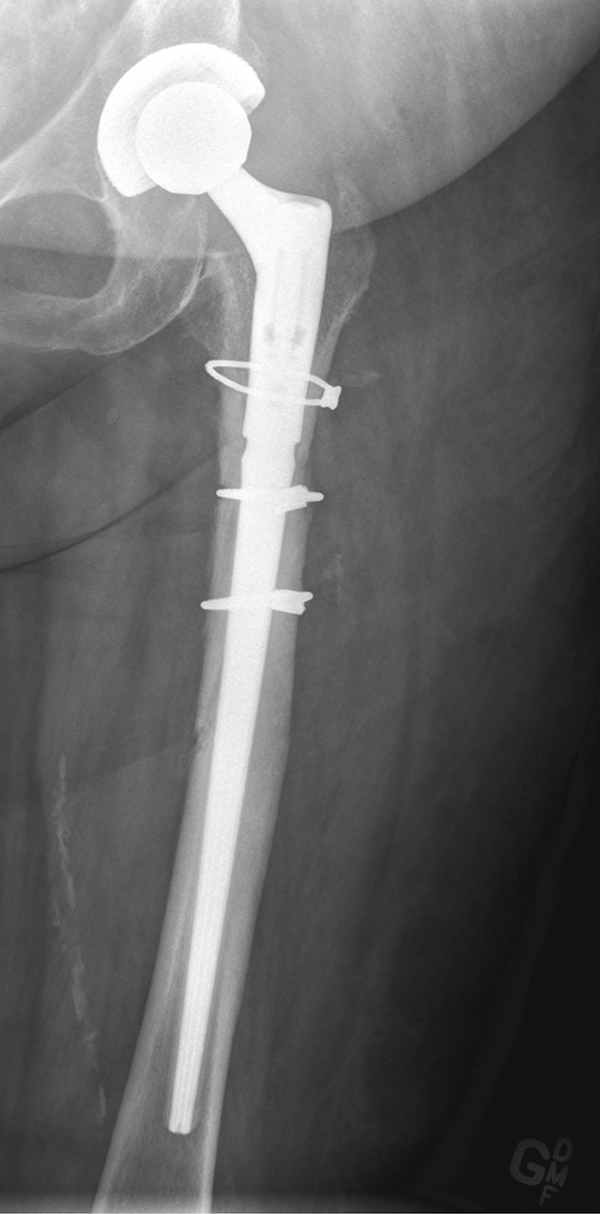
Radiograph at initial presentation. Anteroposterior view of the left hip.
Intravenous cefazolin was initiated empirically at a dosage of 2 g every 8 hours for suspicion of a prosthetic joint infection. The patient underwent a 2-stage revision hip arthroplasty. Irrigation and debridement of the left hip were completed, and an antibiotic cement spacer loaded with 2 g of vancomycin and 10 g of gentamicin was implanted. Synovial fluid and intraoperative cultures grew Pseudomonas aeruginosa susceptible to ciprofloxacin, ceftazidime, gentamicin and piperacillin/tazobactam. Cefazolin was therefore replaced with a 6-week course of both ciprofloxacin 500 mg orally twice daily and ceftazidime 2 g intravenously every 8 hours. The patient’s serum creatinine was measured at 110 μmol/L, with a calculated Cockcroft-Gault clearance of 51 mL/min (actual body weight of 87 kg), corresponding to the patient’s baseline renal function.
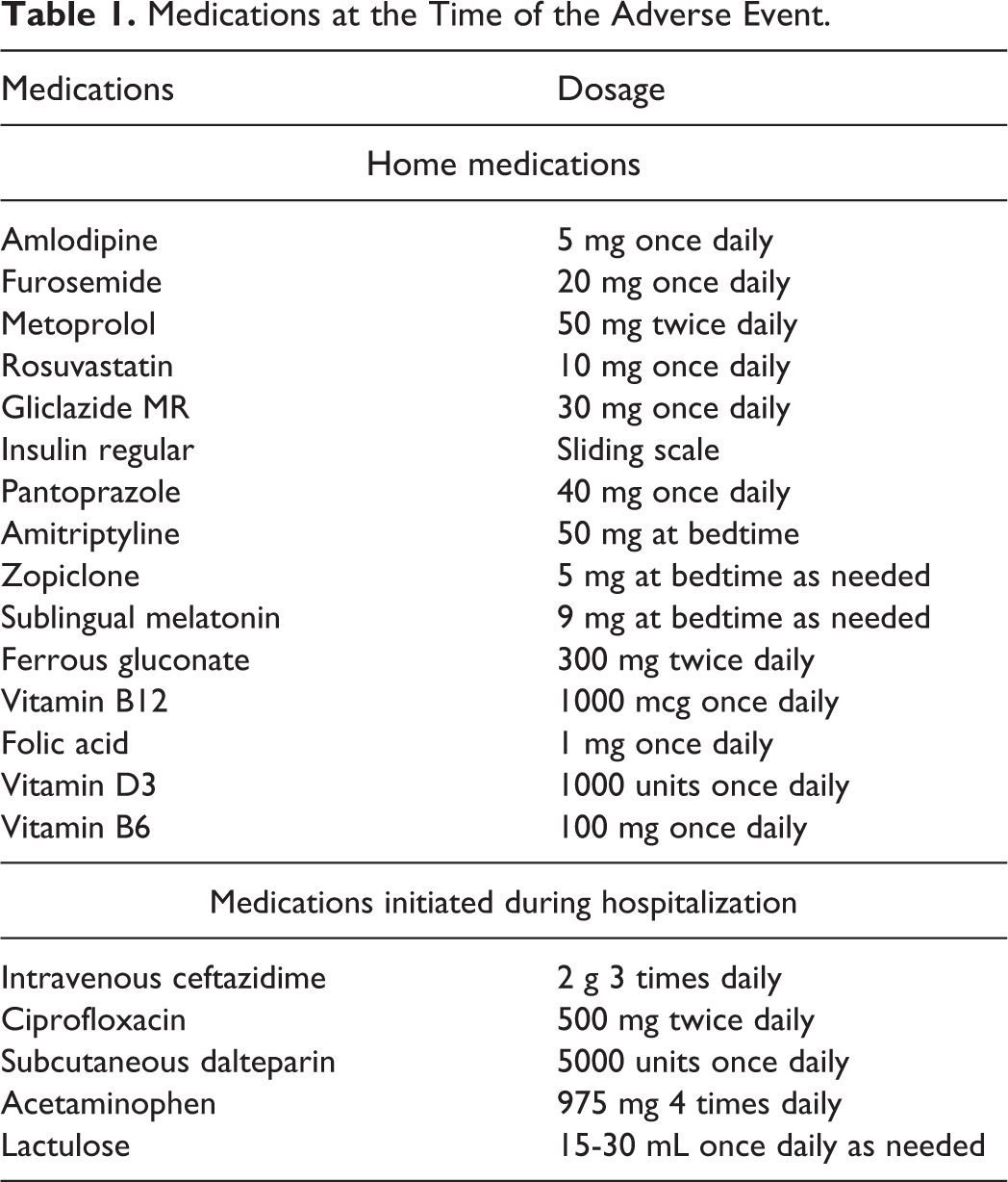
Five days after initiating the ceftazidime and ciprofloxacin regimen, the patient developed sudden confusion and disorientation. The patient was afebrile with improving inflammatory markers. Clopidogrel was started under suspicion of an acute stroke. Magnetic resonance imaging (MRI) could not be done at the time due to the presence of staples at the surgical site; however, computerized tomography (CT) of the head revealed no acute intracranial hemorrhage or ischemia. All other active medications at that time are presented in Table 1.
Medications at the Time of the Adverse Event.
Over the next few days, the patient developed an onset of global aphasia suggestive of a middle cerebral artery (MCA) stroke. A CT of the head was repeated and once again showed no signs of acute stroke. Thyroid function tests, liver function tests, ammonia and vitamin B12 levels were all within normal range; blood urea nitrogen was at the upper limit of normal. While urine analysis revealed mild hematuria, urine cultures were negative. Serum creatinine levels increased to 156 μmol/L and continued to rise thereafter, suggestive of acute kidney injury (AKI) (Figure 2). Furosemide and gliclazide were suspended in this context, and the patient received intravenous fluids to correct dehydration and electrolyte imbalances.
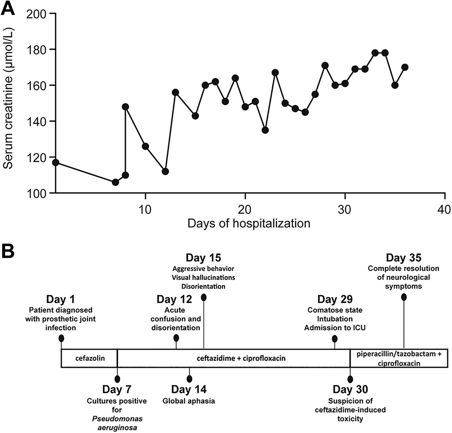
Creatinine levels over time and clinical case presentation. A) Time-course of serum creatinine levels during hospitalization. B) Timeline of the patient’s symptoms and treatment regimens.
The patient’s neurological symptoms worsened despite the discontinuation of anticholinergic medications, including amitriptyline and zopiclone. The electroencephalogram (EEG) demonstrated abnormally slow background activity suggestive of a diffuse encephalopathic state (Figure 3). The patient continued to demonstrate disorientation in time and place, agitation and aggressivity. Multiple oral and intramuscular haloperidol 0.5 mg doses were unsuccessful at alleviating agitation. The patient’s mental status deteriorated rapidly, eventually leading to a comatose state requiring intubation and transfer to the intensive care unit. Oral ciprofloxacin had been changed to intravenous ciprofloxacin, which was dose-adjusted to 400 mg every 24 hours according to a calculated Cockcroft-Gault clearance of less than 30 mL/min.
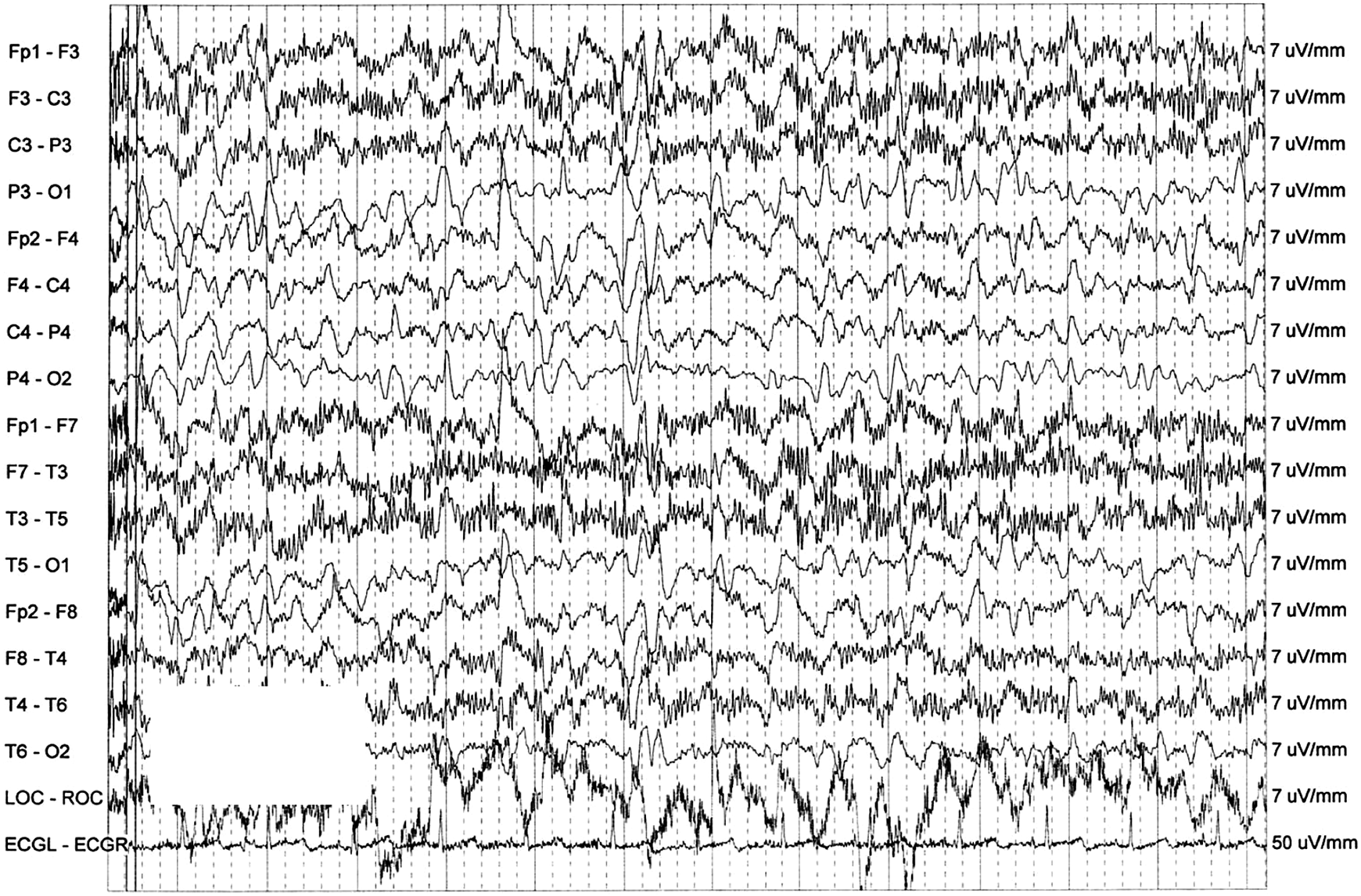
Electroencephalogram showing triphasic waves. The EEG reading was characterized by slow background activity and was poorly organized and polymorphic in the delta-theta range reaching 4.5 to 5 Hz. Generalized sharp wave complexes were seen with a frequency of 0.5 Hz. These discharges occasionally have a triphasic morphology. This is an abnormal EEG suggestive of a diffuse encephalopathic state.
The inflammatory markers were trending down and the patient was afebrile, suggesting that the prosthetic joint infection was adequately treated. The physical exam was not suggestive of meningitis. The patient was assessed by the infectious disease team who found no other infectious cause explaining the patient’s state.
Since no other etiologic factor could explain the patient’s symptoms, ceftazidime-induced neurotoxicity was suspected. Ceftazidime was discontinued and replaced with piperacillin/tazobactam 3.375 g intravenously every 6 hours. The patient’s mental status improved within 72 hours, allowing extubation. Neurological symptoms resolved completely over the next 5 days without needing further therapy. An MRI of the brain was then safe to perform and showed no signs of acute stroke. Considering the availability of other therapeutic alternatives, ceftazidime was not re-challenged. The patient remained on piperacillin/tazobactam and ciprofloxacin to complete 6 weeks total of antibiotic therapy.
Discussion
Ceftazidime is a broad-spectrum cephalosporin that is not metabolized and is primarily excreted unchanged by glomerular filtration. 9 Consequently, dose adjustment based on renal function is crucial in order to prevent ceftazidime accumulation and adverse reactions. 10 Cephalosporin-induced neurotoxicity is a rare but well-described phenomenon in the literature. A recent retrospective study based on data from the French Pharmacovigilance Database identified cefepime, ceftriaxone and ceftazidime as the cephalosporins most frequently associated with neurological adverse events. 2 Not surprisingly, neurotoxicity in patients treated with cephalosporins has mostly been reported in patients with renal dysfunction.3,4,11
Acute kidney injury is a frequent complication of total joint arthroplasty. 12 In the case presented here, the patient’s serum creatinine levels increased significantly during the course of treatment, suggesting that the decreased renal function was most likely the principal factor that led to an increased exposure to ceftazidime (Figure 2). The dosage of ceftazidime was not reduced in the context of renal impairment. Since ceftazidime is not metabolized and is excreted in its active form in the urine, dosage reduction is crucial in patients with underlying renal dysfunction. 8 Considering that this patient's Cockcroft-Gault creatinine clearance decreased below 30 mL/min, the appropriate ceftazidime dose adjustment would have been 2 g intravenously every 24 hours until renal function improved.
The main cause of kidney injury in this patient was likely prerenal and caused by reduced fluid intake. However, we cannot exclude that ceftazidime itself was a contributing factor, as a few studies have highlighted its potential negative impact on renal function.13,14 The severity of this patient’s symptoms resulting from an increased exposure to the drug emphasizes the importance of closely monitoring renal function in patients receiving this antibiotic, 15 particularly in elderly patients with chronic kidney disease.
While the precise mechanism behind cephalosporin-induced neurological adverse effects is still unclear, 16 evidence suggests that it involves their ability to competitively bind and antagonize the gamma aminobutyric acid (GABA) receptors in the central nervous system.1,17 Ceftazidime can cross the blood-brain barrier, 18 but its low lipophilic properties are predicted to limit its accumulation in the cerebrospinal fluid in the absence of meningeal inflammation.5,19 On the other hand, several lines of evidence suggest that kidney injury may affect the integrity of the blood-brain barrier, possibly through the release and reduced clearance of proinflammatory cytokines. 20 Interestingly, it has also been suggested that in the context of renal failure, the active transport of beta-lactam antibiotics from the cerebrospinal fluid may be impaired through competitive inhibition with accumulated toxic organic acids, such as creatinine and guanidino compounds. 21
We calculated a Naranjo score of 7 for this patient, indicating that the observed neurotoxicity was probably a consequence of ceftazidime administration. 22 The rapid improvement of the patient’s condition following the discontinuation of ceftazidime strongly supports this conclusion. This is in line with published reports on ceftazidime-induced neurotoxicity, which indicate that treatment discontinuation usually leads to symptom improvement within days. 4 In certain cases, interventions to control symptoms also included hemodialysis and administration of antiepileptic drugs or benzodiazepines. 23 In this patient’s case, ceftazidime discontinuation led to a complete remission of neurological symptoms after 5 days, without the need for dialysis or any adjunct medications.
Various neurological manifestations of cephalosporin intoxication have been reported, including seizures, myoclonus, aphasia, hallucinations, psychosis and coma.1,2,5 EEG findings in intoxicated patients are typically characteristic of metabolic encephalopathy with the presence of triphasic waves and diffuse slow activity, as observed in this patient (Figure 3). These features are however mostly nonspecific and may result from various causes, such as traumatic brain injury or pre-existing neurodegeneration. 24
Quinolones are also known to cause adverse neurological effects in the elderly. Quinolone-induced neurotoxicity usually presents as seizures, confusion, encephalopathy, myoclonus and psychosis.23,25 However, as ciprofloxacin was appropriately dose-adjusted for renal function, this agent was not considered likely contributive. The patient’s symptoms improved despite continuation of ciprofloxacin, further supporting the hypothesis that ciprofloxacin was not a primary etiological factor of the patient’s symptomatology.
Beta-lactam induced fever has been described as a potential cause of confusion. 26 In this case, the patient remained afebrile after initiation of ceftazidime and inflammatory markers suggested an appropriate response to therapy, which prompted the medical team to evaluate other possible causes of neurological impairment. In addition, delirium is common in frail and elderly inpatients and frequently poses a clinical challenge in terms of both diagnosis and management strategies. 27 These factors likely contribute to the difficulty of rapidly identifying cephalosporin-induced neurological toxicity in frail patients treated for serious infections. It is particularly striking that the significant delay in identifying the cause of this patient’s neurological symptoms led to serious clinical consequences, requiring the patient to be intubated and admitted to the intensive care unit. This further illustrates that cephalosporin-induced neurological adverse effects can easily be overlooked when establishing a differential diagnosis in hospitalized patients.
Furthermore, results from animal studies suggest that cephalosporins can induce neurotoxicity in a dose-dependent manner. 28 Monitoring of cephalosporin drug levels is not always readily available in clinical practice. Therefore, proper dose adjustment in high-risk patients, such as older adults with renal dysfunction, is crucial in order to minimize the potential risks of this adverse event. This population often requires dosing at a decreased frequency. Continuous EEG monitoring may be of value in diagnosing cephalosporin-induced neurological toxicity. 1 Moreover, increasing awareness of this potentially serious adverse event is essential for early detection and diagnosis of such toxicity.
Conclusion
This case report provides new insight on the potential severity of neurological toxicity with the use of ceftazidime if not detected in a timely manner. As demonstrated, delayed diagnosis can lead to an unfavourable patient outcome. This underscores the importance of suspecting drug-induced neurotoxicity in patients treated with this antibiotic and presenting acute onset confusion. Close monitoring of renal function is crucial to avoid such adverse events in the elderly. As beta-lactam therapeutic drug monitoring is starting to emerge, it will be interesting to see if such monitoring can prevent neurological toxicity associated with cephalosporins.
Footnotes
Authors’ Note
Laetissia Amirouche, Alexandra Cerulli-Kanellopoulos, Sébastien Landry and Véronique C. LeBlanc contributed equally to this article.
Acknowledgments
The authors would like to thank the patient presented in this case for allowing the sharing of her experience.
Declaration of Conflicting Interests
The author(s) declared no potential conflicts of interest with respect to the research, authorship, and/or publication of this article.
Funding
The author(s) received no financial support for the research, authorship, and/or publication of this article.