
Abstract
Growth hormone deficiency (GHD) is a prevalent consequence of traumatic brain injury (TBI) that can contribute to prolonged adverse symptoms and impaired quality of life. The overlap between symptoms of GHD and chronic TBI complicates the clinical picture, highlighting the need for precise diagnostic tools. Insulin-like growth factor 1 (IGF-1), a key mediator of growth hormone (GH) activity, is frequently utilized as a surrogate marker of GH status. However, IGF-1 levels may be normal in patients with confirmed GHD. Furthermore, low or low-normal IGF-1 is not specific to GHD, and provocative testing is required to confirm GHD in most patients with suspected deficiency. To provide a pragmatic clinical framework for interpreting IGF-1 z-score <0 in symptomatic patients after TBI, we reviewed the literature in the PubMed and PubMed Central databases addressing IGF-1 and GHD, differential diagnoses associated with low and low-normal IGF-1, and international guidelines on GH testing. Among TBI patients, GHD remains a leading cause of reduced IGF-1, but alternative diagnoses include malnutrition, chronic liver or kidney disease, poorly controlled diabetes mellitus, hypothyroidism, sepsis, and genetic conditions such as Laron syndrome. A systematic and evidence-based approach to IGF-1 interpretation is essential for distinguishing primary neuroendocrine dysfunction from secondary causes. The use of z-scores improves diagnostic precision by standardizing IGF-1 levels for age and sex. Our framework aids clinicians in integrating thorough neuroendocrine evaluation with broader medical assessments, clinical considerations, and comorbidities, thereby improving diagnostic accuracy and patient management. Future research should emphasize refined screening protocols for neuroendocrine dysfunction in TBI.
Keywords
Introduction
Growth hormone deficiency (GHD) is a prevalent consequence of traumatic brain injury (TBI) that can contribute to prolonged symptoms and impaired quality of life. These symptoms include persistent fatigue, mood and sleep disturbances, cognitive difficulties, altered body composition, reduced bone mass, dyslipidemia, and insulin resistance.1,2 The overlap between the symptoms of GHD and those of chronic TBI further complicates the clinical picture, highlighting the need for precise diagnostic tools. 3 The reported prevalence of GHD in the chronic phase of TBI is highly variable, ranging from 8% to 63%.4–10 Insulin-like growth factor 1 (IGF-1), a key mediator of growth hormone (GH) activity, is frequently utilized as a surrogate marker of GH status.11,12 Beyond mediating GH activity, IGF-1 impacts numerous physiological processes, including fetal growth; development of the renal, cardiovascular, and central nervous systems; and immune modulation. 13
There is an increased risk for GHD in any type of TBI, particularly in cases of repeated injury.14–17 Current guidelines identify dynamic GH stimulation testing as the only accepted method for diagnosis. 18 Due to their ease of measurement and relative cost-effectiveness, IGF-1 levels are often utilized as an initial screening tool. 14 Although low IGF-1 levels may suggest GHD, they are not diagnostic, and provocative testing remains necessary to confirm the diagnosis, unless multiple pituitary hormone deficiencies are present. Furthermore, normal IGF-1 values do not reliably exclude the diagnosis.12,19,20
The limited sensitivity and specificity of IGF-1 levels for GHD require multimodal neuroendocrine follow-up and a thorough understanding of the available diagnostic tools. Here, we provide a pragmatic approach for practitioners evaluating potential GHD in symptomatic TBI patients. We conducted a comprehensive literature review using PubMed and PubMed Central (PMC) to identify relevant peer-reviewed studies. Search terms included keywords related to IGF-1 and TBI. We build on this review by discussing current guidelines for screening, exploring common tests for diagnosing GHD, and highlighting key considerations regarding differential diagnoses for low and low-normal IGF-1 levels (IGF-1 z-score <0). By incorporating improved awareness and diagnostic accuracy, clinicians can better identify candidates for confirmatory testing and optimize management strategies for TBI patients with suspected neuroendocrine dysfunction.
Who Should Be Screened for GHD and When?
The decision to perform IGF-1 testing to screen for GHD should be individualized, considering injury severity and the presence of persistent or emerging symptoms suggestive of GHD. However, recommendations regarding the target population and timing vary. Certain expert recommendations support deferring GHD assessment in moderate-to-severe TBI (msTBI) to 12 months post-injury, given that early post-injury abnormalities may be transient and that resource-intensive confirmatory dynamic testing, which may only be offered at specialist centers, should be delayed until early-stage non-specificity has resolved.21–23 In comparison, some guidelines suggest waiting to screen for GHD until at least 6 months after TBI.14,24 Consistent with these recommendations, Tritos et al. emphasize that GH assessment should be deferred to several months post-injury and performed only after recognition and replacement of other pituitary deficiencies. 25 Earlier recommendations promoted assessing for neuroendocrine dysfunction in patients with mild TBI (mTBI) who required hospitalization for >24 h; who demonstrated imaging abnormalities such as brain swelling, intracranial bleeding, or skull fracture; or who displayed symptoms of hypopituitarism. 26 Finally, Yuen et al. test IGF-1 levels when patients with chronic-phase mTBI (i.e., >3 months post-injury) show symptoms of pituitary dysfunction. 27 At our institution, we typically conduct GHD assessments at 3–6 months after injury for patients with mTBI and persistent symptoms of pituitary dysfunction. For adults with msTBI, we find it optimal to assess IGF-1 levels at 3–6 months post-injury, with follow-up testing at 12 months, particularly when clinical features persist or suggest hypopituitarism.28–30 Early measurement helps identify patients who may benefit from closer clinical monitoring, provides early assessment for panhypopituitarism, and facilitates appropriate 12-month follow-up. We recommend additional pituitary evaluation in patients who are notably symptomatic 6–12 months after TBI, particularly those with other pituitary deficiencies and a clear clinical indication for further testing. For those with persistent symptoms and an IGF-1 z-score <0, we perform diagnostic testing with the glucagon stimulation test (GST) at least 12 months post-injury, followed by initiation of treatment when GHD is identified. 31 For patients with a high pre-test probability and an IGF-1 level z-score >0, a diagnostic test such the GST may be considered. 32
Due to physiologic decreases in GH secretion, IGF-1 levels naturally decline with age. 33 Notably, this decline is accompanied by an increase in IGF-1 receptor transcription in the brain, suggesting an attempted compensatory mechanism. 34 To account for this physiologic decline, IGF-1 levels are reported with an associated z-score, which reflects the number of standard deviations an IGF-1 concentration falls above or below the mean for a patient’s age and sex. These z-scores are calculated by clinical laboratories based on large reference population datasets, enabling clinicians to more accurately interpret IGF-1 levels by accounting for age and sex-related variance. IGF-1 assessment using accurate and reliable assays is crucial in optimizing care for this patient population, as substantial inter-assay variability has been observed.18,35 However, distinguishing anticipated aging-related reductions in IGF-1 from pathological reductions due to underlying disease can still be challenging. In older adults with TBI, low and low-normal IGF-1 levels should be interpreted in the context of clinical symptoms and compared against age-adjusted reference ranges to determine whether additional testing for GHD is necessary.
The 2019 guidelines from the American Association of Clinical Endocrinologists and the American College of Endocrinology provide one of several structured approaches to diagnosing adult GHD. According to these guidelines, initial screening with sporadic measurements of serum IGF-1 is insufficient to diagnose GHD. 18 Consequently, confirmatory dynamic (i.e., GH stimulation) testing should be pursued except when there is evidence of multiple (≥3) pituitary hormone deficiencies together with a low IGF-1 level (z-score < –2). GH stimulation tests should be performed only in response to reasonable clinical suspicion of GHD, with the intent to treat after the diagnosis is made. Furthermore, although TBI is a recognized cause of GHD, GH stimulation testing should be deferred until at least 12 months after the event, given that GHD may be transient in these patients. 18
The insulin tolerance test (ITT) is considered the gold standard for diagnosing GHD. The ITT involves administering intravenous insulin to induce hypoglycemia, followed by serial measurements of GH levels. Although this approach is highly sensitive and specific, it is contraindicated in the elderly, patients with ischemic heart disease, cardiac arrhythmias, and seizure disorders and requires close monitoring and experienced personnel due to the potential for severe hypoglycemia.18,36 Given safety concerns regarding ITT, alternatives such as the GST are recommended. 18 Glucagon stimulation testing involves the administration of glucagon, with subsequent measurement of GH levels at specified intervals. The GST is considered safer than the ITT but has a longer duration and requires intramuscular injection.18,31
Macimorelin stimulation testing involves serial GH measurements after oral administration of a GH secretagogue. The macimorelin approach is noted to be safe and well-tolerated, with a high sensitivity and specificity. Limitations to this assay include drug price and the potential for drug interactions that prolong the QT interval or reduce plasma macimorelin concentrations, leading to false positive results. 18 GHRH-arginine testing involves the injection of GHRH, followed by infusion of arginine, and has demonstrated sensitivity and specificity comparable to the ITT.37–39 The macimorelin and GHRH-arginine tests are unavailable for use in the United States currently. Table 1 compares these four tests for confirming GHD in adults.
Three Common Confirmatory Tests for Growth Hormone Deficiency
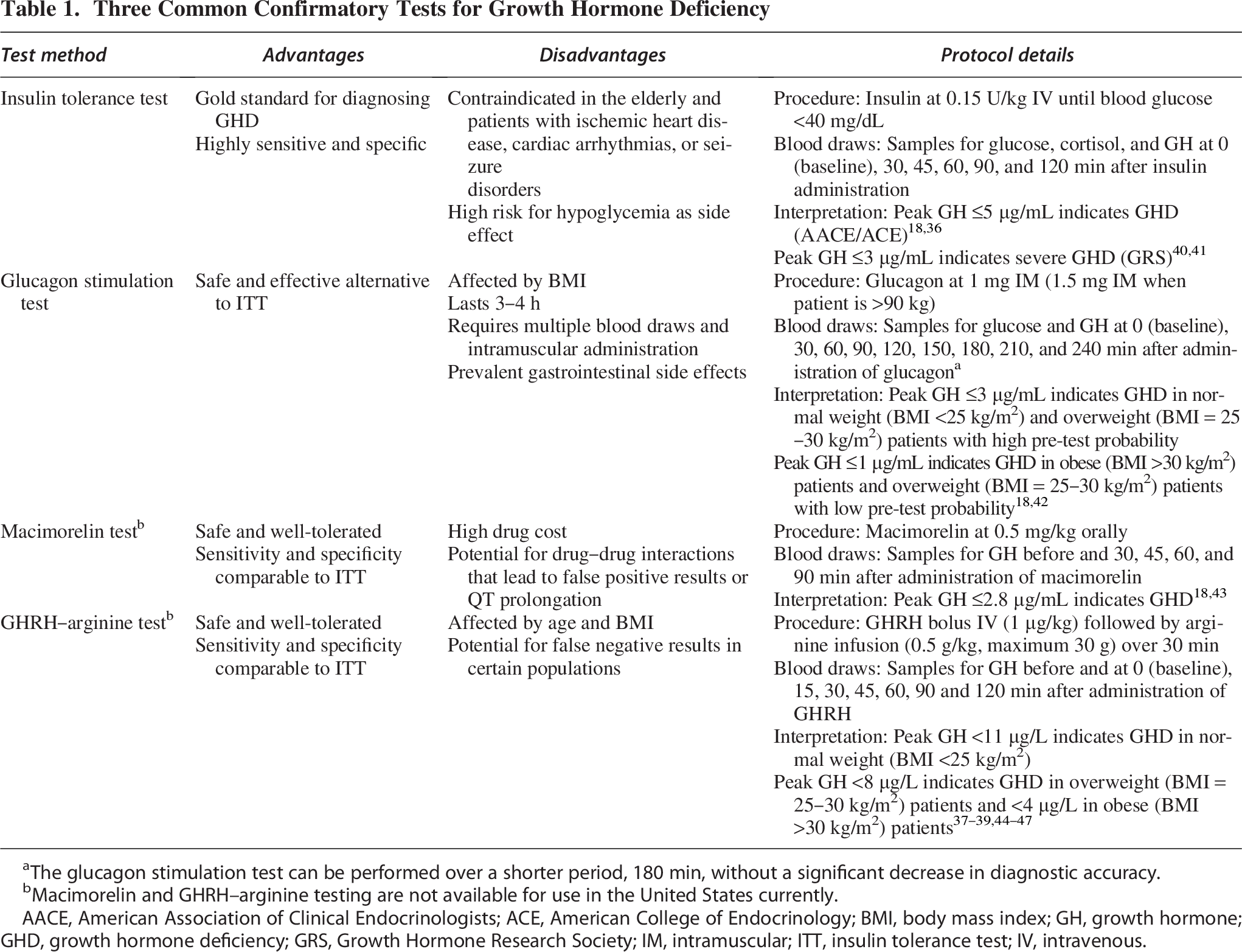
The glucagon stimulation test can be performed over a shorter period, 180 min, without a significant decrease in diagnostic accuracy.
Macimorelin and GHRH–arginine testing are not available for use in the United States currently.
AACE, American Association of Clinical Endocrinologists; ACE, American College of Endocrinology; BMI, body mass index; GH, growth hormone; GHD, growth hormone deficiency; GRS, Growth Hormone Research Society; IM, intramuscular; ITT, insulin tolerance test; IV, intravenous.
Differential Diagnoses for Low and Low-Normal IGF-1 Levels After TBI
IGF-1 levels are considered within the normal range when z-scores are between +2 and −2, with a low IGF-1 defined as a z-score less than −2, and z-scores between 0 and −2 considered low-normal. As demonstrated in the literature and through our own experience, interpreting a low-normal IGF-1 (z-score < 0) level in a TBI patient with symptoms of GHD requires careful consideration of potential underlying causes. Our pragmatic framework for accomplishing this task includes a comprehensive list of differential diagnoses comprising systemic, endocrine, metabolic, and physiological factors that may affect IGF-1 production, clearance, and activity (Fig. 1).
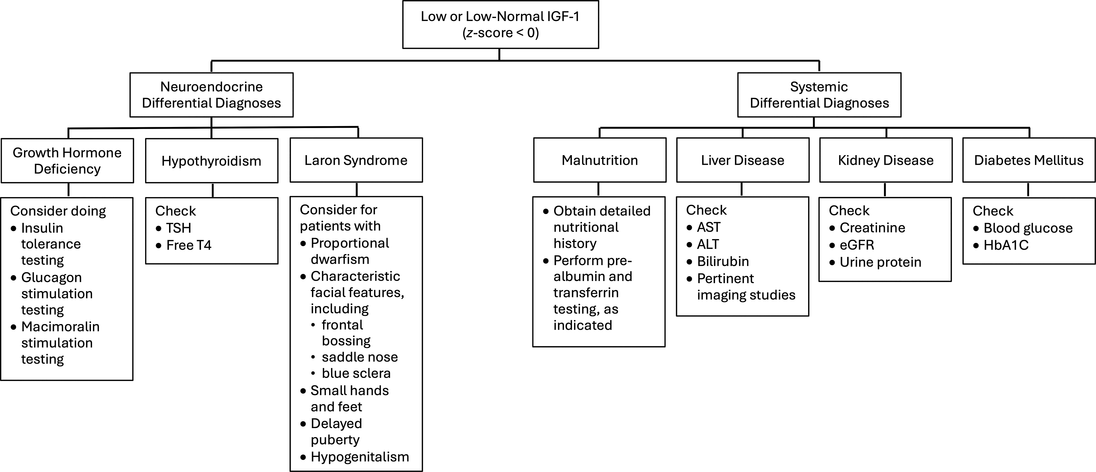
Pragmatic framework for the diagnosis of a low or low-normal IGF-1 level (z-score <0) in patients with TBI and symptoms of hypopituitarism. IGF-1, insulin-like growth factor 1; TBI, traumatic brain injury.
A primary concern regarding a low or low-normal IGF-1 level after TBI is GHD. TBI is a recognized cause of hypopituitarism, which manifests due to a combination of ischemia, cytotoxicity, and neuroinflammation after the injury. 14 A recent meta-analysis indicated that the pooled prevalence of post-traumatic hypopituitarism was 33%, while GHD alone was present in 18%. 48 Symptoms of GHD in adults include fatigue, cognitive impairment, mood and sleep disturbances, impaired quality of life, abnormal body composition, reduced bone mass, and dyslipidemia and insulin resistance. 3 IGF-1 levels have limited sensitivity and specificity for GHD after TBI, and a low IGF-1 is not diagnostic for GHD.2,49 Furthermore, IGF-1 z-scores >0 do not exclude the diagnosis, as prior studies have identified patients with confirmed GHD despite normal or elevated IGF-1 concentrations.20,50 A normal IGF-I level does not rule out GHD and requires confirmatory stimulation testing, while a low IGF-I level, without confounding conditions, suggests GHD and supports further diagnostic evaluation. 32
Thyroid gland hormones exert a regulatory effect on the GH–IGF-1 axis, inducing secretion of GH by the pituitary gland. 51 Hypothyroid states have been associated with significantly reduced concentrations of IGF-1. 52 TBI is associated with an increased risk of deficiencies across all hypothalamic and pituitary axes, including the thyroid axis. Moreover, symptoms of hypothyroidism, including fatigue, weight gain, depression, and sleep changes, can overlap with symptoms of chronic TBI.3,53 Any patient with persistently low IGF-1 levels should undergo a comprehensive assessment of the pituitary axis, including thyroid function testing via measurement of thyroid-stimulating hormone and free thyroxine levels.
Because protein and caloric intake tightly regulate IGF-1 synthesis, nutritional status plays a crucial role in IGF-1 homeostasis. Malnutrition, particularly deficits in protein intake, leads to impaired hepatic IGF-1 synthesis. 54 Oguni et al. describe a condition of GH resistance that is induced in the setting of malnutrition, with suppressed IGF-1 synthesis and increased GH production to prevent hypoglycemia. 55 In addition, various conditions that compromise the nutritional state, including anorexia nervosa, cystic fibrosis, and inflammatory bowel disease have been associated with significantly reduced IGF-1 levels.56–58 In TBI patients, nutritional deficiencies may manifest due to decreased oral intake in the setting of neurological impairment, gastrointestinal dysmotility and dysbiosis, or a hypermetabolic and hypercatabolic state.59,60 Dietary history, body weight trends, and markers of nutritional status, such as prealbumin and transferrin levels, should all be considered when interpreting a low and low-normal IGF-1 level in a TBI patient. 61
Because the liver is the primary site of IGF-1 production, hepatic dysfunction can significantly impact circulating IGF-1 levels. 56 Chronic liver disease due to cirrhosis, non-alcoholic fatty liver disease, or viral hepatitis decreases hepatic functional capacity and thus impairs IGF-1 synthesis.62,63 In cirrhosis, reduced IGF-1 levels correlate with disease severity and have been proposed as a prognostic biomarker.63–65 In patients with TBI, liver dysfunction can be secondary to pre-existing conditions, prolonged use of hepatotoxic medications, or systemic inflammation triggered by trauma. The patient’s medical history should be considered alongside laboratory markers, such as liver function tests, including aspartate aminotransferase, alanine aminotransferase, bilirubin, and albumin. Existing imaging studies, if available, should be reviewed to gain insight into the possible contribution of hepatic dysfunction to low and low-normal IGF-1 levels.
IGF-1 levels may reflect renal dysfunction, as well as hepatic effects. In patients with chronic kidney disease, reduced renal clearance of IGF-binding proteins (IGFBPs) leads to increased binding and sequestration of circulating IGF-1, thereby decreasing its bioavailability. 66 In addition, chronic kidney disease is associated with resistance to GH stimulation, impairing hepatic IGF-1 synthesis.66,67 TBI patients with pre-existing chronic kidney disease, recurrent dehydration, or medication-related nephrotoxicity should be assessed for renal dysfunction as a contributor to low and low-normal IGF-1 levels. Routine evaluation of serum creatinine, estimated glomerular filtration rate, and urine protein levels can help determine the presence and extent of renal impairment and resulting insults to IGF-1.
Diabetes mellitus, particularly when poorly controlled, can disrupt IGF-1 homeostasis. Insulin plays a crucial role in IGF-1 synthesis; by increasing hepatic sensitivity to GH, insulin promotes IGF-1 production and increases its bioavailability by suppressing IGFBP-1. 68 In addition, poorly controlled diabetes mellitus is associated with increased glycation of IGFBP-3, thereby increasing its affinity for IGF-1 and further decreasing bioavailability of IGF-1. 69 Additionally, research using animal models found that hyperglycemia exacerbates neuroinflammation after TBI. 70 Diabetic patients with TBI should undergo regular evaluation of their hemoglobin A1c and fasting glucose levels to assess their glycemic control state and whether impaired insulin signaling may be influencing IGF-1 levels.
Sepsis is another important systemic condition that may affect IGF-1 equilibrium. Both IGF-1 and IGFBP-3 were significantly reduced in septic patients when compared to healthy controls, with a further decrease observed in patients with septic shock. 71 Inflammatory and metabolic alterations during sepsis profoundly impact GH–IGF-1 axis activity, potentially due to endotoxin-mediated resistance to GH and reduction of hepatic IGF-1 synthesis.72–74 Notably, in cases of pediatric sepsis, levels of IGF-1 were lower in non-survivors.75,76 Recognition of recent infection and sepsis as a potential contributor to depressed IGF-1 concentrations is essential to avoid misattributing such findings to underlying GHD.
In the genetic disorder Laron syndrome, deletions or mutations in the GH receptor gene lead to insensitivity to GH. 77 As a consequence, these GH receptor mutations lead to markedly decreased IGF-1 levels despite normal or increased levels of GH. 78 Although severe short stature typically prompts the diagnosis of Laron syndrome during childhood, milder forms may remain unrecognized through adulthood. 79 Though exceedingly rare in the general population, with a prevalence of 1–9 per 1,000,000, Laron syndrome should nevertheless be considered in cases of unexplained low IGF-1 levels in the setting of normal or elevated GH levels, particularly in patients with a history of unexplained growth delays. 80
Conclusions
In patients with TBI and symptoms of hypopituitarism, the decision to evaluate for GHD should be guided by clinical presentation. A low-normal IGF-1 z-score <0 in a person with a TBI is a complex clinical picture that warrants careful interpretation. Although GHD remains the most likely diagnosis, other systemic conditions including malnutrition, chronic liver or kidney disease, uncontrolled diabetes mellitus, hypothyroidism, sepsis, and various genetic conditions must be assessed as potential contributors. Comprehensive clinical evaluation, laboratory testing, and confirmatory endocrine studies are essential to differentiate between primary pituitary dysfunction and secondary causes of low and low-normal IGF-1 levels. By systematically addressing each differential diagnosis, clinicians can ensure appropriate management and optimize recovery for patients with neuroendocrine dysfunction after TBI.
Transparency, Rigor, and Reproducibility Summary
This short communication adheres to principles of transparency, rigor, and reproducibility in clinical research. The article is based on a review of peer-reviewed literature, with all references cited appropriately to ensure traceability of the data. The differential diagnoses for low IGF-1 levels were compiled through established pathophysiological mechanisms, supported by prior research. The utilization of standardized IGF-1 z-scores enhances reproducibility across different age groups and clinical contexts.
Footnotes
Author Disclosure Statement
The authors have no conflicts of interest to declare.
Funding Information
No funding was received for this study.