






Abstract
Episodic memory deficits are frequently encountered following stroke. Rehabilitation of these deficits is often associated with short term effects that do not persist at follow up times. Neuromodulation tools acting on alterations of excitation/inhibition dynamics could be useful for rehabilitation of episodic memory. Prism adaptation with visual field deviation ipsilateral to the affected hemisphere can specifically modulate brain excitability and lead to improvement of cognitive deficits in post-stroke patients. Here we investigated whether prism adaptation followed by digital cognitive training could induce long term improvement of episodic memory deficits in stroke patients as compared with traditional rehabilitation.
Sixty stroke patients were recruited for the study. Thirty patients were treated with prism adaptation combined with serious games targeting executive processes, with a ten days protocol (
The main results showed a significant long term effect of the experimental treatment on both immediate and delayed recall phases of the visual episodic memory task. The effects were not linked to the affected hemisphere (right vs. left), nor to the hemorrhagic or ischemic type of stroke nor to the cortical or subcortical site of lesion.
These findings show for the first time an improvement of long term memory in stroke patients following the use of a medical device combining prism adaptation and digital cognitive training, paving the way to novel rehabilitation techniques for cognitive deficits in stroke.
Introduction
Memory impairment is frequently reported after stroke and it is one of the cognitive disorders that persists more over time (Gallucci et al., 2024; O'Sullivan et al., 2023; Piras et al., 2011; Schaapsmeerders et al., 2013). In fact, the percentage of stroke patients complaining about memory impairments ranges between 23% and 55% at three months after the event and between 11% and 31% at one year follow up times (Snaphaan & de Leeuw, 2007).
Episodic memory deficits revealed by neuropsychological tests are also present as subjective memory deficits in daily life (van Rijsbergen et al., 2017) and have been shown to have a negative effect on the patient's functional independence (Middleton et al., 2014).
A Cochrane review investigated whether stroke patients who received cognitive rehabilitation for memory problems have better outcomes than those given no treatment or a placebo control (das Nair et al., 2016). The review examined the results of 13 clinical trials involving 514 patients. The main results showed an effect of cognitive rehabilitation in the short term (i.e., usually within four weeks), but not in the longer term (i.e., at the second assessment phase after a minimum of three months), thereby suggesting that there is limited evidence to the effectiveness of memory rehabilitation in stroke.
Indeed, although rehabilitation of memory is frequently part of rehabilitation protocols for stroke patients, it is still unclear whether rehabilitation plays a role in the recovery of memory functions per se, or it rather facilitates patients’ adaptation to daily life problems, without a direct action on memory processes. Therefore, there is need for new methods and protocols that have an impact on episodic memory tasks following stroke.
Neuromodulation tools acting on brain excitability could be one of such methods.
Indeed, alterations of excitation/inhibition dynamics have been suggested as a key mechanism occurring after stroke (Harquel et al., 2024) and increased excitation of the affected hemisphere has been reported as a positive marker for rehabilitation improvement in patients with focal stroke (Cicinelli et al., 2003). In addition to techniques that directly impact on brain excitability, such as transcranial magnetic stimulation, recent evidence suggests that a visuomotor adaptation to optical prisms shifting the visual field to the left or right space increases cortical excitability of the brain hemisphere ipsilateral to visual field deviation (Bracco et al., 2017, 2018; Magnani et al., 2014). Activation induced by prisms involves a cerebello-parietal-frontal network (Panico, Sagliano, et al., 2022; Schintu et al., 2022; Wilf et al., 2019). A medical device (Mindlenses Professional) that combines visuomotor adaptation to prisms, aimed to modulate this circuit, with digital cognitive training through serious games, targeting basic processes subserved by that circuit, was recently tested in stroke patients (Danesin et al., 2023) and proved to be able to rehabilitate attention, short term memory, working memory and Stroop-like abilities in stroke, with an effect measured at short term, i.e., immediately after the end of a 10-days rehabilitation protocol (Oliveri et al., 2023).
In the present study we tested the hypothesis that PA and digital cognitive training using serious games could also provide a new non-invasive mean for improving performance on episodic memory tasks at medium and long term follow up times in left and right hemispheric stroke patients. Given the recognized power of rightward prism adaptation in improving visuospatial attention in right-brain-damaged patients (Rossetti et al., 1998), an additional aim was to correlate episodic visuospatial memory deficits with the presence of spatial neglect, and the modulation of visuospatial memory with the modulation of spatial neglect.
Patients and Methods
Sixty patients with first ever ischemic or hemorrhagic stroke involving the right or the left hemisphere were recruited at the Santa Lucia Foundation IRCCS for a randomized controlled trial testing the efficiency of the medical device Mindlenses Professional on different cognitive functions. We report the results regarding verbal and visual episodic memory functions.
The protocol was approved by the Ethical Committee of Fondazione Santa Lucia IRCCS (CE/Prog 891) and it was conducted in compliance with the Declaration of Helsinki. Exclusion criteria were: age below 18 years, previous ischemic or hemorrhagic stroke, presence of comorbid neurological or psychiatric disorders, severe vigilance deficits, severe verbal comprehension deficits restricting the ability to comprehend neuropsychological tests and instructions, impaired motor control of both hands.
Patients enrolled in the trial were randomly assigned to one of two groups: an experimental group, treated with the medical device Mindlenses Professional; a control group, treated with standard cognitive rehabilitation. A block randomized controlled design was adopted.
Table 1 shows the main demographic and clinical characteristics of the enrolled patients at baseline and at the three follow-up times and Figure 1 shows the trial flow diagram. At each evaluation time, no significant differences were observed between groups regarding age, education, lesion side, ischemic vs. hemorrhagic etiology, time from stroke occurrence.

Trial flow diagram (RBD: right brain damaged; LBD: left brain damaged).
Demographic and Clinical Characteristics of Individual Patients.
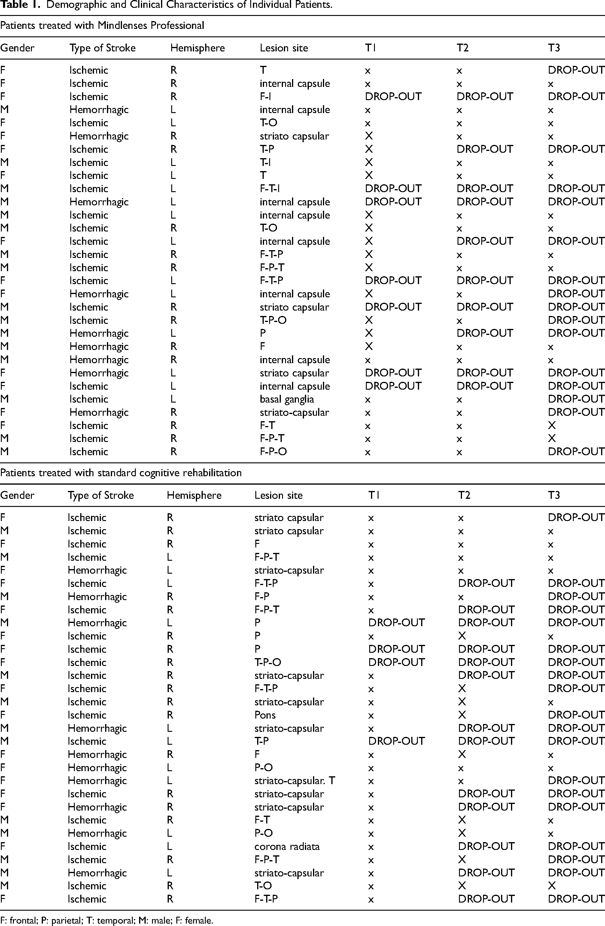
F: frontal; P: parietal; T: temporal; M: male; F: female.
Patients of the group treated with the medical device Mindlenses Professional received a training using this medical device that integrates PA and serious games. The protocol lasted 10 sessions, about 40 min each, distributed over a mean of two weeks.
In each session, the training started with a digitized PA session. Patients were guided by a neuropsychologist to wore prismatic lenses with a power of 20 dioptries. Based on previous results (Oliveri et al., 2023), right-brain-damaged (RBD) patients wore rightward prism lenses and left-brain-damaged (LBD) patients wore leftward prism lenses. The adaptation procedure was done using a 11” tablet, horizontally oriented, that was positioned at a distance of 53 cm from the patients’ eyes, with a position aligned to the patient's sagittal midplane. Visual targets represented by black squares of 1° of visual angle were randomly presented on a white background in one of three spatial positions of the tablet's screen: in the centre of the screen or with a lateralization of 21° to the right or the left space. Following presentation of each target, the patient was asked to point as fast and as accurately as possible toward the target, trying not to correct the movement during its trajectory, using the ipsilesional hand. The visual targets persisted on the screen until patients’ touch or after a fixed interval of 1 s
The procedure used was divided into the following 3 phases:
Serious games were planned as to train basic processes like sustained and divided attention, inhibitory control, visuospatial search, semantic associations, mathematical reasoning and working memory. All games were implemented using a dynamic difficulty algorithm, i.e., adapting velocity of stimulus presentation and game difficulty to the single patient's performance. All games were executed with the supervision of a neuropsychologist. Details of the digital training procedure through serious games are delivered in Oliveri et al. (2023) and Conte et al. (2024).
Patients of the group treated with standard rehabilitation received a routine cognitive rehabilitation program, adjusted to their clinical status, with 10 consecutive sessions distributed over a mean of two weeks, aimed to stimulate the same cognitive functions targeted in the experimental group: sustained and divided attention, inhibitory control, visuospatial search, semantic associations, mathematical reasoning and working memory.
Outcome Measures
All patients were assessed with two episodic memory tests at T0 (before standard or experimental training), at T1 (after rehabilitation) and at two follow up times: 3 months from the end of rehabilitation (T2) and 6 months from the end of rehabilitation (T3).
To evaluate the nature and severity of memory dysfunction and to track changes in memory function over time, two outcome test measures were used. Episodic verbal memory was assessed using the Auditory Verbal Learning task (AVLT) measuring both immediate and delayed recall of a 15 words list after 20’ delay (Carlesimo et al., 1996).
Visual episodic memory was assessed using the Rey-Osterrieth complex figure test, measuring both immediate and 20′ delayed memory reproduction of an abstract, hardly verbalizable complex figure (Carlesimo et al., 1996). Spatial attention was measured using the Line Bisection test (Schenkenberg et al., 1980) and the Bell's cancellation test (Gauthier et al., 1989).
Results
Auditory Verbal Learning Test: Immediate Recall
ANOVA for repeated measures with Group (experimental vs. control) and Hemisphere (right vs. left) as between-subject factors and Time (T0 vs. T1 vs. T2 vs. T3) as within-subjects factor was run on the number of recalled items.
ANOVA showed a significant main effect of Time (F = 4.1; p = 0.01; partial η2: 0.16), while the factors Group (F = 1.6; p = 0.2; partial η2: 0.07) and Hemisphere (F = 0.5; p = 0.4; partial η2: 0.02) were not significant. The interactions Group×Time (F = 1.1; p = 0.3; partial η2: 0.05), Group×Hemisphere (F = 0.4; p = 0.5; partial η2: 0.01), Time×Hemisphere (F = 0.8; p = 0.5; partial η2: 0.03), Group×Time×Hemisphere (F = 1.8; p = 0.1; partial η2: 0.08) were not significant.
Auditory Verbal Learning Test: Delayed Recall
ANOVA for repeated measures with Group (experimental vs. control) and Hemisphere (right vs. left) as a between-subject factors and Time (T0 vs. T1 vs. T2 vs. T3) as within-subjects factor was run on the number of recalled items after a 20′ delay interval.
The main factors of Group (F = 0.6; p = 0.4; partial η2: 0.03), Time (F = 2.4; p = 0.07; partial η2: 0.1), Hemisphere (F = 1.2; p = 0.2; partial η2: 0.05) were not significant. The interactions Group×Time (F = 1.8; p = 0.1; partial η2: 0.08), Group×Hemisphere (F = 0.1; p = 0.7; partial η2: 0.008) were not significant, while a trend towards significance emerged from the interaction Group×Time×Hemisphere (F = 2.3; p = 0.08; partial η2: 0.1).
Inspection of Figure 2 shows a trend of increasing number of recalled items across evaluation times in right brain damaged patients treated with the device Mindlenses Professional (Experimental).

Average number of recalled items by patients treated with the device
Rey-Osterrieth Complex Figure Test: Immediate Recall
ANOVA for repeated measures with Group (experimental vs. control) and Hemisphere as between-subject factors and Time (T0 vs. T1 vs. T2 vs. T3) as within-subjects factor was run on the number of recalled items.
ANOVA showed a significant main effect of Time (F = 14.04; p < 0.001; partial η2: 0.4), while the main effects of Group (F = 0.8; p = 0.4; partial η2: 0.04) and Hemisphere (F = 1.8; p = 0.2; partial η2: 0.08) were not significant.
The interaction Group×Time was significant (F = 9.4; p < 0.001; partial η2: 0.3). The interaction Time×Hemisphere showed a trend towards significance (F = 2.5; p = 0.07; partial η2: 0.1). The interaction Group×Time×Hemisphere was not significant (F = 0.4; p = 0.7; partial η2: 0.02).
Tukey's post hoc comparisons showed that in the group of patients treated with the device Mindlenses Professional (Experimental) the number of recalled items increased significantly across the evaluation time points (T1 vs. T0: t = −4.3; p = 0.007; T2 vs. T0: t = −7.8; p < 0.001; T3 vs. T0: t = −7.2; p < 0.001; T3 vs. T1: t = −3.8; p = 0.02). No significant changes in the number of recalled items were observed in the group of patients treated with standard rehabilitation (Control) (Figure 3).

Average number of recalled items by
Rey-Osterrieth Complex Figure Test: Delayed Recall
ANOVA for repeated measures with Group (experimental vs. control) and Hemisphere as between-subject factors and Time (T0 vs. T1 vs. T2 vs. T3) as within-subjects factor was run on the number of recalled items after a 20′ delay interval.
ANOVA showed a significant main effect of Time (F = 11.8; p < 0.001; partial η2: 0.39) while the main effects of Group (F = 0.2; p = 0.7; partial η2: 0.01) and Hemisphere (F = 0.9; p = 0.3; partial η2: 0.05) were not significant.
The interaction Group×Time was significant (F = 3.8; p = 0.01; partial η2: 0.18). The interactions Time×Hemisphere (F = 0.3; p = 0.8; partial η2: 0.02), Group×Hemisphere (F = 0.9; p = 0.3; partial η2: 0.05) and Group×Time×Hemisphere (F = 0.7; p = 0.6; partial η2: 0.03) were not significant.
Tukey's post hoc comparisons showed that in patients treated with the device Mindlenses Professional (Experimental) the number of recalled items increased significantly across the evaluation time points (T1 vs. T0: t = −3.6; p = 0.03; T2 vs. T0: t = −4.3; p = 0.008; T3 vs. T0: t = −6.1; p < 0.001; T3 vs. T1: t = −3.5; p = 0.04). No significant changes in the number of recalled items were observed in the group of patients treated with standard rehabilitation (Control) (Figure 4).

Average number of recalled items by
Spatial Attention Tasks
To evaluate the potential contribution of spatial attention deficits in the effects on spatial episodic memory, ANOVA was conducted on the scores of line bisection and Bell's cancellation tests, with Group (experimental vs. control) and Hemisphere (right vs. left) as between-subjects factors and Time (T0 vs. T1 vs. T2 vs. T3) as within-subjects factor.
Line Bisection Task
ANOVA showed a significant main effect of Time (F = 6.7; p < 0.001; partial η2: 0.2). The factors Group (F = 0.08; p = 0.8; partial η2: 0.004), Hemisphere (F = 1.9; p = 0.2; partial η2: 0.08) and the interactions Group×Time (F = 0.5; p = 0.7; partial η2: 0.02), Group×Hemisphere (F = 0; p = 1; partial η2: 0), Time×Hemisphere (F = 0.2; p = 0.9; partial η2: 0.01), Group×Time×Hemisphere (F = 0.5; p = 0.7; partial η2: 0.02) were not significant. The scores in the line bisection task increased parallelly across the evaluation times in both patients treated with the device Mindlenses Professional and in patients treated with standard rehabilitation.
Bell's Cancellation Task
The main effects of Group (F = 0; p = 1; partial η2: 0) and Time (F = 1.2; p = 0.3; partial η2: 0.06) were not significant, while the factor Hemisphere showed a trend towards significance (F = 3.3; p = 0.08; partial η2: 0.15).
The interaction Group×Time was significant (F = 3.2; p = 0.02; partial η2: 0.14) while the interactions Group×Hemisphere (F = 1.7; p = 0.02; partial η2: 0.08), Time×Hemisphere (F = 0.2; p = 0.8; partial η2: 0.01), Group×Time×Hemisphere (F = 0.2; p = 0.9; partial η2: 0.01) were not significant.
Despite the trend towards improvement across evaluation times in the group of patients treated with the device Mindlenses Professional (Experimental), Tukey's post hoc comparisons did not show any significant difference in the number of cancelled items in the two groups.
Regression analyses were done to evaluate the contribution of spatial attention performance, as measured with the line bisection and Bell's tasks, to visual episodic memory performance in the two groups.
As to immediate recall of the Rey Osterrieth complex figure (adjusted R square: 0.27), performance at T3 was predicted by performance at the Bell's task at T3 (estimate: 1.7; t = 2.2; p = 0.04), without significant difference between the two patients’ groups.
As to the delayed recall of the Rey Osterrieth complex figure (adjusted R square: 0.32), performance at T3 was again predicted by performance at the Bell's task at T3 (estimate: 2.056; t = 2.8; p = 0.01), without significant difference between the two patients’ groups.
Discussion
The main result of the present study is that prism adaptation with a deviation ipsilateral to the affected hemisphere, combined with digital training of inhibition, selection and set shifting processes, induces a long-term improvement in episodic memory in stroke patients. The rehabilitative effect on episodic memory is increasing over time, starting at the end of the ten sessions protocol and becoming significant at 6 months follow up. No significant effects on episodic memory are observed in stroke patients of the control group, treated with conventional rehabilitation.
The observed improvement mainly regards visual memory, as tested with both immediate and delayed recall of the Rey-Osterrieth Rey figure, while only a trend towards long term improvement of delayed recall is observed in the case of verbal episodic memory, as tested with Auditory verbal learning test.
These results complement recent findings of cognitive improvement in stroke patients treated with the same device (Oliveri et al., 2023). However, differently from what previously reported for attention and working memory functions, where the improvement was just measured after a ten-day session rehabilitative protocol, in the case of episodic memory as tested in the present trial the rehabilitative effect is evident at longer follow up times. Different factors could account for this result. The putative mechanism of action of prism adaptation, relying on increased excitability of parieto-frontal connections (Schintu et al., 2022), could explain a long-term effect on episodic memory through long term modulation of parietal- hippocampal and frontal-hippocampal connections. Further evidence on this issue could be provided by dedicated studies coupling prism adaptation and cognitive training with functional neuroimaging of parieto-frontal as well as cortico-hippocampal networks. Another factor could be related to hemispheric-specific activation for long term retrieval. According to the HERA model (Habib et al., 2003), the right hemisphere is “dominant” for the retrieval phase in long term memory. Although in the present study we did not find hemispheric specific effects in the cognitive modulation, it is worth noting that at T3 follow up time the ratio of right- vs. left brain-damaged patients was 15/9. Since right brain damaged patients in the present protocol are treated with rightward prism deviation, and right brain damaged patients were the majority of those treated at T3 follow up time, one could hypothesize that the long-term effect on episodic memory is linked to a modulation of right-hemispheric retrieval processes. An argument favoring this explanation is also the greater effect observed for visual episodic memory, as tested with the Rey-Osterrieth complex figure, as compared with verbal episodic memory. In fact, although memory improvements at T3 times were observed for both tests, in the case of the visual episodic memory test the improvement is much more significant and it starts even before, i.e., at 3 months follow up. Further findings on the issue of hemispheric specific effects of the medical device and the associated protocol could be provided by future studies better balancing the number of right- and left-brain damage at follow up times.
The use of digital cognitive training following PA would sustain, rather than directly determine, cognitive effects, exploiting the background of modulation of cortical excitability induced by prisms. In each rehabilitation session, serious games were applied in a temporal window immediately following that of prism adaptation. Rather than as a cognitive stimulation boosting a specific cognitive process, the digital cognitive training following prism-adaptation induced neuromodulation is thought as a mean of stimulation of basic trans-domain processes of inhibition, selection and set-shifting. These processes are challenged in all the serious games of the medical device and this executive training is aimed to translate its effects to retrieval of memory traces (Haobo et al., 2024).
One could argue whether the observed improvement of episodic memory does not reflect a modulation/restitution of memory processes per se but rather a better compliance of the patients to life situations, also linked to improvement of neglect symptoms. Indeed, the presence of neglect was balanced in control and experimental groups of the present trial. Moreover, although neglect scores at the cancellation test were significant predictors of the visual episodic memory improvement at follow-up (with higher scores at the Bell's test associated with higher scores in the episodic memory test, for both immediate and delayed recall), this association was valid for both experimental and control groups, while memory improvement was selectively observed in the experimental group.
Although the routine use of prism adaptation for treatment of spatial neglect has recently been questioned (Székeli et al., 2023), it is well known that prism adaptation can modulate spatial neglect in right brain damaged patients (Rossetti et al., 1998). However, this factor alone cannot explain the modulation of episodic visual memory observed in the present study.
The long-term modulation of episodic memory observed in the present study is at odds with the majority of findings reported in neurorehabilitation literature for the application of either conventional or new digital devices for cognitive rehabilitation. Indeed, these studies showed a lack of long-term effectiveness and durability of the effects on episodic memory, a finding that hampers the significance of rehabilitation itself.
As to the effect sizes, our results document large effect sizes for both immediate and delayed recall for either verbal or visual episodic memory tests. This result is in contrast with previous findings on memory rehabilitation on stroke, which report small to moderate effect sizes of the adopted methods (see das Nair et al., 2016 for a review).
New digital therapies and technological tools are expected to increase patients’ compliance and adherence to therapy as compared with traditional ones (Antonenko et al., 2021). A recently published metanalysis (Laver et al., 2017) showed that virtual reality in stroke patients could represent an effective method for rehabilitation of executive functions, visuospatial abilities and even memory (see De Simone et al., 2023). Contradictory findings have been observed as to the duration of its effects and their transfer to activities of daily living. In addition, a general concern regards the presence of side effects, such as cybersickness, that limit the application of such procedure to all patients, requiring careful selection of the patients and individualized procedures. In this sense, although prism adaptation lacks the immersive nature of virtual reality, it does not present with the same side effects and it could be easily applied to the majority of stroke patients, beyond its classical application for treatment of contralesional neglect (see also Panico, Arini, et al., 2022; Panico, Sagliano, et al., 2022).
Footnotes
Funding
The authors disclosed receipt of the following financial support for the research, authorship, and/or publication of this article: This work was supported by the EIT Health,
Declaration of Conflicting Interests
Two of the authors (MO and ADG) are shareholders of the company producing the medical device used in the present study.
Data Availability Statement
The Authors declare the absence of shared data.