






Abstract
Background
Parkinson's disease (PD) is a neurodegenerative disorder characterized by loss of dopaminergic neurons and α-synuclein accumulation. Despite extensive research, there remains a shortage of effective disease-modifying medicines, which is due in part to the failure to translate molecular insights into clinically useful models and diagnostics.
Objectives
This review will summarize recent developments in the PD pathophysiology and diagnostic, therapeutic and experimental models and will focus on the newer in vivo, in vitro and bioengineered in vivo platforms.
Methods
Recent studies on animal models, patient-induced pluripotent stem cell (iPSC) systems, three-dimensional (3D) bioprinting, neuroimaging, and biomarker discovery have been critically examined to determine their translational potential and limitations.
Results
Traditional animal models are effective at replicating dopaminergic degradation but fall short of fully replicating progressive and systemic elements of Parkinson's disease. iPSC-derived neurons and 3D-bioprinted constructs are more genetically specific and cellularly complex, allowing for patient-relevant modeling and medication screening. Advances in imaging and molecular biomarkers aid in earlier detection; nevertheless, no cross-validation or platform standards has been established.
Conclusion
Combining cellular, molecular, and bioengineered models with clinical diagnostics has the potential to improve translational accuracy and accelerate the development of disease-modifying treatments. A cross-platform system is critical for improving the predictive validity of preclinical studies in Parkinson's disease.
Keywords
Introduction
Parkinson disease (PD) is a progressive, chronic, and neurodegenerative disease that mostly influences the functioning of the motor system, and, as the disease progresses, results in important cognitive and behavioral disorders (Permadi et al.; Tong et al., 2025). It is a disease that affects more than 10 million individuals globally, and its occurrence sharply rises with age, as it is the second-most prevalent neurodegenerative disease following Alzheimer (Selvaraj & Piramanayagam, 2019). In addition to being widespread on a global scale, PD is a rising health issue in the population because of its prolonged duration of disease and high cost of treatment, as well as the severe effects it has on the quality of life of patients (Peng et al., 2025). Even though the etiology of PD has remained a conundrum, it is universally accepted that its pathogenesis is a complex interaction of genetic predispositions and environmental exposures (Reynoso et al., 2024). About 10–15% of PD cases are inherited, the rest are sporadic and occur due to interactions between genes and the environment like through pesticide exposure, rural living, or head trauma (Mhyre et al., 2012).
PD can be described at the neuropathological level in terms of progressive neuronal degeneration of dopaminergic neurons in the substantia nigra pars compacta, one of the areas where dopamine is produced, which is essential to motor coordination (Gong et al., 2024; Salamon et al., 2020). This leads to the dopamine deficiency that directly causes the characteristic motor symptoms of PD, i.e., resting tremor, rigidity, bradykinesia, and postural instability (Tai et al., 2024). Nevertheless, PD is not only a movement disorder, as non-motor symptoms include depression, sleep, constipation, and olfactory dysfunctions, which usually develop years before motor symptoms appear, which suggest that the process of neurodegeneration is more complex and systemic (Stoker & Greenland, 2018). The pathogenesis of PD consists of an interaction of such mechanisms as protein aggregation, dysfunction of mitochondria, impaired proteostasis, and chronic neuro inflammation (Fleming et al., 2022; Li et al., 2025). These mechanisms are all responsible to the vulnerability and degeneration of dopaminergic neurons. It is also becoming more probable that peripheral systems, such as the gut-brain axis, can affect central pathology (Ebrahim et al., 2025; Tumpa et al., 2025). The combination of these findings inclines towards a complex and system-level model of PD pathogenesis. The clinical presentation is similar to other parkinsonian syndromes, and biomarkers that are currently available do not have adequate sensitivity and specificity to detect the disease early (Ma et al., 2024; Zhang et al., 2021). These restrictions indicate the necessity of better diagnostic models involving clinical, molecular, and technological methods.
A number of past reviews have examined single factors of PD, including α-syn aggregation or mitochondrial dysfunction, but not many have put these together with new information in microbiome studies, diagnostic novelty, and modeling (Sari & Djuwita, 2025). In contrast to the previous reviews that concentrated on individual mechanisms, it uses a cross-disciplinary approach, which links cellular pathology, peripheral involvement, bio-marker generation, and bioengineered modelling systems to enhance translational applicability. It is necessary to overcome these limitations through greater interconnection between mechanistic study, clinical phenotyping and high-quality modeling platforms. The paper takes a critical and integrative view in assessing the current advances in PD research. Instead of an attempt to give a pure descriptive summary we are comparatively evaluating experimental models, biomarkers and therapeutic strategies according to construct validity, predictive validity, translational relevance and clinical applicability. Specific focus is made on the identification of platforms and ways that can be most effective in bridging mechanistic discovery and patient-centered outcomes. This review seeks to improve the understanding of the most effective clinical progress in PD, by prioritizing the evidence based on translational impact to clarify what is known, as well as those strategies most likely to make meaningful contributions to clinical progress in PD.
Methodological Approach and Integrative Framework
This narrative review is based on structured searches of PubMed, Scopus, and Web of Science, covering literature published between 2002 and 2025. Keywords included “Parkinson's disease,” “biomarkers,” “neuroimaging,” “iPSC,” “3D bioprinting,” “animal models,” and “disease-modifying therapy.” Priority was given to peer-reviewed studies with strong methodology and clear translational relevance. Case reports, non-English articles, and non-peer-reviewed sources were excluded.
An integrative framework was applied to compare evidence across molecular mechanisms, diagnostics, therapeutics, and experimental models. The focus was not only on summarizing findings but on evaluating construct validity, predictive strength, and clinical applicability. Emphasis was placed on approaches with the greatest potential for translational impact.
Clinical Features and Diagnosis
Motor Manifestations
Clinically, PD is mainly diagnosed by its cardinal motor symptoms, bradykinesia, rigidity and tremor as product of underlying loss of dopaminergic neurons and forms the basis of diagnosing the condition. In the course of PD, motor behaviors (including blinking and swallowing) tend to deteriorate automatically; therefore, this condition may lead to too much drooling and dry eyes (Arboleda-Montealegre et al., 2021; Henry & Lai). Other frequent gait problems include shuffling steps and freezing episodes, which also play a major role in impaired mobility (C. Sharma, 2021).
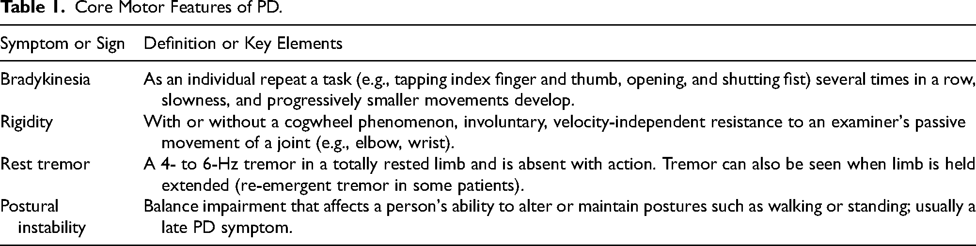
Table 1 provides a brief summary of the major motor features in PD and their clinical definition. The symptoms are assessed on a regular basis and they are assessed using standardized instruments, such as the Unified PD Rating Scale (UPDRS) which is used to establish the severity of the disease and the progression as well. Intersection of motor symptoms with atypical parkinsonian syndromes, including multiple system atrophy and progressive supranuclear palsy, and variability of the age of onset of symptoms and response to treatment may make it difficult to classify PD at an early and accurate stage. Besides, the use of clinical rating scales like the Unified PD Rating Scale (UPDRS) creates subjectivity and variability of inter examiner. These constraints make it clear that objective and quantifiable motor biomarkers are needed to increase the accuracy and standardization of diagnosis.
Core Motor Features of PD.
Non-Motor Manifestations
Non-motor manifestations, which become more and more prominent in the clinical practice, are the indicators of diagnosing the disorder at its early stages and a considerable impact on the quality of life of the patient. First, neuropsychiatric disturbances are some of the most common non-motor characteristics. These are anxiety, apathy, depression, anhedonia and hallucinations. These symptoms are mainly associated with the changes in dopaminergic and serotonergic neurotransmission. In addition, they have serious implications on the emotional well-being and social functioning of patients (Poewe, 2008). Moreover, cognitive dysfunction is also likely to develop with the progression of PD. The patients can develop executive deficit, memory impairment and ultimately, dementia. Later disease stages also report visual hallucinations and psychotic episodes, which are frequently being aggravated by the use of dopaminergic therapy (Tong et al., 2025).
The autonomic dysfunction is also crucial as it shows the prevalence of neurodegeneration typical of PD. Early symptoms include orthostatic hypotension, urinary and bowel issues as well as sexual dysfunction which may become more severe over time (Peña-Zelayeta et al., 2025). The prevalence of these manifestations is often underdiagnosed, even though they are highly prevalent and burden clinical practice. Moreover, the most frequent sleep-related issues are the REM sleep behavior disorder (RBD), insomnia, restless legs syndrome (RLS), and excessive daytime somnolence (Dodet et al., 2024). RBD is especially remarkable among them as a possible prodromal biomarker of synucleinopathies (Martínez-Morales & Liste, 2012).
Besides these, non-motor spectrum encompasses symptoms associated with sense of smell such as olfactory dysfunction (hyposmia), pain, as well as uncomfortable feelings following infection (Naia et al., 2025). It is crucial to highlight that one of the most prominent symptoms of Parkinson's disease is typically a loss of smell, which can be a clinical manifestation in those who are predisposed to the disease (Fullard et al., 2017). Table 2 encompasses the most pertinent non-motor symptoms of PD subdivided into the functional domain. They are now discovering that these characteristics are very relevant to the diagnosis, and that they have a significant impact on their patients’ quality of life. Non-motor symptoms are becoming important in the diagnosis and treatment of Parkinson's disease, despite their difficulty to interpret clinically. The majority of the signs, such as depression, constipation, and sleep difficulties, are not disease-specific and can be found in other neurological or systemic disorders, limiting their diagnostic usefulness. Furthermore, inconsistency in symptom reports and the lack of standardized screening procedures contribute to under diagnosis and conflicting prevalence estimates. These limitations demonstrate the need for verified, measurable non-motor indicators to improve early diagnosis and patient classification.
Non-Motor Features of PD.

Diagnostic Tools and Biomarkers
PD cannot be divided during diagnosis, as it is established on the basis of the definite history and conducting a thorough physical examination (Olegário et al., 2018). The existing diagnostic models are mainly based on clinical criteria that focus on the assessment of motor symptoms (Armstrong & Okun, 2020). Still, early and prodromal stages of the disease remain by far the most problematic ones as they are usually accompanied by diagnostic dilemmas due to the overlapping of the symptoms with those of other neurodegenerative disorders like essential tremor, multiple systems atrophy (MSA) and progressive supranuclear palsy (PSP) (Angelini et al., 2024).
In scenarios whereby clinical manifestation is not exhaustive in the diagnosis process, imaging tools emerge as useful tools. In particular, dopamine transporter (DaT) single-photon emission computed tomography (SPECT) has been approved by the FDA of the United States to differentiate between PD and essential tremor (Wu & Wu, 2020). This technique demonstrate less incorporation of the tracers in striatum and particularly in the putamen in PD and in all cases normal in essential tremor. Other neuroimaging agents such as positron emission tomography (PET), magnetic resonance imaging (MRI), optical coherence tomography (OCT) or transcranial sonography (TCS) have been investigated to rule out progression of the disease, and differentiate between PD and atypical parkinsonisms (J. Y. Lee et al., 2022; Zhang & Liu, 2013).
Although these imaging applications improve diagnostic confidence, they lack sufficient specificity when used independently and may overlap with atypical parkinsonian syndromes. In addition to imaging, biomarker studies have also presented intense advances in the recent past. Monogenic forms of PD have been elucidated in terms of genetic markers that include mutations in the PTEN-induced kinase 1 (PINK1), Parkin, DJ-1 (PARK7), Synuclein-α (SNCA), Leucine-rich repeat kinase 2 (LRRK2), and Glucosylceramidase Beta (GBA) (Kim & Alcalay, 2017; Planas-Ballvé & Vilas, 2021).
These are the genes allegedly involved in mitochondrial dynamics, protein-breakage processes, and lysosomal physiology which are the processes that dominate the pathogenesis of PD. Simultaneously, blood, cerebrospinal fluid (CSF), saliva and tissue biopsies to obtain biochemical biomarkers are eagerly explored (Angius et al., 2023). As an example, the concentration of CSF α-syn, neurofilament light chain (NfL), and DJ-1 proteins have been suggested to be detected as a marker of neuronal injuries and disease developments (Hafsteinsdóttir et al., 2024). As illustrated in Figure 1, accurate diagnosis of PD increasingly depends on a multimodal approach that integrates clinical evaluation, neuroimaging, and molecular biomarkers. Imaging modalities such as DaT-SPECT and PET are constrained by cost, accessibility, and limited ability to distinguish PD from atypical parkinsonian syndromes. Likewise, genetic and biochemical biomarkers are variable across cohorts, do not have standardized assay procedures, and tend to indicate late pathological alterations, but not early pathophysiology. These challenges underscore the necessity of standardized validation frameworks and a multimodal diagnostic strategy integrating clinical, imaging, and molecular data (Räty et al., 2025).

Parkinson's disease biomarkers.
Phosphorylated α-synuclein (α-syn) in cerebrospinal fluid, skin, and salivary gland biopsies is the most promising emerging biomarker because the quality of the assays is currently undergoing significant improvements in improving standardization, and the pathological specificity of the measurement is high. There are also Salivary α-syn species and plasma neurofilament light chain (NfL), which present feasible minimally invasive options, which are associated with disease progression (Rissardo & Fornari Caprara, 2025). Further developments in seed amplification-assays and multiplex biomarker panel enhance these candidates to clinical translation in the future. There is also controversy on the accuracy of DaT-SPECT in early or atypical PD cases.
Furthermore, non-motor symptoms such as hyposmia, REM sleep behavior disorder (RBD), constipation, and depression are now recognized as potential prodromal markers. These symptoms often precede the appearance of motor manifestations by several years and could play a pivotal role in future early diagnostic algorithms (Tolosa et al., 2021). Despite ongoing efforts, Delenclos et al. (2016) note that a definitive diagnosis remains impossible until postmortem histopathological confirmation is made (Delenclos et al., 2016).
Sensitivity and Specificity of Biomarkers and Limitations of Current Diagnostic Modalities
In spite of significant advances, the existing biomarkers of PD have low diagnostic accuracy. CSF α-syn has moderate sensitivity (75% to 85%) and specificity (60% to 80%) in differentiating PD and controls but is not able to differentiate it and atypical parkinsonian syndromes (Delenclos et al., 2016; Rajmohan et al., 2025). Neuronal injury is reported by neurofilament light chain (NfL) and DJ-1 but is not a disease-specific marker, whereas genetic markers, including LRRK2 and SNCA mutations, have high specificity but are only observed in familial PD, which represents 15% of total cases (Angius et al., 2023; Kim & Alcalay, 2017).
Neuroimaging techniques, such as DaT-SPECT, PET, and MRI, increase the confidence of the diagnosis but have significant limitations. DaT-SPECT is not a reliable tool to differentiate PD and multiple system atrophy or progressive supranuclear palsy, whereas PET and MRI have limitations in terms of high cost, technical inconsistency, and accessibility (J.-Y. Lee et al., 2022; Zhang & Liu, 2013). In addition, the majority of the imaging indicators are manifestations of dopaminergic loss at late stages and not at preclinical pathology. Overall, each diagnostic approach contributes differently to PD assessment. Imaging improves diagnostic confidence but lacks disease specificity. Genetic markers are highly specific, yet they apply to only a small proportion of patients. Fluid biomarkers are promising for monitoring disease processes, although their reproducibility remains inconsistent across studies. Prodromal clinical features may support early suspicion, but they are not exclusive to PD. Therefore, multimodal integration currently offers the highest translational potential.
Treatment Options
PD treatment spans pharmacologic and non-pharmacologic domains, including surgical and experimental modalities. This multifaceted approach is essential due to the diverse and progressive nature of both motor and non-motor symptoms (Muleiro Alvarez et al., 2024). Management Large reviews in management can be categorized broadly to consist of non-pharmacological interventions (including physical therapy, exercise, and rehabilitation), pharmacological therapies (to treat the symptoms), surgical and device-assisted treatment (including deep brain stimulation), and new experimental modalities, such as virtual reality-based rehabilitation (McBenedict et al., 2024; Shikama Dias et al., 2024). Some of the agents and devices developed at this time, including Rytary, Duopa, and advanced DBS systems remain part of modern practice in treatment and have influenced future innovations in delivering therapies. In the period between 2021 and 2025, a number of improvements have greatly increased PD management options as indicated in Table 3. The Exablate Neuro system was expanded approved by the FDA to use unilateral and bilateral focused ultrasound pallidotomy to address motor symptom control (Abusrair et al., 2022). The P2B001 Phase III trial in 2022 showed that extended-release pramipexole and rasagiline are effective with fewer side effects, and that it could be used as an early PD treatment (Hauser et al., 2022). ABBV-951 (Vyalev™) was FDA-approved in 2023 as a continuous subcutaneous levodopa/carbidopa infusion to provide stable dopaminergic delivery and reduce “off” periods (NCT04380142). In 2024, Crexont®, an extended-release oral levodopa/carbidopa formulation, gained approval for managing motor fluctuations (Hauser et al., 2025). Ongoing Phase IIb–III trials of Prasinezumab, UB-312, and Exenatide-PD3 highlight the shift toward disease-modifying strategies, including α-syn immunotherapy and GLP-1 receptor agonists (Pagano et al., 2021; Yu et al., 2022).
Therapies to Treat Parkinson's Since 2014.
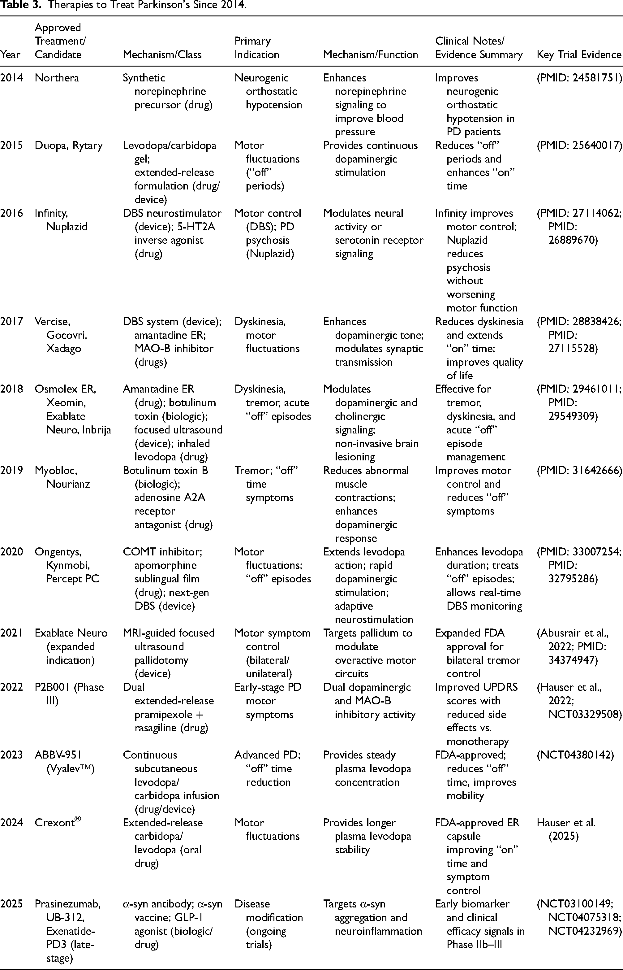
Regimens consisting of levodopa are the most effective interventions to treat symptoms of motor control and still are the hallmark of PD treatment. Adjunct treatments, i.e., MAO-B inhibitors, COMT inhibitors, dopamine agonists, and infusion systems, provide moderate-added value in the sense that they decrease muscle fluctuations and increase the duration of the on time. Conversely, new disease-modifying approaches, including α-syn immunotherapies and GLP-1 receptor agonists, are biologically promising, but have not been proven to have definite clinical efficacy.
Non-Pharmacologic Therapies
Non-pharmacologic treatments are essential in comprehensive management of Parkinson-s disease (PD), especially regarding enhancement of the quality of life of patients and managing the motor and non-motor symptoms (Adamiec et al., 2024). These interventions involving structured exercise, dietary alterations, rehabilitative interventions and supplements, complement pharmacologic management and are increasingly supported as evidence-based adjuncts rather than alternative therapies (Breasail et al., 2022).
Exercise-Based Interventions
It is well known that exercise poses comprehensive improvements in the management of PD, such as mood enhancement, sleep, increase of energy, and cognitive facilitation (Almikhlafi, 2023; Emig et al., 2021). In addition to these, it has a beneficial impact on motor control, improving the factors of gait, grip strength, balance, and coordination, and slowing the loss of dopamine in preclinical models and lowering constipation.
Various research studies have examined certain exercise interventions. As an example, the report of Panunggal et al. (2025) indicated that in a cardiovascular study of nine patients with PD, the combination of treadmill training resulted in persistence of improved motor symptoms (Panunggal et al., 2025). In the same manner, Ridgel et al. (2009) reported a 35-percent gain in motor scores of Unified Parkinson Disease Rating Scale (UPDRS) using forced exercise in ten subjects with mild to moderate Parkinson disease (PD) (Ridgel et al., 2009). Hackney and Earhart (2008) provided evidence that Tai Chi showed a great difference in enhancing balance among 33 PD participants. All this evidence proves the efficacy of exercise as a key element of non-drug therapy (Hackney & Earhart, 2008). Exercise improves mood, sleep, energy, and cognition, while enhancing motor control, balance, and coordination (Almikhlafi, 2023; Emig et al., 2021). Studies show treadmill, forced cycling, and Tai Chi improve motor scores and balance (Panunggal et al., 2025; Ridgel et al., 2009). Among them forced exercise and Tai Chi are always superior in motor and balance enhancement than unstructured exercise, which underscores the importance of training specificity and intensity control.
Dietary Strategies
Besides the physical activity, dietary habits play a significant role in managing the symptoms. Jackson et al. (2019) also pointed out the importance of a moderated diet that is rich in fruits and vegetables. More water and fiber is beneficial to reverse constipation, which is one of the most prevalent non-motor symptoms (Jackson et al., 2019). It is particularly important to note that protein-rich meals may interact with levodopa, so it is advisable that the patient should separate protein consumption at least one hour before taking a drug to maximize its effect (Rusch et al., 2023). Fruits, vegetables, and fiber based balanced diets will relieve constipation and aid in the absorption of medicine (Jackson et al., 2019; Rusch et al., 2023).
Rehabilitative Therapies
Supportive therapies such as physical, occupational, and speech therapy provide essential tools for managing PD-related limitations (Ransmayr, 2011). Physical therapy can help enhance motor functions and movement range as well as endurance and correct problems such as instability when walking, abnormal gait, and bradykinesia (Alameer et al., 2024). Radder et al. (2017) state that occupational therapy positively affects the quality of life through recommendations of adaptive tools and fine motor skills refinement (Radder et al., 2017). McDonnell et al. (2018) revealed that speech therapy namely the Lee Silverman Voice Treatment (LSVT) increases the voice clarity and swallowing (McDonnell et al., 2018). Moreover, such interventions as LSVT BIG and Expiratory Muscle Strength Training (EMST) deliver a focused solution to increase the amplitude of movement and subjects to deglutition, respectively (Saleem et al., 2025). Physical, occupational, and speech therapies improve movement, independence, and communication (Alameer et al., 2024; McDonnell et al., 2018; Radder et al., 2017).
Nutritional Supplementation
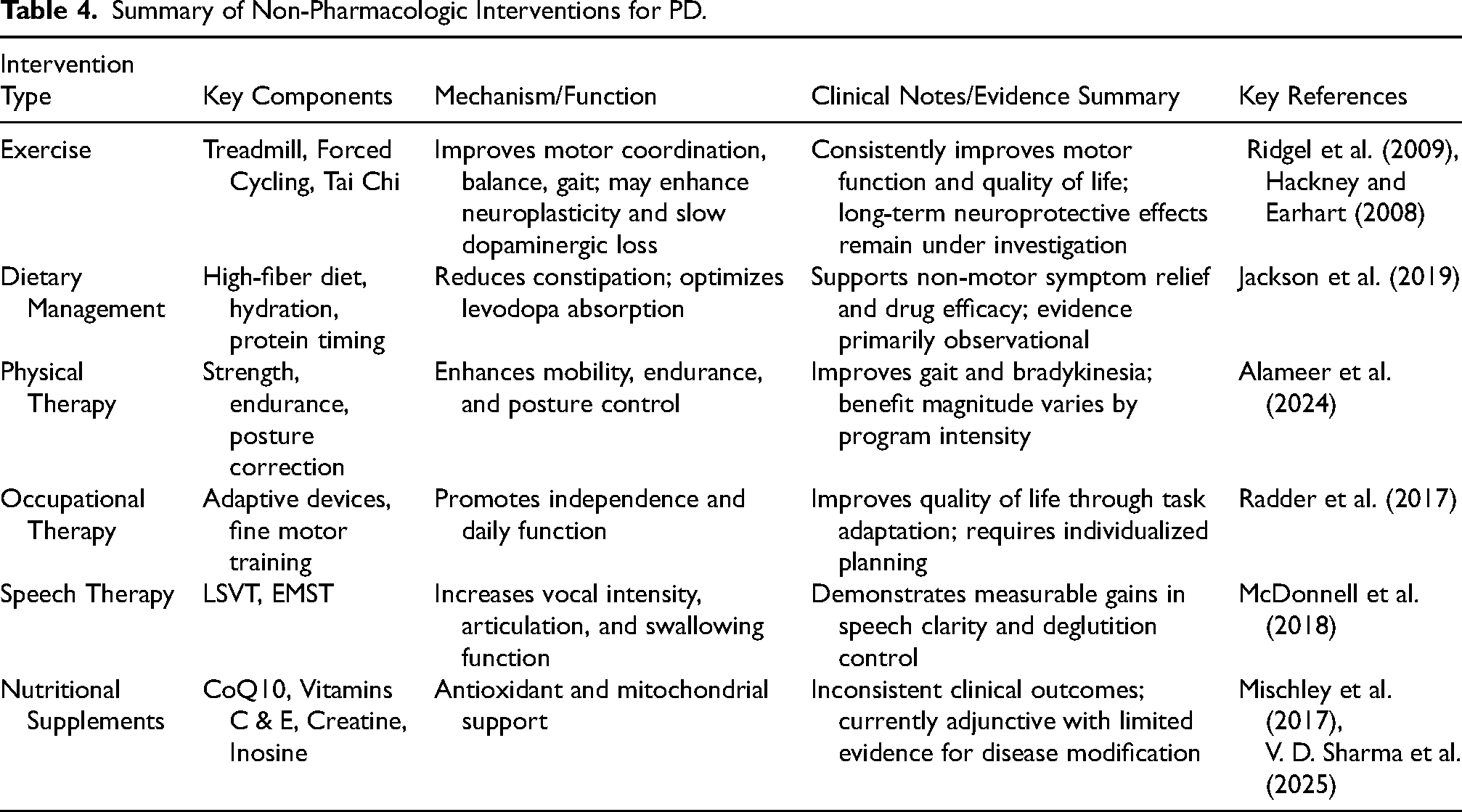
The recent reports on the use of nutritional supplements as adjuncts to slow the progression of PD are in favor of this treatment (Sharma et al., 2025). In a cohort study of 1, 053 self-reported PD patients, it was discovered that there is the potential value in Coenzyme Q10, Vitamin E, Vitamin C, Creatine and Inosine (Mischley et al., 2017). These agents are currently studied intensively and defined as neuroprotective though studies into the matter are still ongoing (Investigators, 2014). Table 4 summarizes major non-pharmacologic interventions, outlining their core components, mechanisms, and supporting evidence. In addition to exercise and rehabilitative strategies, nutritional supplements such as CoQ10, Vitamins C and E, Creatine, and Inosine have been explored as adjunctive approaches in PD management.
Summary of Non-Pharmacologic Interventions for PD.
Pharmacologic
The sequence of pharmacological treatment of Parkinson disease (PD) is mainly to establish dopaminergic balance and to increase motor stability (Tolosa et al., 2021). To start with, levodopa and motor fluctuation–directed agents constitute the major stone in PD treatment (Ferreira et al., 2022).
Rytary, an immediate and extended-release levodopa combination, prolongs symptom management, by evening out the drug levels in the plasma (Mittur et al., 2017). Likewise, Opicapone, a catechol-O-methyltransferase (COMT) inhibitor that can be taken once a day, increases the duration of levodopa and contributes to shortening the periods of the lack of effect. Inhaled levodopa (Inbrija) provides a fast effect in acute stabilization of acute attacks of the off phenomena, first of all, in circumstances when the intake of drugs by mouth is impossible (Hoy, 2019; Paik, 2020). In addition, monoamine oxidase-B (MAO-B) and COMT inhibitors are frequently used as adjunct therapies. Safinamide (Xadago), a newer MAO-B inhibitor, demonstrates particular benefit in patients experiencing motor fluctuations when added to levodopa regimens (Bhidayasiri et al., 2023).
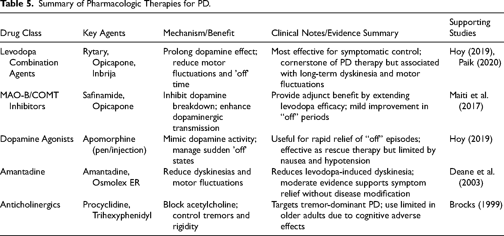
In addition, dopamine agonists, like apomorphine, are of key importance in the management of acute motor symptoms. Apomorphine pens and subcutaneous injections are very efficient in the management of sudden off episodes or early morning akinesia (Ceylan et al., 2022). These prescriptions bypass the absorption in the gastrointestinal tract and are rapid acting, invaluable to patients that display fluctuating symptoms. Other than that, pharmacologic agents classes are employed in order to provide a specific symptom control. Elevated dose of amantadine is an antiviral medication resuming its application in levodopa-induced dyskinesia treatment (Elkurd et al., 2018). Amantadine is also effective with different subtypes of PD as demonstrated by Crosby et al. (2010). It possesses a long-acting formulation, Osmolex ER, that offers long-term symptomatic relief to patients with motor complications (Deane et al., 2003). Meanwhile, anticholinergic agents like Procyclidine and Trihexyphenidyl are employed in treatment of the presentations only in tremor-pre-eminent forms. These drugs reduce spillover production of acetylcholine that is a significant source of tremor in PD. However, they have potential side effects in their mind and therefore; they are very selective in their use thereby restricted to older people (Brocks, 1999). A comparative analysis of the key pharmacologic treatments, mechanisms, and evidence (supporting) is presented in Table 5 (Bhidayasiri et al., 2023; Deane et al., 2003).
Summary of Pharmacologic Therapies for PD.
Surgical and Device-Aided Therapies
The problematic cases that can be treated with surgical and device-assisted approaches are those that cannot be addressed with pharmacologic measures. These modalities focus on motor complications, which do not respond to medications, and they provide better symptom control in moderate to severe PD (V. D. Sharma et al., 2020). Table 6 is a summary of the main surgical and infusion-based approaches, their mechanism and clinical considerations. The best evidence of long-term motor improvement is shown in properly chosen patients using deep brain stimulation (DBS). A non-invasive procedure, focused ultrasound (FUS), is applicable especially to tremor-dominant PD, but there is limited long-term data. Infusion devices can be used to enhance motor fluctuations, but this necessitates constant care of the device. Cautious patient selection on the basis of age, cognitive conditions, and symptoms profile is critical towards best results.
Comparative Summary of Surgical and Device-Aided Therapies in PD.
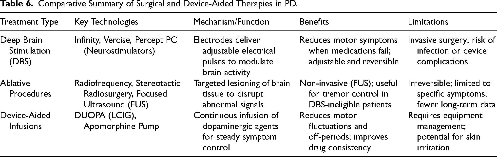
Deep brain stimulation (DBS) currently has the strongest evidence for sustained motor improvement in appropriately selected patients. Focused ultrasound (FUS) is a less intrusive option, especially for tremor-dominant PD, but long-term outcome data are sparse. Device-assisted infusions significantly minimize motor fluctuations, although they are accompanied with equipment burden and administration issues. As a result, thorough patient selection based on age, cognitive state, symptom profile, and comorbidities is critical for improving therapy success.
Deep Brain Stimulation
DBS) is regarded as the best surgical procedure in terms of PD. It is the implantation of electrodes into the precise brain areas (usually, it is the subthalamic nucleus) which are linked with a neurostimulator implanted in the chest with the help of lead wires and extensions (Umemura, 2021). This system provides regulated electrical pulses that will normalize abnormal brain activity and attenuate motor symptoms, including tremor, rigidity, and bradykinesia (D. J. Lee et al., 2018). The key components are depicted in the Figure 2 and they consist of the neurostimulator implanted over the chest which is linked via an extension cable and lead to an electrode that is located into the target part of the brain like the subthalamic nucleus. The system provides regulated electrical stimulations to adjust the activity of neurons and reduce motor symptoms PD causes.

Schematic representation of a DBS system.
DBS is especially useful when drugs are no longer effective or they cause unbearable side effects (Hariz & Blomstedt, 2022). Infinity, Vercise, and Percept PC are commercial systems that are destined to provide accurate and programmable stimulation (Jimenez-Shahed, 2021; Nemade et al., 2021; Paff et al., 2020; Soh et al., 2019).
Ablative Procedures
In the event that DBS is contraindicated or rejected by patients, ablative techniques are used as alternatives. Among them, there are radiofrequency ablation, stereotactic radiosurgery, and focused ultrasound (FUS) (Mahajan et al., 2021). These interventions, unlike DBS, purposefully damage specific brain tissue to relieve symptoms by acting on them in the same way as with DBS (Mahajan et al., 2021). Specifically, Focused ultrasound thalamotomy has become more popular because this procedure is not intrusive, does not involve craniotomy, physical penetration of the brain (Pooja et al., 2021).
Device-Aided Infusions
The device-aided therapies imply the constant use of dopaminergic drugs with the help of special pumps (Tall et al., 2023). Levodopa-carbidopa intestinal gel (LCIG or DUOPA pump) gives sustained levels of medication into the small intestine, keeping plasma levels more stable (Zibetti et al., 2014).
Similarly, continuous stimulation with dopamine agonists such as subcutaneous apomorphine, is also effective in the treatment of severe motor fluctuations and off events (Isaacson et al., 2025). In spite of their invasiveness, they may provide massive quality of life increases to patients with advanced PD and may be of great benefit in terms of motor symptom control in situations where other interventions prove ineffective (Timpka et al., 2017).
Virtual Reality
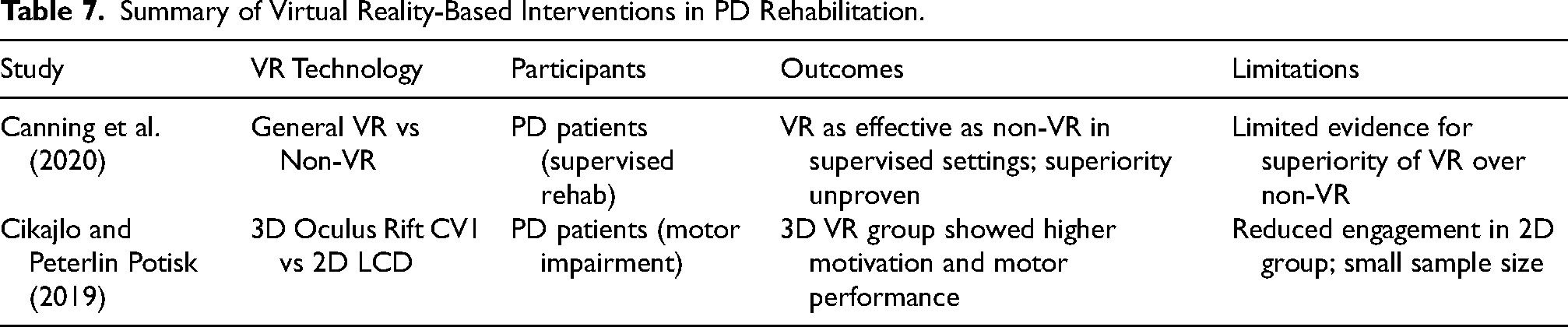
Virtual reality (VR) represents a new adjunctive tool that can be used in the treatment of PD rehabilitation with the objective of improving engagement and functional recovery. It exposes patients to enriched interactive conditions that take the form of recreating real-life tasks at the least amount of physical risk (Ali et al., 2023; Chau et al., 2021). This new method can enhance both motor and non-motor performance of people with PD. In an effort to determine the relative effectiveness of VR, researchers have compared it with traditional methods of rehabilitation. As Canning et al. (2020) stated, both VR and non-VR interventions were found superior to no treatment, but the existing evidence did not provide a sufficient number of studies that prove VR is better than no treatment in terms of gait or balance improvements (Canning et al., 2020). Meta-analyses and randomized trials indicate that VR-based rehabilitation produces comparable outcomes in gait, balance, and motor coordination compared to traditional physiotherapy, with some studies reporting higher patient motivation and adherence (Campo-Prieto et al., 2022; Canning et al., 2020; Cikajlo & Peterlin Potisk, 2019).
The additional information can be gained by clinical research aimed at the improvement of motor functions with the help of immersive VR. In one of the studies by Cikajlo and Peterlin Potisk (2019), individuals who took part in VR training on a 3D Oculus Rift system were observed to have improved upper limb fine motor skills (Cikajlo & Peterlin Potisk, 2019). This was substantiated by clinical evaluations and kinematic studies. Interestingly, the respondents who used 3D systems also stated that they were more motivated and more engaged in a task, which was associated with better performance. On the other hand, participants working on 2D LCD displays showed inferior enthusiasm in the long run, which could influence their performance (Bektic et al., 2024; Campo-Prieto et al., 2022). Such results demonstrate the motivational value of VR, yet also remind us of the difference in the therapeutic effect based on the technology applied.
Table 7 summarizes key virtual reality (VR)-based rehabilitation studies, including their reported outcomes and limitations. VR functions as an extension of conventional physical and occupational therapy by providing immersive, task-specific training environments. It could increase interaction and motor training in well-established rehabilitation programs through interactive feedback. Figure 3 demonstrates the use of immersive VR to train the upper limbs of PD.

A PD patient from the 3D group is seen using the Oculus rift CVI headset for improving motor symptoms (Cikajlo & Peterlin Potisk, 2019).
Summary of Virtual Reality-Based Interventions in PD Rehabilitation.
Comparative Analysis of Therapeutic Approaches
The management of PD needs an integrative approach that involves the use of pharmacologic, non-pharmacologic, and surgical approaches, depending on the disease stage and patient features. Pharmacologic treatment offers a baseline of motor symptom management, whereas non-pharmacologic treatments, such as structured exercise and physiotherapy and diet management, supplement pharmacologic treatment by enhancing balance, mood, and functional performance. Surgical and machine-aided methods have an added advantage in patients with advanced or refractory symptoms that are chosen. New technologies like virtual reality- based rehabilitation extend the range of supportive therapeutic interventions by increasing patient interaction and functional training.
Limitations of Current Therapeutic Strategies
Prolonged levodopa administration is linked with motor variability and dyskinesia. There is limited evidence on non-pharmacologic and virtual reality intervention due to small sample size and short follow-up periods. Surgical and device-assisted techniques are associated with procedural risks, and they are likely to be selected very carefully. The strongest method of motor control is offered by device-assisted therapies, and disease-modifying biologics are investigational. The restrictions underscore the necessity of new disease-controlling measures.
Experimental Models for Investigating PD-Pathogenesis
In Vivo Models (Animal Models)
The in vivo models are important resources of mimicking the pathophysiological and behavioral complications of PD that serves as valuable platform in investigating the pathogenic mechanisms and preclinical drug testing (Santoro et al., 2023; Wal et al., 2024). They provide insightful understanding of certain details of the PD pathology despite their limitations and form a basis of the current translational neuroscience research. The models included in this review were selected according to their experimental robustness, translational relevance, and representation of hallmark PD features such as dopaminergic neurodegeneration, α-syn aggregation, and progressive motor impairment. Preference was given to models with well-documented methodological reproducibility, frequent application in preclinical studies, and established validity across construct, face, and predictive domains. Classical (toxin-based and genetic) and emerging integrative models were both given priority so as to balance the coverage of mechanistic, therapeutic, and translational perspectives in PD research.Toxin-based models are most appropriate to use in the history of dopaminergic neuron loss because of their reproducibility. Mechanistic studies are more adequately tackled by genetic models, especially in the study of mutation-mediated pathways and molecular malfunction. The MitoPark model best describes progressive systems, which are characterized by slow neurodegeneration and motor deterioration. α-syn preformed fibril (PFF) models are especially valuable for investigating prion-like propagation and aggregation dynamics.
Toxin-Based Models
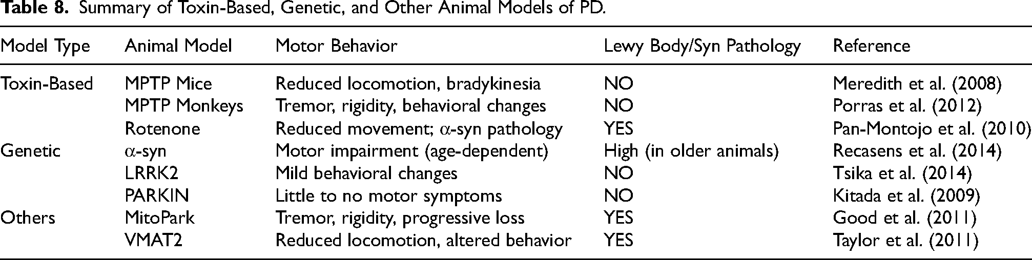
Toxin-based models are commonly used because they recapitulate selective dopaminergic neuronal loss in the substantia nigra pars compacta. These models use neurotoxins to replicate oxidative stress, mitochondrial malfunction and apoptosis which are important characteristics of the human PD (Prasad & Hung, 2020). For example, the MPTP model in mice and monkeys induces bradykinesia and tremor, although it does not replicate Lewy body pathology (Meredith et al., 2008; Porras et al., 2012). The corresponding 6-hydroxydopamine (6-OHDA) rat model also induces unilateral nigrostriatal lesions and is thus suited to such behavioral measures as amphetamine-induced rotations, although it, as well, does not show proteinopathy (Glajch et al., 2012).
Other environmental toxins like rotenone and paraquat/maneb induce both dopaminergic degeneration and α-syn aggregation, providing closer pathological relevance to PD (Bastías-Candia et al., 2015; Pan-Montojo et al., 2010). However, the systemic toxicity and variability in lesion formation limit their reliability. Drugs of abuse such as methamphetamine (METH) and MDMA have also been studied, but their link to PD remains primarily theoretical (Costa et al., 2013; Thrash et al., 2009). These models are especially valuable for screening neuroprotective agents and studying early-stage degeneration mechanisms. Toxin-based systems remain widely used due to their reproducibility and rapid induction of dopaminergic neuron loss; however, their translational value is limited by acute injury patterns, lack of progressive pathology, and incomplete α-syn aggregation.
Genetic Models
Genetic models are designed to simulate familial or monogenic forms of PD by over-expressing mutations on genes related to PD like SNCA, LRRK2, PINK1, PARKIN and DJ-1. With these models, researchers can investigate interaction of genes and the environment and the progressive aspects of diseases. As an example, in transgenic mice in which human α-syn is over-expressed, there are age related behavioural defects and α-syn clumping, especially in aged animals (Recasens et al., 2014).
Although genetic models reproduce PD-related mutations and proteinopathy, they frequently show limited dopaminergic neuron loss and mild motor phenotypes, resulting in high construct validity but reduced face validity (Gispert et al., 2009; Kitada et al., 2009; Tsika et al., 2014).
Emerging and Alternative in Vivo Models of PD
Several other models explore developmental or transcriptional pathways implicated in PD. These include mutations or knockouts in SHH, Nurr1, Engrailed-1, Pitx3, C-Rel-NFκB, and VMAT2 (Gonzalez-Reyes et al., 2012; Zhang et al., 2012). Notably, the MitoPark mouse model recapitulates progressive motor dysfunction, dopaminergic neuronal loss, and α-syn aggregation, making it one of the most comprehensive models available (Good et al., 2011).
A novel consideration is circadian rhythm disruption in PD. Hunt et al. (2022) emphasize the need for melatonin-competent strains such as C3H/HeN over commonly used melatonin-deficient C57BL/6 mice, particularly in sleep-related studies (Hunt et al., 2022). Although the application of melatonin therapy is a common practice in sleep disorders associated with PD, the clinical evidence provided is inconclusive. The predictive validity of toxin-based models is high whereas the construct validity is low, and the genetic models have high construct validity with relatively low motor phenotypes, whereas the integrated models like MitoPark mice have a more balanced combination of progressive neurodegeneration and motor loss. Included in neither of the two categories, α-syn preformed fibril (PFF) inoculation models have recently become essential components of PD studies. These models are analogous to the prion-like infection of intracerebral inoculation of synthetic fibrils, which induces endogenous propagation of α-syn aggregation, (Lewy body-like) inclusions, and progressive nigrostriatal degeneration. PFF models possess strong construct and face validity in the disease propagation and anti-aggregation or immunotherapy testing approaches. The differences in the fibrils preparation, site of injection and dosage, however, decrease the reproducibility and this is why they are not included in the original overview. However, they are increasingly being recognized as adjunctive frameworks that connect toxin and genetic paradigms of PD pathology.
Each in vivo model provides distinct strengths for investigating PD pathogenesis. As summarized in Tables 8 and 9, toxin-based models are well suited for rapid neuroprotective screening, genetic models are valuable for mechanistic studies, and integrative systems such as MitoPark and VMAT2 support longitudinal and translational research.
Summary of Toxin-Based, Genetic, and Other Animal Models of PD.
Comparative Advantages and Disadvantages of in Vivo Models of PD.
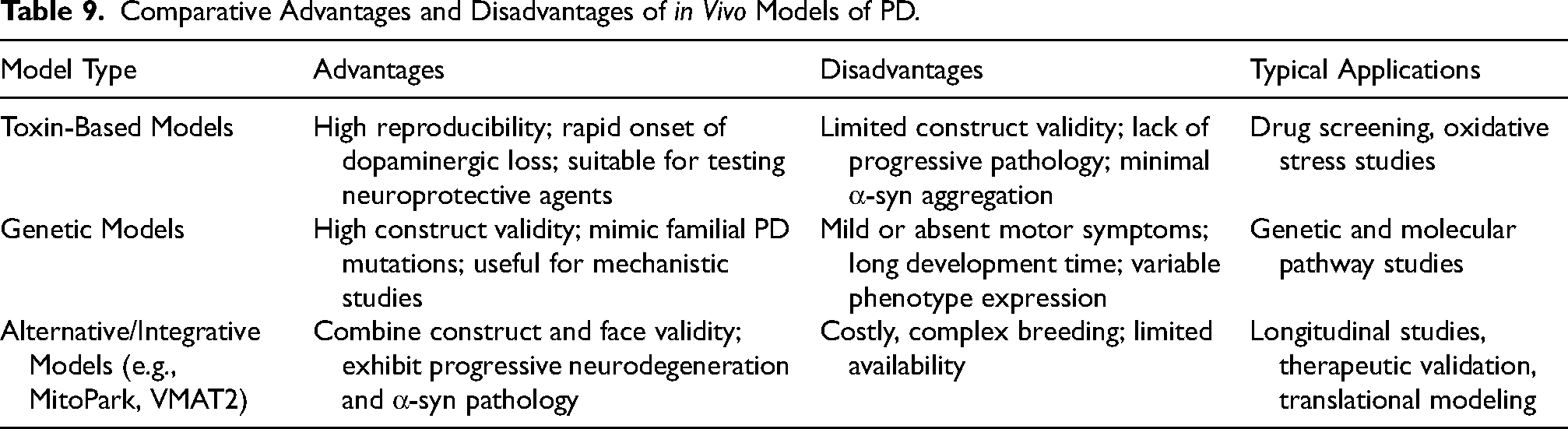
The major problems of the in vivo PD modeling are reproducibility and translational consistency. Inter-laboratory inconsistency is often due to variation in animal strain, toxin preparation, and administration regimen, and behavioral scoring in toxin-based models, lesion extent may vary significantly. Genetic models are more reproducible, but they need longer breeding and are characterized by inconsistency of transgenes expression among colonies. The regulatory frameworks i.e., ARRIVE and OECD are now required to provide standardized reporting, ethical compliance and reproducibility metrics to facilitate the comparability of studies across research centers. Model choice must be consistent with study purpose: toxin-based models (e.g., MPTP, 6-OHDA, rotenone) are best used in neuroprotection studies because the dopaminergic loss can be predicted and is useful in screening therapies; progressive models (e.g., MitoPark and VMAT2 knockouts) are best used in chronic degeneration and longitudinal therapeutic testing; genetic and viral vectors (e.g., 6-OHDA, 6-OHDA overexpression) are best used in genetic therapy testing.
In Vitro Models
In vitro models of PD provide critical platforms to dissect cellular mechanisms, evaluate pharmacological agents, and replicate genetic alterations in a controlled setting (Martínez-Morales & Liste, 2012). Given the complexity of PD pathophysiology, in vitro approaches allow researchers to study specific disease pathways in isolation or combination. Established cell lines offer strong reproducibility and are suitable for high-throughput studies, but their physiological relevance to human PD is limited. The iPSC models that have been derived by patients are highly genetically accurate and disease-specific, but variability in differentiation and maturation is an issue, whereas 3D cultures and organoid systems are more indicative of cellular heterogeneity and micro-environmental interaction but are not universally standardized. The choice of a model must be consistent with certain translational goals, a tradeoff between experimental and biological aspects.
Established Cell Lines
The most commonly used dopaminergic cell lines are SH-SY5Y (a subline of SK-N-SH) and PC12 (derived cells of rat adrenal medulla), as they are readily available, reproducible, and can be used in high-throughput drug screening (Shimohama et al., 2003; Xicoy et al., 2017). SH-SY5Y cells have been especially helpful in the investigation of aggregation of α-syn, mitochondrial stress, and toxin vulnerability such as MPP+ and 6-OHDA. Besides the lines, primary cultures like fetal mesencephalic dopaminergic neurons are highly physiologically relevant to the study of toxin vulnerability and apoptosis (Goulding et al., 2021).
Patient-Derived iPSC Models
The use of patient-derived induced pluripotent stem cells (iPSCs) has transformed the PD modeling by providing the derivation of midbrain dopaminergic (DA) neurons with patient-specific genetic mutations. These neurons have the ability to recapitulate characteristic elements of PD, such as mitochondrial impairment, Lewy body-like inclusions and progressive dopaminergic cell loss (Seibler et al., 2011). Nonetheless, models built on iPSCs have a number of limitations that restrict their translational strength. The process of differentiation can be highly variable both between labs and between protocols and results in the maturation of dopaminergic neurons being inconsistent.
Figure 4 shows that somatic cells of patients can be reprogrammed into iPSCs or directly transformed into induced DA neurons (iDA neurons). Such cells can be genetically engineered, cloned, and developed into neural progenitor cells (NPCs) and eventually develop PD-like phenotypes of DA neurons.

Workflow for generating.
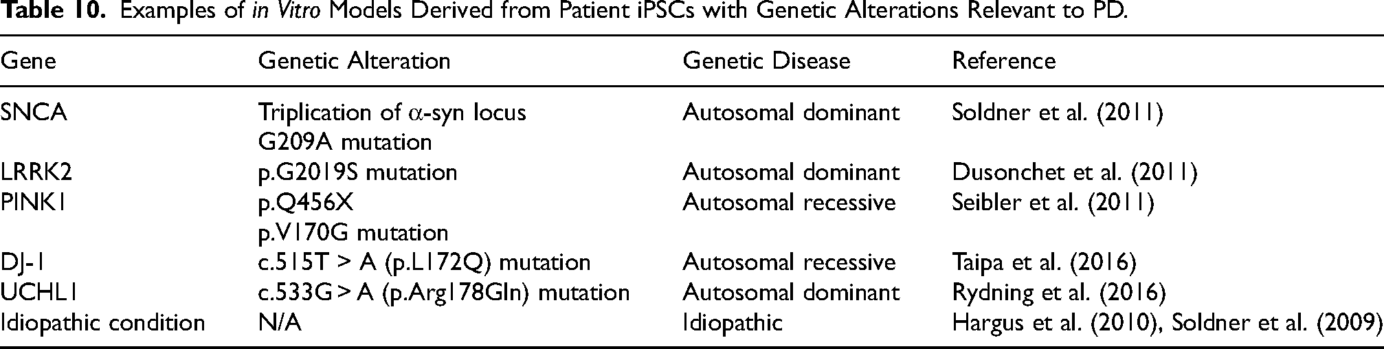
Table 10 emphasizes some genetic changes that have been modeled in iPSC systems of patients and their inheritance patterns. These technologies allow exploring the interactions between genotype and phenotype and contribute to the creation of individual treatment plans.
Examples of in Vitro Models Derived from Patient iPSCs with Genetic Alterations Relevant to PD.
Oxidative Stress and Toxicity Models
In vitro PD models frequently replicate oxidative damage and mitochondrial dysfunction, using agents such as MPP+, 6-OHDA, buthionine sulfoximine (BSO), and bleomycin sulfate (BLM). These compounds induce reactive oxygen species (ROS) and glutathione depletion, mimicking early degenerative changes in PD (Kitamura et al., 2002). Glutamate-induced excitotoxicity also serves as a pharmacological model for endogenous oxidative stress (Andersen et al., 1996).
Reproducibility and Translational Limitations
Although in vitro PD models have considerable mechanistic utility, inter-line variability has been observed including genetic background variations, reprogramming methods, and differentiation regimens (Seibler et al., 2011). Moreover, they are also immature in their development, which restricts the ability to model aging-related PD phenotypes. Neuronal monocultures upon which most systems are based also exclude glial and neuroinflammatory parts of the disease progression. Although traditional 2D cultures are experimentally strong and reproducible, they do not have the complexity of tissue structure found in vivo (Xicoy et al., 2017). On the other hand, more physiological relevant systems such as 3D organoids and more sophisticated co-culture platforms are difficult to standardize across sites (Göksu, 2024).
Bioprinted Models for Neurodegenerative Disease Research
Bioprinting Principles and Bioink Innovations
The neurodegenerative disease models produced with the help of three-dimensional (3D) bioprinting provide the possibility to recreate the brain-like structure and microenvironment. Bio printed constructs, as opposed to traditional 2D cultures, offer both spatial organization and extracellular matrix (ECM) cues required to recapitulate a native central nervous system (CNS) tissue (Fantini et al., 2019). Using the iPSC technology it can be augmented to produce personalized brain organoids, thus enabling disease modeling and drug screening.
Bioinks can be optimized so that the success of bioprinting is ensured. Fantini et al. (2019) incorporated sodium alginate emulsions and gelatin-based formulations to enhance the performance of the print. Playing with gellan gum-RGD bioinks, Lozano et al. (2015) produced brain-like features (Fantini et al., 2019; Lozano et al., 2015). The mentioned bioink enhancements are of special interest to PD modeling since they promote adhesion, survival, and neurite growth of dopaminergic neurons. This is needed in the reconstruction of degeneration of the nigrostratitallopathy. Other attempts encompass the development of bioinks that are preferably dopaminergic differentiation and neural circuitry repair. Such bioinks are typically biochemically cued (e.g., laminin, collagen, hyaluronic acid and neurotrophic factor e.g., GDNF, BDNF) to promote dopaminergic lineage specification, synaptic maturation and network connectivity. These recipes attempt to recap the biochemical and mechanical microenvironment of the midbrain, and allow the formation of organized and physiologically meaningful dopaminergic circuits to model PD.
The bioink rheology, extrusion pressure, and nozzle shear stress determine the survival and maturation of 3D-printed dopaminergic cells. The additional factors affecting cellular development are post-printing conditions such as oxygen levels and neurotrophic factor supplementation. These parameters working together with optimal results in the survival of neurons, extension of neurites and maturation of synapses, therefore, the functional fidelity of dopaminergic constructs. Hsieh and Hsu (2015) suggested polyurethane adjustable stiffness hydrogels that may be used in printing the neural stem cells (NSCs) using fused deposition manufacturing (Hsieh & Hsu, 2015). To PD studies, these tunable hydrogels are fundamental, as mechanical rigidity is a stiff determinant of the efficacy of neuronal differentiation and dopaminergic lineage commitment, which mimic the modulation of biomechanical environment that is present in PD-impacted midbrain tissue. On the same note, Joung et al. (2018) also showed that hiPSC-derived NSCs are viable and can be bioprinted into spinal cord-like constructs (Joung et al., 2018). In spite of the fact that it was first used on spinal tissue, the method demonstrates that hiPSC-derived dopaminergic progenitors could be spatially patterned into PD-relevant designs. This forms a basis on which to reconstruct nigrostriatal networks in vitro.
Applications in Neurodegeneration and Disease Modeling
3D bioprinting, particularly when combined with human iPSC-derived neural cells, enables the generation of physiologically relevant models of neurodegenerative diseases, including PD (Fassina et al., 2023). These systems exhibit improved cellular architecture and more closely resemble in vivo microenvironments compared with conventional 2D cultures (de Leeuw et al., 2021). Joung et al. (2018) demonstrated that hiPSC-derived neural stem cells (NSCs) can be bioprinted with high viability into spinal cord-like structures (Joung et al., 2018). This is particularly relevant to PD, as similar strategies can be used to organize dopaminergic neurons along the nigrostriatal axis. Such patterning supports reconstruction of PD-specific connectivity and degeneration features. Equally, Lozano et al. (2015) used RGD-functionalized gellan gum construct bioinks to bioprint brain-like constructs that contained primary cortical neurons. While not PD-specific, this study demonstrates the feasibility of engineering neuron-rich networks that support neuronal survival and connectivity,key prerequisites for modeling dopaminergic circuit dysfunction in PD. These findings support the feasibility of recreating region-specific neural architectures in vitro (Lozano et al., 2015).
Neural homeostasis depends on astrocytes, microglia, and other glial cells, which are increasingly incorporated into bioprinted constructs to recapitulate neuron–glia interactions. Qiu et al. (2020) demonstrated that, in the absence of specific differentiation cues, bioprinted NSCs develop heterogeneous neuron–glia populations resembling cellular diversity observed in PD (Qiu et al., 2020).
Such heterogeneity is particularly relevant to PD, as it reflects interactions between dopaminergic neurons and glial-driven neuroinflammation central to disease progression. Potjewyd et al. (2018) developed a 3D neurovascular unit (NVU) model using spatially organized bioinks to investigate neurovascular dysfunction (Potjewyd et al., 2018). These platforms are applicable to PD modeling as they allow the examination of blood brain barrier malfunctioning as well as microvascular inflammation that leads to dopaminergic vulnerability. Most recent technologies such as coaxial bioprinting, axonal guidance by chemical gradients, and the incorporation of either vascular or microfluidic systems have enhanced the physiological reproduction of neural constructs. Coaxial printing can be used to produce multi-compartmental neural fibers with a more realistic structure and directional development of axons that is amenable to the study of dopaminergic connectivity. Systems that can be controlled using chemical gradients enable accurate targeted axonal pathfinding and targeted synaptic connections, which allows the reconstruction of functional nigrostriatal networks. Microfluidic channels and vascular channels embedded into printed constructs enhance the diffusion of nutrients and oxygen to the cells, facilitation of removal of wastes, and intercellular signaling. This enhances long-term sustainability and effective stability of PD-specific neural models. These technologies increase structural arrangement, metabolic activity, and functional integration in neural structures related to PD.
Moreover, newer literature now incorporates 3D bioprinting in conjunction with microfluidic and organ-on-chip platforms to provide dynamic, perfused, and physiological responsive neural platforms. With these hybrid biofabrication-microfluidic models continuous medium flow, gradients of chemical concentrations can be maintained under control, and neuronal activity can be monitored in real time, and this is very close to the homeostasis in vivo. Neurovascular on chip and brain-on-chip have demonstrated better conditions of survival and synaptic operations of dopaminergic neurons under the regulated flow conditions. The results indicate the creation of disease-relevant PD-on-chip systems. Taken together, these works point to the fact that 3D bioprinting has allowed recreating PD-relevant microenvironments, consisting of dopaminergic neurons, glia, and vascular components, to study the processes of degeneration, inflammation, and regeneration in a controlled condition. Together, these models allow exploring the PD-specific characteristics, such as nigrostriatal degeneration, drug screening, axonal guidance, and regenerative approaches.
Current Technical Limitations
Nevertheless, a number of technical limitations restrict the translational suitability of 3D bioprinted PD models despite the considerable advancement. Majority of the existing bioinks are based on unstandardized composition, tunable mechanical properties, and lack cell-type specificity undermining reproducibility and long-term stability of constructs (Fantini et al., 2019; Hsieh & Hsu, 2015). Other impediments encompass inadequate vascularization, incompetent microglial integration and incomplete maturation of the neurons. Existing models also have difficulties in preserving axonal polarity, ordered synaptic connections, and proper electrophysiological maturation, which restricts the possibility of re-creating long-range dopaminergic circuits, including the nigrostriatal tract (Qiu et al., 2020). Bio printed constructs are more accurate in their architecture than organoid systems, but are currently less biologically mature.
Future Integration with iPSC and in Vivo Systems
Sensitivity to incorporation of vascular and immune components such as microglia and neurovascular interfaces should be considered in order to provide physiological complexity. In addition, it will be important to develop specific validation metrics, which include structural, molecular and electrophysiological outcomes, to provide objective model evaluation. Combination of patient-specific iPSC-derived dopaminergic progenitors with biofabrication platforms that can be scaled can also be used in the future to support high-throughput drug screening, and precision models. The combination of these strategies will ensure the clinical and translational considerateness of bioprinted PD systems.
Conclusion
The progress in PD modeling has developed to include degeneration models induced by toxins and genetically accurate and patient-derived cellular systems. Although in vivo models are crucial to the study of systemic neurodegeneration and motor dysfunction, in vitro and iPSC-based systems can offer patient-specific information about the molecular processes and therapeutic effects. More recently, spatially organized, multicellular, neural constructs produced by 3D bioprinting have provided an important step toward the study of the native brain structure, providing a bridge between reductionist cell systems and whole-organism science. A combined preclinical system consisting of these methods can help improve translation accuracy. Mechanistic interrogation in this model may be done in 3D-bioprinted structures, patient-specific phenotypes may be assessed by use of iPSC-derived dopaminergic systems, and systemic validation may be done in established in vivo models. The combination of such a coordinated approach with the structural fidelity of bioprinting, the genetic specificity of stem cell technologies and the organism level relevance of animal studies would ensure predictive validity is enhanced.
Moving ahead, it will be necessary to keep the standardization process going, add neurovascular integration, and strongly test them against clinical phenotypes to bring them to translational application. Bioengineering, stem cell biology and disease modeling The intersection of bioengineering, stem cell biology and disease modeling is potentially valuable in hastening the drug discovery process, enhanced patient stratification and development of the disease-modifying treatments of PD.
Footnotes
Author Contribution
Kaushik Sunder: literature review, visualization, writing—original draft, writing – review and editing, conceptualization, project administration. All the authors read and approved the manuscript.
Funding
The author received no financial support for the research, authorship, and/or publication of this article.
Declaration of Conflicting Interests
The author declared no potential conflicts of interest with respect to the research, authorship, and/or publication of this article.
Data Availability
All the data were given in the manuscript.
