
Abstract
A novel method utilizing combination of N, N-dimethylformamide (DMF) and ultraviolet (UV) irradiation to reduce silver (Ag+) ions in situ in ultrafine poly-(
Introduction
Recently, polymer nanocomposites containing metal nanoparticles (NPs) have attracted great interests. 1 Silver (Ag) is a famous disinfectant that has been widely used in infectious diseases treatment. 2 Incorporation of AgNPs into polymer ultrafine fibers can lead to a stronger antimicrobial activity because of high surface area and sterilization. 3 Until now, the incorporation has been achieved either by electrospinning polymer solutions containing AgNPs or reducing the Ag complexes in the electrospun polymer nanofibers. 4,5 In the first method, N, N-dimethylformamide (DMF) is mostly used as a solvent for electrospinning the polymers as well as a reducing agent for the Ag+ ions. The chemical reaction was proposed by Santos and Liz-Marzán as 6 : HCONMe2 + 2Ag+ + H2O → 2Ag0 + Me2NCOOH + 2H+. However, this method always needs a long time. In the second method, Ag+ ions are mostly reduced by ultraviolet (UV) irradiation, 5 heat treatment, 7 ultrasound, 8 reflux, 9 and chemical regent, 1 but the obtained AgNPs are inclined to aggregate. Thus, a new efficient method is required.
Figure 1 shows the structure of poly-(
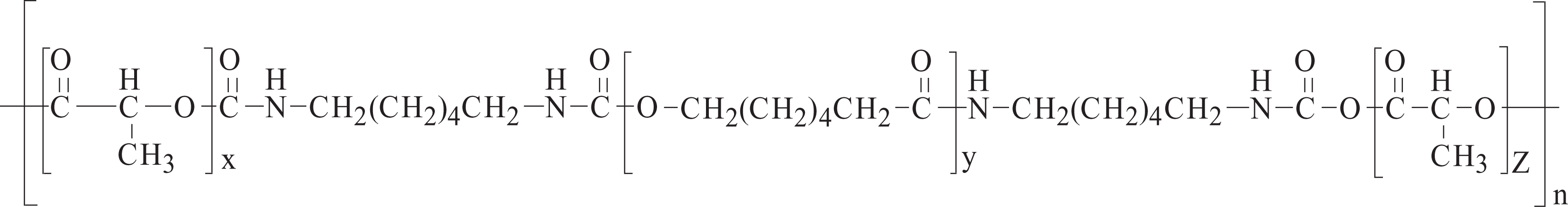
Structure of PCLA. PCLA: poly-(
Electrospinning is a versatile and mature method that can produce polymer fibers in diameter ranging from micrometers to nanometers. 11 It is the most convenient way to produce polymer nanofibers containing AgNPs. In this article, electrospinning was chosen to produce PCLA/silver nitrate (AgNO3) composite nanofibers. Rapid in situ photoreduction of Ag+ ions was carried out in combination with DMF and UV irradiation. The efficiency of the new method was discussed. The morphology, structure, size distribution, and formation of the AgNPs were investigated. It is believed that the product could be used in antimicrobial field in the near future.
Experimental
Materials
AgNO3 was purchased from Sinopharm Chemical Reagent Co. Ltd (China). PCLA (molecular weight = 111,213) was homemade. 10 Dichloromethane (DCM, Tianjin Tiantai Chemical Co. Ltd, China), and DMF (Xilong Chemical Co. Ltd, China) were used as solvents directly.
Preparation of PCLA/AgNO3 composite nanofibers
PCLA and AgNO3 were dissolved in a mixed solvent of DMF/DCM (40/60, v/v) to prepare a 27 wt% polymer solution with 1 wt% AgNO3. After vigorously stirring for 6 h, a transparent solution for electrospinning was obtained. The polymer solution was loaded into a glass dropper that was connected to a high-voltage supply (DW-P303-5 alternating current high voltage (approximately 0–30 kV), Dongwen High Voltage Power Supply Company, China). The voltage for electrospinning was 15 kV. A piece of flat aluminum foil was used to collect the nanofibers. The distance between the tip of the dropper and the collector was 20 cm. All electrospinning processes were carried out at ambient temperature.
Reduction of Ag+ ions
In order to investigate the reductive ability of different methods, the PCLA/AgNO3 composite nanofibers were divided into two parts. One part was aged in a sealed box at room temperature (the obtained AgNPs were called DMF–AgNPs); the other part was irradiated with UV light that had maximum wavelengths of 254 and 365 nm in a dark UV analyzer (the obtained AgNPs were called combined AgNPs).
Characterization
UV–visible (UV-Vis) spectra were recorded on a Shimdzu (Tokyo, Japan) UV-3600 spectrophotometer. Fiber morphology was analyzed using scanning electron microscopy (SEM; model SSX-550, Shimadzu, Tokyo, Japan). Based on the SEM images, the mean diameters of the composite nanofibers were analyzed using image visualization software Image J (about 100 measurements per field) (National Institutes of Health, USA). Transmission electron microscopy (TEM) images were obtained using a JEM-2000EX (JEOL, Japan) TEM for samples deposited on carbon-coated copper grids. The phase structure of the composite nanofibers was identified using x-ray diffraction (XRD). The XRD measurements were performed on a Siemens D5005XRD diffractometer (Germany). X-ray photoelectron spectroscopy (XPS, VG ESCA LAB MK II (Thermo Electron, USA), magnesium Kα, 10−7 Pa) was performed to analyze the chemical bonding states and chemical composition of the nanofibers.
Results and discussion
The formation of AgNPs in electrospun fibers can be detected using UV-Vis spectra. Changes in fiber absorption according to time near 424 nm, which was typical surface plasmon absorption of AgNPs, 12 were shown in Figure 2. In 540 min, the absorbance of the DMF–AgNPs increased very slowly, while the absorbance of the combined–AgNPs increased rapidly and tended to be constant at about 300 min. It was obvious that adding UV irradiation can reduce Ag+ ions efficiently. After naturally aging for 540 min, a yellow coloration was observed with the naked eye and after adding UV irradiation for the same time, a Modena coloration was observed. Color change confirmed the reduction of Ag+ ions and the subsequent formation of metallic Ag nuclei, which demonstrated the high efficiency of the new method once again.
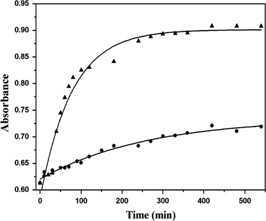
UV–visible absorption changes of the PCLA/AgNO3 composite nanofibers treated with (▴) UV irradiation and (•) aging according to time at 424 nm. UV: ultraviolet; PCLA: poly-(
Based on the results of UV-Vis absorption, the PCLA/AgNO3 composite nanofibers were decided to be treated with 10h of UV irradiation, 24 h of aging, and 240 h of aging (it is reported that almost all of the Ag+ ions can be converted into AgNPs via 240 h aging 13 ). The XRD patterns of the composite nanofibers treated in different ways were shown in Figure 3. Four diffraction peaks were observed in each curve at 37.8, 43.9, 64.2, and 77.4°, which were respectively ascribed to (111), (200), (220), and (311) crystal planes of Ag particles (JCPDS: 04-0783). 14 This indicated that all the three ways can facilitate the production of metallic Ag. However, their peak intensities were different. It was obvious that Figure 3(b) and (c) were almost similar and stronger than Figure 3(a). This illustrated that 240 h aging and 10 h UV irradiation contributed equally on the reduction of Ag+ ions, and their reductive abilities were stronger than 24 h aging.
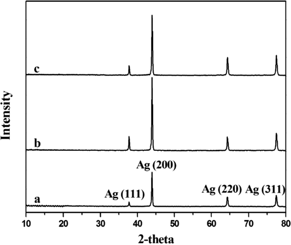
XRD patterns of the PCLA/AgNO3 composite nanofibers treated with (a) 24 h aging, (b) 240 h aging, and (c) 10 h UV irradiation. XRD: x-ray diffraction; PCLA: poly-(
SEM images of the PCLA/AgNO3 composite nanofibers treated in different ways were shown in Figure 4. From SEM images, similar morphologies and homogeneous appearances were observed. The average diameter of the 24 h aging fibers, 240 h aging fibers, and 10 h UV irradiation fibers was 259, 238, and 205 nm, respectively. The difference in diameter was caused by the decrease of solvent amount. In the aging process, DMF gradually started to react with Ag+ ions. While in the combined method, besides the reductive function of DMF and UV themselves, UV irradiation also provided heat to facilitate the reaction between DMF and Ag+ ions, 6 which led to more loss of the solvent. The results intuitively proved that the combined method possesses high efficiency.
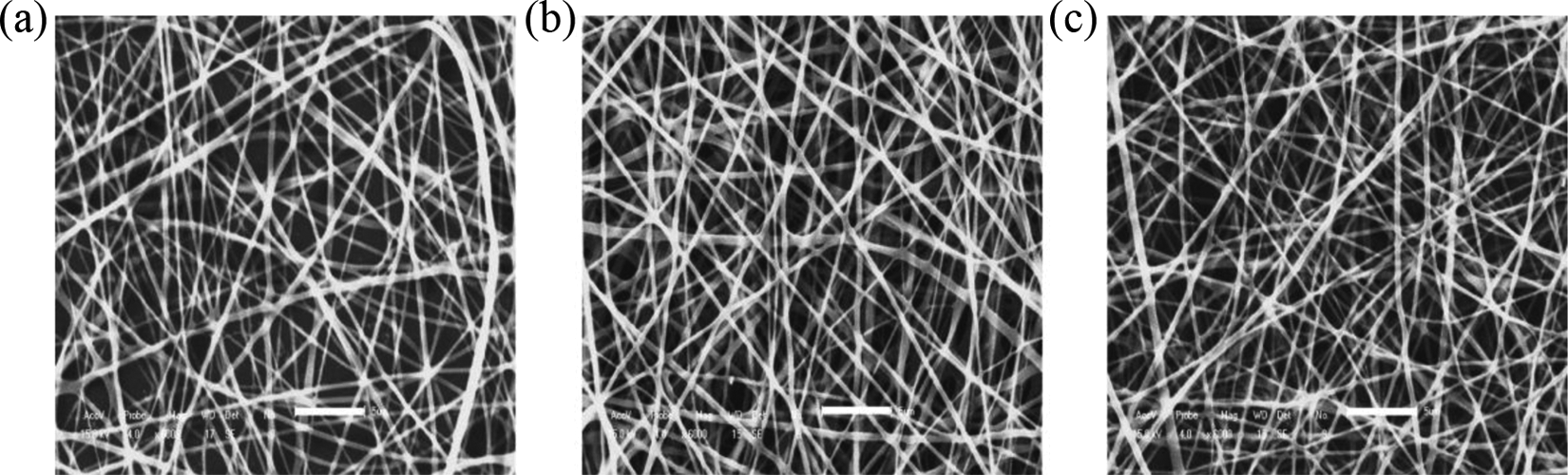
SEM images of PCLA/AgNO3 composite nanofibers treated with (a) 24 h aging, (b) 240 h aging, and (c) 10 h UV irradiation (inset scale bar is 5 µm). SEM: scanning electron microscopy; PCLA: poly-(
TEM images and the particle size distribution were respectively shown in Figures 5 and 6. Ag clusters were observed on the 24 h aging fibers. With increased aging time, round-shaped AgNPs with average size of approximately 10 nm can be seen on the surface of the fibers. This demonstrated that PCLA can act as a stabilizing agent to inhibit the agglomeration of the AgNPs. At the beginning of aging, reductive activity of DMF was stronger. With time increased, the amount of solvent decreased and the concentration of PCLA increased, resulting in a stronger stabilization. 3 The 10 h UV irradiation fibers showed a higher density of AgNPs, implying the formation of a greater number of nuclei. Numerous AgNPs were generated by the rapid photoreduction of the Ag+ ions via UV irradiation. 5 The particles dispersed in the matrix showed much smaller size (6.96 nm) and more uniform distribution than DMF-AgNPs. These results further demonstrated that the new method was beneficial for reducing Ag+ ions.
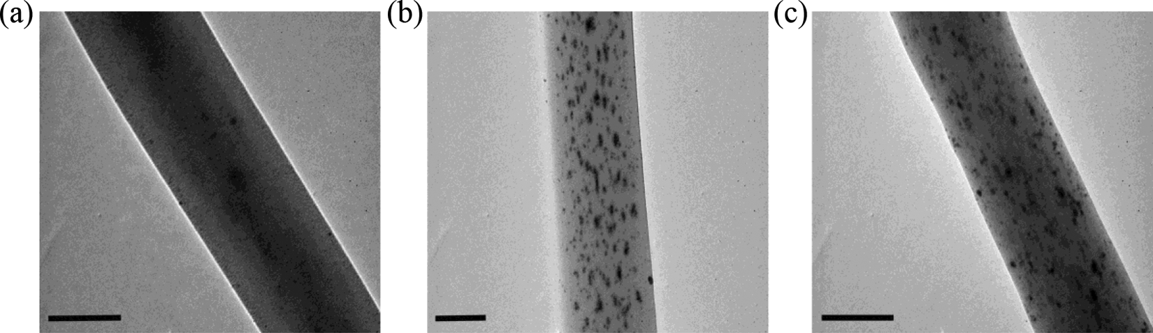
TEM images of PCLA/AgNO3 composite nanofibers treated with (a) 24 h aging, (b) 240 h aging, and (c) 10 h UV irradiation (inset scale bar is 200 nm). TEM: transmission electron microscopy; PCLA: poly-(
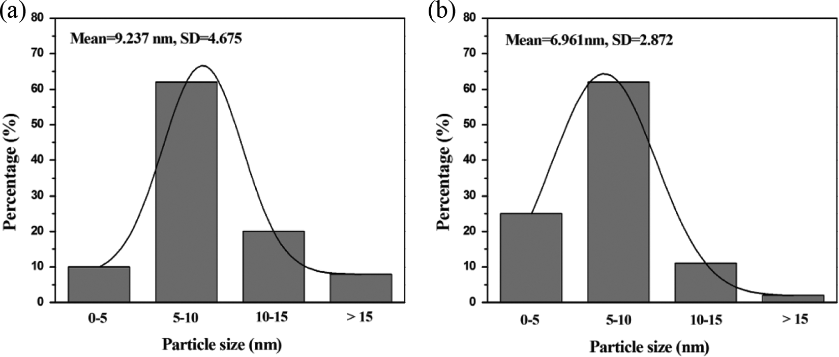
Size distribution of the AgNPs obtained from the PCLA/AgNO3 composite nanofibers treated with (a) 240 h aging and (b) 10 h UV irradiation. AgNPs: silver nanoparticles; PCLA: poly-(
Figure 7 showed the XPS spectra of Ag3d and O1s for PCLA nanofibers containing AgNPs. The binding energies of Ag 3d5/2 and Ag 3d3/2 appeared near 368.6 and 374.6 eV were characteristic peaks of metallic Ag. 15 The Ag0 binding energy of the 24 h aging fibers was slightly shifted to a lower level than that of the 10 h-UV irradiation fibers. It was thought that the reduction of Ag+ ions occurred more actively in the aging fibers. 16 The O1s photoemission spectrum appeared at 532.6 eV was attributed to the interaction between oxygen and silver particle. 17 It seemed that the spectrum of 10 h UV irradiation fibers was shifted to a higher energy, indicating a lower electronic density of hydroxyl oxygen atoms in the fibers. It has been reported by Ryu et al., 18 that the carbonyl groups coordinated with the Ag+ ions was through electron donation from the oxygen atoms to the vacant 5s orbital of the Ag+ ions. Therefore, it can be assumed that aging was in fact a little more beneficial to facilitate the interaction between hydroxyl oxygen atoms and Ag+ ions. However, the peak areas of the 10 h UV irradiation fibers were much bigger than that of 24 h aging fibers, indicating the generation of larger number of AgNPs. 15
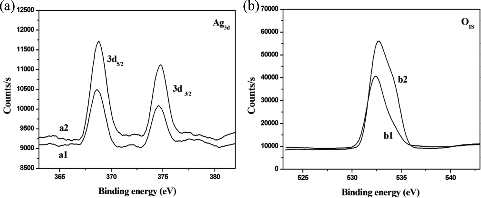
XPS spectra of (a) Ag3d and (b) O1s for 24 h aging PCLA/AgNO3 composite nanofibers (a1, b1) and 10 h UV irradiation composite nanofibers. XPS: x-ray photoelectron spectroscopy; PCLA: poly-(
Conclusion
A new in situ reduction technique, UV irradiation combined with DMF, has been used to reduce Ag+ ions in the electrospun PCLA/AgNO3 nanofibers. In comparison with traditional DMF reductive method, the new method exhibited much higher efficiency. In addition, PCLA was successfully used to carry AgNPs for the first time. It was proved that PCLA can be a stabilizing agent for Ag+ ions. The comprehensive results of this study suggested that PCLA was very appropriate, and the new method was highly effective to prepare polymer nanofibers containing AgNPs. The products were thought to be promising in the field of antimicrobial materials.
Footnotes
Funding
This research received no specific grant from any funding agency in the public, commercial, or not-for-profit sectors.