
Abstract
In the fluoropolymers family, one of the newest members is the terpolymer of tetrafluoroethylene, hexafluoropropylene, and vinylidene fluoride (THV). There are scarce data on THV, and therefore it is necessary to study different properties of THV. In the current research, the detailed surface morphology and water wettability of electrospun microfibers prepared from THV/ethyl acetate solutions in the pure state and with the addition of Wyoming-type low-magnesium montmorillonite are discussed. The morphology of the terpolymer microfibers changes as a function of the polymer solution concentration. For the same polymer concentration, changing the applied voltage in microfiber formation does not alter the morphology. The addition of hydrophilic montmorillonite into the THV solution does not modify the rougher hydrophobic nature and morphology of the final electrospun fiber surfaces. Water contact angle measurements show that both in the pure state and in the mixture of montmorillonite, THV electrospun microfibers exhibit “near superhydrophobic” characteristics with contact angles as high as 145°.
Introduction
Electrospinning is a facile technique with the advantages of producing ultrafine fibers with large surface areas, various surface functionalities, and better mechanical performance. 1,2 The diameter of the fibers can range from micrometers to nanometers, and the electrospun fibers are applied in various areas including biomedical and filtration technology. 2 The technique can be utilized with various polymers and polymer-based mixtures. 1,2 In electrospinning, the polymer solution is pumped through a capillary tube (spinneret) to form a small droplet at the spinneret tip. Applying voltage between the tip of the spinneret and the collector generates surface changes in the polymer droplet. 3 During the flow of the resulting polymer jet toward the collecting substrate, the polymer itself undergoes both stretching and whipping motions. As a result, continuous and randomly oriented fibers are formed. 3 There are several factors including solution viscosity, surface tension, and electrical conductivity that affect the ultimate morphology of the fibers. 1,3,4 Therefore, there are several fiber morphologies observed by the electrospinning technique ranging from thin smooth fibers to porous fibers. 1,3
In the current contribution, electrospinning was applied to form fibers of a fluorinated terpolymer of tetrafluoroethylene (TFE), hexafluoropropylene (HFP), and vinylidene fluoride (VDF), which in short is abbreviated as THV. THV has outstanding properties of low processing temperature, resistance to chemicals including strong acids and bases, and optical clarity. 5,6 There are nine grades of THV available, and due to its solubility in some organic solvents, 7 we selected THV-221 G for this study. In a recent article on THV-221 G by solution fluorine-19 nuclear magnetic resonance (19F NMR), it was shown that THV-221 G is mostly a random copolymer. Quantitative analysis of 19 F NMR spectrum displayed that 50% of TFE units are adjacent to each other while 90% of HFP groups are closer to VDF units. 8 In another recent study on basic characterization in bulk and tuning hydrophobicity of THV-221 G in thin films, mole percentage values of VDF, HFP, and TFE in THV-221 G were calculated as 38.2, 10.4, and 51.4, respectively. 9 The critical molar mass (M c) for the entanglement of random branches of THV equals 2.5 times the molar mass of entanglements (M e), where M e of THV was determined as 4100 g/mol. 10
Different inorganic materials including montmorillonite have been incorporated into polymer fibers using electrospinning. 11,12 Montmorillonite has the typical “smectite” structure with sizes of 1–3 μm. Montmorillonite is a layered aluminosilicate where negatively charged layers are compensated by cations. 13 Montmorillonite is a 2:1-type clay mineral composed of one octahedral central alumina sheet sandwiched by tetrahedral sheets of silica. The oxygen atoms connect the sheets. 14,15 The incorporation of montmorillonite helped to increase the fiber diameters where the fibers were prepared out of polyvinyl alcohol, montmorillonite, and silver (Au) nanoparticles. 11 Since it is thought that montmorillonite may either improve the electrospun THV fibers or the clay may function as processing aid additive, Wyoming-type low-magnesium (Mg) montmorillonite was added into the electrospun THV microfibers.
Despite several attempts at characterizing THV, there is still a lack of adequate data on THV properties especially at solid interphases. Although the electrospinning technique has been employed in polymeric fiber formation, electrospun THV fiber formation was not studied, and electrospun fluoropolymer fiber studies were rare. 2,16,17 In the current contribution, there are two important aims, namely (i) to investigate electrospun THV fiber formation and the properties such as morphology and hydrophobicity and (ii) to prepare electrospun composite fibers out of THV and montmorillonite blends.
Materials and methods
Materials
The terpolymer sample THV-221 G was obtained from Dyneon GmbH (Burgkirchen, Germany) as pellets and used without any further purification. N-type, P-doped [100] silicon wafers with a thickness of 625 μm were purchased from University Wafer (Boston, Massachusetts, USA) and used as substrate. The silicon wafer pieces were cleaned by keeping them in concentrated sulfuric acid overnight prior to use, and rinsing with deionized water. A source clay standard of Wyoming-type montmorillonite with low Mg (SWy-1) was purchased from the Clay Minerals Society, Chantilly, Virgina, USA. Ethyl acetate with 98% purity was purchased from Stockmeier Chemie GmbH (Bielefeld, Germany). The solvents were used as received without any purification.
Sample preparation
THV solutions in ethyl acetate were prepared with the weight percentages of 8.4, 12, 15, and 20. SWy-1 was added into the 8.4 wt% THV/ethyl acetate solution with the weight ratio of polymer:clay (10:1). The mixture of polymer and clay was mixed thoroughly to homogenize the solution prior to use. The nonwoven fibers from the THV/ethyl acetate solution were produced using a NANON-A1 (MEEC Corporation, Japan) electrospinning setup. The polymer solution contained in a 1.6 hypodermic plastic syringe was forced out of a 22-gauge needle by a syringe pump at a feed rate of 1.1 ml/h, while the needle-to-collector (silicon wafer) distance was kept constant at 15 cm. Electrospinning was performed at two different voltage values of 20 and 25 kV.
Sample characterization
Contact angle measurements of the samples were made on a Krüss Drop Shape Analyzer (DSA100; Germany). The volume of the distilled water was 10.0 μl. Three droplets of water were placed on different regions of the films. For each droplet, five different calculations were made by the DSA software of the Krüss device. The average of 15 data points was taken together with the standard deviation. The morphology of the nonwoven electrospun THV fibers was characterized using a Zeiss AURIGA scanning electron microscope (SEM) at the Osnabrück University, Germany. Samples were coated with Au before examining them under the SEM.
Results and discussion
Morphology
There is porous structure observed in the electrospun films (Figure 1). However, the morphology changed when the polymer solution concentration was increased (Figure 2). The electrospun THV films prepared from 8.4 wt% solution depicted hollows with a “well”-type structure, both in the case of THV/ethyl acetate and THV/ethyl acetate and montmorillonite with the weight ratio of polymer:clay (10:1). The addition of the hydrophilic montmorillonite into the polymer solution did not alter the morphology of the electrospun THV fibers. This shows that the hydrophilic clay may act as a processing aid in electrospinning the fluorinated terpolymer. The same morphology of hollows with a well-type structure was observed as the polymer solution concentration was increased to 12 wt%. When the concentration of the THV solution was increased to 15 wt%, the morphology changed to irregularly shaped beaded fibers. Lengthy fibers with “bean”-type structures were prepared from 20 wt% of the THV polymer solution (Figure 2).
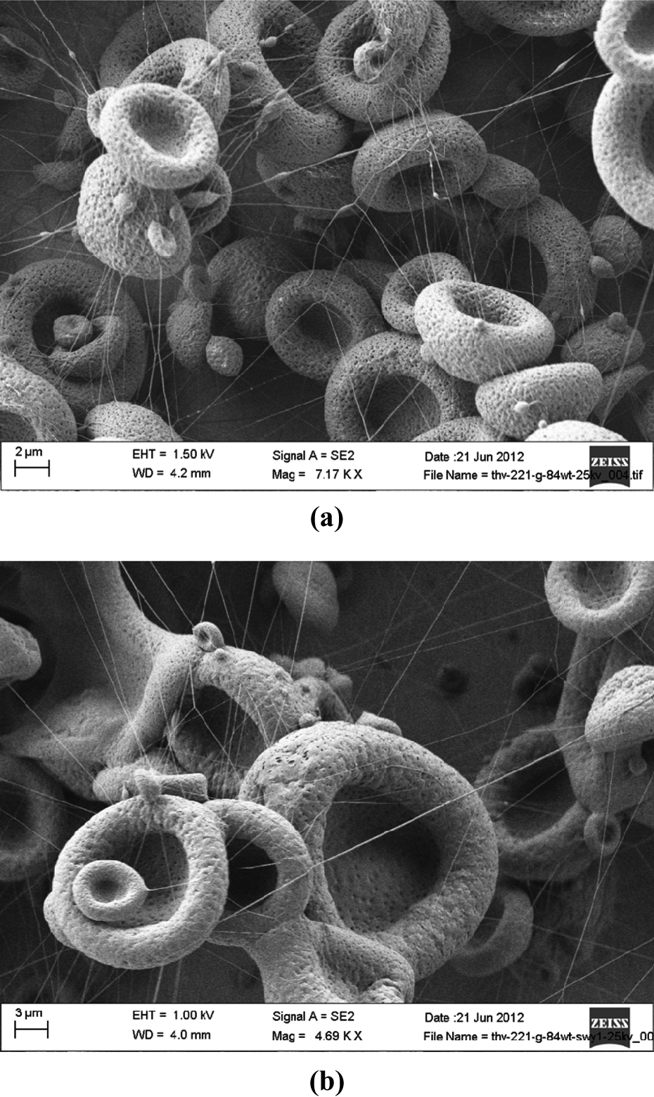
Electrospun THV microfibers with the porous structure prepared from the THV/ethyl acetate solution with the concentration of 8.4 wt% at 25 kV (a), and prepared from the THV/ethyl acetate solution 8.4 wt% at 25 kV with source clay standard SWy-1 (THV:montmorillonite, 10:1) (b). THV: terpolymer of tetrafluoroethylene, hexafluoropropylene, and vinylidene fluoride; SWy-1: Wyoming-type montmorillonite.
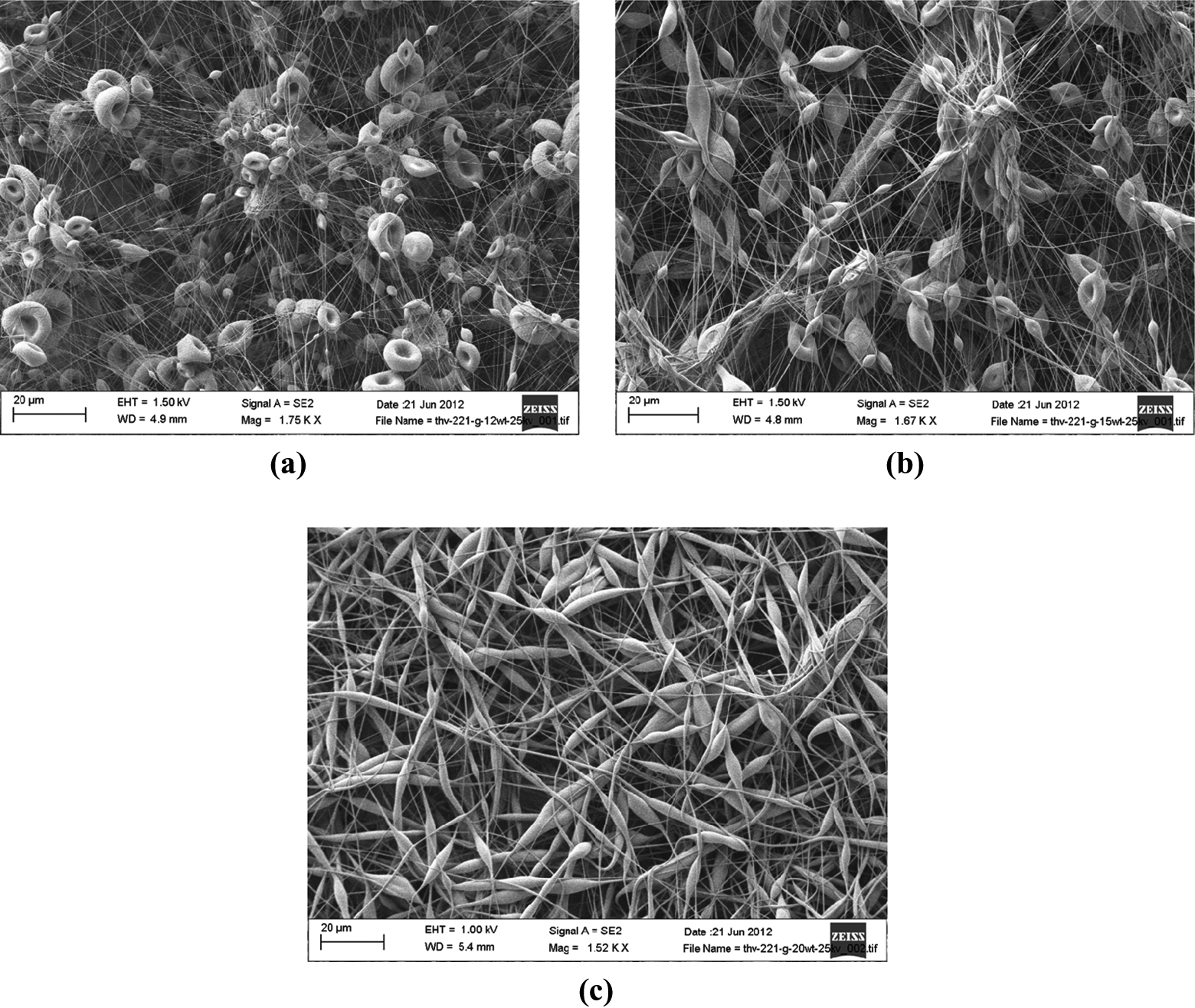
SEM images of electrospun THV microfibers prepared from the following solutions: THV 12 wt% at 25 kV (a), THV 15 wt% at 25 kV, beaded fibers (b), and THV 20 wt% at 25 kV, bean structured fibers (c), same morphology observed at the applied voltage of 20 kV. THV: terpolymer of tetrafluoroethylene, hexafluoropropylene, and vinylidene fluoride; SEM: scanning electron microscope.
In electrospinning, there are two important factors affecting the structure formation, namely, solvent evaporation and elongation of the solidifying fibers. 18 When both of the above-mentioned processes are completed, the fiber is formed. 2 In the electrospinning mechanism, solution viscoelasticity extremely affects both jet formation and stability. The solution itself has to keep a proper viscoelasticity so that the jet could survive stretching, acceleration, and whisking. 19 The relationship between viscoelasticity and the concentration regime of the polymer solution in the electrospinning mechanism needs to be discussed. 20,21 The concentration regime influencing the ultimate electrospun fiber morphology includes dilute, semi-dilute, entangled, and concentrated entangled systems. 20 These regimes are theoretically categorized in order to distinguish the rheological attitudes of polymers. 22 Rosic et al. analyzed both bulk and interfacial rheology of natural polymers to define solution systems for predicting smooth fiber formation.19 Rosic and coworkers figured out that determining bulk properties allows the prediction of jet and fiber formation while the interfacial properties help with the prediction of jet continuation. Depending on the solution regime and rheology, the fiber-like structure proves the existence of enough chain entanglement. Polymer solution either in dilute or semi-dilute regime without chain entanglement by electrospinning yields mainly little particulates rather than fiber-like structures. 23 Possible bead structures, for instance, could be formed from dilute and semi-dilute solutions. 21 The electrospun THV fibers prepared from 15 wt% THV/ethyl acetate solution depicted beaded structures (Figure 2). Yarin et al. modeled that during electrospinning viscoelastic dumbbells with the beads and electrical charges of the charged polymer result in the instabilities leading to the jet path. 24 First, the beads interact among each other via springs and dashpots from the mechanical interactions in the jet and the viscoelastic response acts toward elongation in the electrospinning process. Formation of beads during electrospinning is also explained by the breaking up of the polymer solution jet due to the deficiency of chain entanglements 25 and the formation of spherical droplets arising from surface tension. 26 Based on these works, we can say that our observations of bead and bean-like structures are directly related to these solution regimes. For example, the bead structure arises from a semi-dilute regime while 20 wt% polymer solutions have enough entanglement leading to bean-like fiber formation.
In the current study, the electrospun-fluorinated terpolymer fibers show a porous structure. The first reason is related to the stretching of the polymer chains and a quasi-Newtonian flow in which fully stretched chains flow past each other leading to a constant elongational viscosity. 27 In the study by Costa et al., the orientation of pores along the longitudinal direction of the electrospun fibers is related to the rapid stretching effect during electrospinning. 28 Fast solvent evaporation during the elongation flow state may induce stretching that may yield porous structure. The other explanation of the pore-like structures is phase separation into polymer-rich and polymer-poor regions, which is again related to rapid solvent evaporation. At first, electrospinning of polymer solutions involves fast solvent evaporation. However, as the solvent concentration is reduced, phase separation is initiated. Thus two phases, namely, polymer-rich and polymer-poor, characterized by different concentrations of the polymer will be established. 29 The polymer-poor regions will lead to the formation of the pores. 2 If a volatile solvent is utilized in the electrospinning method, there are solvent-rich regions formed. When the solvent molecules evaporate such polymer-poor regions evolve into pores. 30,31
It is seen that the porosity observed in the electrospun THV fibers is independent of the polymer concentration. Therefore, we think that porous structure is mainly related to the fast solvent evaporation. Further, porous fibers may have applications in various areas such as drug delivery systems and cell growth studies. 2,3 Pores depending on their sizes and arrangements can be utilized to control the release kinetics of drugs. Pores may also affect the degradation rate of biodegradable polymeric nanofibers. 2 Porosity may also favor cell growth. For example, it was shown that mesenchymal stem cells form branches to the pores. 32 Moreover, cell proliferation may be influenced by the degree of porosity and the mean pore dimensions. 33 We believe that these inert and stable electrospun fibers with porous structure may have applications in tissue engineering and filter membranes.
Montmorillonite partial incorporation
The addition of clay into the polymer solution did not affect the morphology of the electrospun fibers significantly compared with the control experiments without clay. There is also a 13° decrease of the contact angle of the fibers (Table 1) prepared from THV/montmorillonite solution toward water with respect to pure THV solution. In a recent report, the effect of montmorillonite addition on the wettability of zein/montmorillonite electrospun nanofibers was studied. By well-dispersed hydrophilic montmorillonite in zein nanofibers, it was possible to increase hydrophobicity systematically. 34 The addition of montmorillonite into the polymer solution leads to a decrease in the contact angle of water on the resulted THV/clay-based electrospun fibers but when the applied voltage is 20 kV. Our observation of a decrease in the contact angle in THV/montmorillonite fibers prepared at 20 kV is consistent with literature, which means that there is physical spreading of montmorillonite on the electrospun fibers. Furthermore, this is an important observation, since montmorillonite is known to improve mechanical, thermal, and barrier properties of polymer composite materials. 35 To disperse the clay into a polymer matrix, modification of the hydrophilic character of the clay might be needed. 36 This is done in order to increase the mutual compatibility of the two phases. 37 The presence of fillers such as clay materials will allow adjusting the properties of the electrospun fibers. 38 In the present study, although the natural clay SWy-1 was not modified, it was possible to disperse the hydrophilic clay in the polymer solution. Since the montmorillonite has swelling ability, physical spreading of montmorillonite on the electrospun fibers gives the advantage of forming fibers with swelling capability in addition to hydrophobicity. THV is a chemically stable and inert polymer, while there are possibilities to enlarge the interlayer space of the montmorillonite from 0 Å up to 140 Å. 39 The combination of these two properties of the polymer (THV) and the clay may be used to intercalate organic or biological molecules in an inert and stable medium. In this way, electrospun THV/montmorillonite composite fibers form inert nano-containers for the storage of bioactive molecules and for the stabilization of nanoparticles. 40
Contact angle values of the electrospun THV fibers prepared from either pure THV-221 G solutions or mixtures of THV-221 G and SWy-1 with low Mg source clay standard.
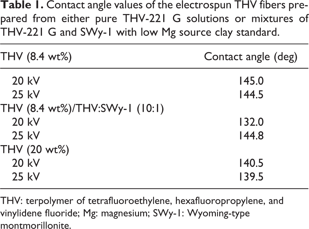
THV: terpolymer of tetrafluoroethylene, hexafluoropropylene, and vinylidene fluoride; Mg: magnesium; SWy-1: Wyoming-type montmorillonite.
Contact angle measurements
In the current contribution, one major focus is testing the hydrophobicity of the electrospun THV microfibers. Contact angles for water were measured only on three groups of electrospun THV fibers prepared from the lowest and the highest concentrated polymer solutions. As seen in Table 1 and shown in Figure 3, the contact angle of water on electrospun THV fibers is closer to but not exceeding 150°. The contact angles of fibers belonging to the lowest and the highest concentrated polymer solutions did not show a significant difference. The addition of hydrophilic montmorillonite into the polymer solution resulted in electrospun fibers with only 13° greater than the case of the fibers prepared from pure polymer solution. The hydrophobic character of the fibers did not change significantly by addition of the clay. This is explained by the physical spreading of the clay on the fibers.
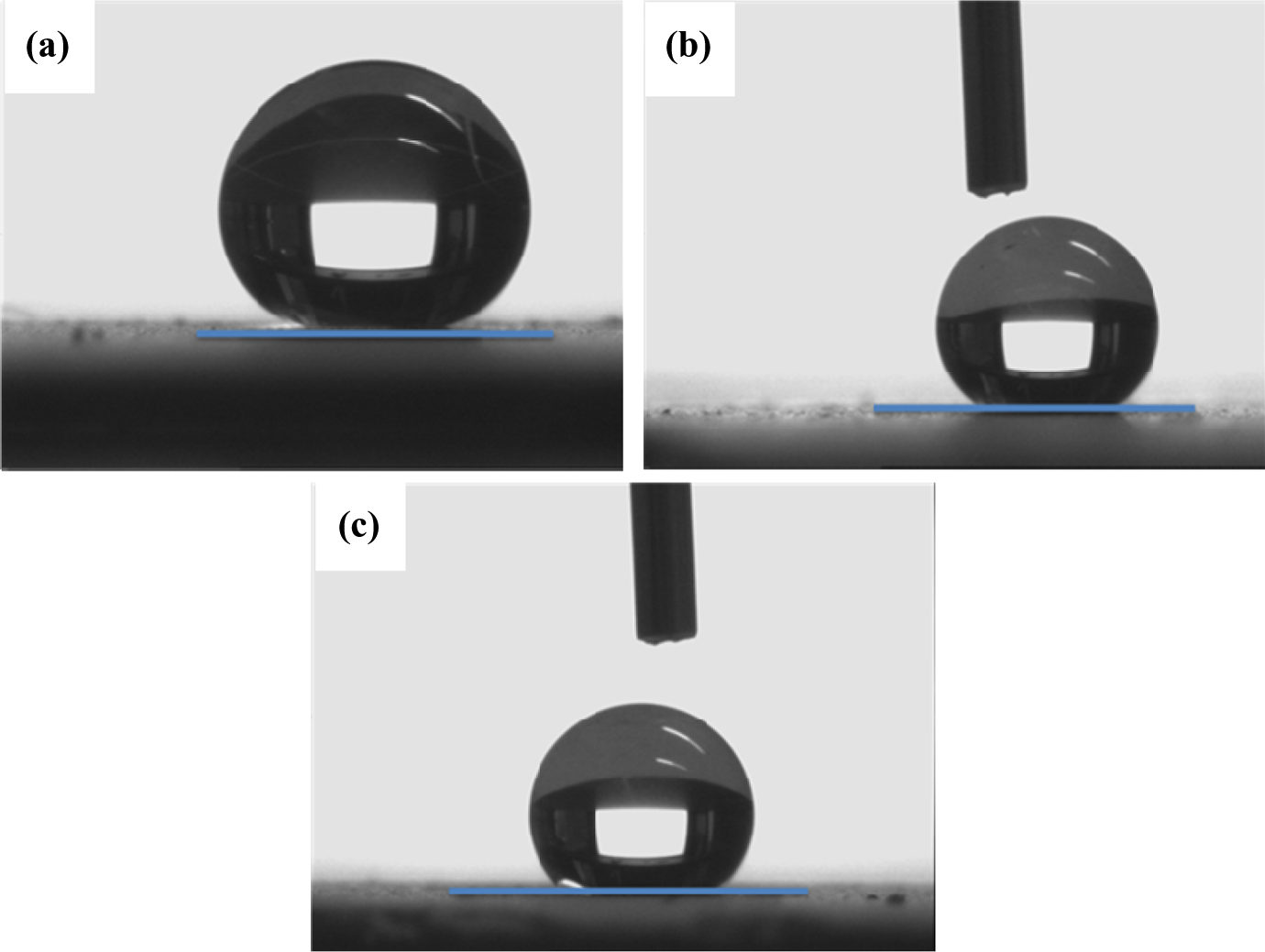
Representative profiles of water droplets on electrospun THV fibers from the following solutions: THV/ethyl acetate 8.4 wt% (a), THV/ethyl acetate 20 wt% (b), and THV/ethyl acetate 8.4 wt% with the weight ratio of THV:montmorillonite (SWy-1) (10:1) (c). THV: terpolymer of tetrafluoroethylene, hexafluoropropylene, and vinylidene fluoride.
Superhydrophobic surfaces attract great interest in different research areas.
41
It is essential to know how to tune the hydrophobicity of a surface by various parameters such as the surface composition and the roughness.
42
Wettability, one of the universally important parameters of solid surfaces, can be controlled by the surface chemistry and the surface geometrical structure.
43
To model the wetting or dewetting behavior of solid surfaces, using water is very common.
44
The wetting degree of a liquid on a solid substrate is quantified by the contact angle (θ), a material property of the liquid–solid interface. The wetting degree of liquids at solid interphases is an important factor in many applications including coatings.
45
Young’s equation
46
gives a value for the contact angle in terms of the interfacial tensions between the three phases:
where γ sv is the solid–vapor interfacial tension, γ sl is the solid–liquid interfacial tension, and γ lv is the liquid–vapor interfacial tension. For the physical and chemical understanding of the roughness-induced hydrophobicity, the works of Wenzel 47 and Cassie 48 have been cited extensively. According to Wenzel’s theory, the liquid on a solid surface is assumed to fill the grooves of the roughened surface. 47 According to Cassie’s theory, the droplet sits on functional areas consisting of the solid surface and air, while the droplet does not fill the grooves. 48 In Wenzel’s theory, only if the initial surface is hydrophobic, achieving greater hydrophobicity by enhancing roughness becomes possible. Cassie mentioned that rather than any waterproofing agents, the surface structure can play a more important role in repelling water. 48
In the study by Ok et al., 9 it was shown that THV-221 G has a contact angle of 102° toward water in solution-cast thin films prepared from 5 wt% THV/ethyl acetate solution. Addition of kaolinite in solution-cast thin films of THV-221 G did not alter the contact angle. In the same study, the highest contact angle of water on non-solvent-casted THV-221 G thin films was recorded as 123°. It was indicated that there was direct correlation between roughness and increase in the contact angle of water. 9 In the present contribution, the highest contact angle value of water on THV was measured as 145°. However, no additive or waterproofing agent was used. Even THV/montmorillonite composite fibers showed similar hydrophobicity with the contact angle of 132° toward water. Due to such observations, the Cassie theory seems to be more valid in explaining the near superhydrophobic character of the electrospun THV fibers. The water droplet does not fill the grooves of the surface of THV fibers. The term “super hydrophobicity” describes the hydrophobicity of a surface when the contact angle is greater than 150°. 41 Since the highest contact angle of water on the electrospun THV fibers is 145°, we prefer to use the expression “near superhydrophobic” in explaining the hydrophobic character of the THV fibers.
Super hydrophobicity expresses itself in low surface energy of the materials used and roughness both in micro and nanometer scales, and heterogeneity of solid surfaces. The higher fluorine content of a polymer leads to lower surface energy. This leads to higher contact angles. 49 Hydrophobic polytetrafluoroethylene is known for its very low surface energy. 41 Rougher surfaces tend to show higher contact angles with water. 50 Contact angles can be varied by roughness in two ways. First, the surface area of the solid is increased by roughness. Thus, when a liquid is placed on a rough surface, the magnitude of solid–vapor interface replaced by solid–liquid interface is larger than it would be on a flat surface. 48 The second effect of roughness arises from energy barriers formed by the local slope of the rough surface changing the local contact angles. These energy barriers block the spreading of the liquid front. 51 The Wenzel state is at a lower energy. There is an energy barrier for a droplet hindering its transition to the Wenzel state. Therefore, the droplet can switch from one equilibrium state to another only if it can overcome the energy barrier between the two states. 52,53 The possible reason for the near superhydrophobic character of the electrospun THV fibers is related to the energy barrier effect discussed in the literature. Although the Wenzel state is favored due to its lower energy, the energy barrier is not overcome so that the water droplet on the electrospun THV fibers remains in the Cassie state. This leads to non-wetting rather than wetting indicated with contact angle values closer to 150°. When the polymer solution viscosity is low, generally micrometer-size beads are formed. The beads introduce roughness, which leads to hydrophobic surfaces if the polymer is hydrophobic. 16 In this regard, our results are consistent with the literature findings.
Comparison with literature
Only a small number of fluoropolymers have been studied by electrospinning. 17,20,54,55 Simsek et al. investigated a poly(styrene-co-perfluoroalkyl ethylacrylate) random copolymer having 13 mol% fluorinated monomer. 55 Bead surfaces were observed in the case of the poly(styrene-co-perfluoroalkyl ethylacrylate) random copolymer. Weakening bead shapes led to nanoscale roughness and in turn to the establishment of hydrophobic surface. A recent study by Ahmed et al. showed the introduction of super hydrophobicity (contact angle >150°) on the poly(vinylidene fluoride-hexafluoropropylene) (PVDF-HFP) electrospun fibers. 20 The electrospinning itself (such as the applied field, the electrode configuration, or the feeding rate of the fluid to be spun) may also be responsible for the observed structures. 3 The morphology of THV electrospun fibers showed differences with respect to the PVDF-HFP. For instance, the PVDF-HFP electrospun fibers did not exhibit pores for the concentrations of 12–18 wt% and applied voltage of 11, 18, and 24 kV. 54 In the study by Li et al., a membrane prepared from PVDF-HFP electrospun fibers with uniform morphology was obtained under the conditions of 16 wt% solution in a solvent mixture of acetone and N,N-dimethylacetamide with 7:3 weight ratio at 18 kV. 54 These experimental conditions yielded an average fiber diameter of 1.0 μm. The morphology and average fiber diameter of the electrospun PVDF-HFP fibers were tested as a function of polymer concentration, applied voltage, and solvent ratio. Among these parameters, the largest influence on the morphology and average fiber diameter arises from concentration followed by applied voltage and solvent ratio. In another study, regular PVDF-HFP electrospun fibers were obtained at 15 wt% polymer solution concentration and 14 kV of applied voltage. 17 The distance to the collector was 15 cm with flow rate of 2 ml/h. The electrospun PVDF-HFP had an average diameter of 0.8–1.0 μm. It was also depicted that porosity of the electrospun PVDF-HFP fibers was not affected by the morphology prepared at different experimental conditions. Comparing our results with respect to the ones belonging to similar fluoropolymer electrospun fibers, 17,54 we can say that the electrospun THV/ethyl acetate fibers have diameters in the micrometer range. In line with previous findings, consistent with the literature, the THV/ethyl acetate concentration has strong influence on the morphology of the fibers. Varying the applied voltage from 20 kV to 25 kV did not change the morphology.
Conclusion
In the present study, the experimental conditions including polymer solution concentration and applied voltage were clarified for forming electrospun THV fibers from THV/ethyl acetate solutions. Both morphology and non-wetting behavior of the THV fibers are studied in detail. In addition to pure polymer solutions, fibers were produced also using the mixtures of polymer and clay low-Mg montmorillonite. The electrospun THV microfibers showed porosity in their morphology. Increasing the polymer solution concentration altered the morphology from a hollow structure to beaded fibers. For each polymer concentration, changing the applied voltage does not alter the morphology of the microfibers. The THV fibers prepared from pure THV/ethyl acetate solutions showed near superhydrophobic character with contact angles toward water as high as 145°. The physical spreading of hydrophilic low-Mg montmorillonite lowered hydrophobicity slightly.
Footnotes
Acknowledgments
Dr Ok gratefully acknowledges the German Science Foundation (DFG) (grant number STE 1127/15-1) for the support of the summer research at Institute for Chemistry of New Materials at Osnabrück University. The authors also acknowledge the Central Microscopy Research Facility at the University of Iowa, USA and Electron Microscopy Facility of Osnabruck University, Germany. For the critical readings of an earlier version of the manuscript, the authors gratefully acknowledge Professors Warren Huff of Cincinnati University, USA and Meral Dogan of Hacettepe University of Turkey and University of Iowa, USA.
Declaration of Conflicting Interests
The author(s) declared no potential conflicts of interest with respect to the research, authorship, and/or publication of this article.
Funding
The author(s) disclosed receipt of the following financial support for the research, authorship, and/or publication of this article: This research received grant from the German Science Foundation (DFG) (grant number STE 1127/15-1).