
Abstract
The understanding of basic surface properties and the relationships between temperature and the configuration of the polymer surface are the key to further understand the first interaction mechanism of the polymer with the body. To this end, some poly(carbonate tetramethylene ether)urethane (PCEU), poly(carbonate siloxane tetramethylene ether)urethane (PCSiEU), and their 1:1 gravimetric mixture (PCEU/PCSiEU) membranes were prepared. In order to establish the relationships between temperature and the chemical structure of polymer surface, the polyurethane (PU) membranes were analyzed by attenuated total reflectance–Fourier transform infrared spectroscopy. The temperature has a significant influence on these PU surface structures. Surface characteristics such as wettability and surface free energy were also analyzed since the interaction between biomaterials and blood occurs at their interface. The preliminary cytotoxicity screening showed no cytotoxicity of these PU membranes. The PU samples do not accelerate the clot formation mechanisms under the tested conditions. The results suggest that these PU membranes are promising materials for the preparation of cardiovascular scaffolds.
Keywords
Introduction
Generally, polyurethanes (PUs) are a versatile class of polymers that could provide matrices with good mechanical and physical properties, 1 –3 with various application in different fields, especially in biomedical applications. 4,5 They have been used in biomedical field for various applications due to the ability to modify their unique properties and due to the easy availability of these materials. 6 Properties such as processability, durability, surface functionality, flexibility, biocompatibility, and biostability make them superior for the development of materials that are useful for drug delivery, tissue engineering, and also for the development of many medical devices. 7,8 Thus, various PU structures have been utilized in blood-contact applications such as heart valves, dialysis membranes, breast implants, aortic grafts, bone adhesives, and so on. 9
Polycarbonate urethanes have attracted attention in the biomaterials market due to enhanced stability against hydrolysis and oxidation in comparison with polyester urethanes. 7 These characteristics make polycarbonate urethane an attractive material for developing cardiovascular devices. 10
Silicones with their unique material properties have found widespread application in health care. Properties attributed to silicone include biocompatibility and biodurability, which can be expressed in terms of other material properties such as hydrophobicity, low surface tension, and chemical and thermal stability. 11 Silicone’s chemical stability and elastic nature are beneficial for many applications involving long-term implantation. Poly(urethane–siloxane) are a class of hybrid polymers situated on a boundary between organic and inorganic materials. They consist of organic segments derived from PU and inorganic siloxane structures. These materials combine the advantages of both polymers used, that is, good tensile strength and abrasion resistance which are specific for PU, with low free surface energy and glass transition, great elasticity (especially at low temperature) as well as good thermal, chemical, and biological stability, which contribute to the system by polysiloxanes. Due to their properties, poly(urethane–siloxane) are widely used as protection coatings, medical implants, and so on. 12,13
The chemical structure from PU surfaces is capable of biological responses, which include protein adhesion, 14,15 antithrombogenic activity, and low platelet activation. 16 Therefore, it is vital to know the surface properties of these polymers. The analysis of the surfaces gives us the possibility to understand the first interaction mechanism of the polymer with the body. No detailed published work focusing on the effect of temperature on the PU surface has been found. Thus, one of the goals of this article was to study the influence of temperature on the chemical structure of PU surface in order to evaluate the stability of these materials at body temperature and also to choose a suitable sterilization method for these types of materials. Another aim was to investigate the surface characteristics such as wettability and surface free energy because the interaction between biomaterials and blood occurs at their interface. Thus, blood compatibility of these PU scaffolds strongly depends on their surface characteristics.
For this research, we analyzed the following PU matrices: poly(carbonate tetramethylene ether)urethane (PCEU), poly(carbonate siloxane tetramethylene ether)urethane – PCSiEU, and their 1:1 gravimetrically mixture. These types of PUs are promising materials for the development of scaffolds for cardiovascular tissue engineering.
Experimental
Materials
Poly(alkyl carbonate)diols were synthesized in our lab, where alkyl was hexametylene or tetramethylene. The polyesters have the hydroxyl number around 56 mg potassium hydroxide (KOH)/g and the number-average molecular weight (M n) around 2000 Da. 4,4′-Diphenylmethane diisocyanate (MDI) was freshly distilled before synthesis. Poly(dimethylsiloxane) bis(hydroxyalkyl)terminated (M n: 5600; hydroxyl value of 20 mg KOH/g) and dimethylformamide (DMF) were purchased from Sigma Aldrich (St Louis, Missouri, USA) and used as received. Other chemicals were of analytical grade and used without further purification.
PU synthesis
The synthesis of PCEU and PCSiEU was previously reported. 17 The main reaction mechanisms are illustrated in Figure 1. The M n values were 102,500 Da for PCEU and 103,500 Da for PCSiEU. The structures of the resulting PUs were confirmed by 1 H NMR (400 MHz, deuterated dimethyl sulfoxide (DMSO-d 6), δ in ppm): PCEU: 1.23–1.70 (–CH 2–), 3.32 (–O–CH 2–), 3.78 (–C6H4–CH 2–C6H4–), 4.03–4.06 (–O–CO–CH2 –), 7.09 and 7.34 (–C6 H4 –), 9.52 (–O–OC–NH–); PCSiEU: 0.05 (–Si–(CH 3)2), 1.31–1.71 (–CH 2–), 3.32–3.34 (–O–CH 2–), 3.78 (–C6H4–CH 2–C6H4–), 4.04–4.10 (–O–CO–CH2 –), 7.09 and 7.35 (–C6 H4 –), 9.51 (–O–OC–NH–).
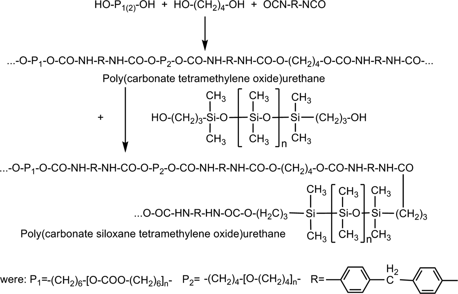
Reaction mechanism of PUs. PU: polyurethane.
Sample preparation
PU solutions (30% w/w) were degassed for 15 min under vacuum (10–15 mmHg) and then were cast onto a glass slide using a doctor blade with a gap of 0.6 mm. The films were precipitated in deionized and distilled water at 45°C for 1 h. 18,19 During this time, the resulted PU films were detached from the glass plate and subsequently washed five times with 1 L of deionized and distilled water. The films were dried at room temperature and low pressure (1–2 mmHg) for 24 h. The thickness of PU films was around 0.33 mm.
ATR-FTIR spectroscopic analysis
The influence of the temperature on surface structures was analyzed using attenuated total reflectance–Fourier transform infrared (ATR-FTIR) spectroscopic analysis. FTIR spectra were recorded in ATR mode on a Bruker VERTEX 70-Specac Golden Gate spectrometer (Billerica, Massachusetts, USA) from 25°C to 180°C at a heating rate of 10°C min−1. At relevant temperature, the sample was held for 5 min until the temperature remained constant and then the IR spectrum was recorded. The sample surfaces were scanned in the 400–4000 cm−1 range at 45° angle. The samples were in direct contact with the heated diamond crystal throughout the measurement and the evanescent wave extends beyond the crystal between 0.5 and 5 μm. The schematic diagram of an ATR accessory with a single reflection is illustrated in Figure 2.
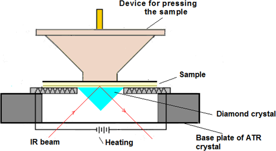
Schematic diagram of an ATR accessory with a single reflection. ATR: attenuated total reflectance.
Static contact angle measurements and surface free energy analysis
PU surface characteristics were evaluated by static contact angles measurements using sessile drop method and a CAM-101 (KSV Instruments, Helsinki, Finland) contact angle measurement system, equipped with a liquid dispenser, video camera, and drop shape analysis software. Double-distilled water and ethylene glycol were used as solvents for these studies. For each kind of liquid, three different regions of the surface were selected to obtain a statistical result. Single drop of test liquids with volumes around 1 µL were deposited on the PU film surface and 10 photographs were recorded at an interval of 0.016 s. All the measurements were performed at room temperature.
The values of the static contact angle (θ) can be used to estimate the wettability and surface tension of a solid surface. Calculations based on these measurements give several parameters such as surface free energy, adhesion work, and interfacial solid–liquid tension. Thus, the surface energy of the films was calculated using Young’s equation:

where
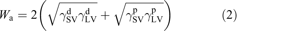
The surface free energy of PU surfaces was calculated according to the Owens–Wendt–Rabel and Kaelbe methods. 20 This method has been widely used to investigate the surface free energy of polymer films. Surface tensions can be divided into two components: the polar component (γ p) including two types of coulomb interactions, dipole–dipole and dipole-induced dipole, and dispersive components (γ d) which represent van der Waals interactions:

where W a is the work of adhesion.
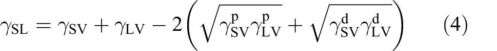
The interfacial solid–liquid tension (γ SL) is given by the following equation:
From equations (1) and (4), the following relation can be derived:

Cytotoxicity screening
Cell viability test was determined by direct contact method, using mouse fibroblast cell line NIH3T3 in 24-well culture plates. This type of cells has been used in cytotoxicity assays to preliminarily test the biocompatibility of biomaterials. The cells were cultured in Eagle’s minimum essential medium (Sigma, Germania), supplemented with 10% fetal bovine serum (FBS, Gibco, UK) and antibiotics solution (penicillin–streptomycin–neomycin solution stabilized; Sigma Aldrich, Germania). To determine the viability of cells, the 3-(4,5-dimethylthiazol-2-yl)-5-(3-carboxymethoxyphenyl)-2-(4-sulfophenyl)-2H-tetrazolium (MTS) reagent (CellTiter 96 Aqueous One Solution Assay; Promega, Madison, Wisconsin, USA) was used. PU samples were cut into small pieces (0.5 × 0.5 cm2), sterilized by dipping in 70% ethanol solution for 2 h. After that, PU samples were washed several times in sterile phosphate-buffered saline (Sigma) and incubated in culture media at 37°C and 5% carbon dioxide (CO2) atmosphere for 48 h.
The cell growth and viability testing protocol was previously reported in literature. 24,25 Briefly, PU pieces were added over the fibroblast monolayer, seeded for 24 h prior to the MTS experiment at initial seeding density of 4.5 × 10⁴ cells/well, in 24-well culture plates. The materials were incubated with the cells at 37°C, 96% humidity, and 5% CO2 incubation atmosphere for 24, 48, and 72 h, when cytotoxicity MTS proliferation assay was performed. MTS assay is based on cellular conversion of MTS in a product, soluble in the culture medium, which can be quantified by reading its absorbance at 490 nm and which is directly proportional to the number of living cells. Cell viability (%) was calculated with respect to the control cell cultures, incubated in the same condition but without any material sample. All experiments were performed in triplicate.
Hemocompatibily (thrombogenicity) analysis
In order to evaluate the hemocompatibility of PU surfaces, blood was withdrawn from healthy adult volunteer by venipuncture and mixed immediately with 3.8% sodium citrate using a ratio of 9:1 (v/v) blood:anticoagulant. This method used to collect and prepare the platelets for this study was approved by the ethics committee of the Grigore T. Popa University of Medicine and Pharmacy, Iasi, Romania, for hemocompatibility studies of PU derivatives with potential biomedical applications. The blood plasma was obtained after blood centrifugation for 15 min at 1000 r min−1 at room temperature. The resulted blood plasma was collected in silicone vials and used further for coagulation tests. PU films with size of 1 × 1 cm2 were immersed in sodium chloride physiological solution at room temperature for hydration. After hydration, the films were incubated in human plasma for 2 h at 37°C. After incubation time, the human plasma was tested to determine the main parameters of blood coagulation (clotting) using extrinsic and intrinsic pathways: fibrinogen concentration (Fb), coagulation time or quick time (TQ), and activated partial thromboplastin time (APTT). The international normalized ratio (INR) was calculated based on coagulation parameters. INR is the ratio of a patient’s prothrombin time to a normal (control) sample, raised to the power of the international sensitivity index value for the analytical system used. All tests were performed by standard methods using specific reagents (Biodevice, Italia). All post-incubated data were compared to the preincubated one and to the normal reference values.
Results and discussion
Temperature influence on the surface structures studied through ATR-FTIR spectroscopy
Spectroscopic methods are widely used to reveal valuable information regarding the constituent elements and chemical structure of the sample surfaces. 26 The aim of this ATR-FTIR work was to study the influences of temperature on the chemical structures of PCEU and PCSiEU developed for medical application, in order to prevent the scaffolds degradation during the dry heat sterilization.
The ATR-FTIR spectra of PCEU and PCSiEU are presented in Figure 3. It can be noticed that the increase in temperature causes modifications of the main absorption bands.
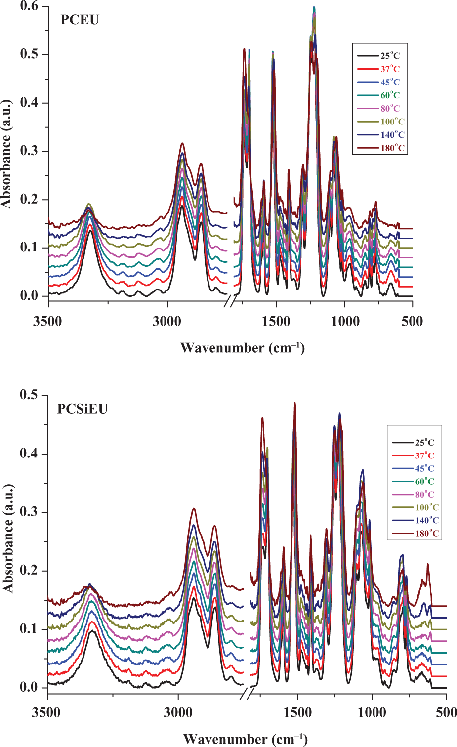
Temperature dependence of ATR-FTIR spectra between 3500–400 cm−1 of PCEU and PCSiEU in the range 25—180°C. ATR: attenuated total reflectance; FTIR: Fourier transform infrared; PCEU: poly(carbonate tetramethylene ether)urethane; PCSiEU: poly(carbonate siloxane tetramethylene ether)urethane.
From this figure, it is observed that the characteristic absorption at 3330 cm−1, which corresponds to –NH– bonded, decreases with the increases in temperature. The same phenomenon occurs at 2940 and 2860 cm−1 which corresponds to the ν(CH2) antisymmetric and symmetric stretching vibration of the soft segment. If we study the spectra in the region 1780–1640 cm−1 (Figure 4), which are the characteristic bands of carbonyl stretching vibrations ν(C=O), we observe that the crystalline structures of urethane groups decrease with increasing temperature while carbonate structure content increases.
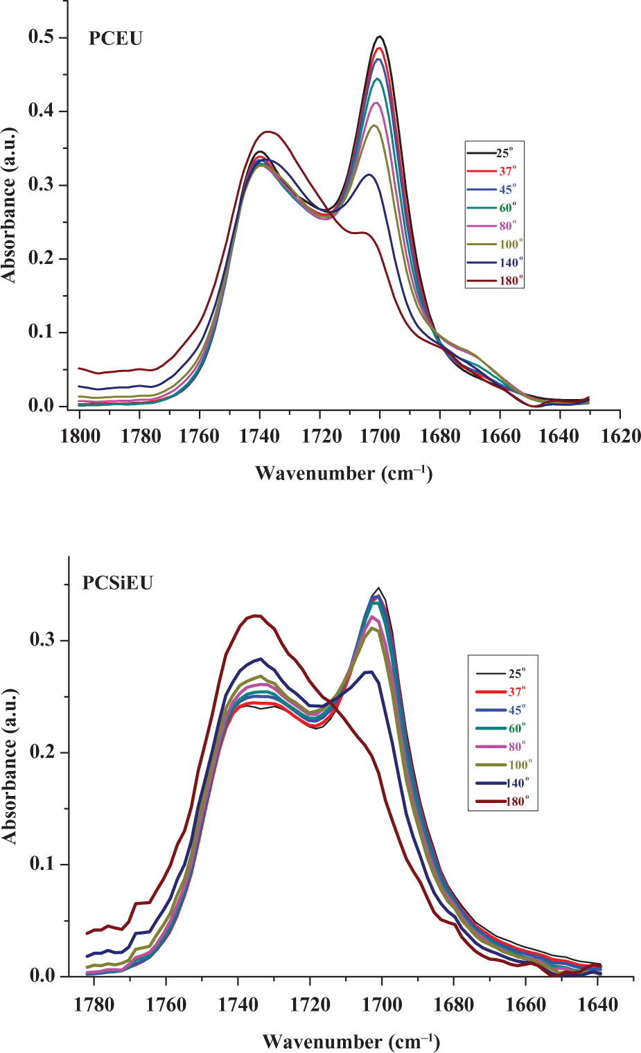
Temperature dependence of carbonyl ATR-FTIR spectra between 1800–1630 cm−1 of PCEU and PCSiEU in the range 25–180°C. ATR: attenuated total reflectance; FTIR: Fourier transform infrared; PCEU: poly(carbonate tetramethylene ether)urethane; PCSiEU: poly(carbonate siloxane tetramethylene ether)urethane.
The modifications of carbonyl surface structures due to the increasing of the temperature can be also noticed if we normalized the total carbonyl peak area (Figure 5(a)), where it is observed that PCSiEU is more stabile than PCEU. This phenomenon is due to the fact that PCSiEU is more flexible and dissipates thermal energy faster than PCEU.
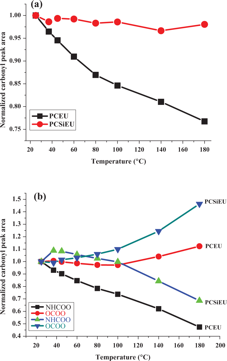
Temperature dependence of normalized (a) total carbonyl peak area (amide I) and (b) carbonyl urethane and carbonyl carbonate peak area of PCEU and PCSiEU. PCEU: poly(carbonate tetramethylene ether)urethane; PCSiEU: poly(carbonate siloxane tetramethylene ether)urethane.
From the temperature dependence of normalized carbonyl urethane and carbonyl carbonate peak area of these two PUs (Figure 5(b)), it is remarked that below 80°C there are no major changes of carbonyl structures. The slight decrease in the concentration of carbonyl urethane groups of PCEU up to 80°C maybe due to the surface changing structure of the polymer. The heated PU surface becomes richer in soft segments and poorer in hard segments so that the energy at the interface between the crystal and polymer to be small. This phenomenon occurs because the polymer molecules have much greater freedom to rearrange themselves at the surface in order to accommodate a change in chemical potential in the surrounding environment. 27
Between 80 and 140°C there appear some modifications of the carbonyl structures such as transesterification reaction between two molecules of PU and intramolecular transposition of the carbon–oxygen bond resulting in an amino ester urethane (Figure 6(a) and (b)). 28 Over 140°C, the decomposition of urethane group in amine, carbon dioxide (CO2), and unsaturated polymers can be observed (Figure 6(c)). 29
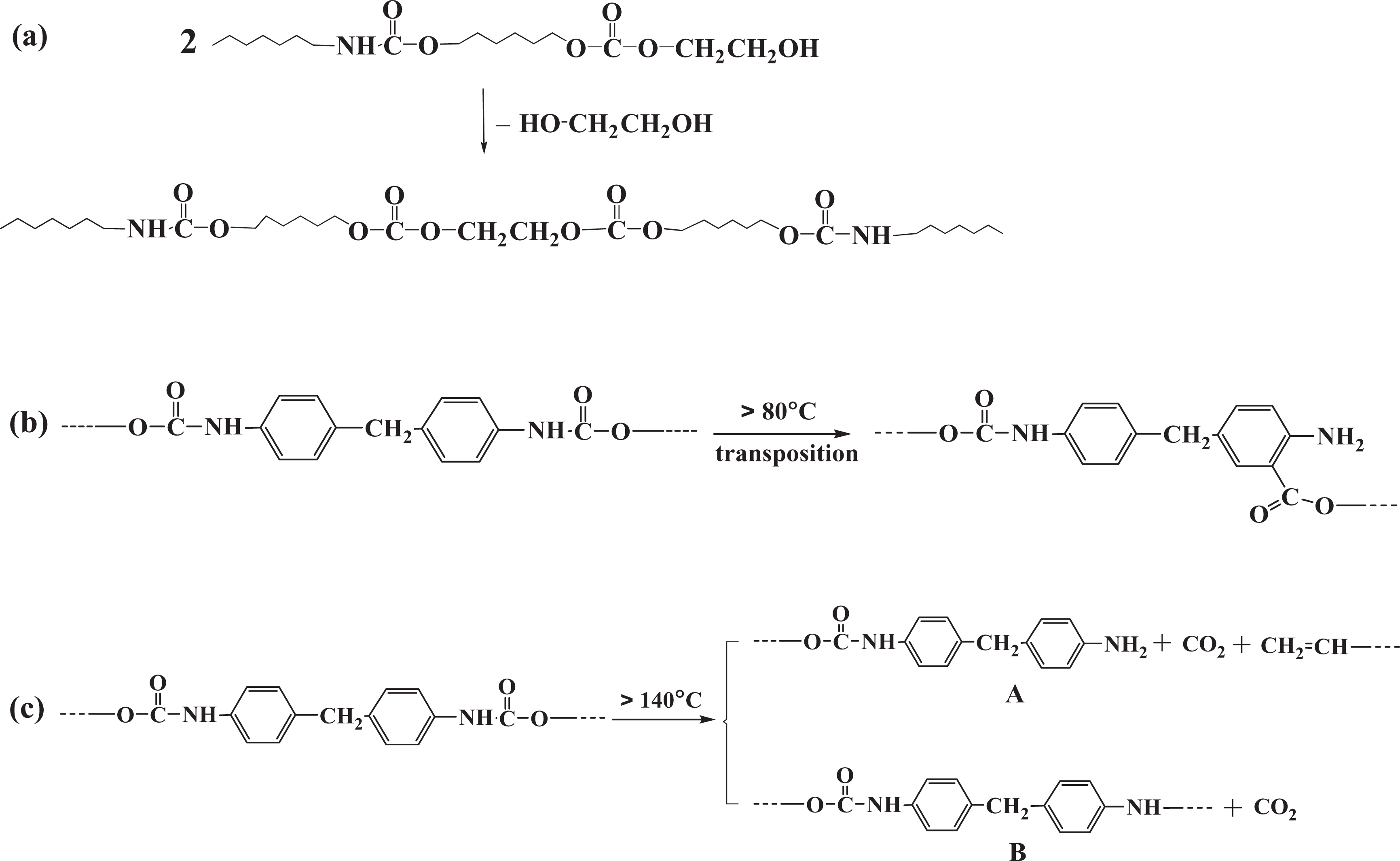
(a) Transesterification reaction between two PU molecules; (b) thermal transposition of urethane groups; and (c) thermal dissociation of urethane groups. PU: polyurethane.
Contact angle and surface free energy
The contact angle measurement technique has evolved from theoretical and empirical approaches to a rapid way to gain information about the surface constitution of materials and its role in interfacial phenomena. This technique can yield information that can be used to understand some aspects of material interaction and can then allow further tailoring of surfaces to favor specific biological responses. 30 Thus, the biocompatibility of materials, especially blood compatibility, depends strongly on their surface characteristics due to the fact that the interaction between blood and biomaterials occurs at their interface. 31 Since these PUs were designed to obtain scaffolds for cardiovascular applications, their wettability and surface energy play a key role.
The results obtained from contact angle measurements and the surface free energy determinations are presented in Table 1.
Surface parameters of PU samples.a
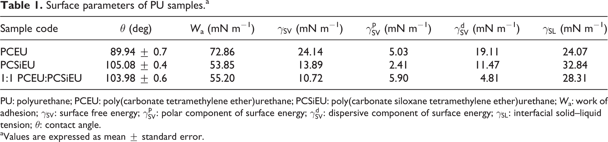
PU: polyurethane; PCEU: poly(carbonate tetramethylene ether)urethane; PCSiEU: poly(carbonate siloxane tetramethylene ether)urethane; Wa: work of adhesion;
aValues are expressed as mean ± standard error.
It is observed from Table 1 that PCSiEU (105.08) has a higher value of the water contact angle than PCEU (89.94). Therefore, the incorporation of the siloxane structure caused an important increase of the water contact angle, making this surface hydrophobic. When these two polymers were mixed, there was a decrease in the contact angle value (103.98) when compared to pure PCSiEU.
Based on this contact angle values, some parameters such as surface free energy, work of adhesion, interfacial solid−liquid tension can be calculated (Table 1). Thus, surface free energy was determined from the contact angle measurements using Owens–Wendt–Rabel and Kaelbe methods. 20 –23 The results reveal that the total surface energy of PCSiEU was reduced when compared with PCEU. This behavior is ascribed to the low surface energy of siloxane structure, 11,32 which leads to the migration of these segments at the PU surface and further resulted the lowering of the surface tension. It is also known from the Young’s equation that the higher the contact angle, the lower the solid surface energy. The calculation of polar (γ p) and dispersive (γ d) components of surface free energy gives more detailed information on the surface properties of the studied samples. It is clear from the results that the dispersive components are much higher than the polar components. It is well known 33 that polar forces are permanent dipoles, while dispersion forces are temporary dipoles that arise from dipole moments induced by nearby molecules. If dispersion forces are strong, molecules will be significantly attracted to each other. These forces are also stronger when intermolecular attractions are maximized through the total contact area between polymer chains. For the 1:1 mixture of these two polymers, it is observed that the values of both components (polar and dispersive) are quite similar, 5.9 and 4.81 mN m−1, respectively.
The work of adhesion (W a) of liquid to polymer surface can also be calculated from the contact angle data. The values of W a are decreased for the PU sample, while the values of their contact angle increase. The interfacial solid–liquid tension (γ SL) values increase with increasing contact angle values. It is well known that these values could be high or low, depending on the attractive forces between molecules of the liquid and solid. 31
Cytotoxicity analysis
In this study, mouse fibroblast cell line NIH3T3 was used for preliminarily toxicity screen of PU samples. Thus, cell viability based on MTS results for all the samples is illustrated in Figure 7.
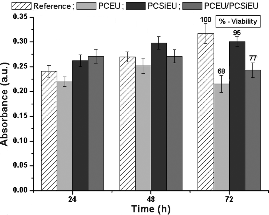
MTS absorbance and cell viability calculated for 72 h of cell incubation with PU samples. MTS: 3-(4,5-dimethylthiazol-2-yl)-5-(3-carboxymethoxyphenyl)-2-(4-sulfophenyl)-2H-tetrazolium; PU: polyurethane.
From these results, it is observed that none of the three materials affect the cellular viability after 24 and 48 h of incubation in cell culture. In some cases, the number of viable cells after incubation with PU samples was higher than that of the cells in the control wells (without polymer). After 72 h of incubation, a decrease of viability is observed (approximately 30%) for PCEU. For PCSiEU, the viability is unchanged as compared to the control and for the blends this decreases (20-25%). Taking into consideration the evolution of cellular viability during the first 24–48 h of incubation, the reductions in cellular viability do not show the toxicity of the studied materials. Therefore, these preliminary results show higher biocompatibility of PCSiEU and PCEU/PCSiEU in comparison with PCEU.
Hemocompatibily (thrombogenicity) analysis
In order to evaluate the hemocompatibility of PU samples, the main parameters related to blood coagulability were determined (extrinsic and intrinsic pathways): Fb, TQ, and APTT. Based on coagulation parameters, the INR was calculated. The experimental results are presented in Table 2.
Results of hemocompatibility tests.
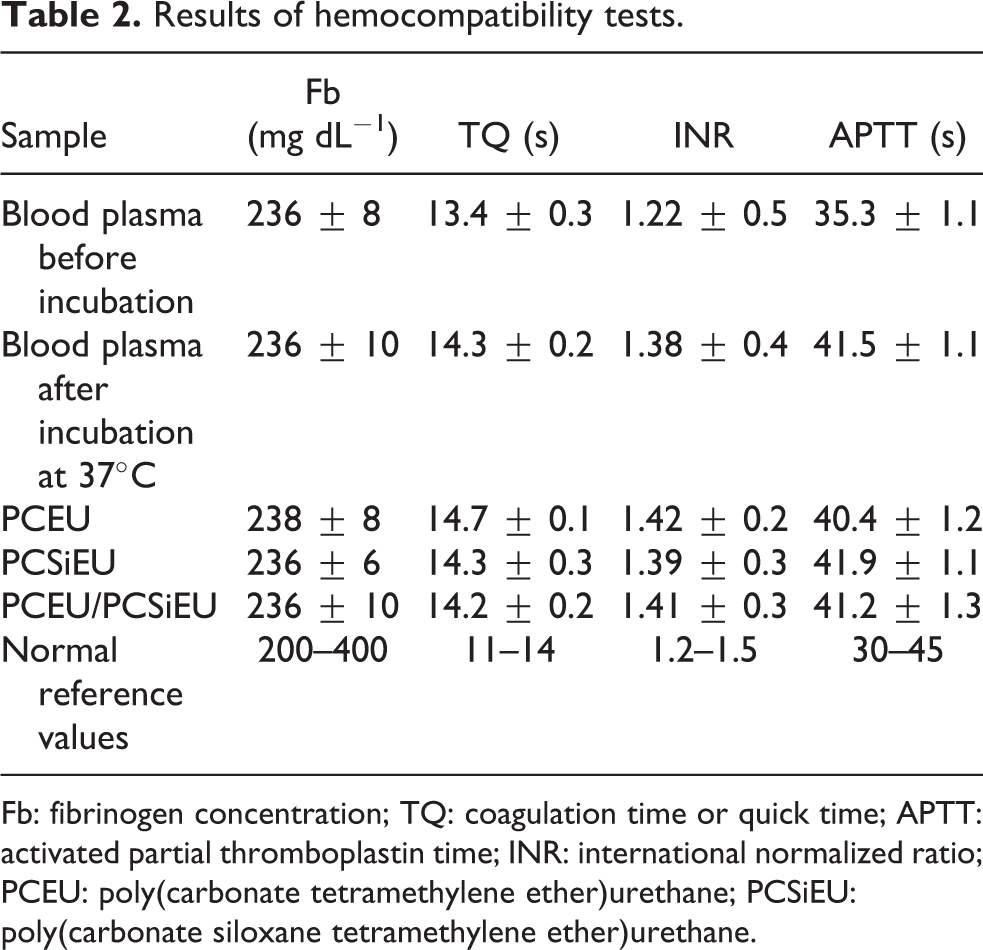
Fb: fibrinogen concentration; TQ: coagulation time or quick time; APTT: activated partial thromboplastin time; INR: international normalized ratio; PCEU: poly(carbonate tetramethylene ether)urethane; PCSiEU: poly(carbonate siloxane tetramethylene ether)urethane.
It is important to notice that none of the studied materials alter the blood clotting ability after incubation, as compared to the control (blood plasma incubated at 37°C without material). All the coagulation parameters did not differ significantly from the negative control incubated under the same conditions. Moreover, the TQ slightly increased, as compared to the control blood plasma. These data suggest that under the tested conditions, the PU samples (PCEU, PCSiEU, and PCEU/PCSiEU) did not accelerate clot formation mechanisms.
Conclusion
Since the chemical structures of the PU surfaces are capable of biological responses, it is vital to know the surface properties of these polymers. The analysis of these surface properties gives us the possibility to understand the first interaction mechanism of polymer with the body. Thus, some PCEU, PCSiEU, and their 1:1 gravimetric mixture (PCEU/PCSiEU) membranes were prepared and their surface properties were analyzed. The effect of temperature on the chemical structure of PU surfaces was investigated by ATR-FTIR spectroscopy. The analysis of the spectra reveals that the temperature has a significant influence on these PU surface structures. At body temperature, these materials are stable, but at high temperatures when sterilization methods are employed, PCSiEU is more stable than PCEU.
Increase in the water contact angle value and decrease in surface energy demonstrate the hydrophobic nature of PCSiEU and PCEU/PCSiEU surfaces which is in favor of polymer biocompatibility. This can be attributed to the low surface energy characteristic of siloxane structure, which promotes its migration toward the surface of PUs. The preliminary cytotoxicity screening showed noncytotoxic effect of these PU membranes over NIH3T3 mouse fibroblast cells. It was also shown that PU samples did not accelerate clot formation mechanisms under the tested conditions. Therefore, the surface and biological properties suggested that these PU membranes were promising materials for the preparation of cardiovascular scaffolds.
Footnotes
Funding
The authors are grateful for the financial support of BIOSCENT Project, grant agreement no. ID 214539 and of Romanian National Authority for Scientific Research, CNCS-UEFISCDI, project number PN-II-ID-PCE-2011-3-0199 (contract 300/2011).