






Abstract
Pneumocystis jirovecii pneumonia (PJP) is one of the leading opportunistic infections seen in people living with HIV. Bilateral infiltrations characterize PJP and miliary involvement is very rare. In this article, we report a 32-year-old person living with HIV who was followed up with a diagnosis of miliary PJP. Our patient was admitted to the hospital with complaints of cough, sputum, and weight loss. Initially, miliary tuberculosis was considered due to the presence of miliary involvement on chest radiography, but the diagnosis was made with P. jirovecii PCR positivity. This article aims to report a case of miliary PJP, a rare clinical form of PJP.
Introduction
Pneumocystis jirovecii pneumonia (PJP) is among the leading opportunistic infections seen in people living with human immunodeficiency virus (HIV). 1 PJP typically begins with nonspecific symptoms such as fever, dry cough, and dyspnea. 2 The most common radiological appearance is bilateral interstitial infiltration or alveolar ground-glass infiltrates. 3 This article presents an atypical case of PJP that emerged with bilateral miliary involvement in a person living with HIV, mimicking miliary tuberculosis.
Case report
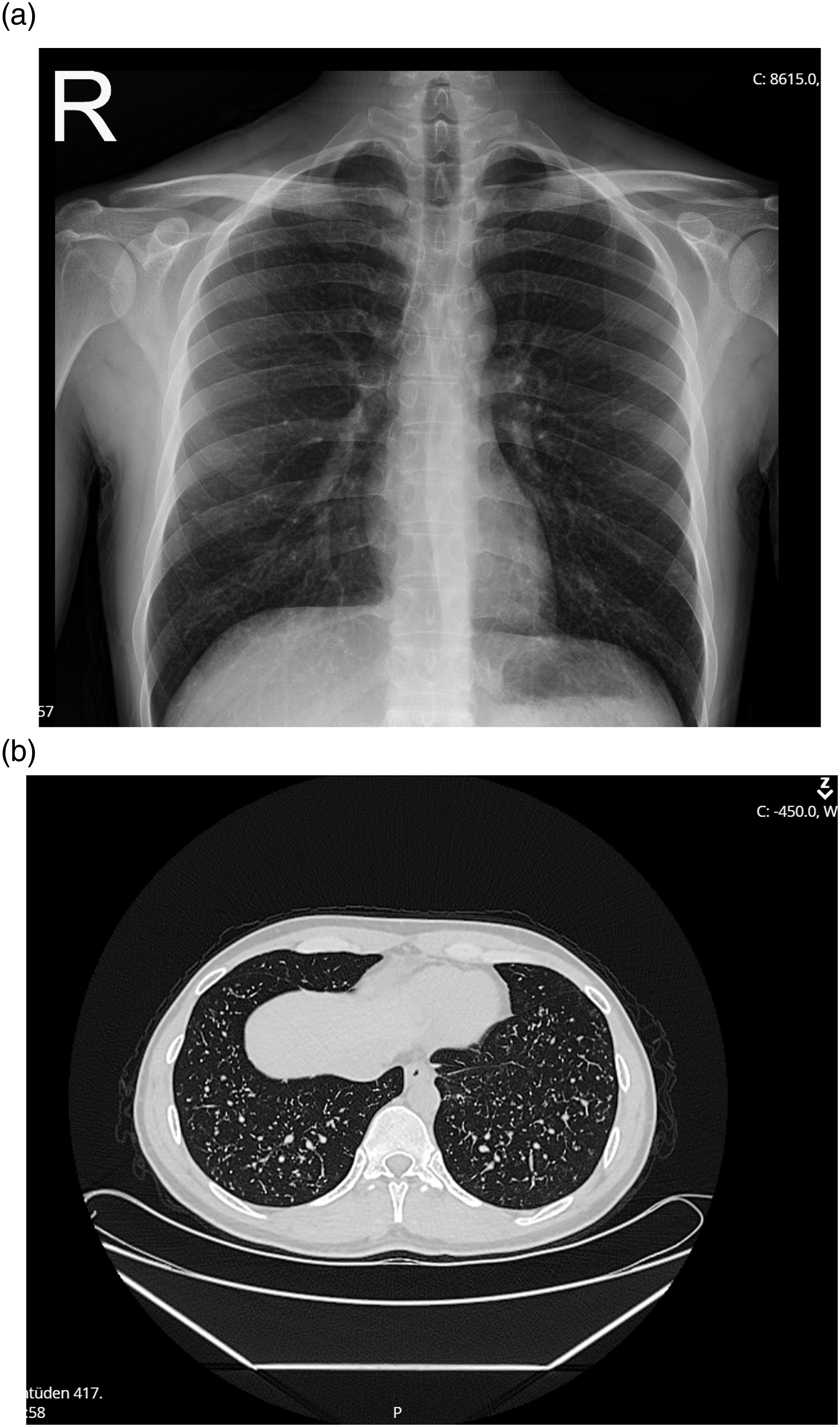
A 32-year-old male patient with no known medical history presented with cough, sputum, and weight loss (7–8 kg) lasting about a week. The patient was in good general condition, oriented, and cooperative. A miliary pattern was detected on the chest X-ray (Figure 1), and the patient was admitted with a preliminary diagnosis of miliary tuberculosis. Upon finding the anti-HIV test positive, a confirmatory test was requested. Thoracic computed tomography (CT) reported “multiple centriacinar nodules scattered throughout both lung parenchyma, more prominent in the lower lobes” (Figure 1). “ HIV RNA was 7.4 × 10⁵ copies/mL, and CD4 count was 45 cells/mm³ (5%). Trimethoprim-sulfamethoxazole (TMP/SMX) 5 mg/kg was started for PJP prophylaxis. The serum cryptococcal antigen was negative. PJP positivity was detected in the sputum PCR sample. It was considered colonization. A bronchoscopy was performed. Bronchoalveolar lavage (BAL) aside fast stain (AFS) was negative, and a bone marrow biopsy was performed. Anti-tuberculosis treatment (isoniazid, rifampicin, ethambutol, and pyrazinamide) was started. No growth was observed in the BAL culture. BAL tuberculosis PCR was negative, BAL PJP PCR was positive, and TMP/SMX 160/800 mg 3 × 2 IV was continued. On the third day of TMP/SMX dose adjustment, he had leukopenia and thrombocytopenia. TMP/SMX treatment was discontinued. TMP/SMX was restarted at a prophylactic dose upon resolution of leukopenia and thrombocytopenia after 48 h. However, 41°C fever, maculopapular rash, and hypotension were detected, and prophylaxis was discontinued. No growth was observed in the bone marrow culture, tuberculosis PCR was negative, AFS and mycobacterium culture were negative. The existing miliary involvement was felt to be due to PJP, and primaquine 15 mg/kg PO and clindamycin 600 mg 3 × 1 IV was started. Tenofovir disoproxil fumarate 250 mg, emtricitabine 150 mg, and dolutegravir 2 × 50 mg were started. Clinical and radiological findings regressed after PJP treatment (Figure 2). Chest X-ray showing nodular infiltrations at the time of presentation (a), Thorax CT showing nodular infiltrations at the time of presentation (b). Improved chest radiograph after treatment (a), Thorax CT findings improved after treatment (b).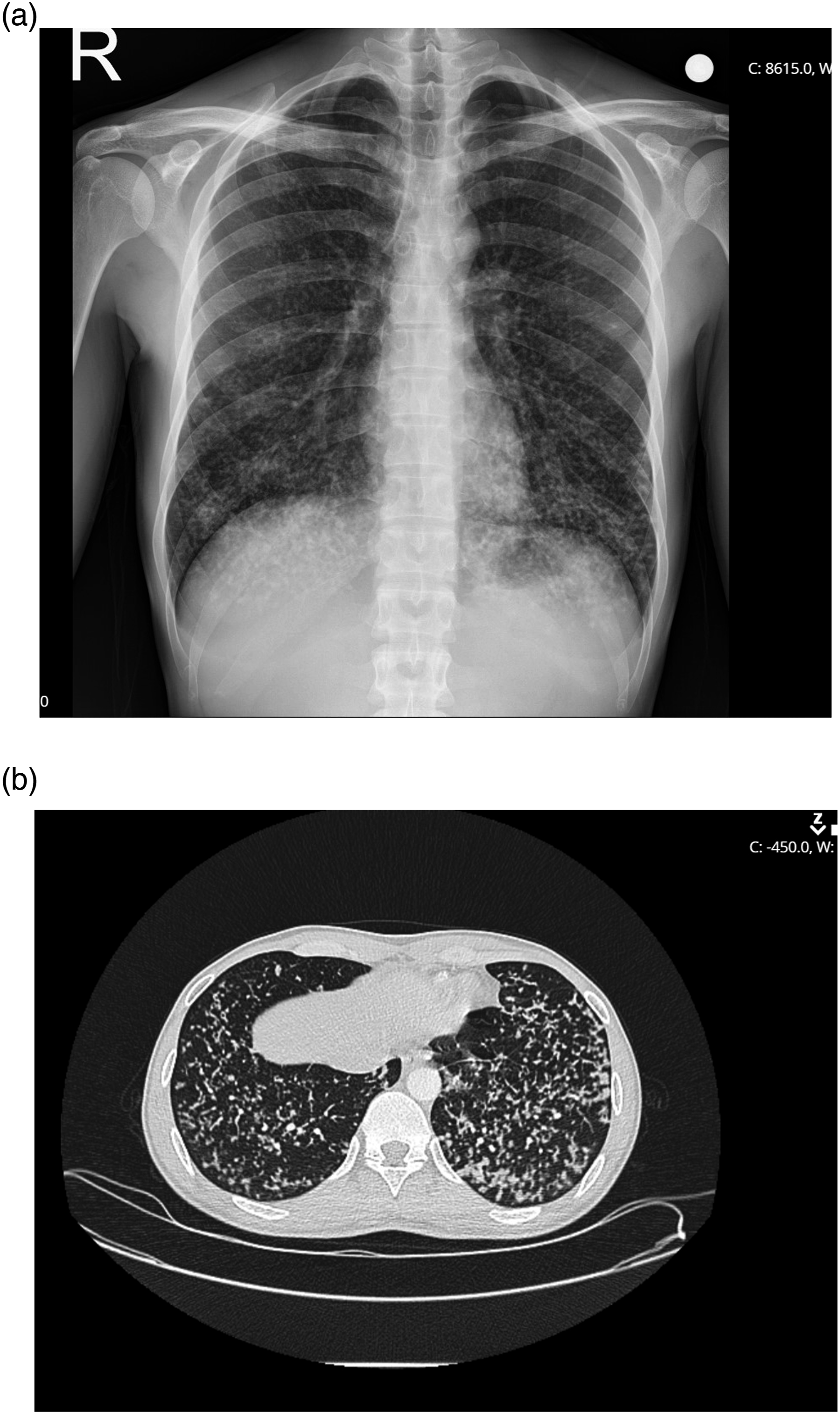
Discussion
P. jirovecii can cause severe pneumonia in people living with HIV (PLWH). The most common clinical findings of PJP are fever, cough, and dyspnea. 2 The typical radiographic findings on thoracic CT are bilateral interstitial ground-glass opacities. However, radiological images can vary from normal to lobar or nodular infiltrates, cavities, pleural effusion, pneumatoceles, and pneumothorax. 3 Our case was a person living with HIV with a low CD4 T count that presented with cough, sputum, and weight loss. Multiple nodules scattered throughout both lung parenchyma were observed on thoracic CT, displaying a miliary pneumonia pattern.
The definitive diagnosis of PJP is demonstrating cysts/trophozoites or PCR. The sensitivity of PCR positivity in induced sputum/BAL is higher than staining and immunohistochemistry. 4 In our case, the preliminary diagnosis was miliary tuberculosis and P. jirovecii PCR positivity was initially considered colonization. However, the positivity of BAL PCR and the absence of microbiological evidence of tuberculosis supported the diagnosis of PJP.
The miliary pattern of PJP is a very atypical presentation, with only a few cases reported in the literature.5–8 The first case was a person living with HIV reported in 1989. 5 The second case was reported in 1992. 6 Among the other cases, there is a PJP case reported by Jung et al. in 2009 in a kidney transplant patient and a PJP case reported by Kenjiro et al. in 2023 in a person living with HIV, both of which were mistaken for miliary tuberculosis. 8 To our knowledge, this is the first miliary PJP case reported from our country.
TMP/SMX is the approved primary treatment regimen for PJP treatment and prophylaxis. 9 A systematic review of 29 studies evaluating the use of primaquine/clindamycin as a second-line agent in PJP treatment. They found that the response rates were not lower than those of trimethoprim/sulfamethoxazole; however, IV pentamidine was significantly less effective. 10 Based on the findings of this systematic review, earlier initiation of primaquine/clindamycin or trimethoprim/sulfamethoxazole during treatment could have prevented worsening outcomes.
The miliary pattern of PJP is very unusual compared to miliary tuberculosis. In severe cases, anti-tuberculosis treatment may sometimes be started based on clinical diagnosis alone, without microbiological evidence. In our case, anti-tuberculosis treatment was initiated before confirming the diagnosis because CT findings supported miliary tuberculosis. Another possibility was co-infection with M. tuberculosis and PJP, however, there was no evidence of TB as AFS, mycobacterium cultures, and tuberculosis PCR were all negative. This case demonstrates that microbiological diagnosis is necessary even if clinical and/or radiological findings suggest a specific etiology.
Footnotes
Author’s note
The institution where the study was conducted: Sivas Cumhuriyet University Research Hospital.
Declaration of conflicting interests
The author(s) declared no potential conflicts of interest with respect to the research, authorship, and/or publication of this article.
Funding
The author(s) received no financial support for the research, authorship, and/or publication of this article.
