
Abstract
We report two cases of secondary syphilis with an isolated papule on the palm as the initial presentation. The clinical manifestations of secondary syphilis can be diverse, with a high rate of misdiagnosis and underdiagnosis. This article presents two patients with a purpose to alert clinicians not to forget the great imitator of syphilis for lesions of uncertain diagnosis.
Keywords
Patient A
A 54-year-old man presented with a 3-month history of a papule on his right palm. The nodule evolved into a pustule and subsequently formed a crust. Two months prior, he began to notice the formation of papulopustules on his head and face, followed a month later by ulcers on his penis. Upon phisical examination, a well-defined hyperkeratotic clavus-like papule approximately the size of a soybean, with scaling, was observed on his right palm (Figure 1). Additionally, multiple papulopustules with crusts were present on his face and head, and multiple ulcers were found on his penis. A positive toluidine red unheated serum test (TRUST 1:16) and a reactive Treponema pallidum particle agglutination (TPPA) assay were obtained. The quantitative polymerase chain reaction (qPCR) were positive for Treponema pallidum in the lesion samples from head, hand and penis. The HIV test was negative. Based on these findings, the diagnosis of secondary syphilis was made. The patient was treated with two doses of intramuscular benzathine benzylpenicillin (2,400,000 U). Four weeks later, all lesions had completely resolved. A well-defined hyperkeratotic clavus-like papule approximately the size of a soybean, with scaling, was observed on his right palm.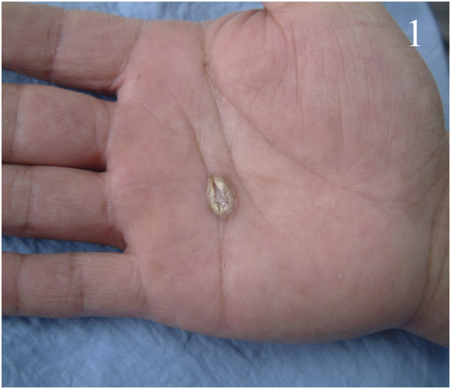
Patient B
A 44-year-old man presented with a painful papule on his left palm for 20 days, along with painful perineal ulcers and perianal nodule developed 15 days prior. Twenty months ago, he was diagnosed with primary syphilis indicated by genital ulcers and positive serologic tests, with a TRUST titer of 1:16. The lesions resolved completely after he received two intramuscular injections of benzathine benzylpenicillin. Follow-up testing 11 months later showed a decreased titer of 1:4. However, a routine test 3 months ago indicated a recurrence, with the titer returning to 1:16, and he was subsequently treated with oral doxycycline for 28 days. Physical examination found an isolated clavus-like papule with a yellowish-white crusted center on his left palm (Figure 2(a)). An ulcer was observed in the perineum and an exudative pink nodule in the perianal region. Syphilis serology was positive with a TRUST titer of 1:16. Real-time qPCR on a perineum ulcer swab tested positive for Treponema pallidum. The HIV test was negative. Histopathological analysis of skin lesions from the left palm and perianal area revealed epidermal hyperkeratosis, parakeratosis, neutrophilic migration, and plasma cell and lymphocyte infiltration around blood vessels in the superficial middle layer of dermis (Figure 2(b) and (c)). Immunohistochemical staining confirmed the presence of syphilis spirochetes (Figure 2(d) and (e)). The diagnosis was recurrent secondary syphilis. Treatment consisted of three intramuscular doses of benzathine benzylpenicillin (2,400,000 U). The lesions resolved completely 3 weeks later. (a) An isolated clavus-like papule with a yellowish-white crusted center was found on his left palm. (b) Histopathological analysis of skin lesions from the left palm revealed epidermal hyperkeratosis, parakeratosis, neutrophilic migration, and many plasma cell and lymphocyte infiltration around blood vessels in the superficial middle layer of dermis. (c) Histopathological analysis of skin lesions from the perianal area revealed epidermal hyperkeratosis, parakeratosis, neutrophilic migration, and many plasma cell and lymphocyte infiltration around blood vessels in the superficial middle layer of dermis. (d) Immunohistochemical examination of a lesion from the left palm confirmed the presence of syphilis spirochetes. (e) Immunohistochemical examination of a lesion from the perianal area confirmed the presence of syphilis spirochetes.
Discussion
Secondary syphilis is notoriously diverse in its clinical presentation, with symptoms such as rashes, papules, pustules, flat warts, and hair loss mimicking a myriad of skin conditions. This resemblance to other diseases significantly increases the risk of underdiagnosis or misdiagnosis. Few reports describe secondary syphilis presenting solely as isolated clavus-like papules on the palm. Often, hyperkeratotic papules or plaques in the palmoplantar region are misdiagnosed as viral warts or calluses. 1 These lesions, known as clavi syphilitici, 2 always lead to clinical confusion. Thus, suspected cases of syphilis warrant serological tests, and, if necessary, histopathological examination to detect the spirochetes.
These two cases presented with atypical syphilis, initially showing a rare clavus-like rash on the palm. Patient A’s diagnosis of secondary syphilis was confirmed by positive serology, qPCR, and a rapid response to benzathine benzylpenicillin. Patient B, with a history of syphilis, experienced a recurrence confirmed by positive serology, qPCR, and immunohistochemistry. These cases underscore the importance of considering syphilis in atypical lesions.
Footnotes
Declaration of conflicting interests
The author(s) declared no potential conflicts of interest with respect to the research, authorship, and/or publication of this article.
Funding
The author(s) received no financial support for the research, authorship, and/or publication of this article.