
Abstract
A subtle yet ubiquitous feature of the human face is eye glint—specular reflections from the surface of the eye that vary with the position of light sources in the environment. This study tested whether eye glint influences face perception, particularly in how observers perceive the gaze direction of a person they are viewing. Adult participants viewed computer-rendered face images that varied in eye direction, head rotation, and illumination. The presence of eye glint had little influence on the accuracy or precision of perceived gaze direction when faces were viewed under simplified conditions. However, biases in perceived gaze direction caused by changes in head orientation or illumination direction were reduced when eye glint was present relative to when it was absent. This suggests that eye glint can help an observer to maintain constancy in gaze perception despite variability in the appearance of the eye region that occurs across viewing conditions.
Keywords
Gaze perception is an essential component of social interactions. Movement of the eyes can reveal a person’s intent and focus of attention, and correspondingly, as observers, we tend to track the gaze direction of others to help understand, anticipate, and coordinate with their speech and behavior. Behavioral sensitivity to the direction of other people’s gaze is a key social-cognitive milestone that develops early in life (Striano & Reid, 2006), including reflexive orienting of attention to where others are looking and sensitivity to gaze directed toward oneself (Farroni et al., 2002, 2004; Senju & Johnson, 2009). A key visual feature that we rely on to determine the gaze direction of others is the coarse pattern of luminance contrast visible within their eye opening, which varies systematically with the position of the (darker) iris and pupil relative to the (lighter) sclera of the human eye (Ando, 2002). However, the relationship between luminance cues and veridical gaze direction is influenced strongly by contextual variables, such as head orientation and illumination direction (Otsuka & Clifford, 2018; Palmer et al., 2020). Accurate gaze perception thus requires a surprisingly complex visual analysis of facial features both within and outside of the eye region, likely mediated by specialized neural mechanisms in later stages of the visual pathway (Carlin & Calder, 2013; Clifford & Palmer, 2018).
A subtle feature of the human eye is the occurrence of mirror-like specular reflections from the eye surface (eye glint) arising from the unique material properties of the cornea and the glossy surface of the sclera. Although inconspicuous, eye glint is almost always present in faces we view. Does this facial feature play any role in face perception? More generally, specular reflections can influence the perception of object properties, including color (Yang & Maloney, 2001), lightness (Boyaci et al., 2006), curvature (Adams & Elder, 2014), and material composition (Schmid et al., 2023). In the context of face perception, one study found that artificially manipulating the position of specular reflections within the eye region of 3D-rendered faces can be sufficient to alter observers’ judgments about when the faces are making eye contact with them (Palmer et al., 2020). Similarly, eye glint is commonly drawn stylistically in cartoon faces, and its position within the eye can be redrawn to suggest different gaze directions or give an impression of eye movement (Anstis, 2018; Kitaoka, 2007). However, it is unknown whether eye glint contributes to gaze perception in naturalistic conditions and what the perceptual mechanisms are that underlie any such effect.
Interestingly, many commercial video-based eye-tracking systems estimate the rotation of the subject’s eyes partly by comparing the position of the pupil in a recorded image to one or more specular reflections visible on the cornea that serve as landmarks within the eye region (Hansen & Ji, 2010). This typically involves the eye tracker detecting specular reflections produced by an infrared light source controlled by the system, whereas in natural environments the position of light sources relative to the face is typically uncontrolled and highly variable. Hence it is unclear to what extent the human visual system exploits information provided by eye glint for gaze perception.
Here, we proposed and tested three distinct hypotheses about the potential role of eye glint in gaze perception. In the first experiment, we tested whether the presence of eye glint alters the accuracy and precision of perceived gaze direction in human observers. As illustrated in Figure 1a, the position of glint within the eye opening can remain relatively constant when a face is viewed under fixed lighting conditions, whereas changes in gaze direction produce a shift in the relative position of the iris. The position of the glint relative to the iris could thus serve as a cue for detecting changes in gaze direction. For example, human observers tend to judge a range of gaze directions centered around veridically direct gaze as making eye contact (Gamer & Hecht, 2007; Palmer et al., 2022). This provides scope for changes in glint-iris position that occur under fixed lighting conditions to enhance a viewer’s sensitivity to subtle deviations in eye direction away from direct gaze—at least when viewing faces from a (near) distance at which glint is visible.
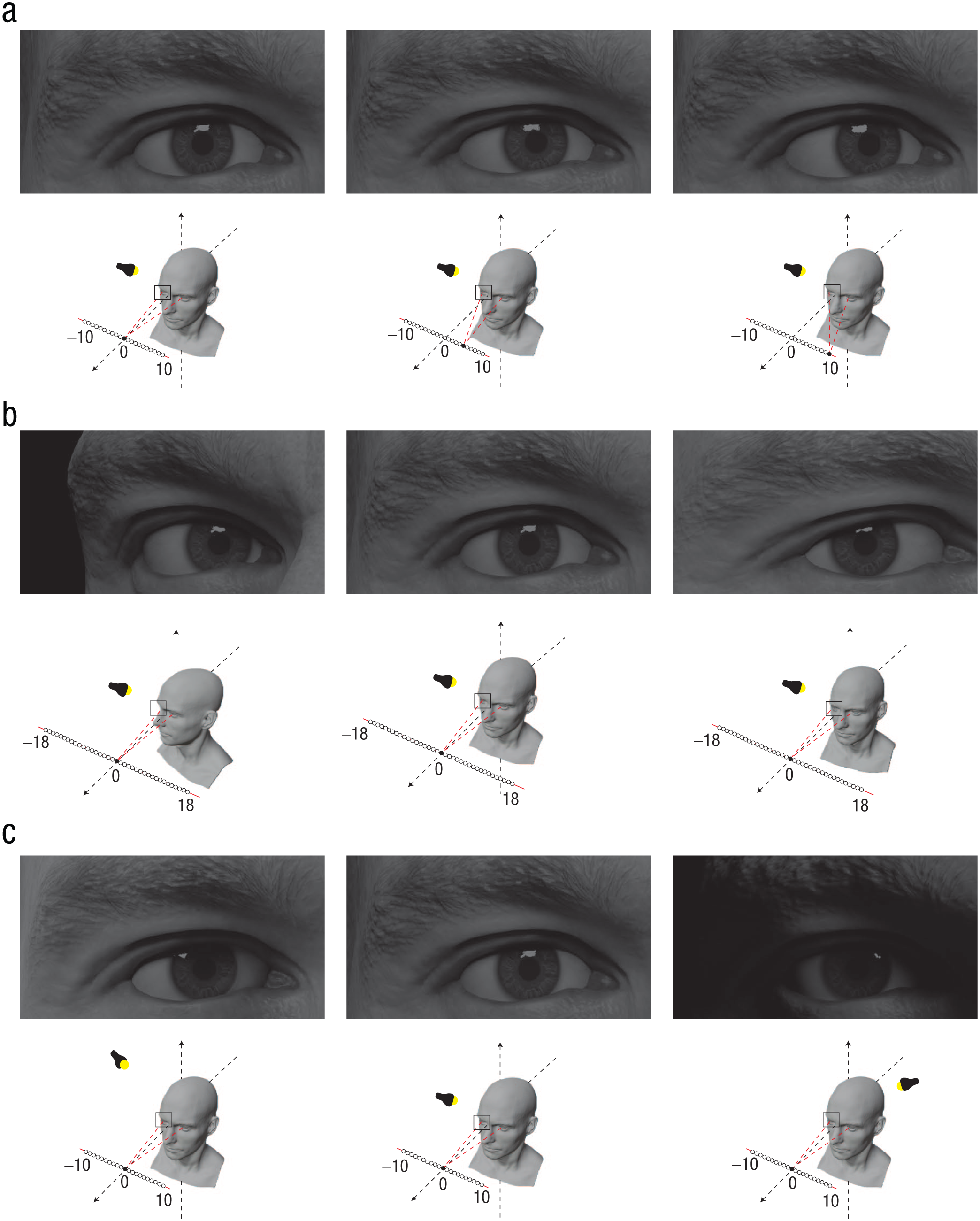
An illustration of how eye glint varies with eye direction, head orientation, and lighting direction. In (a) we show a shift in eye direction while the head position and lighting direction remain constant. Notice that eye glint is central on the iris when the eye is directed toward the observer with a central light source (left) but falls on the left of the iris as gaze is directed 5° (middle) and 10° (right) rightward. In (b) we show a shift in head orientation while the gaze remains directed at the observer. Glint remains central on the iris, despite the drastic shift in iris position within the eye opening caused by leftward (left) and rightward (right) head rotations (middle: no rotation). Hence, under fixed lighting conditions, the glint position on the iris might provide a cue to gaze direction that is relatively immune to changes in head orientation. In (c) we illustrate changes in the position of the light source—that is, to the left, directly above, and to the right of a face—while eye and head orientations remain constant. Eye glint varies in position across the iris depending on lighting direction, as does the pattern of shading and shadows across the eye region. In principle, glint position might serve as a cue to illumination direction.
In the second experiment, we tested whether eye glint helps an observer to maintain a robust estimate of gaze direction across changes in head orientation. A key challenge in object vision is perceptual constancy—that is, maintaining accurate perception of an object despite image variability related to factors like viewpoint and illumination direction. In the context of gaze perception, a given direction of gaze relative to the observer (e.g., the sense of eye contact) can be conveyed by many different combinations of head and eye direction. Perceptual constancy is maintained largely through the implicit integration of cues to eye direction and head orientation, which can be modeled as a form of linear cue combination (Otsuka et al., 2014, 2016).
Nevertheless, systematic biases in perceived gaze direction occur because of changes in the appearance of the eye region across head orientations, most notably changes in the apparent position of the iris and sclera within the eye opening when a face is viewed from different angles (Otsuka et al., 2015). For instance, gaze direction is perceived to shift leftward when a head that is directly facing the observer turns to the right while maintaining the same veridical eye orientation relative to the observer. Interestingly, the position of eye glint on the iris remains relatively stable across changes in head orientation when the direction of gaze is fixed relative to the observer (Fig. 1b). However, it is unknown whether the human visual system tracks glint displacement on the iris to help maintain perceptual constancy when estimating others’ gaze direction.
In the third experiment, we tested whether eye glint influences gaze perception by providing a cue to the direction of illumination. Noncentral illumination of a face can cause systematic biases in perceived gaze direction—for example, leftward illumination of a face can shift the apparent angle of gaze toward the right because of asymmetric darkening of the sclera by shading and shadows that appears to interfere with luminance cues central to gaze perception (Palmer et al., 2020; West, 2013). Although illumination can present a confound to accurately estimating gaze direction, there is evidence that human observers implicitly use other contextual cues produced by illumination (e.g., shading across the face) to effectively minimize gross errors in gaze perception that would otherwise be caused by changes in the appearance of the eye region (Ando, 2004; Palmer et al., 2020). More generally, specular highlights can provide information about lighting direction that influences judgments about object properties such as lightness (Boyaci et al., 2006). Because the position of eye glint visible to an observer varies depending on the position of light sources in the surrounding environment, observers could potentially use glint position to better estimate the lighting direction, which may help to minimize the confounding effects of shading and shadows on gaze estimation across lighting conditions (Fig. 1c).
Research Transparency Statement
General disclosures
Disclosures for Studies 1, 2, and 3
Experiment 1
Method
Participants
A total of 40 participants (12 men, 28 women) with an average age of 20.1 years (SD = 1.5 years) completed this experiment. All reported normal or corrected-to-normal vision and were naive to the purposes of the experiment. Participants received course credit for their participation. The protocol for all three experiments was approved by the National University of Singapore Departmental Ethics Review Committee (Department of Psychology, ID No. 2023-January-09), and all participants gave written informed consent.
Because there was little prior research to inform expectations about the effect size of the manipulations tested in Experiment 1, we chose the target sample size of 40 on the basis of the sample sizes of earlier studies that employed a similar method for measuring perception of eye contact (e.g., Palmer & Clifford, 2018; Palmer et al., 2020, 2022). A sensitivity analysis using G*Power, version 3.1.9.6 (Faul et al., 2007), suggested that the minimum effect size that can be reliably detected with 40 participants with 95% power is 0.58 (Cohen’s d).
Face stimuli
The face stimuli used in this study were produced using 3D models of human faces created by Ten24, a professional 3D-scanning company (https://ten24.info/3d-scanning/), and made available via their online store (https://www.3dscanstore.com/). The 3D models were generated by scanning human faces using a multicamera array combined with photogrammetry to produce high-resolution models of face shape and surface texture. Models of four men and two women were used to generate the face images, and the same six models were used across all experiments. Each of these faces bore a neutral expression.
The faces were rendered as 2D images in Blender 3.6.1 (The Blender Foundation, Amsterdam, The Netherlands) using Cycles, which is a physically based rendering engine that traces light paths within a modeled 3D space. This involves simulating interactions between light and object surfaces using customizable shaders to configure surface characteristics such as diffuse and specular reflectance. Blender’s Principled Bidirectional Scattering Distribution Function shader was used to render the images, driven in part by texture maps created using the 3D scans of individuals described above. The shader properties of the faces incorporated a combination of diffuse reflectance, specular reflectance, and subsurface scattering. In particular, the eyes of each face were modeled to be roughly spherical objects encased by a transparent surface that produced specular highlights, similar to the cornea and the wet surface of the human eyeball. Face images were rendered both with and without the transparent casing of the eyeball models, allowing the presence of eye glint to be manipulated while all other generative properties of the images were kept the same (e.g., diffuse reflectance of the eyeball, lighting, shading; see Fig. 2a). We will refer to these as glint-present images and glint-absent images, respectively. The diffuse reflectance of the eyeball was driven by a high-resolution texture that depicted a detailed iris and sclera. Additional facial features such as the eyelashes, eyebrows, teeth, and clothing were modeled separately for some identities. The image-production process was similar to that described in Palmer and Clifford (2020) and Palmer et al. (2020).
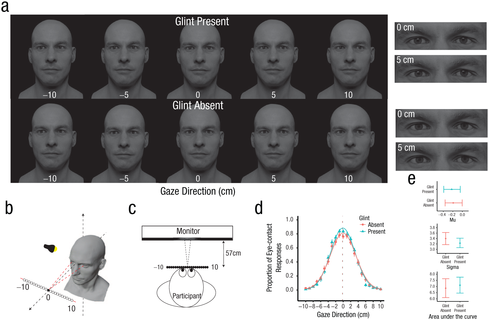
Stimuli, setup, and results of eye-contact task in Experiment 1. Faces were rendered looking in 21 different gaze directions (a, b), corresponding to a set of fixation locations at a depth of 57 cm from the face and varying in the horizontal dimension in 1-cm intervals from −10 to 10 cm (only specific intervals and one model are shown here). Negative coordinates indicate a leftward gaze relative to the observer. Each face image was rendered consistent with a single light source positioned centrally above the face, with and without eye glint. Faces were presented on screen such that they appeared to be gazing at a point between the eyes of the participants when looking directly ahead (c). Participants were seated 57 cm away from the monitor, consistent with the fixation depth of the face models. In (d) we show mean sample data representing the gaze directions that participants perceived as making eye contact with them. Error bars represent the standard errors of the mean (SEMs). Curves are Gaussian functions fitted to the super-subject data of each condition. Vertical dashed lines correspond to the mean (μ) values of each curve. In (e) we show the effect of eye glint on the sense of eye contact, quantified as the difference between the mu (μ), sigma (σ), and area-under-the-curve (AUC) values of the best-fitting Gaussian functions across conditions. Error bars represent 95% confidence intervals computed using bootstrapping.
A single light source, a square plane measuring 60 cm × 60 cm that emitted light, was used in the generation of each image. Within Blender, the light source was positioned 75 cm away from the face and was always elevated at an angle of 30° above the face at 0° along the x-axis, so that the faces were illuminated centrally from the front.
Image production in Blender was controlled using custom scripts in Python, version 3.10.12 (Python Core Team, 2017). Face images were rendered from the perspective of a camera situated 57 cm in front of each model, which matched the viewing distance of the participant during the experiment. The camera was aligned with the midpoint of the two eyes of the face model. The face images were presented at approximately life size on screen with an interpupillary distance of 6.3 cm (Fesharaki et al., 2012). Because the eyes were modeled separately from the rest of the face model, the gaze direction could be controlled precisely. In particular, gaze directions were varied by rotating each eyeball so that its pupillary axis intersected the desired point of fixation within the 3D scene. The fixation distance of the eyes was matched to the viewing distance of the participants. This meant, for example, that when the face models were rendered looking directly ahead, the two eyes of the face model were converged to fixate a point in between the viewer’s eyes. For each image generated, we also included a horizontally flipped version (with the horizontal gaze direction recoded accordingly) to control for any effect of left-right asymmetries in facial appearance (e.g., face morphology or texturing).
Design
This experiment was designed to examine whether the presence of eye glint affects the perception of gaze direction under simple viewing conditions. This question was tested across two tasks. The first task tested the effect of eye glint on the perception of eye contact. Participants were presented with faces on a computer screen with a range of horizontal gaze directions centered around direct gaze and indicated whether each face was making eye contact with them or not. The second task tested the effect of eye glint on the perceived angle of gaze across a wider range of gaze directions. Participants were presented with faces and rotated a pointer on the screen to report the exact angle that each face appeared to be looking. In both tasks, the presence or absence of eye glint was varied while we controlled all other features of the face images, so that any effect of eye glint on the precision or accuracy of gaze estimation could be measured. Participants completed both the eye contact and pointer tasks in a single session with the task order fixed across all participants. The session took approximately an hour to complete.
Procedure
Eye-contact task
The gaze direction of the face models varied to fixate a range of locations between −10 cm and 10 cm in 1-cm intervals (21 locations) along a horizontal plane centered between the participant’s two eyes, with negative values indicating leftward gaze relative to the participant (Fig. 2b and 2c). This task consisted of 10 practice trials and two main experiment blocks, one of which consisted only of glint-present images, whereas the other consisted only of glint-absent images (Fig. 2a). Block order was counterbalanced across participants, with trial order randomized for each participant within each block. Each block consisted of 252 trials (21 gaze directions × 12 repetitions for each direction consisting of the horizontally flipped and nonflipped versions of each image across six face models). A fixation cross was first presented within each trial for 0.5 s before each image was presented for 2 s. Participants were then prompted to respond whether the face presented made eye contact with them or not by pressing one of two keys, with the assignment of response keys counterbalanced across participants. Participants could make their response as soon as the faces appeared on the screen, with no response time limit. Each trial was initiated only after the participant had entered their response for the previous trial.
Pointer task
The gaze directions used in this task varied across −25° to 25° in 5° intervals (11 directions). The convergence of the two eyes was consistent with a fixation distance of 57 cm (Fig. 3b). Participants first completed 20 practice trials before moving on to two main blocks. Similar to the eye-contact task, glint-present trials and glint-absent trials were blocked and counterbalanced across participants. Each block consisted of 132 trials (11 gaze directions × 12 repeats of each direction). Each image was presented for 1 s, after which a spherical pointer was shown on the screen (Fig. 3c). The horizontal angle of the pointer was randomly selected within the range of ±90° for each trial, and the vertical angle was fixed at 0°. Participants reported the perceived gaze direction of each face stimulus by rotating the pointer using the mouse and were encouraged to be as accurate as possible. The pointer remained visible until the participant indicated completion of the adjustment with a mouse click.
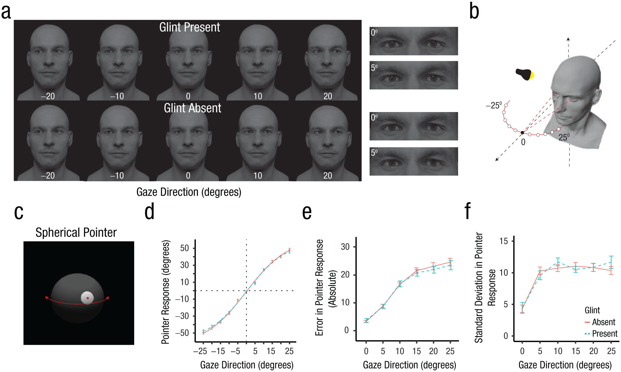
Stimuli, setup, and results of the pointer task in Experiment 1. Faces were rendered with 11 different gaze directions within the range of −25° to 25° in 5° intervals using a central illumination source (a, b). In each trial of the task (c), a spherical pointer was displayed on the screen after the face stimulus. Participants were required to rotate the pointer along the horizontal dimension using the computer mouse to report the gaze direction of the face. In (d) are shown mean pointer response data with error bars (representing standard errors of the mean) across gaze directions for the glint-present and glint-absent conditions. Curves represent logistic functions fitted to the data. Participant responses were collapsed across the leftward and rightward gaze directions (e.g., −5° and 5° were combined) in plots (e) and (f). In (e) we show the mean angular difference (absolute values used) between perceived and actual gaze directions (i.e., response error); in (f) we illustrate mean standard deviations of pointer responses at each gaze direction (i.e., response variability).
In both tasks, image presentation was controlled using MATLAB (Version R2023a; The MathWorks, Natick, MA), and the Psychophysics Toolbox (Version 3.0.19; Brainard, 1997) on a Lenovo ThinkVision monitor (2,560 × 1,440 resolution, 36 pixels per cm, 60-Hz refresh rate). Participants were seated approximately 57 cm away from the monitor to match the distance of the camera from the face models when generating the face images in Blender. Participants viewed the images using a chin rest and were also asked to adjust the height of the chair so that the eyes of the face images were level with theirs, so the faces on the screen would appear to be gazing at a point between the participants’ eyes when looking directly ahead (at 0 cm or 0°). Participants completed the experiments in a dark room.
Analyses
Statistical analyses were done using MATLAB and RStudio (Version 2024.12.1; R Foundation for Statistical Computing) running R version 4.5.0. For analysis of variance (ANOVA) tests, Greenhouse-Geisser corrections were applied to F and p values whenever Mauchley’s sphericity test indicated a significant violation. Bonferroni correction was used where appropriate, with p values multiplied by the number of post hoc comparisons. One-tailed t tests were used where appropriate to test the directional hypotheses outlined in the introduction.
Across all experiments, trials in which participants took less than 10 ms to respond were excluded, because this indicated that participants made their response before viewing the stimulus. Trials with response timings longer than 10 s were also excluded from analysis except for the pointer task, because participants might have needed more time to rotate the pointer.
Results
Eye-contact task
Consistent with previous findings (Palmer et al., 2022), participants tended to perceive eye contact when viewing faces with a range of horizontal gaze directions that landed on a series of locations distributed horizontally across their faces (Fig. 2d). As seen in Figure 2d, this zone of eye contact was very similar regardless of whether the faces were rendered with eye glint visible within the eye region or not. The effect of eye glint on the perception of eye contact was quantified by fitting Gaussian functions both to the pooled mean eye-contact responses of each condition separately (i.e., the super-subject data) and to the response data for individual subjects. Previous research has found that Gaussian functions are well suited for capturing the distribution of eye-contact responses across a range of horizontal gaze directions; such responses commonly exhibit a Gaussian-like profile even at the level of individual subjects (e.g., Linke & Horstmann, 2024; Palmer et al., 2022). The approach of fitting functions to super-subject data was included because it may help to reduce the noise inherent in individual-participant data, leading to more stable parameter estimates.
Gaussian functions included mean (μ), standard deviation (σ), and peak-height parameters, and were fitted to the data using the fminsearch function in MATLAB by minimizing the sum of squared errors. The fitted functions were constrained to the range of possible values for proportion data [0,1] by applying a penalty to the computed error value if the height of the peak fell outside this range. We expected that if eye glint improved the accuracy or precision of eye-contact judgments, the spread of eye-contact judgments across horizontal gaze directions would be significantly narrower in the glint-present condition compared with the glint-absent condition. In other words, a zone of eye contact with narrower width would suggest higher precision or less variability in eye-contact judgments. In addition to using the standard deviation and AUC of the best-fitting Gaussian functions as an indicator of the precision of eye-contact responses, the mean of each curve was used as an estimate of the center of the zone of eye contact. Bootstrapping was used to calculate the 95% confidence intervals (CIs) on the parameters of the best-fitting functions. This was done by resampling the response data with replacement 10,000 times, with the function refitted to the resampled super-subject data on each iteration. The same resampling of participants across conditions was used on each iteration to control for between-subject variance.
The super-subject response data were well approximated by the Gaussian functions with 99.1% and 99.5% variance explained for the glint-present and glint-absent conditions, respectively. Accordingly, the mean (μ) values of the best-fitting Gaussian functions were −0.22 cm, 95% CI = [−0.39, −0.04], when glint was present and −0.19 cm, 95% CI = [−0.36, −0.01], when glint was absent. In addition, the standard deviations of the best-fitting Gaussian functions were similar across the glint-present condition (σ = 3.23, 95% CI = [3.06, 3.41]), and the glint-absent condition (σ = 3.40, 95% CI = [3.17, 3.62]), indicating that the precision of gaze perception remained unaffected irrespective of whether eye glint was visible within the eye region or not. Although the glint-present condition appeared to have a higher proportion of eye-contact responses with a higher peak at 0 cm compared with the glint-absent condition, the AUCs for the glint-present condition (AUC = 7.12, 95% CI = [6.46, 7.76]) and glint-absent condition (AUC = 6.86, 95% CI = [6.09, 7.64]) did not significantly differ from each other, suggesting a similar proportion of eye-contact responses across conditions. A similar pattern was observed when Gaussian functions were fitted to individual participant data and the mean parameter values were compared as a function of glint presence using paired-samples t tests. The hypothesis that there would be a narrower distribution of eye-contact responses across horizontal gaze directions when eye glint was present was not supported—σpresent: M = 3.16 cm, SD = 0.65; σabsent: M = 3.23 cm, SD = 0.84; t(39) = 0.91, p = .18, one-tailed, Cohen’s d = 0.14; AUCpresent: M = 7.14, SD = 2.12; AUCabsent: M = 6.92, SD = 2.51; t(39) = −1.13, p = .87, one-tailed, Cohen’s d = −0.18. There was also no significant difference in the center point of the zone of eye contact across glint conditions—μpresent: M = −0.16 cm, SD = 0.53; μabsent: M = −0.13 cm, SD = 0.64; t(39) = 0.40, p = .69, two-tailed, Cohen’s d = 0.06. The Gaussian functions fit the individual participant data well, with the variance explained 92% on average (SD = 5%) across participants and conditions.
The centroid of eye-contact responses across horizontal gaze directions was also calculated for each participant and compared between the glint-present (M = −0.19 cm, SD = 0.53) and glint-absent (M = −0.17 cm, SD = 0.62) conditions using a paired-samples t test. Again, no significant difference was found between glint conditions, t(39) = 0.24, p = .81, two-tailed, Cohen’s d = 0.04. Overall, the results indicate that the presence of eye glint affected neither the precision nor overall proportion of eye-contact responses under the simplified viewing conditions tested in Experiment 1.
Pointer task
The pointer task was used to quantify the accuracy and precision of perceived gaze direction when viewing faces with a range of horizontal gaze directions. One can see in Figure 3d, 3e, and 3f that participants showed an overestimation in the perceived angle of gaze when viewing faces with averted gaze, which has also been reported in several previous studies (e.g., Alais et al., 2018; Balsdon & Clifford, 2018). These data also illustrate very little difference in gaze perception when comparing faces rendered with eye glint visible within the eye region to faces rendered without eye glint, in either the accuracy or precision of responses.
To test the effect of eye glint on gaze perception statistically, participant responses were first averaged across gaze directions for each glint condition. Logistic functions were fitted to the super-subject data of each condition to capture the relationship between pointer responses and the veridical gaze direction of the faces presented (Fig. 3d). Functions were fitted to the data using the fminsearch function in MATLAB by minimizing the sum of squared errors. These functions were fitted with four parameters: (a) inflection point (
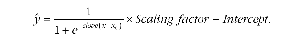
Bootstrapping was used to determine the 95% CIs on the parameters of the best-fitting functions. The logistic functions fitted the data very well, with 99.8% variance explained by the functions’ fit to the data in both the glint-present and glint-absent conditions. The best-fitting slope values across the glint-present condition (slope = 0.09, 95% CI = [0.07, 0.10]) and the glint-absent condition (slope = 0.08, 95% CI = [0.07, 0.09]) did not significantly differ from each other, and neither did the scaling factor (glint present: 118, 95% CI = [107, 133]; glint absent: 127, 95% CI = [115, 142]) nor the intercept values (glint present: −59.56, 95% CI = [−66.82, −54.03]; glint absent: −64.65, 95% CI = [−72.14, −58.51]), supporting the observation that the presence of eye glint within the eye region did not affect gaze perception in this task. Participants’ responses were centered on 0 degrees for the glint-present condition (
To estimate the effect of eye glint on the accuracy of gaze perception, the absolute difference between stimulus gaze direction and pointer responses was calculated as a measure of response error and compared across conditions (Fig. 3e). The standard deviation of the pointer position at each gaze angle was also calculated as an estimate of the (inverse) precision of responses (Fig. 3e). Participant responses were collapsed across the leftward and rightward gaze directions (e.g., −5° and 5° were combined). Data were first averaged across trials for each participant and then across the sample. Separate two-way repeated-measures analyses of variance (ANOVAs) with gaze direction and glint presence as within-subjects factors were conducted on the response error and standard deviation scores. There was a significant main effect of gaze direction on response error, F(2, 62) = 186.54, p < .001, η2 g = .55. Post hoc pairwise t tests revealed significant increases in response errors as gaze direction deviated further from 0° for both the glint-present and glint-absent conditions. This is consistent with an overestimation bias in perceived gaze direction that has been reported in several previous studies (e.g., Alais et al., 2018; Balsdon & Clifford, 2018). There was also a significant main effect of gaze direction on the standard deviation of responses, F(4, 142) = 65.66, p < .001, η2 g = .25. In particular, the precision of participants’ responses significantly decreased as gaze deviated away from the center. However, no significant differences were observed across glint conditions in participants’ response accuracy or precision.
In summary, gaze perception was very similar between the glint-present and glint-absent conditions in both the eye-contact task and the pointer task. This suggests that eye glint neither enhances one’s sensitivity to subtle deviations in gaze direction away from eye contact nor improves one’s judgment about the direction of another person’s gaze in terms of accuracy and precision. However, the face images used in both tasks were rendered under simplified viewing conditions, namely with central illumination of the faces and with the faces oriented directly toward the observer. Two main forms of variability in the appearance of faces in natural environments are changes in viewpoint (head orientation relative to the viewer) and illumination. Both factors are known to have a significant impact on the perception of gaze direction. In Experiments 2 and 3, we examined whether eye glint contributes to gaze perception by allowing the viewer to better tolerate these natural forms of variability in the appearance of faces.
Experiment 2
Method
Participants
Forty participants (22 men, 18 women; M = 23 years, SD = 3 years) were recruited for this experiment. The data of two participants were excluded and replaced, because they responded to the head orientation instead of gaze direction 100% of the time, resulting in no determinable centroid (described below). Five participants participated in both Experiments 2 and 3; each task was completed on a separate day, and all participants completed Experiment 3 first. The sample size of 40 was decided before data collection to match that of Experiment 1. Participants were compensated with $10 cash.
Design
This experiment was designed to measure the effect of head orientation on perceived gaze direction and whether this varies depending on the presence of eye glint. Head orientation strongly influences gaze perception, because identical eye regions can appear to be looking in different directions when seen in the context of different head orientations (Otsuka et al., 2015). For example, perceived gaze direction can be attracted toward head orientation, a phenomenon previously observed by Wollaston (1824). However, a simultaneous repulsive effect of head orientation on the perceived angle of gaze also occurs because of differences in the viewer’s perspective of the eye region. As a result of these opposing attractive and repulsive effects, perceptual constancy in gaze perception is imperfect when faces are viewed across different head rotations. Under typical viewing conditions, there is a net repulsive effect of head orientation on perceived gaze direction, so that faces angled to the right of the observer (for example) will tend to be perceived as looking more leftward than they really are.
In this experiment, we asked whether eye glint can improve the accuracy of gaze perception by acting as a stable reference point within the eye region, thereby reducing the influence of head rotation on gaze perception. As shown in Figure 1b, for a given gaze direction relative to the observer, glint position relative to the position of the pupil remains relatively constant across significant changes in head orientation. Participants were asked to judge the direction that faces were looking across varying gaze directions and lateral head rotations. Responses in glint-absent and -present conditions were then compared, to evaluate whether the presence of eye glint reduces the repulsive effect of head rotation on perceived gaze direction, thereby contributing to perceptual constancy in gaze perception.
Procedure
The procedure and setup were similar to the eye-contact task in Experiment 1 except for the face stimuli used and the instructions given. Specifically, in addition to the images with heads that were rendered to be looking straight ahead (0°), heads that were turned to the left (−20°) and right (20°) of participants were also generated (Fig. 5a). When generating the images in Blender, the head was rotated around the center of the eyes so that the eyes would always be in the same position on the screen regardless of the head rotation, similar to the images generated in previous studies that have measured the effect of head orientation on perceived gaze direction (e.g., Otsuka et al., 2015). For each head orientation, gaze directions varied to fixate a range of locations distributed horizontally across the participant’s face. In particular, the gaze of the face model fixated locations distributed between −18 cm and 18 cm in 3-cm intervals (13 points) at a distance of 57 cm from the face model. Two sets of images were generated—one set of images with eye glint rendered within the eye region of each face model, and another set that was identical but without eye glint, to allow us to compare the effect of eye glint on gaze perception when viewing faces that differed in head rotation.
Each participant completed 20 practice trials and two main experiment blocks with the order of glint-present and -absent blocks counterbalanced across participants. Each block consisted of 468 trials (13 gaze directions × 12 repeats of each gaze direction × 3 head orientations). Each image was presented for 0.5 s, similar to the paradigm used in Otsuka et al. (2015). Participants pressed one of three keys to indicate whether the face was looking to their left, looking directly at them, or looking to their right.
Analyses
The aims of the behavioral analyses were to quantify the effect of head rotation on perceived gaze direction and to examine whether eye glint contributes to perceptual constancy in gaze perception across head rotations. Two kinds of analyses, parametric and nonparametric, were conducted to measure the effect of head rotation on gaze perception. In addition to fitting Gaussian functions to the distribution of direct responses across horizontal gaze directions, similar to Experiment 1, we compared the centroids of the direct responses between the two head-rotation conditions.
Centroids representing the center point of the distribution of each participant’s response data were used to assess the magnitude and direction of shifts in perceived gaze direction across conditions. We first calculated the proportion of trials in which participants reported the face as looking directly at them for each gaze direction, to obtain a distribution of direct responses across gaze direction and to visualize each participant’s cone of direct gaze. Each centroid, which is a weighted average of the distribution, was then determined using the following formula:

where gi represents the gaze direction of the stimulus presented and pi represents the proportion of direct responses the participant made when viewing faces with that gaze direction across trials. Individual centroids were calculated for each participant before being averaged across participants for each condition to obtain a sample mean. To quantify the repulsive effect of head rotation on gaze perception, we calculated the centroid half-difference (CHD) for each participant. This was the difference in centroids between the left and right head-rotation conditions, divided by two. CHDs were compared between the glint-present and glint-absent conditions to evaluate whether the presence of eye glint reduces the effect of head rotation on perceived gaze direction (which would be reflected as smaller CHDs in the presence of eye glint), hence contributing to perceptual constancy in gaze perception.
Results
Image analysis: the influence of head orientation on glint and pupil positions
The current experiment was motivated by the idea that, when viewing a face under fixed lighting conditions, the position of the glint on the iris might serve as a cue to gaze direction that is relatively robust to changes in head orientation (Fig. 1b). To test this assumption, we performed an image analysis to quantify the position of the glint relative to the position of the pupil in the face images used in the behavioral experiment. We extracted the positions of the glint and pupil within each eye for the glint-present images and measured the distance between them (Fig. 4b), then plotted how this metric varied across gaze directions and head orientations.
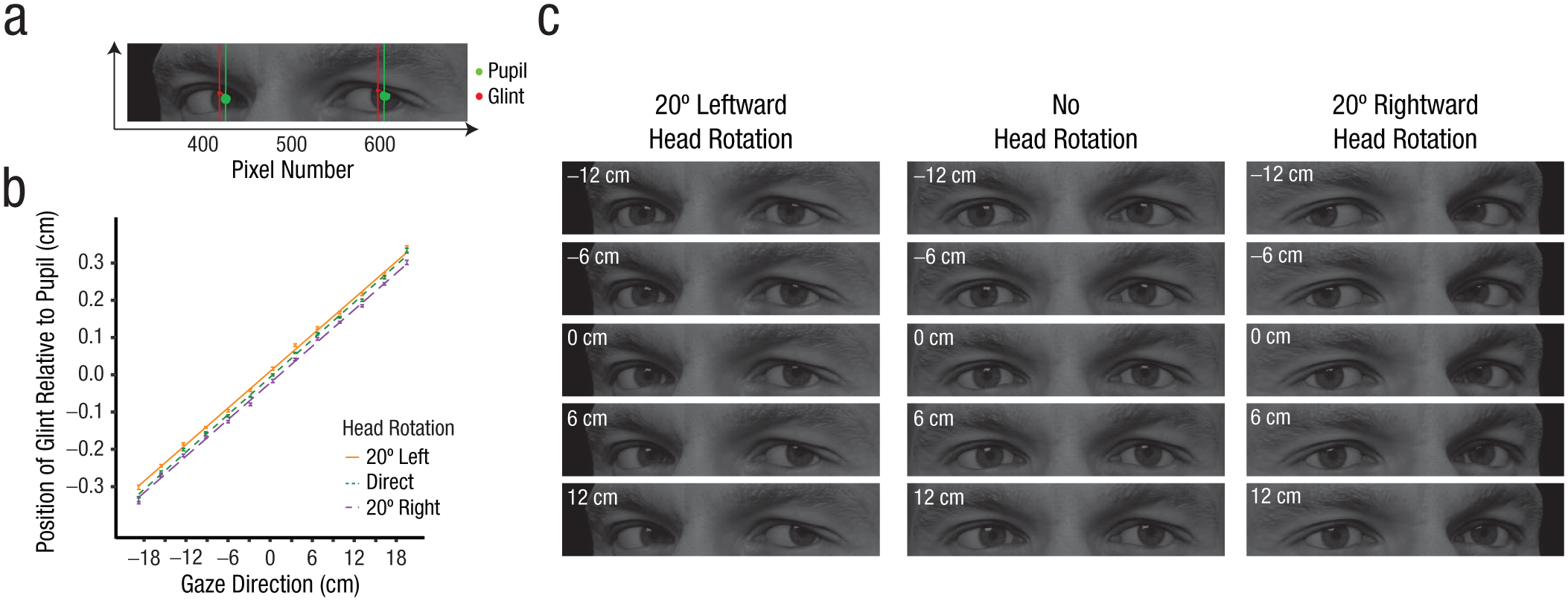
Image analysis comparing the position of eye glint relative to the pupil as a function of gaze direction and head orientation. In (a), we show the isolated glint (in red) and the pupil (in green) as well as the corresponding centroids of these features defined by pixel intensity and cluster volume, respectively. In (b) we show the horizontal distance between the glint and pupil (pupil position – glint position) as computed across the face images used in Experiment 2 that varied in face identity, gaze direction, and head orientation and that were illuminated by a fixed light source. Distances were first calculated in terms of the number of pixels, as in Figure 4a, and then converted to centimeters relative to image-presentation size during the behavioral experiment. These values were averaged across the left and right eyes of each image, and across the faces shown for each gaze direction. The mean distance between pupil and glint position varied linearly with gaze direction. Linear fits are shown separately for each head orientation. Error bars represent standard error of the mean (SEM) at each gaze direction. In (c) we show close-up examples of the eye region highlighting how the position of eye glint relative to the pupil varies with head rotation and gaze direction. Note how, for a given gaze direction relative to the viewer, the position of glint relative to the pupil remains relatively consistent across significant changes in head orientation.
To determine the glint and pupil positions for each image, we generated new versions of the image that exploited the 3D rendering process to isolate the glint or pupil within the image. To isolate eye glint, we disabled the reflectance of the face and eyeballs in Blender, leaving only the transparent layer of the eyeball that contributed specular reflections to the original images. The glint-only images thus consisted of the specular reflections within the eye region appearing as white clusters against a black background. The location of the glint was determined by calculating a centroid of the luminance distribution in the horizontal dimension of the glint-only image (separately for each eye), on the basis of the summed pixel values of each column of the image. Essentially, the centroid represented the point within the glint cluster with the highest luminance, reminiscent of how the brightest part of the glint would likely be perceived as the most salient point to a human observer. Pupil-only images were generated by editing the texture that was wrapped to the eyeball in Blender so that the eyeball texture consisted of a white circle overlaying the pupil in the original texture, with the rest of the eyeball set to black. New images were rendered with the reflectance of the face disabled, so that the pupils appeared as white clusters against a black background. The images were then binarized using the imbinarise function in MATLAB, and centroids were calculated to find the center of the pupil volume for each eye. This process was repeated across the same rendering parameters as the original images (i.e., different face identities, head orientations, and eye orientations). For each gaze direction, centroid differences between the glint and pupil positions obtained for each eye were averaged across the left and right eyes, then averaged across the faces shown during the behavioral experiment for each combination of head orientation and gaze direction (i.e., the different face identities and the flipped and nonflipped versions of each).
Figure 4b illustrates how the position of the glint relative to the pupil (pupil position – glint position) varies as a function of gaze direction. Linear functions were fitted to these data separately for each head rotation. The results show that the distance between the glint and the pupil varies systematically across gaze directions, which indicates that the glint could in principle be used as a cue to gaze direction. A small shift in the intercept of the linear fits is also apparent when comparing across head orientations. The directions in which the linear functions shift across head orientations is consistent with a repulsive influence of head orientation on estimated gaze direction: For example, the linear function shifts slightly upward for leftward head rotation, which is in the same direction as that caused by an actual shift in gaze direction toward the right. Notably, however, a small change in gaze direction (e.g., 3°) was sufficient to produce a shift in glint and pupil positions larger than that produced by a head rotation of 20°, suggesting that the glint-pupil distance is relatively robust to changes in head position. Together, these results suggest that sensitivity to glint position could in principle help an observer to maintain perceptual constancy in gaze perception when viewing faces that differ in head rotation.
The results shown in Figure 4b were based on pupil-only images that were rendered with the whole face included, resulting in the pupils being partially occluded by the eyelids of the face models, particularly for the more averted gaze directions (e.g., Fig. 4a). This version of images corresponds to those seen by participants during the behavioral experiment reported below. The same image analysis was run on another version of pupil-only images that did not include the face, so that there was no occlusion of the iris caused by the boundaries of the eye opening. The trend of results was the same across both versions of the image analysis.
Behavioral analyses
Consistent with earlier studies (Otsuka et al., 2014, 2015), a repulsive effect of head rotation on gaze perception was observed. Specifically, centroids of the direct responses shifted in the direction that the head was turned, consistent with the perceived direction of gaze being biased in the opposite direction (Figs. 5b and 5c). This suggests that participants tended to perceive a slightly leftward gaze (e.g., −2 cm) to be looking directly at them (0 cm) when the head was turned leftward, as perceived gaze direction was being repulsed away from its veridical direction. Centroid half-differences (CHDs) were used to quantify the effect of head rotation. CHDs were calculated by subtracting leftward head-rotation centroids from the rightward head-rotation centroids and dividing the difference by two to obtain the average difference. This was done first for each participant and condition and then averaged across participants to get the mean CHD for each glint condition. This measure was significantly greater than zero for both the glint-absent condition, t(39) = 10.0, p < .001, one-tailed, Cohen’s d = 1.59, M = 3.19 cm, SD = 2.01 cm, and the glint-present condition, t(39) = 9.40, p < .001, one-tailed, Cohen’s d = 1.49, M = 2.92 cm, SD = 1.96 cm, highlighting a strong repulsive effect of head rotation on gaze perception regardless of whether glint was present or not. In contrast to our hypothesis that the presence of eye glint would improve perceptual constancy in gaze perception, a paired samples t test indicated that the CHD was not significantly lower in the glint-present condition compared with the glint-absent condition, t(39) = 1.10, p = .14, one-tailed, Cohen’s d = 0.17, mean difference = 0.27 cm, SD = 1.55 cm (Fig. 5d).
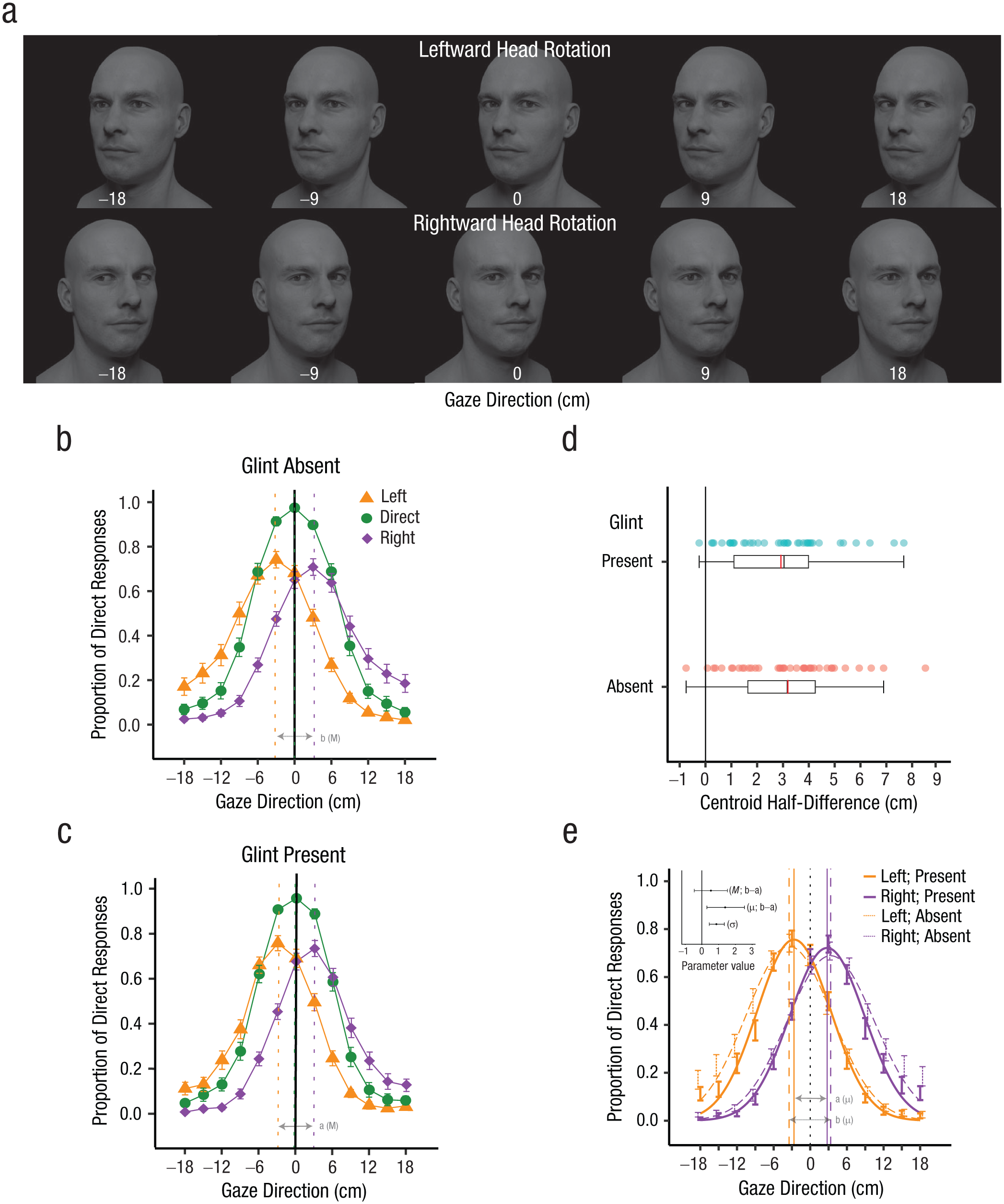
Stimuli and results of head-rotation task in Experiment 2. Faces were rendered (a) with head orientation 20° to the left, straight ahead (0°), or 20° to the right. For each head orientation, the eyes were rotated to fixate on a set of points that were between 18 cm to the left and right of the viewer in intervals of 3 cm. Images for alternate intervals and the glint-absent condition are not shown. In (b) and (c), we show mean response data across participants for each gaze direction showing the proportion of trials in which the face stimuli were perceived as looking direct. Mean centroids of direct responses are indicated by the vertical dashed lines, separately for faces differing in head orientation and the presence of eye glint. The difference in responses when the head is oriented 20° to the left and the right illustrates the repulsive effect of head orientation on perceived gaze direction. The boxplots (d) represent the distribution of centroid half-differences (CHDs) between the leftward and rightward head orientations across participants, quantifying the repulsive effect of head orientation on perceived gaze direction. The red line represents the mean of the distribution, which corresponds to the centroids in (b) and (c); the black line represents the median value. The lower and upper edges of the boxes correspond to the 25th and 75th percentiles. The upper and lower whiskers extend from the edges to the largest and smallest values no further than 1.5 × IQR (the interquartile range) from each edge, respectively. In (e) are shown mean response data across the leftward and rightward head rotations for the glint-present and -absent conditions; error bars represent the standard errors of the mean. These points correspond to the same data plotted in (b) and (c). Curves plotted correspond to the best-fitting Gaussian function for each condition, with vertical lines corresponding to the mean (μ) values of each curve. The figure inset illustrates the mean difference between the leftward and rightward head rotation centroids for the glint-present versus glint-absent conditions—glint absent(rightward-leftward) − glint present(rightward-leftward)—and equivalent comparisons using the mean parameter (μ) of the Gaussian fits. The mean difference between the sigma (σ) values of the Gaussian fits for the glint-present versus glint-absent conditions across all head rotations is also plotted. Error bars represent 95% confidence intervals.
The centroid is a useful measure of changes in perceived gaze direction, partly because it makes no assumption about how the response data are distributed. However, the centroid is dependent on the range of the recorded data and can in principle lead to an underestimation of changes in gaze perception across conditions if the range of gaze directions tested is not adequate. In this regard, it is significant to note that the proportion of direct responses for the leftward and rightward head orientations does not reach zero at the limits of the range of gaze directions tested in the current study (Figs. 5b and 5c). In other words, the distribution of data for these conditions appears truncated. This means that the centroid measure may underestimate the true center of the response distributions for these conditions, and hence the CHD may underestimate the effect of head orientation on perceived gaze direction. To complement the centroid analysis, we fitted Gaussian functions to the response data for each head-rotation condition to provide an alternative measure of central tendency appropriate for the truncated range. The mean (μ) parameter of the best-fitting functions were compared across head-rotation conditions to quantify the effect of head rotation on gaze perception. The functions were first fitted to the super-subject data, and the difference in the mean (μ) parameter across head-rotation conditions was compared statistically between the glint-present and glint-absent conditions by bootstrapping confidence intervals on this metric.
Gaussian functions fitted the data well, with more than 96% of the variance explained across all conditions (Fig. 5e). There was a difference in μ values between the rightward and leftward head-rotation conditions for both the glint-absent condition (μAbsent(R-L) = 6.82 cm, 95% CI = [5.32, 8.49]) and the glint-present condition (μPresent(R-L) = 5.43 cm, 95% CI = [4.25, 6.79]). The difference between the μ values of the rightward and leftward head rotations differed between glint conditions (μAbsent(R-L)-Present(R-L) = 1.39 cm; 95% CI = [0.31, 2.50]; see figure inset of Fig. 5e), indicating that head rotation caused a greater shift in perceived gaze direction when glint was absent compared with when it was present. In other words, when viewing faces that varied in head orientation, the apparent gaze direction of the faces was generally repulsed away from the direction of head rotation, but this effect was reduced when eye glint was visible within the eye region. Gaussian functions were also fitted to individual participants’ data. Data from 3 participants were excluded because of poor fits. The mean μAbsent(R-L)-Present(R-L) value for the remaining participants was 0.97 cm (SD = 3.29), suggesting a similar trend to that observed in the super-subject analysis. Consistent with the hypothesis that the presence of eye glint would improve perceptual constancy in gaze perception, a one-sample t test showed that the repulsive effect of head rotation on gaze perception was significantly lower when glint was present compared with when glint was absent, t(36) = 1.80, p = .04, one-tailed, Cohen’s d = 0.30.
To assess whether eye glint influenced the precision of gaze estimates, the sigma (σ) parameter of the Gaussian functions were also compared across the glint-present versus -absent conditions. The values for the leftward, direct, and rightward head rotations within each glint condition were first averaged for functions fitted to the super-subject data before being compared across conditions (σAbsent-Present = 0.87 cm; 95% CI = [0.43, 1.34]; see figure inset of Fig. 5e). It was found that glint presence significantly improved the precision of gaze perception when participants were required to judge gaze direction across changes in head rotation. Correspondingly, the AUC of the best-fitting Gaussian functions was also compared across glint conditions and was found to be significantly reduced when glint was present (AUCAbsent-Present = 1.38; 95% CI = [0.50, 2.38]). When Gaussian functions were instead fitted to individual subject data, the mean σAbsent-Present across participants was 0.68 cm (SD = 1.28) and the mean AUCAbsent-Present was 1.47 (SD = 3.24). Both values were significantly greater than zero—σ: t(36) = 3.21, p < .005, one-tailed, Cohen’s d = 0.53; AUC: t(36) = 2.75, p < .005, one-tailed, Cohen’s d = 0.45—consistent with the hypothesis that the presence of eye glint can improve the precision of gaze estimates. The finding that glint can enhance the precision of gaze estimates is analogous to what has been observed for multisensory integration (Ernst & Banks, 2002): Additional sensory cues help to increase the precision of perceptual estimates. This further suggests that participants used the glint as an additional visual cue to gaze direction.
Overall, the image analysis reported above indicates that the position of eye glint relative to the pupil in our image set carries information about gaze direction that is relatively robust to changes in head orientation; our behavioral results are surprising in indicating that gaze perception in human observers is actually sensitive, in part, to this subtle visual cue. First, we tested whether the repulsive effect of head rotation on perceived gaze direction is reduced when eye glint is present compared with when it is absent. The outcome was sensitive to the method used to quantify the effect of head rotation on gaze perception: The presence of eye glint was associated with a reduced repulsive effect of head rotation when this was quantified by fitting Gaussian functions to the data (whether super-subject or individual-subject data), but not when it was quantified by calculating differences in the centroids of the data.
This difference between analysis methods may reflect an interaction between the centroid measure and the range of gaze directions used in the task, which in principle could lead to an underestimation of effects, as discussed above. In addition, we found evidence that eye glint improves the precision of gaze estimates, and this finding was consistent across analysis methods. Experiment 3 built on this finding by testing whether eye glint contributes to perceptual constancy in gaze perception when viewing faces under varying illumination.
Experiment 3
Method
Participants
Another 40 participants (16 men, 24 women; M = 22.5, SD = 4.7 years) were recruited for this experiment. Participants were either reimbursed $10 in cash for their time or participated for course credit.
The sample size for this experiment was determined a priori on the basis of the results from an earlier study by Palmer et al. (2020), in which a similar approach was used to evaluate the effect of illumination on gaze perception. An initial power analysis (Faul et al., 2007) indicated that 10 or fewer participants would be needed to replicate the effect size of 1.38 (Cohen’s d) with 95% power found previously for the effect of horizontal lighting on perceived gaze direction. A second power analysis was run to estimate the number of participants needed to detect a difference in this effect between the glint-present and -absent conditions. This analysis indicated that 30 participants would be required, assuming that the presence of eye glint would allow an observer to compensate for at least one third of the bias in gaze perception usually caused by asymmetrical illumination of the face. We chose to test 40 participants to err on the side of greater power, taking into consideration variability across individuals and experimental designs.
Design
This experiment was based on the design of an earlier study by Palmer et al. (2020), which examined the effect of illumination on perceived gaze direction. In the current study, we compared the effect of illumination between conditions when eye glint was present versus absent. Asymmetries in horizontal lighting of a face can result in shading and shadows within the eye region that produce systematic biases in how observers perceive the gaze direction of the face (Palmer et al., 2020; West, 2013). Specifically, changing the azimuthal angle of the light source tends to shift the apparent gaze direction of the face away from the position of the light source. This effect of illumination on gaze perception is reminiscent of the “bloodshot illusion,” which occurs when artificially darkening one side of the sclera of both eyes in an image of a face shifts our perception of its gaze toward the darkened side of the eyes even though the iris position remains unchanged (Ando, 2004). A form of perceptual constancy has been observed, however, wherein contextual cues to the lighting direction can partially reduce the biases in perceived gaze direction that occur across illumination conditions (Ando, 2004; Palmer et al., 2020). On the basis of these findings, we tested the idea that eye glint might serve as a perceptual cue to illumination direction that facilitates constancy in gaze perception. Given that the visible position of eye glint varies directly with the position of dominant light sources in the surrounding environment, as observed in Figure 1c (i.e., the glint appears to the left of the pupil when the light source is located to the left of the face), eye glint could potentially provide information about the lighting direction that helps an observer to better interpret gaze direction from the appearance of the eye region despite the changes in shading and shadows across the eye region that occur when faces are lit from different angles. Following the design of Palmer et al. (2020), we quantified the influence of illumination direction on gaze perception by comparing the perception of eye contact between faces with varying gaze direction when the faces were lit from the left versus the right. We then compared this measure between faces rendered with eye glint present versus eye glint absent to assess whether gaze perception is more robust to changes in illumination direction when eye glint is visible.
Procedure
The same procedure and setup as the eye-contact task in Experiment 1 was used, save for the position of the light source in the images generated. Instead of a central light source (0°), as in Experiment 1, the light source was located either 45° to the left or 45° to the right of the face (Fig. 6a and 6b). As in Experiment 1, the light source was always positioned 75 cm away from the face and elevated at an angle of 30° above the face. Images were rendered both with and without eye glint. Glint-present and -absent trials were blocked, and block order was counterbalanced across participants. Each block consisted of 504 trials (21 gaze directions × 12 repeats of each trial type × 2 illumination conditions) presented in a random order. In each trial, participants indicated whether the face was making eye contact with them or not by pressing one of two keys on a keyboard, with keyboard mappings counterbalanced across participants.
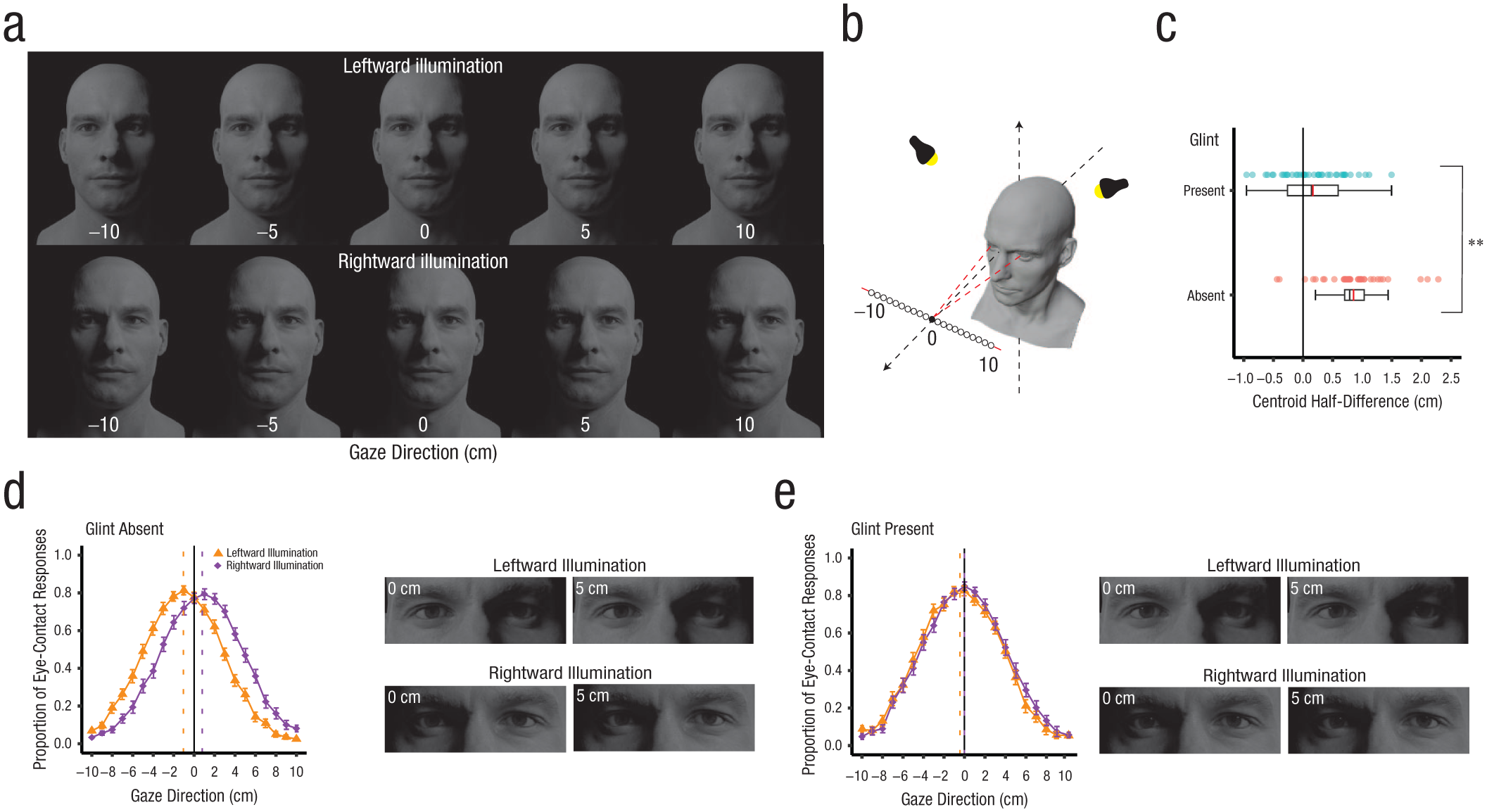
Stimuli and results of illumination task in Experiment 3. Similar to Experiment 1, faces were rendered (a) with gaze direction varying to fixate a range of locations between −10 to 10 cm either side of the observer. Faces were either illuminated from the left or right and rendered with or without specular reflections within the eye region (the glint-absent condition is not shown). When visible, the eye glint was rendered consistent with the prevailing illumination conditions in such a way that the glint appeared toward the left of the irises for leftward illumination and toward the right for rightward illumination. In (b), we illustrate the locations of the illumination source used to generate the images. The light source was located either 45° to the left or 45° to the right of the face in Blender to create leftward and rightward illuminated faces. In (c), the boxplots represent the distribution of centroid half-difference (CHD) values across individuals for the glint-present and -absent conditions. There was a strong effect of lighting direction on perceived gaze direction, but this effect was significantly reduced when eye glint was visible within the eye region of the face. Red lines within the boxplot represent the mean CHD, whereas the black lines represent the median CHD values. The mean distribution of eye-contact responses across participants for each illumination condition (in the absence and presence of eye glint, respectively) are shown in (d) and (e). Dashed lines show the centroids of the response data for each condition. **p < .01.
Analyses
Similar to Experiment 2, centroids and CHDs were calculated for each participant to quantify shifts in perceived gaze direction across conditions. In addition, Gaussian curves were fitted to the super-subject and individual-subject data for each condition, with shifts in perceived gaze direction across conditions quantified using the μ parameter. One-tailed t tests were used to test the hypotheses that changes in illumination direction would shift perceived gaze direction away from the direction of the light source and that this bias would be reduced when eye glint is present.
Results
The gaze directions perceived as making eye contact shifted systematically depending on the position of the light source when eye glint was absent (Fig. 6d). Leftward illumination of the face shifted the range of gaze directions perceived as making eye contact with the viewer toward the left, whereas rightward illumination of the face shifted the gaze directions perceived as making eye contact in the opposite direction. In other words, in the case of leftward illumination, leftward gaze directions were perceived as making eye contact, but veridically direct gaze was perceived as more rightward-looking, highlighting a strong repulsive effect of illumination on perceived gaze direction. This replicates previous work (e.g., Palmer et al., 2020), which has also shown that these effects of illumination on perceived gaze direction result from changes in shading and shadows falling across the eye region when the horizontal lighting direction varies, presumably because these asymmetric changes in the appearance of the eye region interfere with luminance cues that drive perceived gaze direction. In contrast, in the current study, participants were less likely to be biased by the illumination direction when specular reflections were visible within the eye region, rendered to be consistent with the prevailing illumination conditions (Fig. 6e). This is consistent with the notion that specular reflections from the eye surface can provide information about the lighting direction that helps observers to better interpret the appearance of the eye region across varying lighting conditions (Fig. 1c).
To test these observations statistically, centroids of the eye-contact responses across horizontal gaze directions were calculated for each condition as a measure of the center of the zone of eye contact. To evaluate the effect of horizontal illumination on gaze perception, we calculated CHD values as the difference in centroids between the leftward and rightward illumination conditions divided by two. The CHDs were then averaged across the sample and compared across the glint-present and glint-absent conditions to test whether the effect of illumination in biasing perceived gaze direction was stronger when glint was absent compared with when it was present (Fig. 6c). The mean CHD was significantly greater than zero for the glint-absent condition, t(39) = 9.83, p < .001, one-tailed, Cohen’s d = 1.55, with a mean of 0.85 cm (SD = 0.55 cm). Similarly, the mean CHD was significantly greater than zero for the glint-present condition, t(39) = 1.88, p = .03, one-tailed, Cohen’s d = 0.30, M = 0.17 cm, SD = 0.56 cm. A paired-samples t test indicated that the absence of specular reflections within the eye region led to a significantly greater difference in centroids between the leftward- and rightward-illumination conditions, t(39) = 8.84, p < .001, one-tailed, Cohen’s d = 1.40, mean difference = 0.69 cm, SD = 0.49 cm, reflecting a greater effect of lighting direction on the perception of eye contact compared with when eye glint was present.
In a further analysis, Gaussian functions were fitted to the super-subject data for each glint condition, and the μ values of each function were bootstrapped to obtain CIs. Gaussian functions were found to fit the data well, accounting for at least 99% of the variance in each condition. A similar trend to that observed in the centroid analysis occurred when the difference in μ values of Gaussian functions fitted to the response data for leftward and rightward illuminated faces was compared across glint conditions. There was a difference in μ values between the rightward and leftward illumination conditions for the glint-absent condition (μAbsent(R-L) = 1.83 cm; 95% CI = 1.46, 2.20), but not the glint-present condition (μPresent(R-L) = 0.36 cm; 95% CI = −0.05, 0.77). Most important, the effect of lighting direction on the perception of eye contact was significantly greater when viewing faces that lacked specular reflections within the eye region, compared with viewing faces that were rendered with specular reflections from the eye surface that were consistent with the prevailing illumination conditions (μAbsent(R-L)-Present(R-L) = 1.47 cm; 95% CI = [1.12, 1.83]). The same analysis was repeated by fitting Gaussian functions to individual subject data. The mean μAbsent(R-L)-Present(R-L) value obtained across all participants was 1.51 cm (SD = 1.29). A one-sample t test revealed that this difference was significantly greater than zero, t(39) = 7.39, p < .001, one-tailed, Cohen’s d = 1.17, which is consistent with the results obtained from the super-subject Gaussian fits. Together, these results indicate that the changes in the horizontal illumination of the face produce systematic biases in the perception of gaze direction, consistent with previous work (e.g., Palmer et al., 2020), but that human observers are less susceptible to these biases in gaze perception when specular reflections are visible within the eye region that are consistent with the lighting direction. This supports our hypothesis that eye glint can provide information about the lighting direction that helps an observer to better tolerate changes in the appearance of faces (related to the illumination direction) when interpreting facial cues.
Were participants aware of eye glint affecting their judgments about gaze direction? We asked participants at the end of each session whether they noticed any differences in the appearance of the eyes across the face images presented during the experiments. Approximately half of the participants in each experiment reported noticing the eye glint: 60% in Experiment 1, 45% in Experiment 2, and 52.5% in Experiment 3. Those who were aware of the glint described it as a “reflection of light” without additional prompts, with some responding that the presence of eye glint helped them better judge the gaze direction of the faces. In contrast, participants who did not report noticing any manipulation of the eye region often remained unsure of what we were referring to even when asked specifically about whether they had seen a “white circle” or “light reflection” in the eyes of certain faces presented.
As an exploratory analysis, we tested whether the effects of eye glint on gaze perception observed in our experiments depended on participants explicitly noticing the eye glint. Because there were no significant effects of eye glint on gaze perception in Experiments 1a and 1b—even when considering the full sample—our quantitative analyses focused on Experiments 2 and 3. For each participant, we computed the CHD difference between the glint-absent and glint-present conditions (i.e., CHDAbsent(R-L) − Present(R-L)) to represent the effect of eye glint on gaze perception. Participants were then grouped according to whether they reported noticing the eye glint. One can see in Figure S1 in the Supplemental Material (available online) that the effect of eye glint on gaze perception was similar across groups. Mean CHD differences quantifying the effect of eye glint on gaze perception were compared between groups using independent-samples t tests.
In Experiment 2 (Fig. S1a), performance did not differ significantly as a function of glint awareness. An independent-samples t test indicated that the groups did not significantly differ in mean CHD difference values, t(38) = 0.36, p = .72, Cohen’s d = 0.11. In addition, group mean CHD differences did not differ from zero for either participants who noticed the eye glint, M = 0.37 cm, SD = 1.51, t(17) = 1.03, p = .32, Cohen’s d = 0.24, or for participants who did not notice the eye glint, M = 0.19 cm, SD = 1.61, t(21) = 0.55, p = .59, Cohen’s d = 0.12. A similar trend was observed when individual μAbsent(R-L)-Present(R-L) values were used, with the exclusion of 3 participants for whom Gaussian functions fit poorly to their data: These values were found to be comparable across the two groups, t(35) = 0.31, p = .76, Cohen’s d = 0.10, and neither group differed significantly from zero on this measure— participants who noticed the glint: M = 1.16 cm, SD = 3.66, t(16) = 1.31, p = .21, Cohen’s d = 0.32; participants who did not notice the glint: M = 0.81 cm, SD = 3.04, t(19) = 1.20, p = .25, Cohen’s d = 0.27.
In contrast, Experiment 3 (Fig. S1b) revealed a robust effect of eye glint on gaze-perception stability across illumination conditions, regardless of whether participants were aware of the eye glint. An independent-samples t test revealed no significant difference in the mean CHD difference between groups, t(38) = −0.42, p = .68, Cohen’s d = −0.13. Group mean CHD differences differed significantly from zero both for participants who noticed the eye glint, M = 0.65 cm, SD = 0.43, t(20) = 7.00, p < .001, Cohen’s d = 1.53, and participants who did not notice the eye glint, M = 0.72 cm, SD = 0.56, t(18) = 5.60, p < .001, Cohen’s d = 1.28. Because the observed effects of eye glint on gaze perception did not depend on explicit awareness of the eye glint, these effects appear more likely to reflect perceptual processes sensitive to eye glint rather than a conscious strategy. Our results therefore suggest some spontaneous sensitivity to eye glint in human gaze perception, which we speculate might be enhanced if observers were explicitly trained to use it as a cue.
Discussion
Eye glint is a subtle yet ubiquitous feature of the human face, related to the unique material properties of the eye surface. Here, we investigate whether eye glint plays any role in how faces are perceived in the context of gaze perception. Our experiments revealed that eye glint has little impact on the accuracy or precision of gaze perception when human observers view faces under simplified viewing conditions. Surprisingly, however, the presence of eye glint helped observers to maintain perceptual constancy in their judgments about eye contact and gaze direction. This occurred when viewing faces across changes in viewpoint and illumination, basic sources of variability in the appearance of faces that are known to produce systematic biases in gaze perception.
How does eye glint provide information useful for gaze perception? The present results suggest two key mechanisms at work. First, when lighting conditions are fixed, the position of eye glint relative to the iris can vary systematically with gaze direction in a way that is relatively stable across changes in head rotation (Fig. 4). This contrasts notably with the coarse pattern of luminance contrast visible within the eye opening (related to the position of the iris and sclera), which varies systematically with eye rotation but is also strongly influenced by how the orientation of the head determines the viewer’s perspective of the eye region (Fig. 1b). Consequently, gaze perception relies on the perceptual integration of visual cues to eye and head direction, which helps to maintain an imperfect perceptual constancy associated with a measurable repulsion of the perceived gaze direction away from the veridical gaze direction when viewing faces angled away from the observer (Otsuka et al., 2014, 2015). Our behavioral results suggest that glint-pupil distance can provide additional information about gaze direction that partially reduces the confounding influence of head orientation on perceptual judgments. Although the effect of eye glint in reducing the magnitude of the repulsive effect of head orientation on gaze perception was sensitive to the analysis method used to quantify this bias, more consistent was the finding that the presence of eye glint sharpens the precision of gaze estimates in this context.
Is this influence of eye glint likely to extend to everyday social interactions? A potential limitation of eye glint as a cue in ecological settings is that specular reflections from the eye surface vary with the relative position of light sources in the surrounding environment. Although lighting conditions are highly variable in everyday environments, eye movements are frequent and rapid (e.g., saccades often occur several times per second). Hence, in a typical face-to-face interaction, the lighting conditions that a face is viewed under will often be fixed over the timescale of eye movements. Similarly, basic qualities of human vision rely on implicit estimates of lighting direction (e.g., lightness, 3D shape perception; Boyaci et al., 2006; Morgenstern et al., 2011), so it is plausible that glint-iris position could be interpreted by human observers across changes in environmental illumination with respect to an internal model of lighting direction, allowing changes in glint-iris position caused by eye rotation to be distinguished from those caused by changes in lighting direction.
A second mechanism through which eye glint appears to contribute to perceptual constancy in gaze perception is by providing information about lighting direction. Previous studies have found that changes in shading and shadows across the eye region can systematically bias gaze estimation in human observers, likely by interfering with luminance cues central to gaze perception related to the relative positions of the iris and sclera (Palmer et al., 2020; West, 2013). Interestingly, this bias in gaze perception is partially eliminated when there are contextual cues available that provide information about the lighting direction, such as asymmetric shading across the face outside of the eye region, potentially reflecting a lightness-constancy effect in gaze perception (Ando, 2004; Palmer et al., 2020). As noted above, eye glint also varies in position across the eye surface depending on the position of the dominant light sources in the surrounding environment (Fig. 1c); hence, in principle, eye glint might provide observers with an additional cue to lighting direction when viewing a person’s face. In the current study, we found that biases in perceived gaze direction caused by asymmetric lighting of faces are significantly reduced when faces are rendered with specular highlights within the eye region (i.e., highlights that are consistent with the prevailing illumination conditions). This suggests that glint position within the eye may provide a cue to lighting direction that facilitates perceptual constancy in gaze perception.
Our perception of basic social cues, such as detecting when we share eye contact with another person, typically feels effortless, yet it relies on a sophisticated visual analysis of facial features supported by specialized neural mechanisms (Carlin & Calder, 2013; Clifford & Palmer, 2018). Here we found that one of the most subtle visual features of the human face, specular reflections from the eye surface, can contribute to an observer’s perception of when they share eye contact with that person. In particular, eye glint may help us deal with common forms of environmental variability in the appearance of faces that are known to interfere with the accuracy of gaze perception—namely, changes in viewpoint and lighting. Although the contribution of eye glint to gaze perception is likely to be most relevant at closer viewing distances, it is a striking example of how the human brain learns to exploit whatever information is available, no matter how subtle, for important cognitive tasks like social perception. Future research could explore whether eye glint contributes to other social judgments (social traits, attractiveness, emotion recognition, etc.), the role of specular reflections in person perception more generally (e.g., skin texture and age perception; Arce-Lopera et al., 2012), and whether observers can be trained to use eye glint as an explicit cue to subtle deviations in gaze direction, including in naturalistic settings.
Supplemental Material
sj-pdf-1-pss-10.1177_09567976261446892 – Supplemental material for Eye Glint as a Novel Perceptual Cue in Human Vision
Supplemental material, sj-pdf-1-pss-10.1177_09567976261446892 for Eye Glint as a Novel Perceptual Cue in Human Vision by Gwenisha J. Liaw and Colin J. Palmer in Psychological Science
Footnotes
Acknowledgements
The authors thank Zhehan Zhong for assistance with data collection. This research was supported by a National University of Singapore Faculty of Arts and Social Sciences Start-up Grant to C. J. Palmer.
Transparency
Action Editor: Zhicheng Lin
Editor: Simine Vazire
Author Contributions
Declaration of Conflicting Interests
The authors declared that there were no conflicts of interest with respect to the authorship or the publication of this article.
Funding
This research was supported by a National University of Singapore Faculty of Arts and Social Sciences Start-Up Grant to C. J. Palmer.
Artificial Intelligence
No artificial-intelligence-assisted technologies were used in this research or the creation of this article.
Ethics
This research received approval from the National University of Singapore (NUS) Departmental Ethics Review Committee (Department of Psychology ID No. 2023-January-09).
Open Practices
Open practices for this article are described in the Research Transparency Statement section, which appears at the end of the Introduction section in the main text.