






Abstract
Background
Lactate is an important biomarker for monitoring metabolic conditions and is widely used as an indicator in clinical diagnostics. It plays a critical role in the early diagnosis and monitoring of neonatal sepsis, where elevated lactate levels indicate tissue hypoxia and metabolic imbalance. Conventional detection methods often involve enzymatic systems, which may suffer from stability and reproducibility issues.
Objective
This study aims to develop a reagent-free, non-enzymatic molecularly imprinted electrochemical sensor for the selective detection of lactate.
Methods
A screen-printed electrode (SPE) was modified with reduced graphene oxide (RGO) and gold nanoparticles (AuNP) via electrodeposition. A molecularly imprinted polymer (MIP) layer was fabricated through electropolymerization of 3-aminophenylboronic acid (3-APBA) in the presence of lactic acid as the template. The stepwise fabrication, including template removal and rebinding, was characterized using cyclic voltammetry (CV) and electrochemical impedance spectroscopy (EIS). Sensor performance was evaluated using differential pulse voltammetry (DPV).
Results
The sensor exhibited a linear response to lactate over the range of 0.05 µM to 6.0 mM, with a limit of detection (LOD) of 0.361 µM. The MIP sensor demonstrated high selectivity against common interfering species, including glucose, urea, ascorbic acid, and acetic acid, along with good reproducibility and stability.
Conclusion
The developed reagent-free, non-enzymatic MIP-based sensor provides a simple and effective platform for selective lactate detection, with potential applicability to clinical monitoring, following further validation on real samples.
Keywords
Introduction
Neonatal sepsis is a bloodstream infection caused by a dysregulated host immune response to an infection. About 1.3 million sepsis-related cases have been reported globally each year, 1 and the mortality rate is reported to be 11% to 19% due to neonatal sepsis. 2 Major organisms causing sepsis include Klebsiella spp., Group B streptococcus (GBS), Escherichia coli (E. coli), and Staphylococcus aureus (S. aureus), respectively.3,4 Currently, microbial methods such as blood culture are used to identify sepsis pathogens. Blood cultures, the gold standard for diagnosis, are slow (taking 24–48 h) and may yield false negatives due to low bacterial loads in neonates. The first clinical response to neonatal infection is the administration of empirical antibiotics to control the disease. The use of antibiotics even in non-sepsis infections before blood culture confirmation leads to the development of multidrug resistance in neonates. The limitations of the above methods require a rapid, point-of-care electrochemical sensor for the early diagnosis of sepsis biomarkers. 5 Developing point-of-care devices for early, specific diagnosis of sepsis can prevent false-positive cases and prevent the use of unwarranted antibiotics. Sepsis can be diagnosed using either pathogen-specific or host-response-specific biomarkers.
Host-derived biomarkers can aid in the early diagnosis of sepsis and help differentiate it from non-specific disease conditions. When an infection enters the human body, pattern recognition receptors identify the infection and produce a series of chemokines and cytokines to remove the pathogens from the body. 6 These biomarkers include Tumor Necrosis Factor-alpha (TNF-α), Interleukin-1 beta (IL-1β), Interleukin-6 (IL-6), C-reactive Protein (CRP), Procalcitonin (PCT), Lactate, Serum amyloid A (SAA), Lipopolysaccharide-binding protein, and Presepsin. Lactate is an essential biomarker in neonatal sepsis.7–9 Blood lactate is widely used as a metabolic indicator of tissue hypoxia and impaired perfusion. In healthy neonates, the resting lactate level typically ranges from 1.0 to 2.0 mM; however, in septic conditions, it can rise significantly, often exceeding 4.0 mM, indicating metabolic acidosis and circulatory failure. 10 Elevated lactate levels (>2.0 mM) indicate tissue hypoxia and serve as an early marker of sepsis progression. A lactate level above 4.0 mM is considered to be associated with a high mortality risk.11–13 Lactate clearance, or the rate at which lactate levels normalize with treatment, is also a critical indicator of therapeutic response. Increased lactate levels can also indicate other disease conditions. But, in neonates, sepsis can rapidly lead to impaired oxygen delivery and mitochondrial dysfunction, resulting in elevated lactate levels even before overt clinical symptoms emerge. Monitoring lactate concentrations offers valuable insight into the infant's hemodynamic status and helps identify early signs of septic shock or organ dysfunction. Therefore, lactate measurement is an important tool for the prognosis and management of neonatal sepsis.14,15
Several commercially available devices such as blood gas analyzers and portable enzyme-linked immunosorbent assay (ELISA) kits, are widely used in the hospital setting; however, they require multiple incubation and washing steps and trained professionals, making the process highly time-consuming. 16 To overcome these challenges, many sensing devices were developed. Most of these devices depend on enzyme-based amperometric electrochemical sensors that utilize lactate oxidase or lactate dehydrogenase enzymes.17,18 However, these enzyme-based sensors suffer from stability issues due to the denaturation of enzymes during long-term storage conditions.19,20 To overcome these problems, Molecularly Imprinted Polymer (MIP) was used as an alternative biorecognition element due to its better stability, selectivity, cost-effectiveness, and sensitivity to the target molecule.21–23 MIP-based sensors are developed by polymerizing monomers around a target molecule, and after template removal, they create specific recognition cavities. Here, electropolymerization of MIP on the working electrode provides strong adhesion and controlled film thickness.24,25
Many researchers have focused on developing an electrochemical sensor for lactate detection. However, studies on MIP-based sensors for lactate are limited. Recently reported MIP-based lactate sensors utilize traditional functional monomers such as methacrylic acid (MAA), acrylamide, or o-phenylenediamine (o-PD), often polymerized on rigid glassy carbon or gold electrodes, and often involve multi-step fabrication and limited suitability for point-of-care use. MIP-based sensor was developed using MAA and ethylene glycol dimethacrylate (EGDMA) as functional monomer and crosslinker with a limit of detection of 0.162 mM. Despite their significant sensitivity, the merging of the cavity and reduced binding capacity may affect the sensor's performance. 26 Until now, only a few studies have focused on the MIP-based lactate sensor for neonatal sepsis monitoring. In this work, 3-aminophenylboronic acid (3-APBA) is used to enable reversible boronate–diol interactions to improve lactate recognition. This is further enhanced by integrating the MIP layer onto an AuNP/RGO-modified screen-printed carbon electrode, providing improved conductivity, higher surface area, and controlled electropolymerization. Importantly, the sensor operates within the clinically relevant lactate range (0.05 µM −6 mM) and is validated in complex matrices such as artificial serum and microbial culture media. The proposed sensor is reagent-free and non-enzymatic, avoiding issues associated with enzyme denaturation and instability. Moreover, systematic optimization of the MIP layer ensured efficient template extraction and reproducible binding performance, addressing key limitations not reported in previous studies.
In the present work, MIP-modified Screen-Printed Electrode (SPE) was developed as an electrochemical sensing device for the rapid detection of lactate molecules. Here, 3-aminophenylboronic acid (3-APBA) was used as a functional monomer, and the novelty lies in the specific boronic acid–diol formation, in which 3-APBA forms reversible covalent boronate ester interactions with the cis-diol groups of lactate, enabling more selective and stable recognition compared to conventional MIP methods. The surface of SPE was first coated with electrochemically reduced graphene oxide (RGO) and subsequently decorated with gold nanoparticles (AuNPs) to increase electrode conductivity and effective surface area. The MIP was then electropolymerized onto this nanostructured layer for the selective recognition of lactate molecules. RGO significantly improves the electrochemical properties and stability of the electrode surface,27–30 while AuNPs further enhance electron-transfer kinetics due to their high conductivity, biocompatibility, and catalytic activity.31–33 The high surface area of the RGO and the excellent electrical conductivity of gold nanoparticles help develop a highly sensitive and selective sensing platform for lactate diagnosis. At the same time, the boronic acid functionality enhances molecular recognition of lactate. Importantly, the developed MIP sensor operates within the clinically relevant lactate range (0.05 µM −6 mM) associated with neonatal sepsis, highlighting its practical applicability for point-of-care diagnostics. This combined approach represents a targeted advancement over existing MIP and enzyme-free lactate sensing systems. This integrated sensor provides an enzyme-free, structurally stable platform for selective lactate detection, with potential suitability for point-of-care applications.
Experimental
Chemicals and reagents
Graphite powder (150 μm, synthetic), Chloroauric acid (HAuCl4), Sulfuric acid (H2SO4), Potassium permanganate (KMnO4), Hydrogen peroxide solution (H2O2), Potassium chloride (KCl), Potassium ferrocyanide, Potassium ferricyanide, Acetic acid, L-Lactic acid, 3-aminophenylboronic acid (3-APBA), Fetal bovine serum was purchased from Sigma Aldrich. De Man, Rogosa, and Sharpe (MRS) broth powder was purchased from HiMedia, India. Screen-printed electrodes with carbon as the working electrode (3 mm diameter), a carbon counter electrode, and an Ag reference electrode were purchased from Zensor, India. FTIR analysis was performed using a SHIMADZU IRTracer-100 spectrometer, and surface morphology was characterized using HRSEM (Thermo Scientific Apreo S). The electrochemical analysis was obtained from the Zive SP1 potentiostat.
Media broth and culture conditions
The homofermentative Lactobacillus casei strain was obtained commercially and confirmed for viability in MRS broth (HiMedia, India). The strain was stored at −20°C in MRS broth with 20% (v/v) glycerol. MRS broth was prepared following standard composition and adjusted to pH 6.2 before sterilization. Inoculum (10% v/v) was prepared from the culture and incubated aerobically at 37°C to promote lactate accumulation.
Cleaning the screen-printed electrode (SPE)
The surface of the screen-printed electrode was cleaned using 0.5 M H2SO4 by CV over the range −1 to 1 V at a scan rate of 100 mV/s for 10 cycles. This completely removed any particles or residue on the surface of the SPE. Then, the SPE was allowed to dry at room temperature before being used for further analysis.
Synthesis of AuNP/RGO modified SPE
Graphene oxide (GO) was synthesized as described in previous work by our research group. 34 A suspension of 0.5 mg/mL GO was first prepared in distilled water. 50 µL of the solution was drop-cast onto the surface of the working electrode and left to dry overnight. Then, it was reduced to RGO by CV over a potential range of −2 to 0 V in 0.1 M KCl and PBS at pH 7.4 at a scan rate of 50 mV/s for 11 cycles. The SPE was then dried at room temperature.
The gold suspension was prepared by mixing 0.5 mM HAuCl4 in 0.5 M H2SO4 and was electrodeposited onto the RGO layer. The electrodeposition was carried out by applying a potential of −0.4 to 1.2 V at a scan rate of 50 mV/s for 20 cycles using CV. This technique enabled controlled deposition of both RGO and AuNPs on SPE. The strong interaction between Au and the residual oxygen functional groups on RGO, combined with physical adsorption and π–metal interaction, provides uniform distribution and binding of RGO and AuNPs, as shown in the SEM/EDX characterization.
Fabrication of MIP/AuNP/RGO-SPE
After nanomaterial deposition, 3-APBA was electropolymerized on top of the gold nanoparticles. A 50μL mixture of 3-APBA and lactate at a specific ratio was placed on the WE surface and electropolymerized using CV. CV was conducted over the range of −0.4 to 0.8 V in PBS at pH 7.4 at a scan rate of 50 mV/s for a specified number of cycles. Following electropolymerization, the template was removed using 8% acetic acid for 2 h to form the recognition cavities. 35 Then, the electrodes were washed thoroughly to remove residual molecules and left to air dry.
Electrochemical analysis
The electrochemical analysis of MIP/AuNP/RGO-SPE was conducted by CV, EIS, and DPV in 0.5 mM ferro/ferricyanide solution. CV was used to analyze the different steps involved in the fabrication process. The MIP and NIPs template removal and rebinding studies were analyzed using DPV within a potential range of −0.2 V to +0.5 V, with a pulse width of 0.1 s, amplitude of 0.0025 V, and pulse period of 0.5 s. Initially, a sample of approximately 90 μL of blank was placed on the WE, and CV, EIS, and DPV measurements were taken. The sample was then spiked with different concentrations of lactate solutions (0.05 µM to 6 mM). The electrode was incubated for 20 min at each sample concentration. 35 The interferent study was conducted using different interferent agents at specific concentrations, such as Ascorbic acid, glucose, urea, and acetic acid. Figure 1 shows the schematic of steps involved in the fabrication of MIP/AuNP/RGO-SPE. Recovery analysis was performed by spiking PBS with a known concentration of lactate (3 mM). The spiked sample was analyzed using the developed sensor under identical conditions, and the measured concentration was obtained from the calibration curve. For PBS-based analysis, lactate solutions of different concentrations were prepared in PBS (pH 8) and used directly for DPV measurements. Fetal bovine serum (FBS) was used as a complex biological matrix to preliminarily evaluate sensor performance under protein-rich conditions whereas artificial serum is the simulated serum with electrolyte composition used for controlled electrochemical measurements. For FBS analysis, serum samples were diluted tenfold with PBS to reduce matrix effects and improve signal stability prior to lactate spiking and electrochemical measurement.
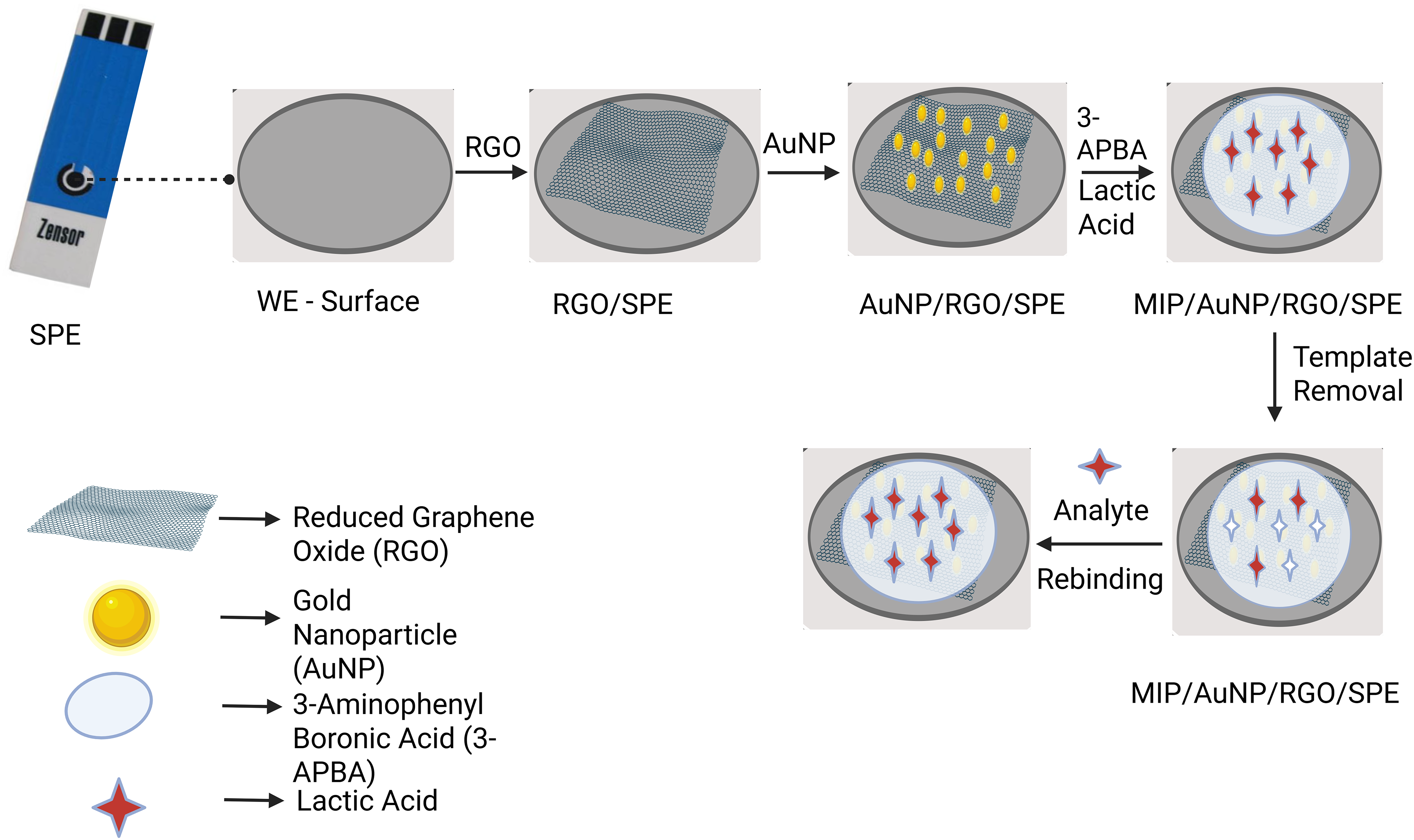
Schematic representation includes SPE surface modification with nanomaterial, MIP deposition, template removal, and analyte rebinding.
Statistical analysis
Statistical analysis was performed in triplicate, and the significance was evaluated using analysis of variance (ANOVA) and Tukey's post hoc multiple comparison test. It was observed from the analysis that the results were statistically significant at p < 0.05. The analysis was performed using Origin software 2025b version 10.25.
Results and discussion
Surface morphological study
The SEM morphologies of the bare and modified SPE reveal distinct surface characteristics corresponding to each modification stage. The bare SPE shows a flaky and porous rough surface, as shown in Figure 2(a). After the electrodeposition of RGO, the entire surface of the working electrode was covered with a wrinkled and wavy layer of RGO, which can increase the surface area and the active sites of the electrode (Figure 2(b)). Subsequent AuNP deposition introduces spherical, well-dispersed particles over the RGO sheets, as shown in Figure 2(c), further enhancing the surface roughness and providing active sites for electron transfer. After electropolymerization, the MIP and NIP-modified SPE exhibit a uniform, porous polymer layer coating on the electrode (Figure 2(d) and (e)). These morphological differences support the sequential modification and the selective binding capacity of the MIP layer. The EDX analysis of AuNP/RGO-SPE confirms the presence of gold as well as carbon and oxygen on the layers of RGO (Figure 2(f)).
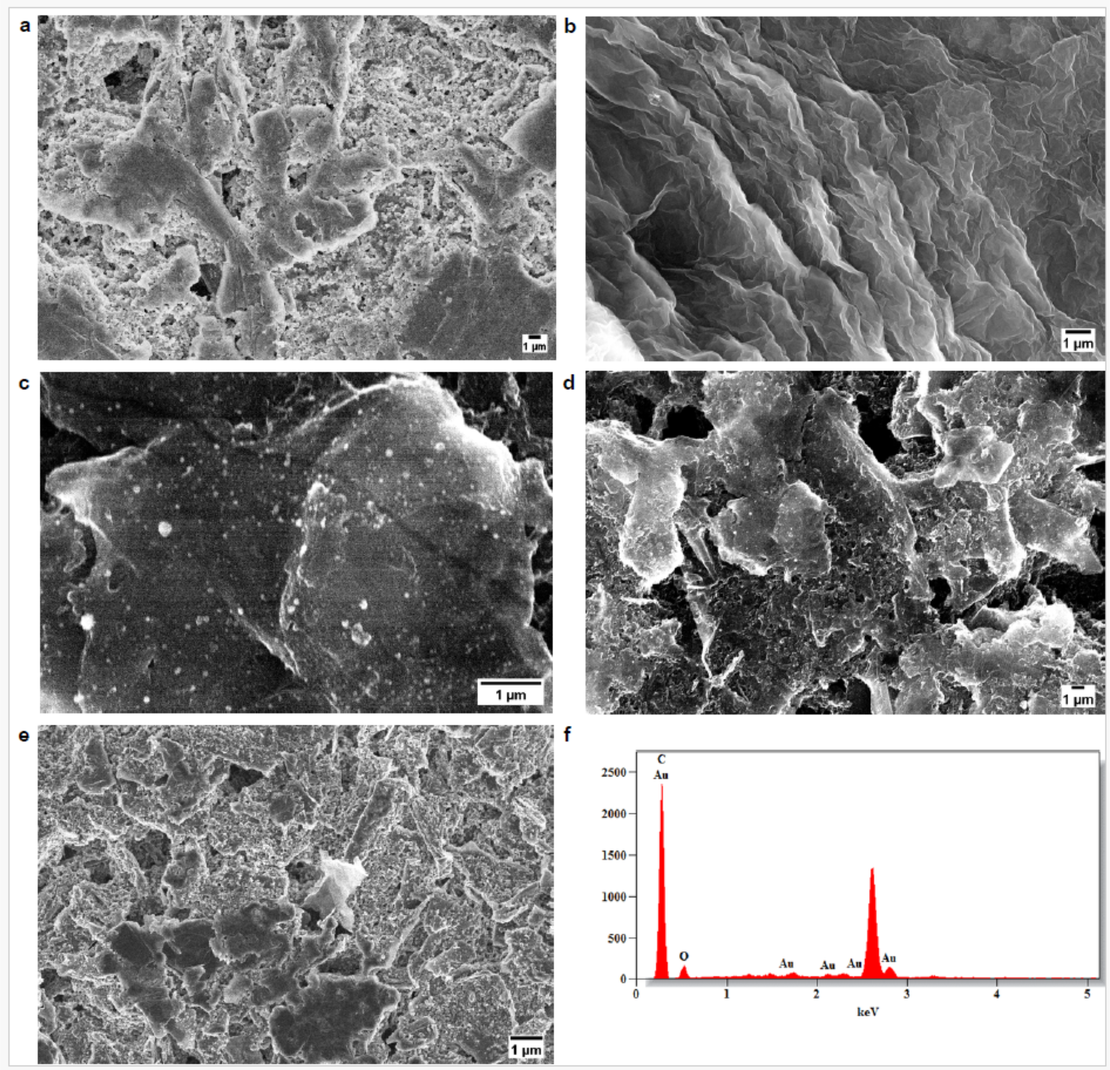
SEM images of (a). Bare SPE. (b). RGO-SPE. (c). AuNP/RGO-SPE. (d). MIP/AuNP/RGO-SPE. (e). NIP/AuNPs/RGO-SPE. (f). EDX Spectrum of AuNP/RGO-SPE.
Fourier transform infrared spectroscopy (FTIR)
The FTIR analysis of the modified electrode confirms the presence of functional groups associated with the polymeric MIP layer and 3-APBA incorporation. A broad absorption band observed around 3250 cm−1 corresponds to O–H stretching, indicating hydroxyl groups in the boronic acid and the polymer matrix. The characteristic peak near 1650 cm−1 is due to C = O stretching, while the band around 1550 cm−1 is attributed to N–H bending and/or asymmetric stretching of COO− groups, supporting the presence of amine-containing functional groups and lactate-related carboxylate interaction. The peak at around 1500 cm−1 corresponds to aromatic C = C stretching, confirming the presence of a phenyl ring in 3-APBA. In addition, the peak near 1300 cm−1 is attributed to B–O stretching, providing evidence of boronic acid functionality. A band observed around 2800–3000 cm−1 is associated with C–H stretching vibrations, indicating the polymer backbone and organic components of the MIP film. These peaks collectively confirm successful formation of the 3-APBA-based MIP layer on the electrode surface (Figure 3).
Electrochemical analysis of MIP/AuNP/RGO-SPE
Each step involved in the fabrication of MIP/AuNP/RGO-SPE was analyzed by CV and EIS in a ferro/ferricyanide solution of 0.5 mM. As shown in Figure 4(a), redox peaks for the bare SPE can be observed. After the deposition of RGO, the peak current increased due to higher electrical conductivity and a higher surface-to-volume ratio. When gold nanoparticles were deposited on top of RGO, a further increase in the peak current was observed because of the inherent nature of gold nanomaterial (Figure 4(a)).
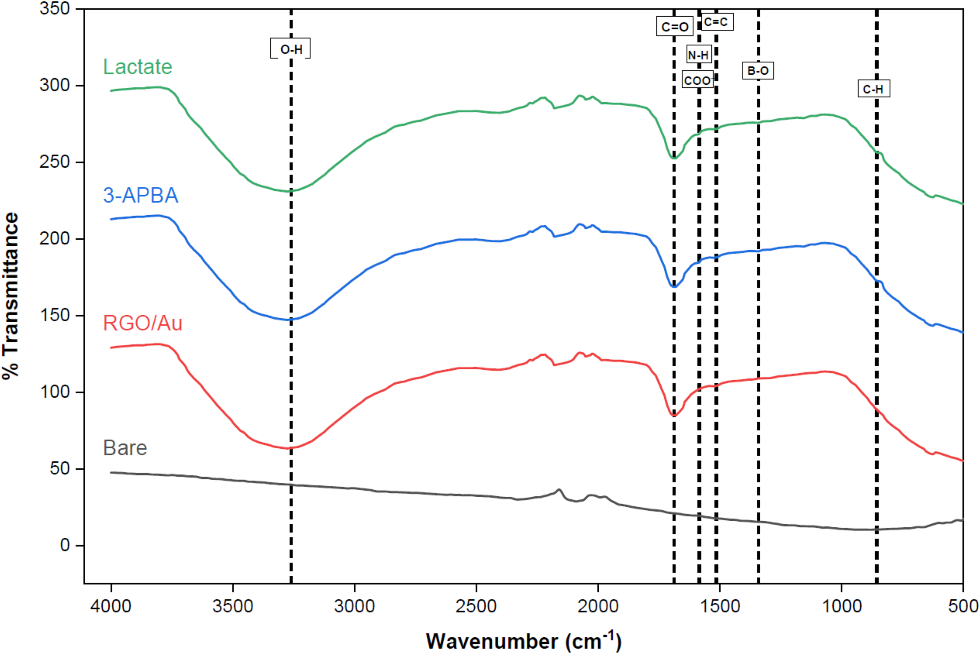
ATR-FTIR spectra of bare SPE, RGO/AuNP-modified SPE, MIP/AuNP/RGO-SPE, and lactate-rebound MIP electrode.
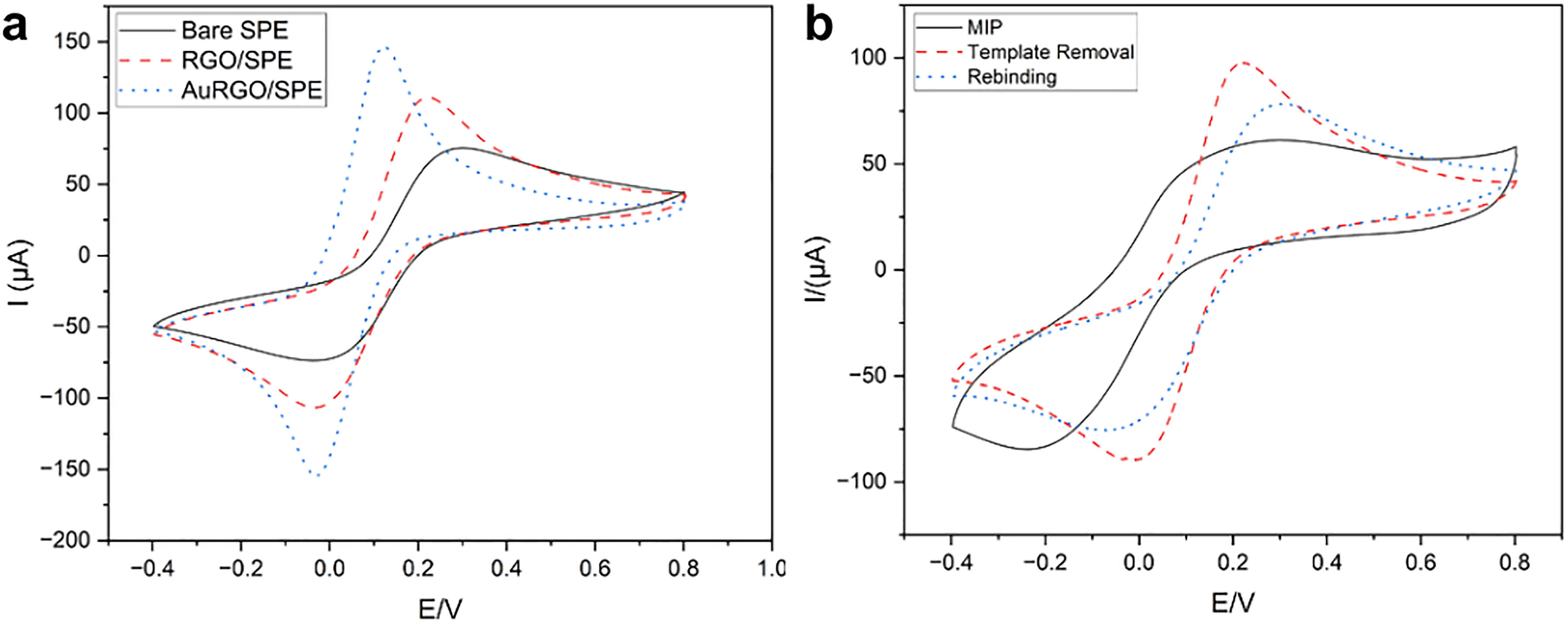
(a). Cyclic voltammograms of Bare electrode, after RGO deposition, and after gold nanomaterial deposition at the scan rate of 100 mV/s. (b). CV of AuNP/RGO-SPE after electropolymerization, template removal, and rebinding of lactic acid.
Alternatively, the EIS measurements for the same were analyzed using the charge transfer resistance (Rct) obtained from the Nyquist plot. Rct was found to decrease as the SPE was deposited using nanomaterials. Following AuNP/RGO deposition, the SPE surface was modified by electropolymerizing 3-APBA in the presence of specific concentrations of lactate. In the CV analysis, no redox peaks were observed after MIP deposition, suggesting that the even and uniform layer formation of the poorly conductive polymer affects the electron transfer. Thus, confirming the homogenous formation of the polymer layer on the electrode surface (Figure 4(b)). The sensing mechanism is based on the reversible covalent interaction between the boronic acid groups of 3-APBA and the cis-diol group of lactate, resulting in the formation of cyclic boronate ester complexes. Under slightly alkaline conditions, the boronic acid is partially converted to its anionic boronate form, which exhibits a higher affinity toward diol groups. This facilitates strong and selective binding of lactate within the imprinted cavities, which are structurally and functionally complementary to the target molecule. The formation of the boronate ester alters the local charge distribution and interfacial electron-transfer characteristics at the electrode surface, leading to a measurable change in current. The interaction mechanism between 3-APBA and lactate is illustrated in Figure 5. Upon rebinding, lactate selectively occupies these cavities, forming boronate ester interactions and increasing charge transfer resistance, thereby leading to a decrease in DPV current. Therefore, the sensing mechanism is governed by the synergistic effect of dynamic boronic acid–diol chemistry and molecular imprinting-induced shape selectivity. The interaction between boronic acid and diol groups is pH-dependent, as boronic acid exists in equilibrium between its neutral and anionic (boronate) forms. At near-physiological pH, the formation of the boronate species occurs, enhancing the binding affinity toward α-hydroxy compounds such as lactate. In this study, the electrochemical measurements were carried out in PBS, which is optimal for boronate ester formation.
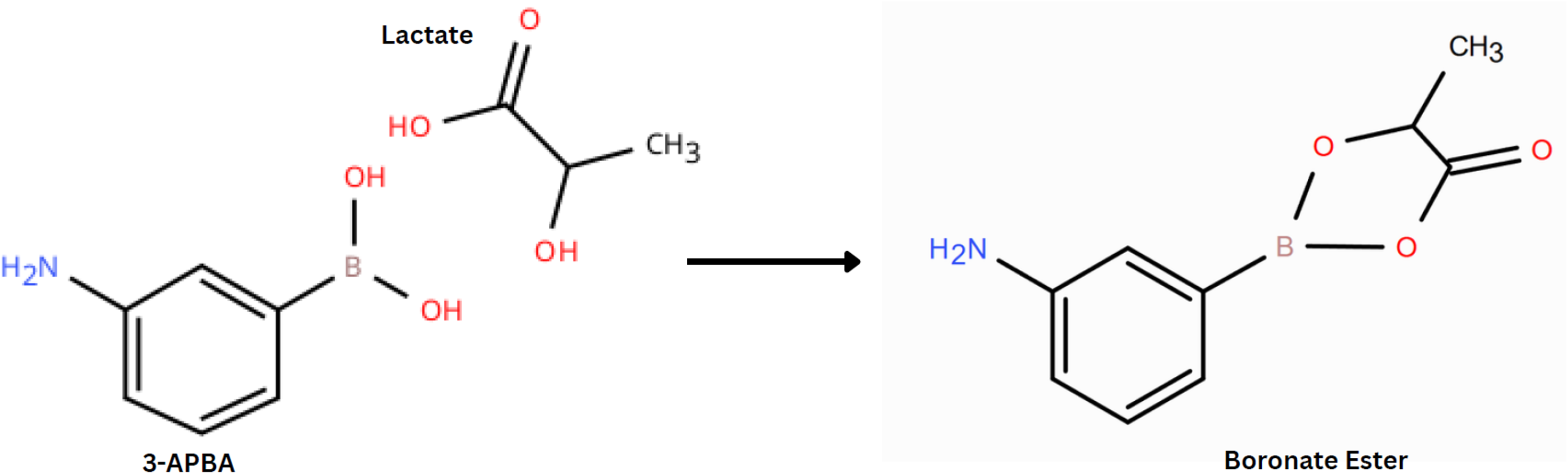
Mechanism of 3-APBA–lactate interaction.
After the polymerization, the template molecule was removed using acetic acid as an effluent solvent. The CV of template removal shows a pair of redox peaks due to electron transfer, suggesting the formation of molecular cavities on the polymer layer. Now, the prepared MIP/AuNP/RGO-SPE was subjected to different concentrations of lactate molecules for an incubation period of 20 min. 35 After incubation, the current decreased as lactate molecules occupied the molecular cavities in the polymer layer (Figure 6(a)).
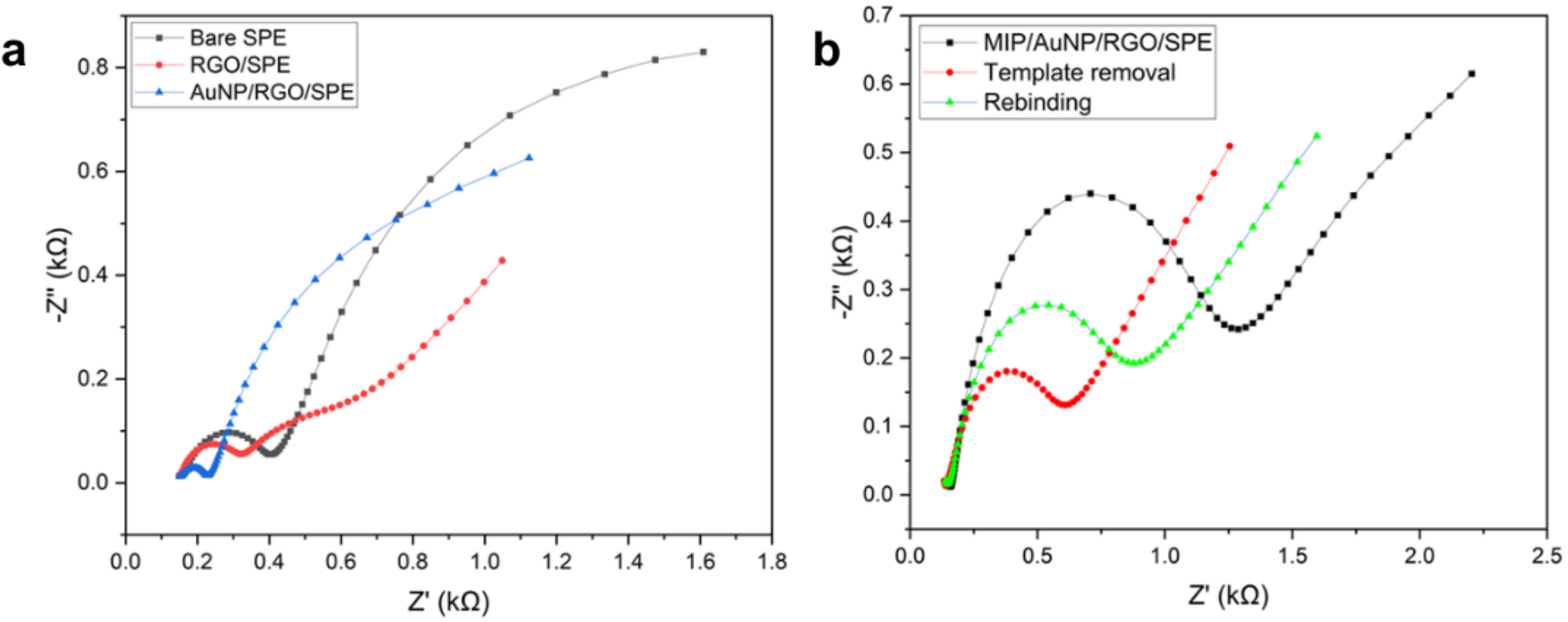
(a). Nyquist diagrams show changes in charge transfer resistance of bare, after RGO deposition, and AuNP deposition. (b). Nyquist diagrams for AuNP/RGO-SPE after MIP deposition, template removal, and lactic acid rebinding, confirming surface modification and target recognition.
The Nyquist diagram, on the other hand, has shown an increased Rct after electropolymerization, indicating an increase in resistance due to the poor electron transfer of the polymer. After the removal of lactate from the polymer, the Rct value decreased due to the formation of cavities in the polymer layer. Then, after rebinding, the Rct value increased, leading to the conclusion that the cavities are occupied by the lactate molecules, which further hinder the electron transfer (Figure 6(b)).
Optimizing conditions for MIP deposition on SPE
The monomer and the polymerization parameters influence the performance of the MIP. Here, the monomer-template ratio and the number of polymerization cycles are the two important parameters that were optimized. First, the number of polymerization cycles was analyzed by assigning the monomer-template ratio as 1:2. The fabricated sensors were assessed by polymerization cycles of 10, 15, 20, and 25 polymerization cycles (n = 3), respectively. Comparatively, polymerization with 20 cycles was found to exhibit a higher normalized current response than others, as shown in Figure 7(a), with good reproducibility (RSD < 3%). Statistical analysis revealed a significant difference among the tested cycle numbers (p < 0.0001), confirming that the polymerization cycle critically affects sensor performance. Here, the normalized DPV response is taken to evaluate the sensor performance and to avoid baseline variability as the peak current for the blank varies for different SPEs due to inherent changes in the commercial sensors.
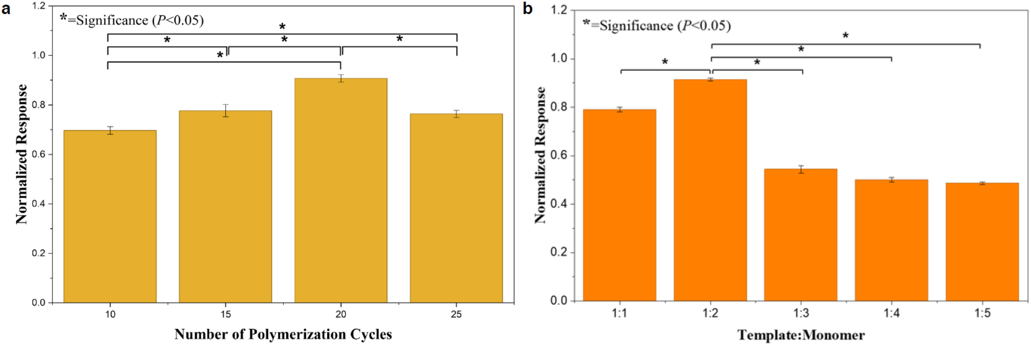
Normalized DPV response of MIP/AuNP/RGO-SPE (a). for polymerization cycles 10, 15, 20, and 25 (b). for the different molar ratios of lactic acid and 3-APBA at 20 cycles.
After optimizing the number of polymerization cycles, the template-to-monomer ratio was investigated. A series of template-to-monomer ratios was examined from 1:1 to 1:5. The study revealed the highest current response at a template-to-monomer ratio of 1:2, as shown in Figure 7(b), with good repeatability (n = 3, RSD < 3%). Statistical comparison demonstrated a significant difference among the tested ratios (p < 0.0001), confirming that the imprinting efficiency strongly depends on the composition ratio. When the number of cycles and the template-to-monomer increased, the thickness of the polymer layer on the SPE also increased, which led to a decrease in the electron transfer. The thicker monomer layer also makes it difficult to remove the lactate molecule imprinted on the matrix. When the number of polymerization cycles and monomer ratio is reduced, a thin polymer matrix is formed on the SPE, leading to a smaller number of cavities for lactate recognition.
The influence of pH on lactate recognition was investigated to validate the boronic acid–diol interaction mechanism. As shown in Figure 8, the DPV response (ΔI) increased gradually from pH 6.0 to 8.5 and reached a plateau beyond pH 8.0. This behavior confirms that lactate binding is favored under mildly alkaline conditions due to the conversion of boronic acid to the boronate form, which forms stronger, reversible ester complexes with α-hydroxy acids such as lactate. Therefore, the pH-dependent response experimentally supports the proposed binding mechanism.
Analytical study on the detection of lactate
After template elution, the sensor performance against lactate molecules was studied using DPV analysis in PBS. It was analyzed against different concentrations of lactic acid from 0.05 µM to 6000 µM (6 mM) for an incubation period of 20 min. As shown in Figure 9(a), as the concentration increases, the current decreases. This is due to the fact that the imprinted cavities on the polymer matrix were occupied by the lactate molecules, which hinders electron transfer. The calibration curve has shown two linear concentration regions, 0.05–100 µM and 250–6000 µM, with correlation coefficients of R2 = 0.99679 and R2 = 0.99618, respectively (Figure 9(b)). Since the LOD must be evaluated near the blank response, the LOD and LOQ were calculated using the slope of the lower concentration range (0.05–100 µM), according to the equation LOD = 3.3σ/S and LOQ = 10σ/S where σ represents the standard deviation of the blank signal (n = 3) and S is the slope of the calibration curve. Based on this, the LOD and LOQ were found to be 0.361 μM and 1.093 μM. This analytical range (0.05 µM to 6 mM) includes the clinically relevant neonatal lactate levels, including normal concentrations (1–2 mM), elevated levels (>2 mM), and critical thresholds (≥4 mM), thereby showing potential for clinical monitoring. The LOD of 0.361 μM is below the normal physiological lactate range of 1–2 mM, indicating that the sensor can detect even a small change in lactate levels, allowing timely therapeutic intervention. The sensor exhibits measurable responses from 0.05 µM, which lies below the normal physiological range, suggesting that it is sufficiently sensitive to detect early-stage increases in lactate concentration.
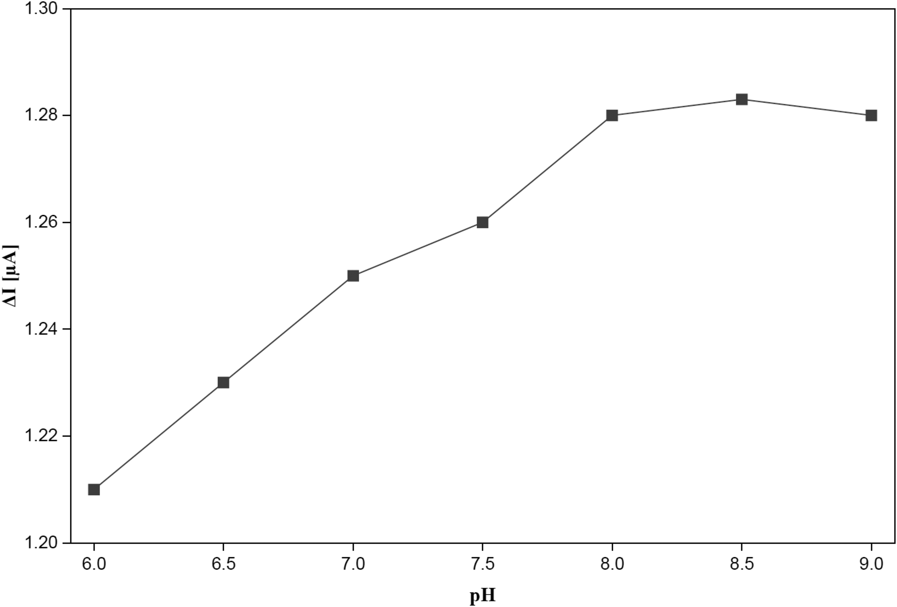
Effect of pH on the performance of the MIP/AuNP/RGO-SPE in PBS.
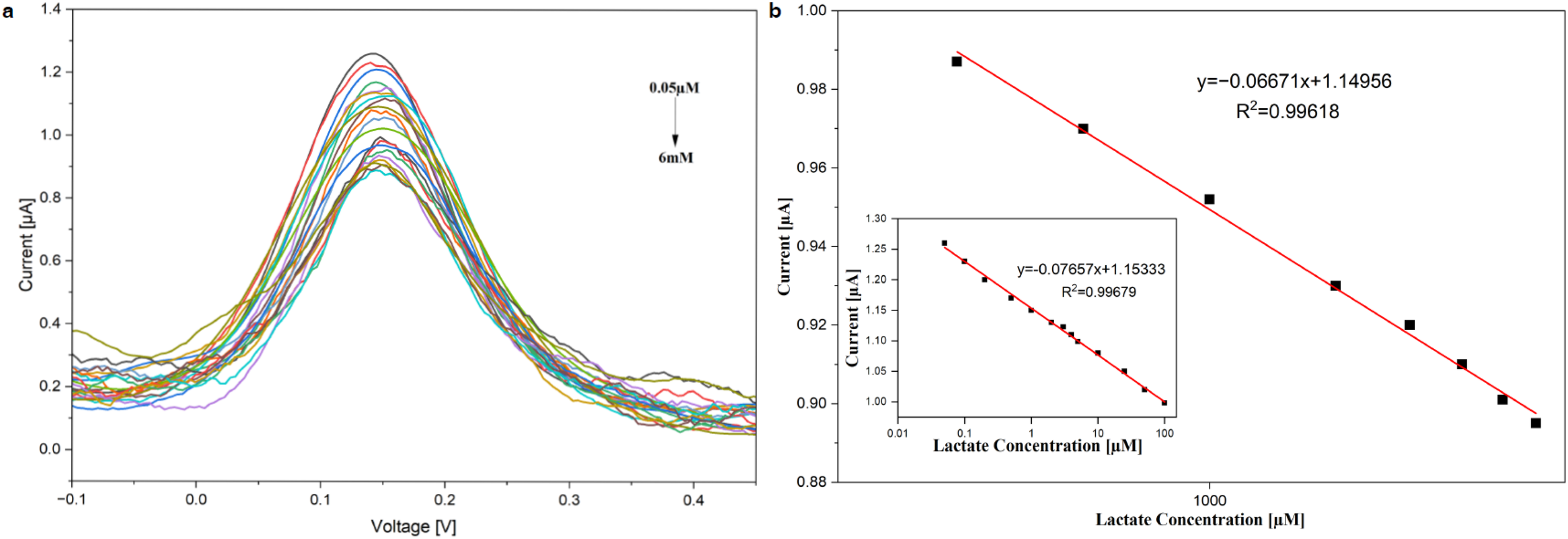
(a). DPV response to various lactate concentrations (b). Calibration curve for lactic acid rebinding using MIP/AuNP/RGO-SPE for different concentrations of lactic acid from 0.05 µM to 6 mM in PBS solution for an incubation period of 20min.
To quantify the affinity of lactate toward the imprinted cavities, a Langmuir adsorption model was applied using DPV response data. The Langmuir linear plot was constructed by plotting Ce/ΔI versus Ce, where Ce is the lactate concentration and ΔI = I0−I represents the current change. The obtained linear fit (Figure 10) indicates monolayer-type adsorption behavior and confirms specific binding at the MIP recognition sites. The binding constant was calculated from the slope-to-intercept ratio, yielding Ka = 2.99 × 10⁴ M−1, demonstrating strong affinity between lactate and the MIP binding cavities.
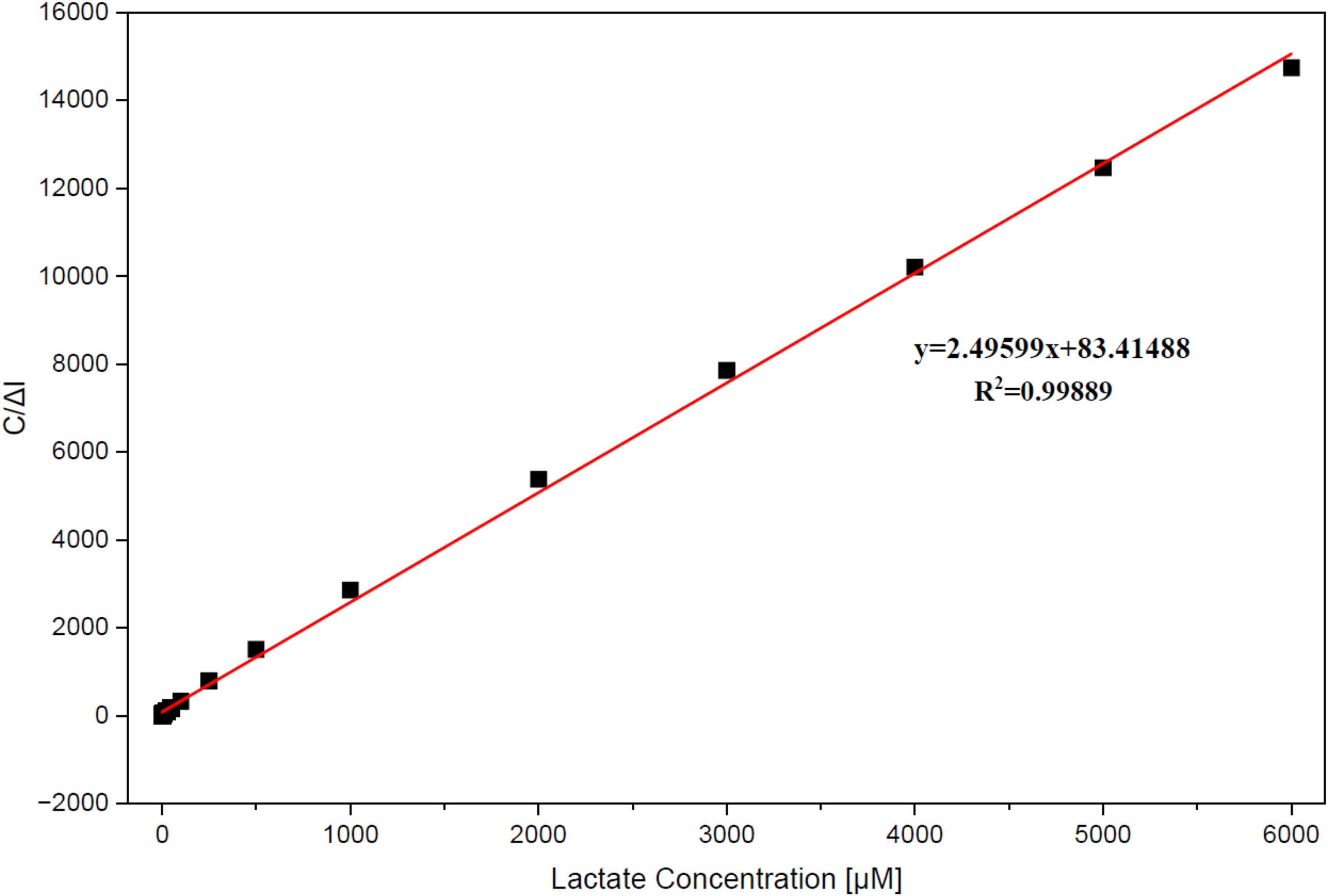
Langmuir adsorption isotherm plot for lactate binding on the MIP/AuNP/RGO-SPE.
For practical applications, the sensor was further studied using artificial serum with different concentrations of spiked lactate ranging from 0.5 mM to 6 mM. Artificial serum was used for the analysis as it mimics the biological matrix, though it does not fully contain the complex biological components like the serum. Therefore, further evaluation using real clinical samples is needed. A calibration plot was developed from the DPV response for different concentrations ranging from 0.5 mM to 6 mM. As shown in Figure 11(a), the DPV response shows that the current response decreases as the lactate concentration increases, indicating efficient electrocatalytic interaction between the electrode surface and lactate molecules and showed a linear calibration response from 0.5 mM to 6 mM with a correlation coefficient of 0.99405 (Figure 11(b)). The limit of detection for developed sensor in artificial serum was found to be 0.482 mM, demonstrating the sensor's high sensitivity in complex biological-like media. Compared with PBS, the sensor showed a similar linear response but a slightly reduced current response, due to the presence of serum proteins and electrolytes that introduce minor matrix effects. In particular, protein binding plays a significant role, as lactate can interact with serum proteins or become partially associated within the protein matrix formed on the electrode surface. This interaction may reduce the freely available lactate concentration and hinder its diffusion toward the imprinted cavities, leading to a slight attenuation in signal. Additionally, nonspecific adsorption of proteins onto the electrode surface can create a diffusion barrier and partially block active recognition sites. Despite these effects, the linear response of the sensing platform indicates that the recognition sites from MIP remain effective, demonstrating the stability of the sensor in protein-rich environments.
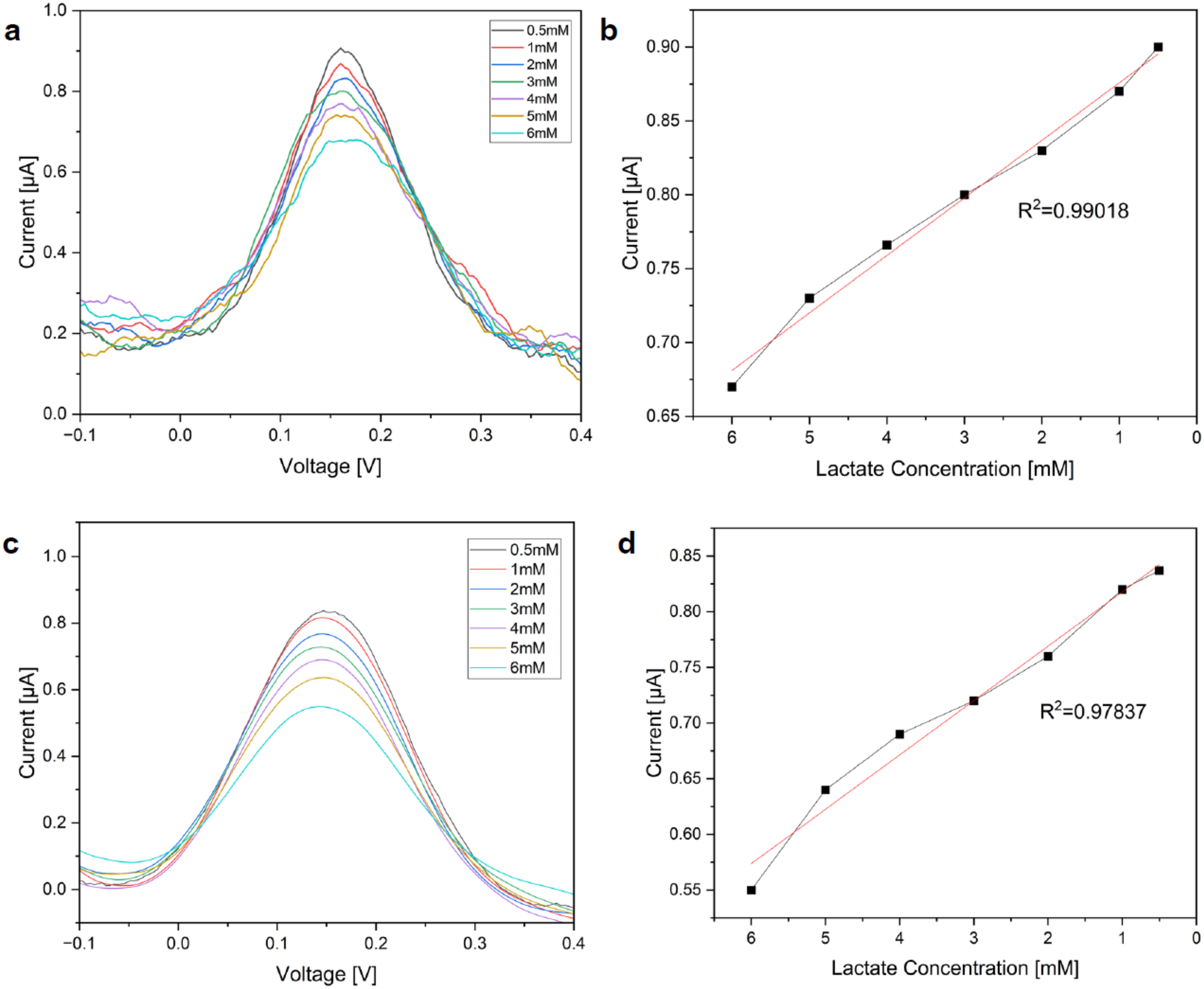
(a). DPV response of various lactate concentrations in artificial serum (b). Calibration curve for lactic acid rebinding in artificial serum (c). DPV response of lactate concentrations in Lactobacillus culture supernatant (d). Calibration curve for lactic acid rebinding in Lactobacillus culture supernatant.
An analytical evaluation was carried out to detect lactate produced during the growth of Lactobacillus casei in MRS broth using DPV. Freshly prepared MRS broth was inoculated with Lactobacillus casei and incubated at 37°C for 6 to 12 h. At every two-hour time intervals, the sample was taken and diluted further using PBS and centrifuged. The culture supernatant was collected and directly tested with the developed SPCE-based lactate sensor. The DPV response exhibited a progressive change in peak current with increasing incubation time, reflecting the accumulation of lactate in the medium with approximate concentrations ranging from 0.5 mM to 6 mM (Figure 11(c)). The calibration curve, obtained using standard lactate solutions, was applied to convert the DPV current signals into equivalent lactate concentrations (Figure 11(d)). The measured values indicated lactate accumulation within the physiologically relevant range for sepsis studies, with a calculated LOD of 0.72 mM. Compared to artificial serum and PBS, the DPV signals from Lactobacillus culture were slightly attenuated, likely due to interference from other fermentation by-products. Although the results confirmed that the sensor is capable of directly quantifying lactate in complex microbial growth media without prior extensive sample preparation, demonstrating its applicability for biological matrix testing in preliminary sepsis-related lactate studies. The calibration slopes obtained in PBS and artificial serum were statistically compared using a t-test. The calculated t-value (2.36) was lower than the critical value, indicating no significant difference between the slopes (p > 0.05). This confirms that the sensor maintains comparable sensitivity in complex biological media.
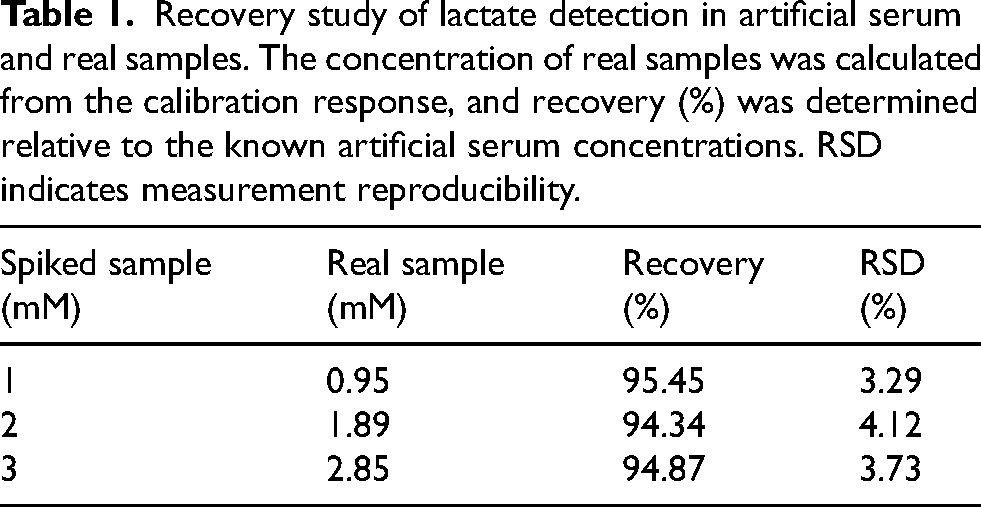
To further validate the analytical performance in complex matrices, a recovery study was performed by comparing the responses obtained in artificial serum and real samples at different lactate concentrations (1–3 mM). As shown in Table 1, the recovery values were found to be 95.45%, 94.34%, and 94.87%, respectively, with RSD values below 5%. These results show good accuracy and reproducibility of the sensor, confirming its reliability in biologically relevant conditions.
Recovery study of lactate detection in artificial serum and real samples. The concentration of real samples was calculated from the calibration response, and recovery (%) was determined relative to the known artificial serum concentrations. RSD indicates measurement reproducibility.
To further validate performance in a protein-rich matrix, lactate detection was carried out in FBS spiked with lactate concentrations ranging from 0.05 µM to 6 mM. As shown in Figure 12, the DPV peak current decreased with increasing lactate concentration, confirming that the imprinted recognition sites remain functional under serum conditions. The calibration plot showed good linearity with a correlation coefficient of R2 = 0.98965, with minor signal attenuation compared to PBS due to protein adsorption on the electrode surface and matrix effects. Although the sensor demonstrated good performance in PBS, fetal bovine serum (FBS), and culture supernatant, validation using human clinical blood/serum samples was not performed in this study due to limited sample accessibility. Therefore, further clinical evaluation is required before translation to patient testing.
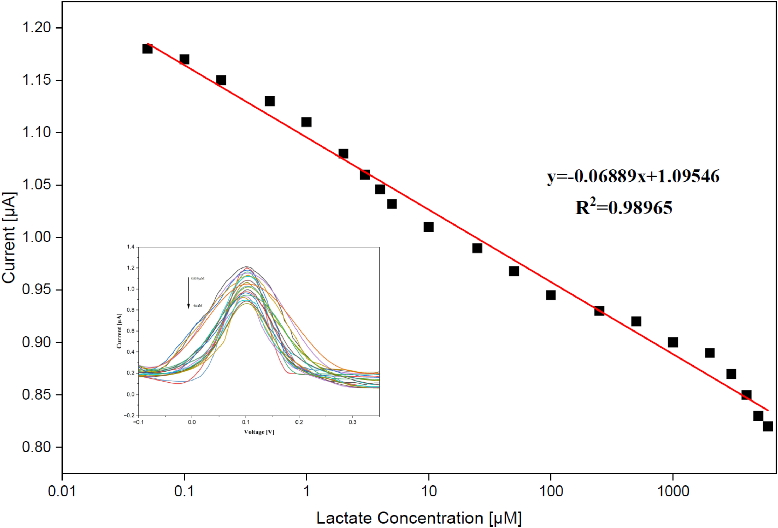
DPV response and calibration curve of lactate in FBS (0.05 µM–6 mM).
Selectivity of MIP/AuNP/RGO-SPE
MIP acts as a selective recognition element due to its specific imprinted cavities, which resemble the analyte, enabling them to be highly selective towards any analyte of interest. The selectivity of the designed sensor was analyzed by DPV response against specific interferents such as Ascorbic acid, glucose, urea, acetic acid, pyruvate, albumin, NaCl, and citric acid, each in different physiological concentrations (1 mM, 5 mM, 5 mM, 1 mM, 0.1 mM, 0.1 mM, 0.6 mM, 0.1 M, 0.1 mM). The peak currents of the potential interferents showed no significant difference from that of the blank solution. Although interference experiments were performed at single representative concentrations, the sensor was additionally validated in artificial serum and microbial culture media, which simulate complex biological environments containing proteins, electrolytes, and metabolic by-products. The preservation of linearity and acceptable sensitivity under these conditions demonstrates the robustness of the sensing platform against matrix effects and supports its potential applicability in biologically relevant samples. The calculated cross-reactivity percentages and selectivity coefficients are summarized in Table 2.
Selectivity evaluation of the MIP sensor toward lactate in the presence of potential interfering species.
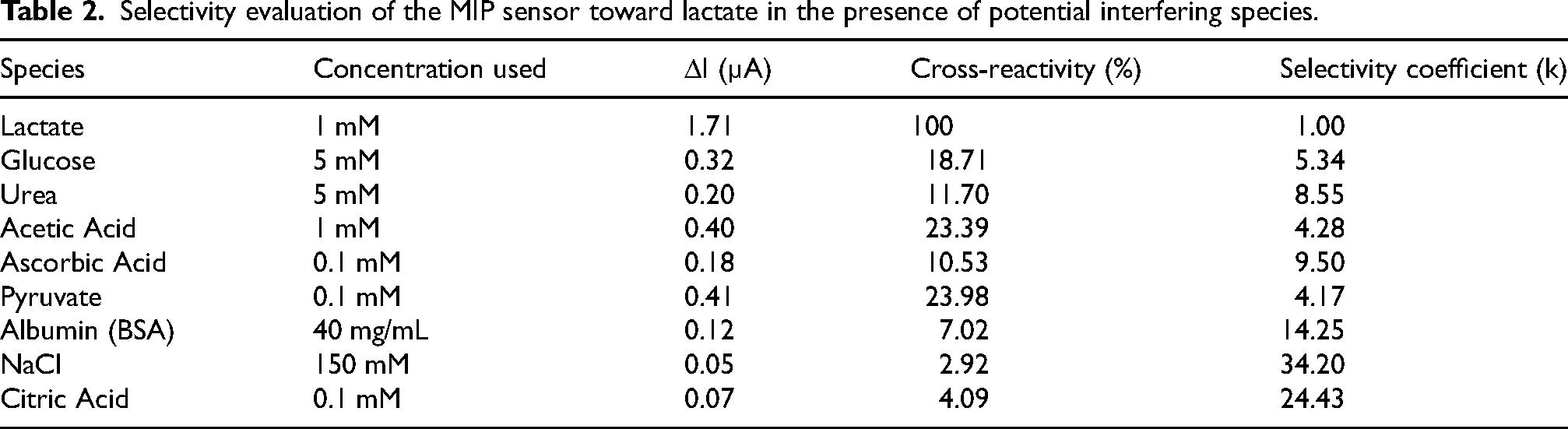
Figure 13(a) shows the peak current response of lactic acid and other interferents after 20 min of incubation on the sensor. From the response, it was observed that the rebinding of lactic acid exhibited a significantly higher peak current compared to other potential interferents, indicating selective recognition capability of the fabricated sensor toward lactic acid. The relatively higher response toward pyruvate may be attributed to its structural similarity to lactic acid, leading to partial interaction with the imprinted cavities. Furthermore, in the presence of interferents (Figure 13(b)), the sensor maintained a distinguishable and statistically significant response (*p < 0.05), demonstrating its ability to selectively recognize lactic acid even in complex environments.
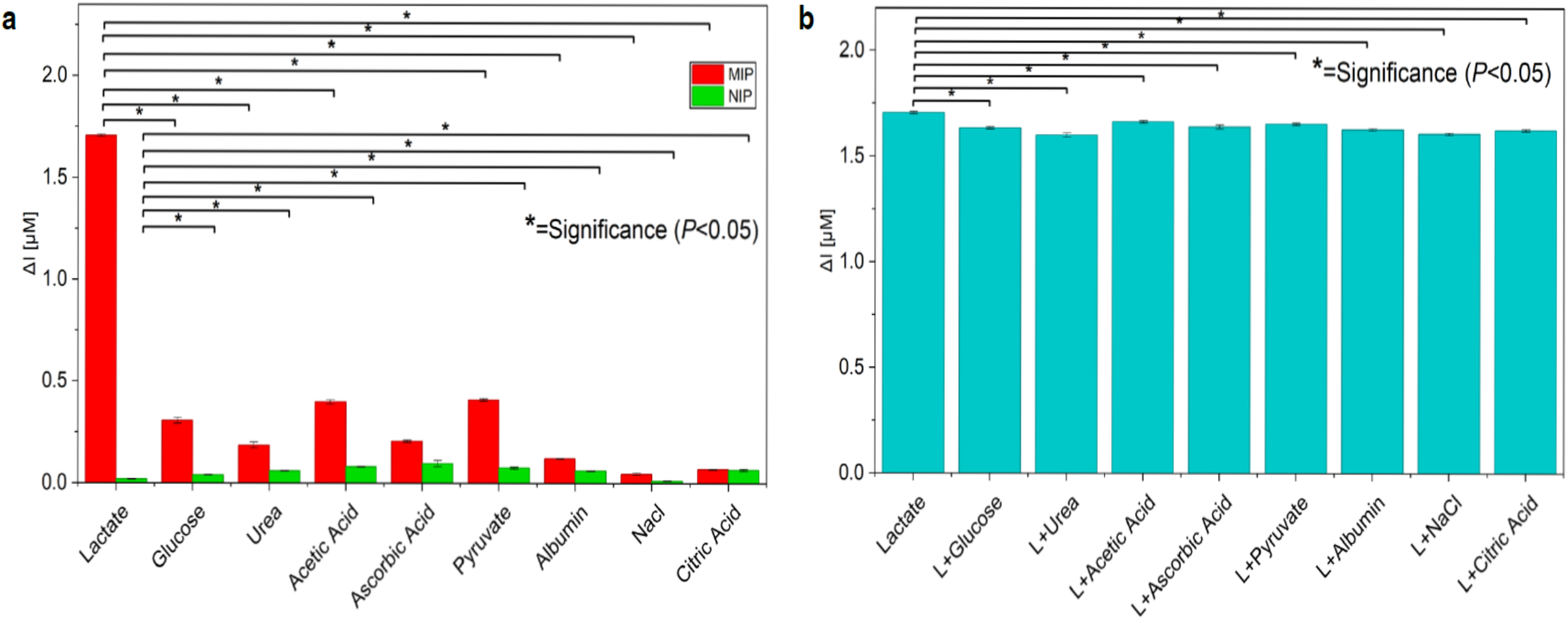
(a). MIP/AuNP/RGO-SPE and NIP/AuNP/RGO-SPE sensor responses toward lactate and interference molecules (b). Selectivity study of the MIP/AuNP/RGO-SPE in the presence of interfering molecules where L denotes lactate (glucose, urea, acetic acid, ascorbic acid, pyruvate, albumin, NaCl and Citric Acid).
The specificity of the developed sensor was studied using the calculation of the imprinting factor (IF), which provides the ability of the sensor to selectively recognize lactate over other interferents. When compared to the IF values of interferents, such as glucose, urea, acetic acid, and ascorbic acid, which range from 1.14 to 1.43, the MIP exhibited a strong affinity toward lactate, as indicated by the lactate IF value of 8.53. This significant difference in IF values demonstrates the strong molecular recognition capacity of the imprinted cavities developed during the polymerization process and validates the sensor's exceptional specificity even in the presence of structurally and chemically similar compounds.
Analytical performance of the proposed sensor is compared with the previous works, as outlined in Table 3. The developed sensor has shown a wide linear range and lower limit of detection. The sensor has shown good sensitivity and specificity towards the target analyte and can be developed as a compact portable device.
Comparison of the reported lactate sensing platforms and the present work.
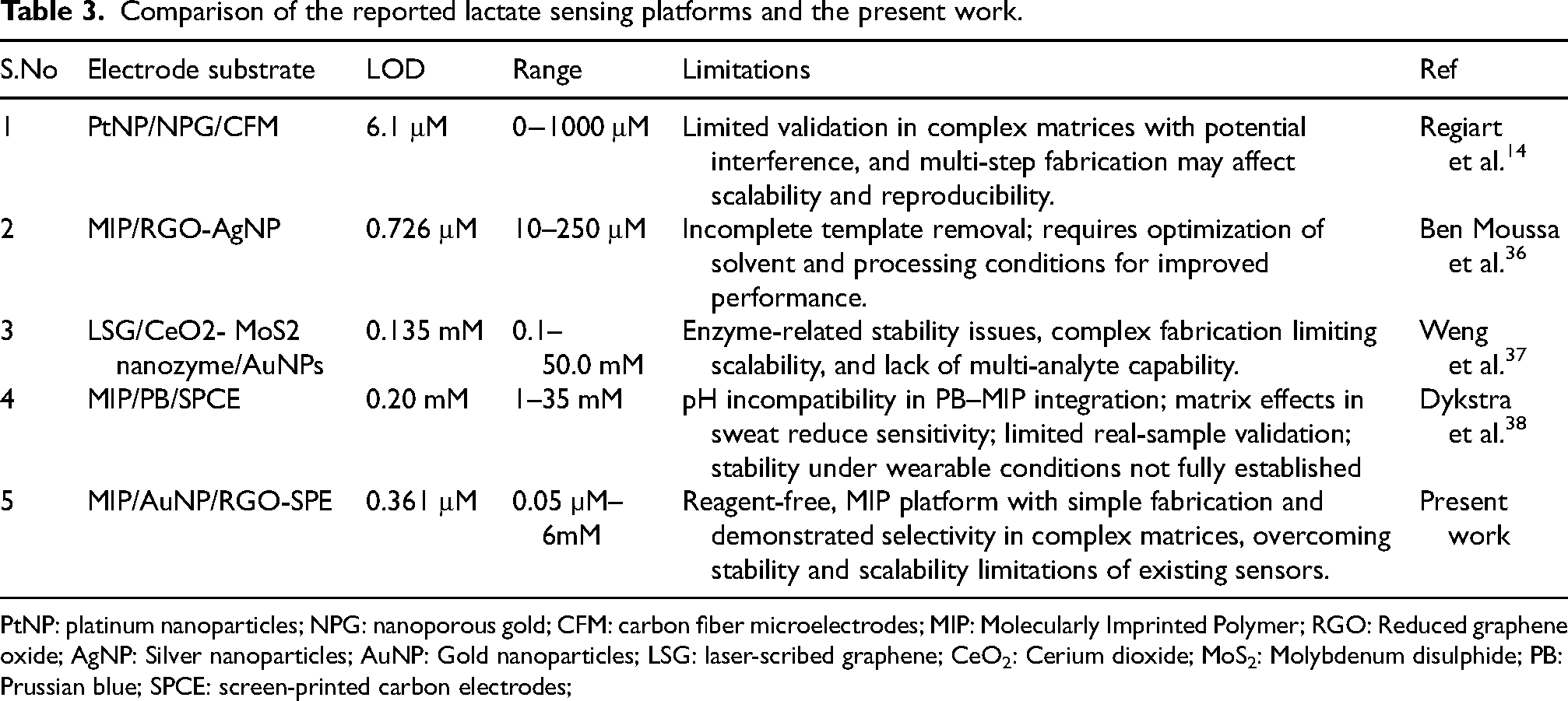
PtNP: platinum nanoparticles; NPG: nanoporous gold; CFM: carbon fiber microelectrodes; MIP: Molecularly Imprinted Polymer; RGO: Reduced graphene oxide; AgNP: Silver nanoparticles; AuNP: Gold nanoparticles; LSG: laser-scribed graphene; CeO2: Cerium dioxide; MoS2: Molybdenum disulphide; PB: Prussian blue; SPCE: screen-printed carbon electrodes;
Reproducibility and stability of the sensor
The repeatability of the MIP/AuNP/RGO-SPE sensor was analyzed using five identically prepared electrodes using similar conditions. The current variation among these electrodes was minimal and the relative standard deviation was found to be 1.29%. The stability of the sensor was measured after a month of storage in a dry condition at 4 °C. The normalized response remains 95.3% after 1 month of storage. From the results obtained, the reproducibility and long-term stability make the sensor potentially suitable for real time applications.
Conclusions
In this work, MIP-based electrochemical sensor was successfully developed and optimized for the detection of lactic acid. The steps involved in the fabrication of lactate sensor were evaluated using CV and EIS and the performance analyzed using DPV against different concentrations of lactate and other interferents. This sensor demonstrated good sensitivity and selectivity towards lactate molecule. The rebinding of the lactate molecule in buffer solutions has provided insight into template-monomer interactions in biofluids (such as perspiration, saliva, and interstitial fluid), which is promising for upcoming non-invasive diagnostic medical applications. The developed sensor has shown a lower limit of detection of 0.361 μM and a wide linear range between 0.05 µM and 6 mM. Although the reported detection limit is higher than that of conventional blood gas analyzers (∼0.1 mM resolution), these systems require bulky instrumentation and trained personnel. In contrast, the proposed sensor offers a simpler and potentially portable alternative with adequate sensitivity for clinically relevant lactate monitoring. Therefore, the operational range of the sensor platform, sensitivity, and ease of fabrication make it a promising platform for further development toward point-of-care lactate monitoring following clinical validation. This study represents a proof-of-concept for an enzyme-free, boronic acid-based MIP sensing for lactate detection, and further evaluation in clinically relevant samples will be essential to advance its translational applicability. This developed sensor could potentially be integrated with a portable potentiostat for rapid lactate monitoring using small sample volumes, demonstrating its feasibility for future point-of-care applications following further clinical validation. The electrochemical detection method allows fast signal acquisition within minutes, showing potential for neonatal sepsis-related lactate monitoring of metabolic stress, hypoxia, or sepsis-related complications. Furthermore, the low-cost fabrication of screen-printed electrodes combined with electropolymerized MIP films supports disposable sensor formats, which are advantageous for decentralized diagnostics and resource-limited healthcare environments. In clinical samples, factors such as individual variability in protein content, ionic strength, pH, and coexisting metabolites may influence sensor response; hence, systematic clinical validation across diverse patient samples is necessary. Future work will focus on validating the sensor response against gold-standard clinical lactate measurement techniques such as blood gas analyzers and enzymatic lactate assays, to establish accuracy under real clinical conditions.
Footnotes
Acknowledgments
We acknowledge the Biomaterials laboratory, SRMIST, for providing the research facilities.

Ethical considerations
This article does not contain any studies with human participants or animals performed by any of the authors.
Author contributions
Backiyalakshmi Gnanasekaran: Conceptualization, Validation, Methodology, Investigation, Formal analysis, Data curation, Writing – Drafting and Editing. Snekhalatha Umapathy: Writing – Drafting, Editing & Supervision, Methodology, Formal analysis, Data curation.
Funding
The authors received no financial support for the research, authorship, and/or publication of this article.
Declaration of conflicting interests
The authors declared no potential conflicts of interest with respect to the research, authorship, and/or publication of this article.
Data availability statement
The data that support the findings of this study are available from the corresponding author upon reasonable request.