
Abstract
Objectives
To describe the incidence, diagnostic clinical manifestations and severity of juvenile systemic lupus erythematosus (jSLE) in a cohort of New Zealand Maori and Pacific Island children compared to European children.
Methods
A chart review was conducted of children with jSLE seen by the Starship paediatric rheumatology and/or renal services between January 2000 and November 2010. Diagnostic clinical data and lupus nephritis data at anytime were collated while classic British Isles Lupus Assessment Group (BILAG) and Systemic Lupus Erythematosus Disease Activity Index (SLEDAI) scores were derived retrospectively.
Results
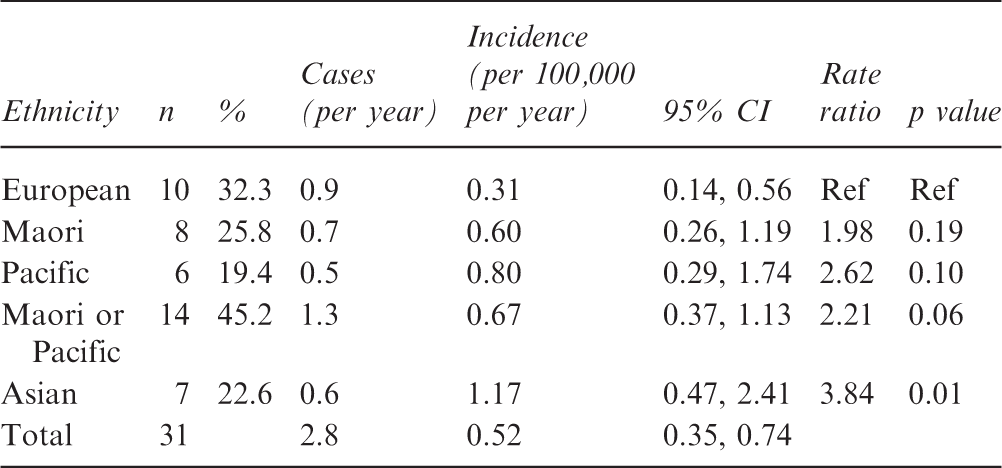
Thirty-two children were diagnosed with jSLE with an annual incidence of 0.52 per 100,000 per year. Compared with European children (0.31 per 100,000 per year) the incidence of jSLE was higher among Maori and Pacific (0.67 per 100,000 per year, p = 0.06) and significantly higher among Asian children (1.17 per 100,000 per year, p = 0.01). Compared with European children, Maori and Pacific children were more frequently diagnosed with lupus nephritis (80% vs 40%, p = 0.09) and severe (WHO class 4 or 5) renal lesions (60% vs 40%, p = 0.43) at presentation. Similarly, at any time during the study, lupus nephritis (100% vs 40%, p = 0.001) and severe (WHO class 4 or 5) renal lesions (73.3% vs 40%, p = 0.12) were more frequent among Maori and Pacific compared with European children. Furthermore, retrospective BILAG assessment of diagnostic disease severity demonstrated that Maori and Pacific children experienced the majority of severe “Category A” disease (56.8% vs 22.7%, p = 0.17) which was predominantly renal (73.3% vs 40%, p = 0.12) in nature.
Conclusions
This is the first description of the incidence and clinical manifestations of jSLE in a cohort of New Zealand children. Although limited by the small numbers involved it confirmed anecdotal suspicions that the incidence of jSLE among Maori, Pacific and Asian children is higher than European children. Lupus nephritis is also more frequent and severe in Maori and Pacific children.
Introduction
The incidence, demographics, and clinical manifestations of juvenile systemic lupus erythematosus (jSLE)1−4 are well established in European and North American paediatric populations. This study will present data on the ethnic incidence and diagnostic clinical manifestations of a cohort of New Zealand children with jSLE and examine the severity of jSLE in Maori and Pacific Island New Zealand children compared with European children.
Materials and method
Starship Children’s Hospital is located in Auckland, New Zealand providing tertiary paediatric healthcare to a diverse multicultural paediatric population including European, Pacific, Asian and Maori children. Children diagnosed with jSLE and seen by the Starship Hospital paediatric rheumatology and/or renal services between January 2000 and December 2010 were included in this study. All were referred from within the Starship rheumatology catchment area which includes multiple district health boards located within the greater North Island of New Zealand and were aged 15 years or less at the time of their review.
Children were identified using the paediatric rheumatology, renal biopsy and Starship inpatient databases. Children aged less than 20 years admitted to Starship hospital after 1995 were identified using the key search words “juvenile systemic lupus erythematosus” and “systemic connective tissue disorders”.
All clinic letters, correspondence, inpatient clinical notes, discharge summaries and investigations were reviewed using the Clinical Records Information System (CRIS) and Concerto computerised administration systems.
Juvenile SLE was defined using the American College of Rheumatology (ACR) 1997 revised lupus criteria. 5 Children were considered to have a diagnosis of jSLE once they fulfilled the ACR criteria and/or had histological evidence of lupus nephritis. Mucocutaneous features (malar rash, photosensitivity, oral ulcers and discoid lesions), arthritis, nephritis, serositis, haematological and neuropsychiatric clinical manifestations were identified. Biochemical (complement, renal function, albumin, liver function), haematological (full blood count, haptoglobin and Coomb’s test) and immunological markers (anti nuclear antibodies, extractable nuclear antigens, double stranded DNA and antiphospholipid antibodies) associated with jSLE were recorded. The World Health Organization (WHO) classification criteria (1974) 6 was used to further categorise lupus nephritis. These included WHO class 2 (purely mesangial disease), class 3 (focal proliferative), class 4 (diffuse proliferative) and class 5 (membranous) disease. Features were considered to have occurred at the time of jSLE diagnosis if they occurred within 1 month of diagnosis.
The British Isles Lupus Assessment Group (BILAG) and Systemic Lupus Erythematosus Disease Activity Index (SLEDAI) were used to assess severity of jSLE at diagnosis by retrospectively reviewing the signs, symptoms and laboratory makers documented within 1 month of jSLE diagnosis. The eight organ systems were assigned an alphabetical and numerical value (Category A = 9, B = 3 and C = 1). 7,8
The overall and ethnic incidence of jSLE per 100,000 per year was calculated using the 2006 New Zealand census and collected data of children diagnosed with jSLE aged 0−14 years. Children were assigned to four ethnic groups: European, Maori, Pacific and Asian. The ethnic incidence was evaluated by comparing European to Maori, Pacific and Asian groups using exact Poisson 95% confidence intervals and Chi square p values. As is convention for incidence data analysis of census data the largest group (European, n = 297,857) included minority groups (Latin American and African, n = 2643). Single proportion, binomial 2-sided test was used to compare the “category A” disease systems while Fisher’s exact test was used to compare lupus nephritis and American College of Rheumatology diagnostic lupus criteria in Maori or Pacific versus European children. The population of New Zealand has grown at a steady increase of 1.2% (50,000) per year from 2001 to 2011 and therefore the 2006 census was used as a reasonable estimate of the annual catchment over the entire study period.
Results
The incidence of Juvenile Systemic Erythematosus (jSLE) in children aged 0−14 years
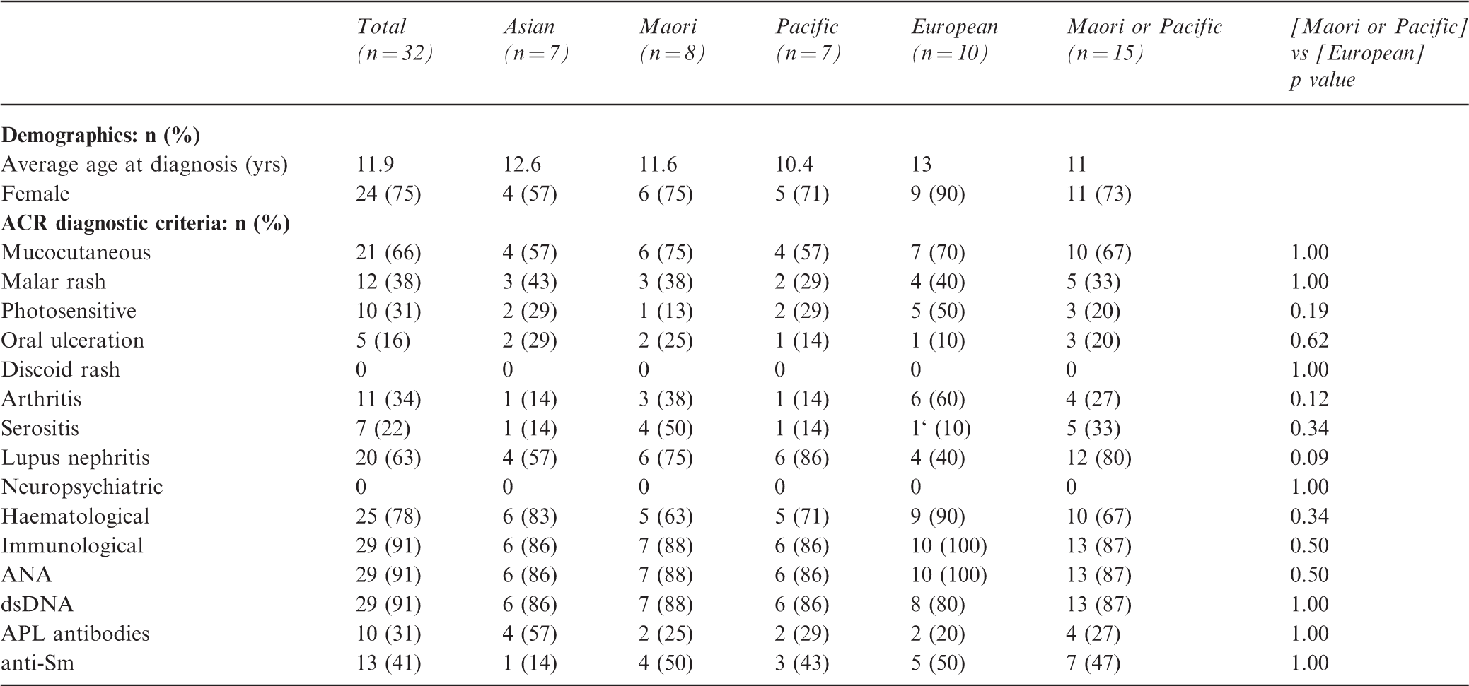
The demographics and diagnostic clinical manifestations of jSLE
Juvenile SLE severity
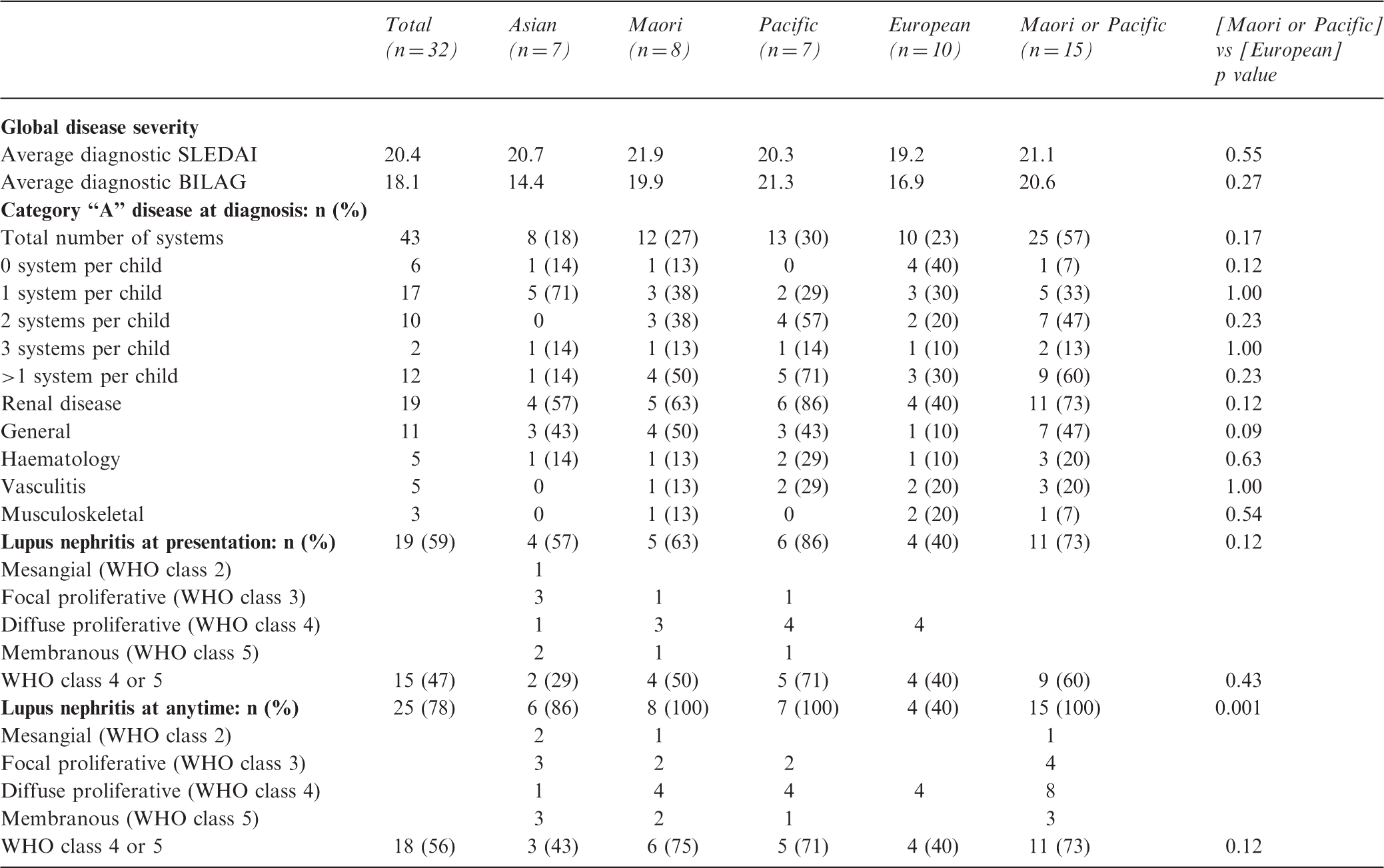
The average diagnostic BILAG (20.6 vs 16.9, p = 0.27) and SLEDAI (21.1 vs 19.2, p = 0.55) were higher among Maori and Pacific compared with European children. Category “A” disease was present in 43 systems. Compared with European children severe disease was more frequent among Maori and Pacific children (56.8% vs 22.7%, p = 0.17). In addition, more Maori and Pacific children experienced greater than one category “A” system per child (60% vs 30%, p = 0.23) while more European children experienced no “category A” systems per child (40% vs 6.7%, p = 0.12).
The kidneys were the most commonly affected category “A” system (n = 19, 44%) followed by general (n = 11, 25.6%), haematological (n = 5, 11.6%), vasculitis (n = 5, 11.6%) and musculoskeletal (n = 3, 7%). In addition, more Maori and Pacific children experienced severe renal (73.3% vs 40%, p = 0.12) or general disease (46.7% vs 10%, p = 0.09) compared with European children (see Table 3).
Discussion
Systemic lupus erythematosus has been described to be more common and of greater severity in people of Australian Aboriginal, Asian, Polynesian and African American ethnicity. 9 There is no data evaluating the incidence and severity of jSLE among Maori and Pacific children.
The incidence among children aged less than 16 years has been estimated in small retrospective studies in the United States (0.53 per 100,000 per year), Canada (0.36 per 100,000 per year), Europe (0.37 to 0.48 per 100,000 per year) and a large Japanese series (0.47 per 100,000 per year). 10 The only previous New Zealand prevalence data of SLE in Auckland estimated the prevalence of Polynesian adult SLE in Auckland to be 3.5 times greater than that of Europeans (50.6 vs 14.6 per 100,000). 11 While the overall incidence in our study was comparable to other similarly sized retrospective studies, ethnic subgroup analysis confirmed a higher Maori and Pacific and significantly higher Asian incidence when compared with European children.
Lupus nephritis is a common clinical manifestation and important marker of morbidity and mortality in children. Compared with those with adult onset disease, a significantly higher proportion of children experience kidney disease and require dialysis and renal transplantation. 12 A 23-year predominantly prospective, study of 256 children demonstrated lupus nephritis at diagnosis in 37% and 55% at any time. The most common subtype was diffuse proliferative disease present in 47% at diagnosis and 46% at any time with membranous disease present in 16% at diagnosis and 21% at any time. 2 These lesions are considered severe as diffuse proliferative nephritis conveys an estimated 5-year renal survival of between 88 and 93% with a reported 10-year renal survival of only 85%. 1 Membranous nephritis is associated with persistent nephrotic syndrome with the majority experiencing eventual renal insufficiency. 13
In a recent retrospective Auckland study of 170 adults with SLE, Maori and Pacific patients constituted a high proportion of the 24 diagnosed with lupus nephritis at presentation (n = 15, 62%) and the 56 diagnosed at anytime (n = 31, 54.4%). 14 Similarly we found that the Maori and Pacific children constituted a high proportion of the 20 diagnosed with lupus nephritis at presentation (n = 12, 60%) and the 25 diagnosed at any time (n = 15, 60%). The numbers involved in this study were small. However, all Maori and Pacific children developing histological evidence of lupus nephritis during the study period was a significant finding. In addition, subgroup analysis confirmed that severe (class 4 or 5) lupus nephritis was more prevalent among Maori and Pacific children at diagnosis or any time, although this did not reach statistical significance.
While average diagnostic BILAG and SLEDAI global severity scores were higher among Maori and Pacific compared with European children the retrospective BILAG application was felt by the authors to be more informative as an organ-based single measure of lupus severity. This further confirmed anecdotal clinical suspicion that more Maori and Pacific children experience jSLE of greater severity than European children with a higher number of severe “category A” systems involvement at diagnosis. These were often multiple and predominantly renal in nature further reinforcing the burden of significant renal disease amongst Maori and Pacific children.
Large prospective studies of juvenile SLE are rare. Despite data representing a 10-year period the small numbers involved and historical or chart review nature of this study were the major limitations .The catchment population was representative containing 65% of all New Zealand children including European (57%), Maori (73%), Pacific (80%) and Asian (78%) children. Fifty-three percent of the catchment population lived within the Auckland region; including Pacific (82%), Asian (84%), European (48%) and the minority of Maori children (24%). Geography in addition to other barriers, including low socioeconomic status has been shown to potentially reduce access to healthcare services for Maori 15 and may provide similar barriers to accessing healthcare for Pacific children. Renal and rheumatology services provide outreach clinics within the catchment area to centres outside Auckland. Children residing in these or poorly accessible regions with less severe disease may have been less likely to be seen at Starship. These factors may potentially contribute to selection bias and underestimation of incidence, overestimation of disease severity and potential for delayed presentation contributing further to disease severity in those seen.
When further assessing severity, the BILAG has been validated in children. 7 As this was retrospectively applied variation of clinical assessment between clinicians may have influenced the results. However, some organ systems such as haematological and renal disease were largely dependent on objective data making the BILAG and SLEDAI systems easily applicable retrospectively.
Conclusions
In this representative cohort of New Zealand children we demonstrated a higher overall incidence of jSLE and frequency and severity of renal disease among Maori and Pacific children compared with European children. Larger prospective studies are required to replicate these findings.
Key points
There is paucity of juvenile SLE data worldwide. This is the first data available from New Zealand children, particularly those of Maori and Pacific ethnicity. The incidence of juvenile SLE is higher among Maori and Pacific and significantly higher among Asian compared with European children in New Zealand. In New Zealand Maori and Pacific children, lupus nephritis is more common and severe than European children.
Footnotes
Acknowledgement
The authors would like to thank The Starship Hospital Paediatric Renal service.
Conflict of interest
None declared.
Funding
This research received no specific grant from any funding agency in the public, commercial, or not-for-profit sectors and local ethical approval was obtained.